Embed Size (px)
Citation preview

Neurochemical Research, Vol. 13, No. 4, 1988, pp. 369-376
Spermine Binding to Subsynaptosomal Fractions of Rat Brain Cortex
Vittorio Genti le , 1 Carla Esposito, 1 Antonio Fusco, ~ Mauriz io Popol i , 2 and Raffaele Porta 1
(Accepted August 12, 1987)
Binding sites for [14C]spermine have been identified in rat brain cortex subcellular frac- tions. The binding, characterized by using synaptosomal membranes, is specific for sperm- ine. It was not detected below 20~ and increased about three/four-fold with a temperature rise of 10~ Binding occurred only in the presence of - S H reducing agents. It was com- pletely suppressed by metal chelating agents, and was stimulated about four-fold by 1-5 x 10 -5 M Fe 2+. Smaller increases were observed in the presence of Mn 2+, Ni 2+, Ca 2+, Mg 2§ and Zn2+; in contrast, millimolar concentrations of most divalent cations inhibited the binding differently (Mn 2§ = Ni 2§ = Zn 2+ = Co 2+ >> Mg 2+ > Ca2+).
Bound radioactive spermine was not displaced by the addition of high concentrations of unlabelled polyamine or chelating agents, nor by precipitation and washing of the mem- branes with 10 percent trichloroacetic acid, or by boiling of the precipitate in the presence of 1.0 percent SDS and 10 percent [3-mercaptoethanol. The trichloroacetic acid precipitate showed two radioactive bands, corresponding to low Mr (< 8,000) components, after SDS- polyacrylamide gel electrophoresis and fluorography. The Fe2+-stimulated [14C]spermine binding was neither influenced by a previous heating of the membranes at 100~ for 30 min nor by trypsin or pronase digestion, whereas the heat,treatment increased the binding occurring in the absence of Fe 2+ by about two fold. A non-enzymatic formation of a spermine-metal complex tightly bound to some membrane peptide(s) is suggested.
KEY WORDS: Spermine; polyamines; synaptosomal membranes.
I N T R O D U C T I O N
The polyamines spermidine (Spd) and spermine (Spm) are heterogeneously distributed at high con- centrat ions in neural tissue, which is an especially rich source of their biosynthetic enzymes, the ami- nopropyl t ransferases (39, 41).
Specific functional roles of these polycations in the brain have frequently been postulated. It is well
x Istituto di Chimica e Chimica Biologica, 1 a Facolt~ di Medicina e Chirurgia,
2 Istituto delle Malattie del Sistema Nervoso, 2 a Facolt& di Med- icina e Chirurgia, Universit& di Napoli, Napoli, Italy.
Address reprint request to: Prof. RaffaelePorta, Istituto di Chim- ica e Chimica Biologica, Via Costantinopoli 16, 80138 Napoli, Italy, Tel. 081/341568.
369
known that alterations of the cerebral endogenous polyamine pools, which possess exci tatory prop- erties, lead to marked behavioral changes. It is fur- thermore established that intraventricularly injected polyamines produce convulsions in mice, rabbits (3), and chicks (31). They are also effective in caus- ing insomnia and suppressing feeding behavior in rats (36). In addition, the intraperitoneal adminis- tration of the long-latency convulsant agent L-me- thionine-dl-sulfoximine was shown to have a stim- ulatory effect on the biosynthesis of both Spd and Spm in mouse brain (34).
The rapid movemen t o fpo lyamines along axons (22, 26, 39, 41) suggests that their specific role in the cerebral tissue might be played at the synapse level. It is notable that about 25 percent of the total
0364-3190/88/0400-0369506.00/0 �9 1988 Plenum Publishing Corporation

370 Gentile, Esposito, Fusco, Popoli, and Porta
p o l y a m i n e c o n t e n t of the rat cerebra l cor tex was f o u n d in the ne rve ending- r ich f rac t ion (38).
P r e l im ina ry data have b e e n pub l i shed on the b i n d i n g of the po lyamines to cerebra l t i ssue com- p o n e n t s . Sei ler and Decka rd t (37, 38) repor ted evi- d e n c e for a high aff ini ty b ind ing of bo th Spd and
Sp in to m e m b r a n e s of the synap t ic complex , and
H a r m a n and Shaw (17) descr ibed two high-aff ini ty s o d i u m - d e p e n d e n t up take sys tems for Spm in rat c e r eb ra l co r t ex slices. T r a n s g l u t a m i n a s e (E.C.
2.3.2.13.) was recen t ly shown to be p re sen t in mam- m a l i a n b r a in (14, 40); in addi t ion , pro te ins able to
ac t as a m i n e - a c c e p t o r subs t ra tes for t r ansg lu tamin- ase, and thus able to cova len t ly l ink po lya mi ne s , h a v e b e e n repor ted in h u m a n bra in homoge na t e s (40), in Ap lys i a ne rvous t issue (1), and on the sur-
face of m o u s e n e u r o b l a s t o m a cells (8). In the present paper we describe and partially
characterize the binding of Spm to subcellular and subsynaptosomal fractions of rat brain cortex.
E X P E R I M E N T A L P R O C E D U R E
Materials. Putrescine diHCl, Spd triHCl, Spm tetraHCl, di- thiothreitol, horse heart cytochrome c, and porcine pancreas trypsin (type IX) were purchased from Sigma Chemicals Co.. Streptomyces griseus pronase was purchased from Calbiochem- Behring Corp.. 1,4-[14C1 Putrescine diHC1 (118 mCi/mmol), [a4C]Spd triHC1 (118 mCi/mmol), and [I4C] Spm tetraHC1 (118 mCi/mmol) were obtained from Amersham. Acrylamide and methylenebisacrylamide were purchased from Fluka. Desfer- rioxamine mesylate (Desferal) was purchased from Ciba-Geigy. All other chemicals were the purest available grades from stan- dard commercial sources.
Homogenization and Subcellular Fractionation. Wistar rats (150-200 g), fed ad libitum, were decapitated and brain cortex, kidney, heart, lung, and liver were homogenized in 5 volumes of 20 mM Tris-HCl buffer, pH 7.4. For subcellular fractionation the cerebral cortex was homogenized in 10 volumes of ice-cold 0.32 M sucrose solution. Synaptosomes were prepared according to Hajos ~6. The homogenate (H1) was centrifuged (1,500 g) for 10 rain at 4~ The resulting pellet (P1) was rehomogenized in half the volume of the original solution with a hand-held teflon ho- mogenizer (5 up-and-down strokes), and centrifuged as described above, and the supernatant fractions were combined and cen- trifuged (9,000 g) for 20 min at 4~ The resulting pellet (P2) was resuspended in 0.32 M sucrose solution (3 : 1 vol/wt of the starting material), homogenized with a hand-held teflon homogenizer (5 up-and-down strokes), and layered onto a 0.8 M sucrose solution, which was centrifuged at 9,000 g for 30 rain. The 0.32 M sucrose fraction and the myelin layer (Psi) at the 0.32-0.8 M sucrose interface were discarded. The synaptosomes in the 0.8 M sucrose fraction (P2II) and the mitochondrial pellet (P2III) were collected. Synaptic vesicle-enriched fractions (SV) were then prepared ac- cording to De Lorenzo and Freedman (11). The PzlI fraction was washed with saline (1 : 1 vol/wt of the starting material) and cen-
trifuged at 9,750 x g for 10 min. The pellet was resuspended in distilled water (3:1 vol/wt of the starting material) and quickly homogenized in a Dounce all-glass homogenizer. The suspension was immediately diluted with 0.25 volumes of 50 mM Tris- maleate buffer, pH 7.5, containing 50 mM KC1, and centrifuged at 20,000 g for 30 rain. The resulting pellet (P3), containing syn- aptosomal membrane fragments, was suspended in 10 mM Tris- maleate buffer, pH 6.5, containing 10 mM KCI, and stored at - 80~ until used. The supernatant was centrifuged at 135,000 g for 45 min; the obtained pellet (SV) was suspended in 10 mM Tris-maleate buffer, pH 6.5, containing 10 mM KC1, and stored at - 80~ until use, The synaptic vesicle content of the SV frac- tion, determined by electron microscopy, was about 70 percent, with a yield of 0.25-0.35 mg/g of tissue wet weight.
[14C]Spermine Binding Assay. Unless otherwise stated, the biological samples were incubated for different times (10-60 min) at 37~ in 50 mM Tris-HC1 buffer, pH 7.4, with 10 mM dithio- threitol and 1.8 fxM (100,000 d.p.m.) [14C]Spm in the absence or presence of 25 IxM FeC12 (final volume, 0.25 ml). Blanks were simultaneously run either in the absence of dithiothreitol or in the presence of 5 mM EGTA. At the end of the incubation period, 0.25 ml of 10 mM unlabelled Spm was added and the protein was precipitated by the addition of 10 percent trichloroacetic acid (0.5 ml). The pellet, recovered by centrifugation and washed several times with 10 percent trichloroacetic acid containing 5 mM un- labelled Spm until no radioactivity was found in the supernatant, was dissolved in 1.0 ml of 0.2 N NaOH and counted in 10 ml of Pico-Fluor 30 (United Technologies, Packard).
Sodium Dodecylsulfate Polyacrylamide Gel Electrophoresis (SDS-PAGE). Electrophoretic analyses were carried out after the synaptosomal membrane aliquots (containing about 50 p.g of pro- tein) were incubated (2 h at 37~ in 50 mM Tris-HC1 buffer, pH 7.4, with 250,000 d.p.m. [14C]Spm in the absence or presence of either 25 ~tM FeC12, 10 mM dithiothreitol, 10 mM unlabelled Spm, or various concentrations of chelating agents (final volume 0.125 ml). The assays were stopped by addition of 5 txl of 0.5 M unlabelled Spm, 15 tzl ~-mercaptoethanol, and 10 ixl of 15 percent SDS. The samples were heated for 5 min at 100~ and then sub- jected to electrophoresis on 15 percent polyacrylamide gels in 0.1 percent SDS (24). At the end of the run the gels were subjected to a scintillation autography (fluorography) procedure (6). Uter- oglobin (Mr of a single subunit, 8,000) purified from rabbit uterus as previously described (30) and horse heart cytochrome c (Mr, 12,300) were used for molecular weight evaluation.
Protein Determination. Proteins were determined by the method of Lowry et al. (28).
R E S U L T S
Localization o f the Spermine Binding Sites. Pre l imina ry expe r imen t s showed high concen t r a - t ions of b ind ing sites for the na tura l ly occur r ing po- l yamine Spm in the rat b ra in cor tex h o m o g e n a t e (Table I). The b ind ing was appa ren t ly specific for Spin, s ince the a m o u n t of Spd b o u n d was less than 15 pe rcen t of that of S pm and pu t re sc ine was no t s ignif icant ly b o u n d at all. Spin b ind ing was on ly de- tec ted in the p r e s e n c e of - S H reduc ing agents (di-

Spermine Binding in Rat Brain
Table I. Spermine Binding to Homogenates of Rat Tissues
pmol Spin bound/h per p~g protein 1
Tissue - Fe 2+ + Fe 2+ (25 ~M)
Brain cortex 0.818 _+ 0.092 1.195 _+ 0.121 Kidney 0.076 +-- 0.008 0.496 _+ 0.051 Heart 0.017 _+ 0.002 0.144 _+ 0.015 Lung 0.021 _+ 0.002 0.136 ___ 0.014 Liver ND 0.097 _+ 0.014
The assays were performed in triplicate as described in the text by using variable amounts of tissue samples (5-50 ~g of protein). i Values are expressed as means -- SEM of five different ex-
periments. ND, not detectable.
thiothreitol or 13-mercaptoethanol) and was signifi- cantly stimulated by low concentrations of Fe z+ . Addition of millimolar concentrations of unlabelled Spin at the end of the incubation period, followed by a further 2 h incubation at 37~ did not displace the bound radioactive Spm.
Subcellular fractionation of the cerebral cortex homogenate was achieved, and the Spm binding ac- tivity was determined in the obtained fractions. As shown in Figure 1, a particularly high value of spe- cific activity was detected by using the synaptoso- mal membranes (P3) and performing the binding in
r 6 . 0
O .
: 3 .
~- 3.0
O
E O .
H1 Pl P 2 PI I P 2 III P SV 3
Fig, 1. Spermine binding to subcellular fractions of rat cerebral cortex. The assays were performed in triplicate as described in the text, by using different amounts of tissue preparations (5-50 Ixg of protein), in the absence (~) or presence (D) of 25 txM FeCI2. H1, homogenate; P~, crude nuclear pellet; P2, myelin, synapto- somal and mitochondrial pellet; P2II, synaptosomal fraction; P2III, mitochondrial fraction; P3, synaptosomal membranes; SV, synaptic vesicle enriched fraction.
371
"2 2.0
el
o E Q.
A
15 30 45 60
T i m e ,
B 2.0
1,0
15 30
Fig. 2. Effect of incubation temperature on Spm binding in the absence (A) or presence (B) of FeC12 (25 ~M). The assays, with incubation at 27~ (�9 or at 37~ (e), were performed as de- scribed in the text by using 25 p~g of synaptosomal membrane proteins.
the presence of Fe 2+. It is worth noting that the supernatant obtained at 9,000 g was completely de- void of Spm binding components.
In Figure 2 the influence of incubation temper- ature on the binding is reported. Under the assay conditions used the binding was undetectable when incubation was performed at a temperature lower than 20~ in both the absence and presence of Fe 2 +. A temperature enhancing of 10~ from 27~ at 37~ caused increases of the binding of about three fold in the absence of Fe z+ and of four fold in the presence of Fe z+ .
Effect of Different Cations on the Spermine Binding. Figure 3 shows the activating effect of mi- cromolar concentrations of F e z + on the binding ac- tivity exhibited by the P3 fraction. The intensity of this effect was maximal between 10 and 50 ~M under the assay conditions used, decreasing in par- allel with further enhancement of the ion concen- tration. As shown in Figure 4, some other metals mimic the iron effect at low concentrations, even though with minor effectiveness; in contrast, many of them strongly inhibit the Spin binding at milli- molar concentrations, whereas Na + and K § were completely ineffective at all concentrations.
The well-known metal chelating agents EGTA and desferal prevented the binding when present in the incubation mixture (Table II). Desferal was more effective, completely inhibiting the Fe z--stim- ulated binding at a concentration of 10 ~M. Lower concentrations of chelators were needed to prevent the binding that occurs in the absence of iron. It is

372 Gentile, Esposito, Fusco, Popoli, and Porta
A
o 400
0
Z 3oo
200
= I00 23
E
+ 5- +
D 1
D 5 10
Fe 2* t
25
pM
100
Fig. 3. Effect of FeZ---concentrations on spermine binding. The assays were performed in triplicate at 37~ for 10 min as de- scribed in the text, using amounts of synaptosomal membranes varying between 5 and 50 tzg of protein. The control value ob- tained in the absence of Fe 2+ was 2.20 • 0.22 pmol Spm bound/h per txg of protein.
noteworthy that the addition of high concentrations (5 mM) of chelating agents at the end of the incu- bation did not cause the displacement of the pre- viously bound radioactivity.
Effect of pH, heat treatment, and proteolytic enzymes on spermine binding. Figure 5 shows the influence of pH on the Spm binding in the presence and absence of exogenous Fe 2 § The binding with- out addition of iron showed a pH optimum within the physiological range. When 25 ~M FeC12 was present in the assay mixture the binding of Spm in- creased with the increase of the pH.
-6
40C
c 3 0 0
- - 2 0 s
" o
._= "~ 1 0 0
E GO
M z+ Z n 2 + Fe z * Ca ;~* g M n z+ Ni z§ Co z+ Na ~ K +
Fig. 4. Effect of different cations on spermine binding. The as- says were performed in triplicate at 37~ for 10 min as described in the text by using variable amounts of synaptosomal mem- branes, that varied between 5 and 50 txg of protein, in the pres- ence of either 25 ~M (O) or 2.5 mM([]) of file indicated cations. The control value obtained in the absence of metal was 2.02 • 0.22 pmol Spm bound/h per txg of protein.
Table II. Effect of Chelating Agents on the Spermine Binding
pmol Spin bound/h per ~g protein 1
Addition - Fe 2 + + Fe ~ + (25 IxM)
None Desferal,
EGTA,
1.98 • 0.20 6.93 _+ 0.71 0.1 txM 2.01 ~ 0.21 6.81 _ 0.69 1.0 IxM ND 2.72 • 0.21 10 ~M ND ND 5 IxM 1.85 • 0.19 7.01 • 0.72
50 IxM ND 5.61 _+ 0.60 500 txM ND ND
The assays were performed in triplicate as described in the text by using 10 ~xg of synaptosomal membrane proteins. i Values are expressed as means _+ SEM of five different ex-
periments. ND, not detectable.
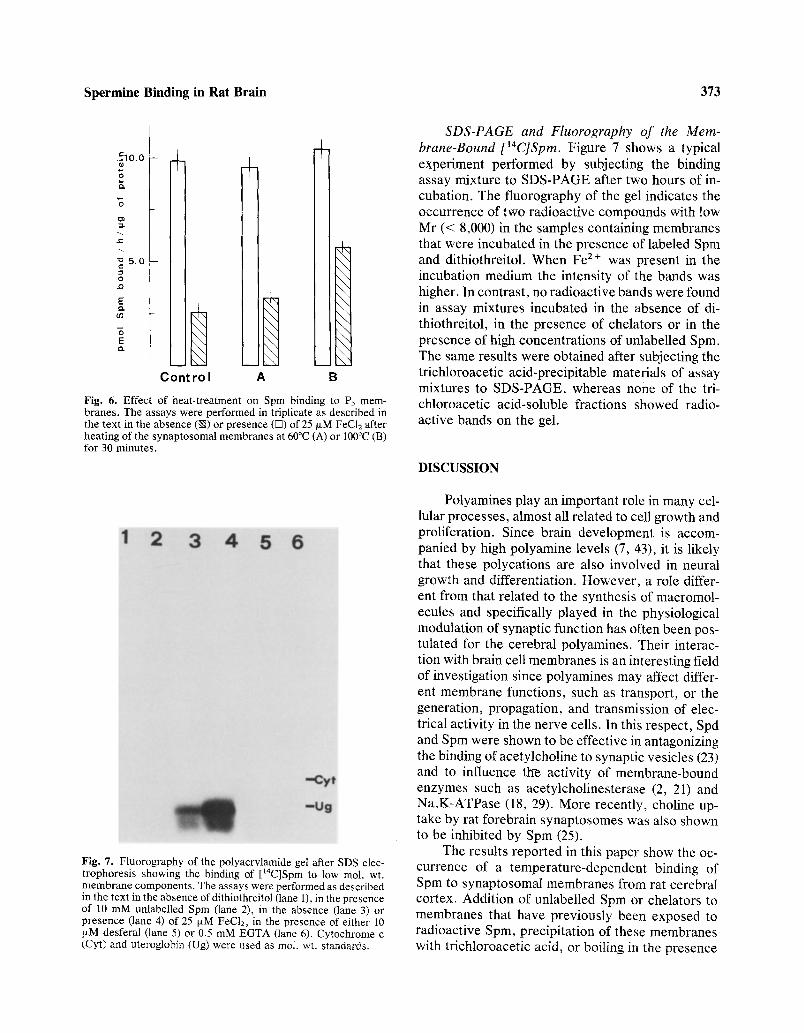
Figure 6 shows the effect of the heat treatment of the P3 membrane fraction on Spm binding. Ex- posure of the membranes to a temperature of 60~ or 100~ for 30 min before the binding assay did not affect the Fe2§ binding of the mem- branes, whereas the binding that occurs in the ab- sence of Fe 2 + was positively influenced.
Preincubation of the membranes with either trypsin or pronase for different times (1-18 h) and with different amounts of enzyme (1 : 100, 1 : 10, 1/1, w/w with respect to membrane proteins), did not affect Spm binding significantly (data not shown).
9 . 0
,_=
o
~- 6 , 0
x: 3 . 0
3 E
6.0 7 . 0 8 . 0
p H
Fig. 5. Effect of pH on the spermine binding. The assays were performed in 50 mM Tris-HCl buffer in triplicate as described in the text, using synaptosomal membranes (10 txg of protein) in the presence (e) or absence (O) of 25 ixM FeCI~.
Spermine Binding in Rat Brain 373
.51o.o
Q.
~ 5 . 0
o e~
E O~
E e~
+ +
Contro l A
+
\ \
\ \
\ \
\ \
\ \
\ \
\ \
\ \
\ \
\ \
\ \
\ \
B
Fig. 6. Effect of heat-treatment on Spm binding to P~ mem- branes. The assays were performed in triplicate as described in the text in the absence (k~) or presence ([]) of 25 I~M FeC12 after heating of the synaptosomal membranes at 60~ (A) or 100~ (B) for 30 minutes.
Fig. 7. Fluorography of the polyacrylamide gel after SDS elec- trophoresis showing the binding of [t4C]Spm to low mol. wt. membrane components. The assays were performed as described in the text in the absence of dithiothreitol (lane 1), in the presence of 10 mM unlabelled Spin (lane 2), in the absence (lane 3) or presence (lane 4) of 25 txM FeC12, in the presence of either 10 ixM desferal (lane 5) or 0.5 mM EGTA (lane 6). Cytochrome c (Cyt) and uteroglobin (Ug) were used as tool. wt. standards.
SDS-PAGE and Fluorography of the Mem- brane-Bound [14C]Spm. Figure 7 shows a typical experiment performed by subjecting the binding assay mixture to SDS-PAGE after two hours of in- cubation. The fluorography of the gel indicates the occurrence of two radioactive compounds with low Mr (< 8,000) in the samples containing membranes that were incubated in the presence of labeled Spin and dithiothreitol. When Fe 2+ was present in the incubation medium the intensity of the bands was higher. In contrast, no radioactive bands were found in assay mixtures incubated in the absence of di- thiothreitol, in the presence of chelators or in the presence of high concentrations of unlabelled Spm. The same results were obtained after subjecting the trichloroacetic acid-precipitable materials of assay mixtures to SDS-PAGE, whereas none of the tri- chloroacetic acid-soluble fractions showed radio- active bands on the gel.
DISCUSSION
Polyamines play an important role in many cel- lular processes, almost all related to cell growth and proliferation. Since brain development is accom- panied by high polyamine levels (7, 43), it is likely that these polycations are also involved in neural growth and differentiation. However, a role differ- ent from that related to the synthesis of macromol- ecules and specifically played in the physiological modulation of synaptic function has often been pos- tulated for the cerebral polyamines. Their interac- tion with brain cell membranes is an interesting field of investigation since polyamines may affect differ- ent membrane functions, such as transport, or the generation, propagation, and transmission of elec- trical activity in the nerve cells. In this respect, Spd and Spm were shown to be effective in antagonizing the binding of acetylcholine to synaptic vesicles (23) and to influence the activity of membrane-bound enzymes such as acetylcholinesterase (2, 21) and Na,K-ATPase (18, 29). More recently, choline up- take by rat forebrain synaptosomes was also shown to be inhibited by Spm (25).
The results reported in this paper show the oc- currence of a temperature-dependent binding of Spm to synaptosomal membranes from rat cerebral cortex. Addition of unlabelled Spin or chelators to membranes that have previously been exposed to radioactive Spin, precipitation of these membranes with trichloroacetic acid, or boiling in the presence

374 Gentile, Esposito, Fusco, Popoli, and Porta
of detergents and reducing agents proved to be in- effective to displace the polyamine from its binding sites. These observations, together with the fact that at least two radioactive bands are observed follow- ing electrophoretic separation of incubated binding assay mixtures, prompted us to do a preliminary analysis of the possibility of enzymatic formation of a covalent linkage between Spin and the membrane proteins. There are several possibilities how amines may be covalently bound. It is well known that pu- trescine, Spd, and Spm serve as acyl acceptor sub- strates in both protein labelling and cross-linking reactions catalyzed by transglutaminases (9, 13, 27, 47). Moreover, Park et al. (32) demonstrated that the 4-amino-2-hydroxybutyl portion of the protein- bound hypusine (42) derives from enzymatic trans- formations of the butylamine moiety of Spd. Thomas et al. (46) pointed out the possibility that the myeloperoxidases are also able to catalyze the incorporation of primary amines into proteins in vitro by first forming mono- and dichloroamine de- rivatives. Covalent incorporation of a labeled ma- terial originating from amines may also occur in the presence of amine oxidases; the derived aldehyde is able to produce Shift bases by reacting with pri- mary amino groups of some amino acid residues, like e-amino group of lysine, contained in proteins. Our experiments show that Spm binding was af- fected neither by heating the membranes at 100~ for 30 min. nor by incubation with trypsin and pron- ase. This indicates that the tenacious bond between the polyamine and the synaptosomal membrane is not enzymatically formed. The positive influence of the incubation temperature on the Spm binding could be explained by assuming a temperature-sen- sitive unmasking of specific reactive groups in the membrane.
Further studies showed that the binding occurs only in the presence of - S H reducing agents (di- thiothreitol or [3-mercaptoethanol), is markedly stimulated by Fe 2+, and is completely suppressed by metal chelators. The strict dependence of the binding on the presence of dithiothreitol (or [3-mer- captoethanol) in the assay medium suggests a pos- sible involvement of one or more - S H groups in the binding site. In this respect, the possibility of the existence of reactive y-glutamyl thioester bonds in some synaptosomal membrane peptides that could non-enzymatically react with the primary amino group of Spm, as occurs with methylamine in the case of oL2-macroglobulin (5) and the third and
fourth components of human complement (45), should not be ruled out.
The activation of the binding by iron allows us to hypothesize that the formation of a Spm-metal complex could be important as a step activating the entire process. The pH influence on the binding in the presence of Fe 2+ appears to be in line with this conjecture. The formation of a Spm-Fe 2+ complex should be facilitated by increasing the pH of the medium to values close to the pK of the polyamine primary amino groups.
The other hypothesis worth considering is that iron may be incorporated in the membrane binding site and thus confer a special conformation to it. This metal is an important constituent of the lipid- protein bilayer in biological membranes. The lower binding activity observed in the absence o f iron could be due to the loss of a certain amount of the metal during the tissue manipulation.
Concerning the possible physiological signifi- cance of the activating effect of iron, it should be considered that (i) iron is the most abundant trace metal in the body, (ii) its distribution in the brain is highly localized (19, 49), and (iii) the major portion of the cerebral non-heme iron is associated with the crude mitochondrial-synaptosomal fraction and myelin (35, 48). Moreover, although the functional role of brain iron is not known, its excess accu- mulation has been associated with a number of neu- rological and psychiatric disorders (10, 12, 15, 20, 44), whereas the nutritional iron-deficiency was shown to affect behavior (33, 48) and to cause a significant and apparently selective reduction in the brain dopamine D2 receptors which were restored after iron repletion therapy (4). Our studies are pres- ently addressed to determining the regional distri- bution pattern of the Spm binding sites in rat brain and to examining their possible reduction in iron- deficient animals.
ACKNOWLEDGMENTS
This research was supported by grants from the Italian Min- istry of Public Education (M.P.I. 40 percent-1985). We are grate- ful to J. Sepe for his valuable assistance.
REFERENCES
1. Ambron, R. T., and Kremzner, L. T. 1982. Post-translational modification of neuronal proteins: evidence for transglutam-

Spermine Binding in Rat Brain 375
inase activity in R2, the giant cholinergic neuron of Aplysia, Proc. Natl. Acad. Sci. USA 79:3442-3446.
2. Anand, R., Gore, M. G., and Kerkut, G. A. 1976. The effect of spermine and spermidine on the hydrolysis of acetylcho- line in the presence of rat candate nucleus homogenate or acetylcholinesterase from Electrophorus electricus. J. Neu- rochem. 27:381-386.
3. Anderson, D. J., Crossland, J., and Shaw, G. G. 1975. The actions of spermidine and spermine on the central nervous system. Neuropharmacology 14:571-577.
4. Ashkenazi, R., Ben-Shachar, D., and Youdim, M. B. H. 1982. Nutritional iron and dopamine binding sites in the rat brain. Pharmacol. Biochem. Behav. 17:S1, 43-47.
5. Barret, A. J. 1981. az-Macroglobulin. Meth. Enzymol. 80:737-755.
6. Bonner, W. M., and Laskey, R. A. 1974. A film detection method for tritium-labelled proteins and nucleic acids in poly- acrylamide ges. Eur. J. Biochem. 46:83-88.
7. Caldarera, C. M., Moruzzi, M. S., Rossoni, C., and Barbiroli C. 1969. Polyamines and nucleic acid metabolism during de- velopment of chick embrio brain. J. Neurochem. 16:309-316.
8. Chen K. Y. 1984. Transglutaminase catalyzed incorporation of putrescine into surface proteins of mouse neuroblastoma cells. Mol. Cell. Biochem. 58:91-97.
9. Clarke, D. D., Mycek, M. J., Neidle, A., and Waelsch, H. 1959. The incorporation of amines into protein. Arch. Biochem. Biophys. 79:338-354.
10. Courvflle, C. B., Nusbaum, R. E., and Butt, E. M. 1963. Changes in trace metals in brain in Huntington's Chorea. Arch. Neurol. 8:481-489.
11. De Lorenzo, R. J., and Freedman, S. D. 1977. Calcium-de- pendent phosphorylation of synaptic vesicle protein and its possible role in mediating neurotransmitter release and ves- icle function. Biochem. Biophys. Res. Commun. 77:1036- 1043.
12. Dooling, E. C., Snijdewint, G. M., and Buijis, R. M. 1974. Hallenvorden-Spatz syndrom. Arch. Neurol. 30:70-83.
13. Folk, J. E., Park, M. H., Chung, S. I., Schrode, J., Lester, E. P., and Cooper, H. L. 1980. Polyamines as physiological substrates for transglutaminases. J. Biol. Chem. 255:3695- 3700.
14. Gilad, M. G., and Varon L. E. 1985. Transglutaminase ac- tivity in rat brain: characterization, distribution, and changes with age. J. Neurochem. 45:1522-1526.
15. Goodman, L. 1953. Alzheimer's disease. A clinico-patho- logic of twenty-three eases with a theory on pathogenesis. J. Nerv. Ment. Dis., 117:97-103.
16. Hajos, F. 1975. An improved method for the preparation of synaptosomal fraction in high purity. Brain Res. 93:485-489.
17. Harman, R. J., and Shaw, G. G. 1981. High-affinity uptake of spermine by slices of rat cerebral cortex. J. Neurochem. 36:1609-1615.
I8. Heinrich-Hirsch, B., Ahlers, J., and Peter, H. W. 1977. In- hibition of Na,K-ATPase from chick brain by polyamines. Enzyme 22:235-241.
19. Hill J. M., and Switzer, R. C. 1984. The regional distribution and cellular localization of iron in the rat brain. Neuroscience 11:595-603.
20. Hsiang, M. S. 1941. Brain pathology of Kaschin-Becks dis- ease. J. Orient. Med. 33:119-162.
21. Kossorotow, A., Wolf, H. U., and Seiler, N. 1974. Regu- latory effects of polyamines on membrane-bound acetylcho- linesterase. Biochem. J. 144:21-27.
22. Kremzner, L. T., and Ambron, R. T. 1982. Metabolism and axonal transport of polyamines in a single identified neuron of Aplysia Californica. J. Neurochem. 38:1719-1727.
23. Kuriyama, K., Roberts, E., and Vos, J. 1968. Some char- acteristics of binding of ~/-aminobutyric acid and acetylcho-
line to a synaptic vesicle fraction from mouse brain. Brain Res. 9:231-252.
24. Laemmli, U. K. 1970. Cleavage of structural proteins during assembly of the head of bacteriophage T4. Nature 227:680- 685.
25. Law, C. L., Wong, P. C. L., and Fong, W. F. 1984. Effects of polyamines on the uptake of neurotransmitters by rat brain synaptosomes. J. Neurochem. 42:870-872.
26. Lindquist, T. D., Sturman, J. A., Gould, R. M., and Ingoglia, N. A. 1985. Axonal transport of polyamines in intact and regenerating axons of the rat sciatic nerve. J. Neurochem. 44:1913-1919.
27. Lorand, L., and Conrad, S. M. 1984. Transglutaminases. Mol. Cell Biochem. 58:9-35.
28. Lowry, O. H., Rosebrough, N. J., Farr, A. L., and Randall, R. J. 1951. Protein measurement with the Folin phenol re- agent. J. Biol. Chem. 193:265-275.
29. Nag, D., and Ghosh, J. J. 1973. Imipramine-induced changes of brain adenosine triphosphatase activity: the role of sperm- ine in counteracting the disorganizing effect of the drug on membrane ATPase. J. Neurochem 20:1021-1027.
30. Nieto, A., Ponstigl, H., and Beato, M. 1977. Purification and quaternary structure of the hormonally induced protein uter- oglobin. Arch. Biochem. Biophys. 180:82-92.
31. Nistic6, G., Ientile, R., Rotiroti, D., and Di Giorgio, R. M. 1980. GABA depletion and behavioural changes produced by intraventricular putrescine in chicks. Biochem. Pharma- col. 29:954-957.
32. Park, M. H., Cooper, H. L., and Folk, J. E. 1981. Identi- fication of hypusine, an unusual amino acid, in a protein from human lymphocytes and of spermidine as its biosynthetic precursor. Proc. Natl. Acad. Sci. USA 78:2869-2873.
33. Pollit, E., and Leibel, R. L. 1976. Iron deficiency and be- haviour. J. Pediatr. 88:372-381.
34. Porta, R., Schatz, R. A., Tatter, S. B., and Sellinger, O. Z. 1983. The biosynthesis of polyamines in mouse brain: effects of methionine sulfoximine and adenosylhomocysteine. J. Neurochem. 40:836-841.
35. Rajan, K. S., Colburn, R. W., and Davis, J. M. 1976. Dis- tribution of metal ions in the subcellular fractions of several rat brain areas. Life Sci. 18:423-431.
36. Sakurada, T., and Kisara, K. 1978. Effects of intraventric- ularly administered polyamines spermidine and spermine on sleepwakefulness cycles in rats. Jap. J. Pharmacol. 28:125- 132.
37. Seller, N. and Deckardt, K. 1976. Association of putrescine, spermidine, spermine and GABA with structural elements of brain cells. Neurochem. Res. 1:469-499.
38. Seiler, N.,andDeckardt, K. 1978. Associationofpolyamines and GABA with structural elements of brain cells, pages 145- 159 in R. A., Campbell, D. R. Morris, D. Bartos, G. D. Daves G. Jr. and F. Bartos (eds.) Advances in Polyamine Research, Vol. 2, Raven Press, New York.
39. Seiler, N. 1981. Polyamine metabolism and function in brain. Neurochem. Int. 3:95-110.
40. Selkoe, D. J., Abraham, C., and Ihara, Y. 1982. Brain trans- glutaminase: in vitro crosslinking of human neurofilament proteins into insoluble polymers. Proc. Natl. Acad. Sci. USA 79:6070-6074.
41. Shaw, G. G. 1979. The polyamines in the nervous system. Biochem. Pharmacol. 28:1-6.
42. Shiba T., Mizote H., Kaneko T., Nakajima T., Kakimoto T., and Sano I. 1971. Hypusine, a new amino acid occurring in bovine brain: isolation and structural determination. Biochim. Biophys. Acta 244:523-531.
43. Shimizu, H., Kakimoto, Y., and Sano, I. 1965. Changes in concentrations of polyamines in developing mouse brain. Na- ture, Lond. 207:1196.
376 Gentile, Esposito, Fusco, Popoli, and Porta
44. Szanto, J., and Gallijas, F. 1955. A study of iron metabolism in neuropsychiatric patient. Arch. Neurol. 14:438-442.
45. Tack, B. F., Janatova, J., Thomas, M. L., Harrison, R. A., and Hammer, C. H. 1981. The third, fourth, and fifth com- ponents of human complement: isolation and biochemical properties. Meth. Enzymol. 80:64-101.
46. Thomas, E. L., Jefferson, M. M., and Grisham, M. B. 1982. Myeloperoxidase-catalyzed incorporation of amines into proteins: role of hypochlorous acid and dichloroamines. Bio- chemistry 21:6299-6308.
47. Williams-Ashman, H. G., and Canellakis, Z. N. 1980. Trans- glutaminase-mediated covalent attachment of polyamines to
proteins: mechanism and potential physiological signifi- cance. Physiol. Chem. Physics 12:457-472.
48. Youdim, M. B. H., and Green, A. R. 1977. Biogenic mon- oamine metabolism and functional activity in iron-deficient rats: behavioural correlates, pages 201-225 in R., Porter and D. W., Fitzsimons (eds), Iron Metabolism, Elsevier, Am- sterdam.
49. Youdim, M. B. H. 1985. Brain iron metabolism:-biochemical and behavioural aspects in relation to dopaminergic neuro- transmission, pages 731-755 in A., Lajtha (ed.), Handbook of Neurochemistry, vol. 10 Plenum Press, New York.