Embed Size (px)
Citation preview
European Journal of Neuroscience, Vol. 9, pp. 1530-1535, 1997 0 European Neuroscience Association
SHORT COMMUNICATION Neurons in the Rat Occipital Cortex Co-expressing the Substance P-Receptor and GABA: a Comparison Between In Wvo and Organotypic Cultures
D. Echevarrial, C. Matute2 and K. Albus’ ’Department of Neurobiology, Max Planck Institute for Biophysical Chemistry, PO Box 2841, D-37070 Gottingen, Germany 2Departamento de Neurociencias, Universidad del Pais Vasco, E-48940 Leioa, Spain
Keywords: colocalizations, immunohistochemistry, inhibitory interneurons, neocortex, tachykinin receptor
Abstract
The morphology and the distribution of neurons expressing the NK1 -receptor (NKl R) and the co-expression of y-aminobutyric acid (GABA) in these neurons were studied in the rat occipital cortex and in organotypic cultures (OTCs) derived from this structure. By employing immunohistochemistry, we demonstrate that the NK1 R- expressing neurons are non-pyramidal neurons and co-express GABA. Some differences were noted between in vivo and OTCs. NK1 R-expressing neurons in OTCs had larger somata and longer dendrites and the proportion stained with an anti-GABA-antibody (-50%) was smaller than in vivo (90%). The preferential location of NK1 R-expressing neurons in layers 11/111 and VI, seen in vivo is not present in OTCs where these neurons distribute rather homogeneously through layers Il-VI. Our findings imply that in contrast to the cat and monkey, in the rat occipital cortex the effects of substance P are almost exclusively mediated via inhibitory interneurons.
Introduction
The tachykinin peptide family whose members are known to be present in the neocortex comprise substance P (SP), neurokinin A (NKA), neuropeptide K (NPK), neuropeptide y and neurokinin B (NKB) (Arai and Emson, 1986; Takeda et al., 1990). The tachykinin peptides interact differentially with three tachykinin receptors: the NK1-receptor (NKIR) prefers interaction with SP, the NK2-receptor with NKA, NPK and neuropeptide y and the NK3-receptor with NKB (Helke et al., 1990). Radioimmunoassay and binding studies have demonstrated the presence of these receptors in the cortex (Dam et al., 1988). The presence of mRNA coding for functional NKlR in the visual cortex has been demonstrated by means of the oocyte expression system (Matute et al., 1993a). By means of microionto- phoretic techniques it has been shown that in the cortex exogenous tachykinins depolarize, i.e. excite, neurons (for review see Albus et al., 1992). Tachykinin-induced suppressions, or hyperpolarizations, were attributed to an indirect action of the tachykinins mediated via inhibitory interneurons (Olpe et al. 1987; Albus et al., 1992).
Recent investigations employing in situ hybridization histo- chemistry (Maeno et al., 1993; Matute et al., 1993b) or immunohisto- chemistry (Liu et al., 1994; Nakaya et al., 1994) have revealed species differences in the morphology and laminar distribution of neurons expressing the NKlR. In the cat visual cortex most of these neurons are pyramidal cells in layers 11, I11 and V; in the rat neocortex they are non-pyramidal neurons which distribute through all layers. In
order to substantiate and extend the latter findings we have studied the morphology, transmitter identity and intracortical location of neurons expressing the NKlR in the rat occipital cortex. In addition, as a first step to analyse NKIR-receptor functions at a cellular level, we wondered whether the characteristic properties of NKlR- expressing neurons are maintained in organotypic cultures (OTCs) of the rat occipital cortex. Due to a high degree of accessibility these long term culture systems have been claimed to be particularly well suited to study basic cellular mechanisms of neuronal functions (Giihwiler, 1981).
Materials and methods
Sprague-Dawley rats and Wistar rats (240-260 g) were deeply anaesthetized with sodium pentobarbitone (60 mgkg) and perfused through the ascending aorta with PBS (145 mM NaCl, 10 mh4 sodium phosphate, pH 7.4), followed by a solution of 4% paraformaldehyde and 0.1% glutaraldehyde in PBS. After perfusion, the brain was removed and postfixed in 4% paraformaldehyde in PBS for a period of 2-6 h. Vibratome sections of neocortex were cut at 45-50 pm thick.
OTCs of the occipital cortex of rats (Wistar) were prepared and maintained in vitro by employing the protocol described by Giihwiler (1981). Culture tubes were supplied with 0.75 ml semi-artificial medium (50% Eagle’s basal medium, 25% Hank’s balanced salt
Correspondence to: D. Echevm’a, as above
Received 20 January 1997, revised 4 February 1997, accepted 12 February I997
GABA and the NK1 receptor in neocortical neurons 1531
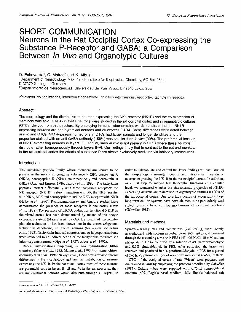
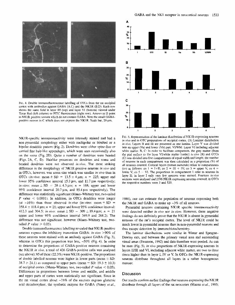
FIG. 1. Morphology of non-pyramidal neurons in the rat occipital cortex expressing the NKlR. Intensely stained neurons and weakly stained neurons (arrowheads) in layers 111 (A), V (B), I1 (C) and VI and adjacent white matter (D). Open arrows point toward cortical surface. Scale bar, 50 pm.
solution, 25% inactivated horse serum, 1 mM L-glutamine, 0.65% D-
glucose and 1.26mM Ca2+). All procedures were carried out in sterile conditions. The medium was changed every 4-5 days. For immunohistochemical experiments OTCs about 3 weeks in vitro were immersed in 4% paraformaldehyde and 0.1 % glutaraldehyde for 1 day.
For immunolocalization of the NKlR, a rabbit polyclonal NKlR antibody (Vigna etal., 1994) was used. Sections (free floating) of occipital cortex and OTCs (on coverslips) were pre-incubated with H202 in PBS for a period of 20min and then blocked over 30- 45 min in 2% normal goat serum with 0.5% Triton X-100 (or in the case of OTCs 1% Triton X-100) in PBS. Next, they were incubated overnight at 4°C with the NKlR primary antibody diluted 1500.
After washing thoroughly with PBS, a biotinylated goat anti-rabbit (dilution 1:200) secondary antibody was added for 60 min and later incubated for 60 min in avidin-biotin-peroxidase complex (Vectas- tain, Burlingame, CA). The immunoreaction product was visualized with diaminobenzidine and Hz02. Sections and cultures were then washed in PBS, mounted onto gelatinized slides, dehydrated and coverslipped.
For the colocalization experiments, a double immunofluorescence staining method was performed. Following the preincubation and blocking procedure (see above), sections and OTCs were incubated overnight with the NKlR primary antibody at 1:500 dilution. Labelling was achieved by using a fluorescein isothiocyanate-labelled goat anti-
1532 GABA and the NKl receptor in neocortical neurons
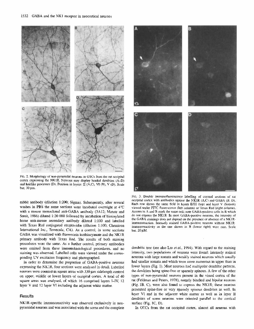
FIG. 2. Morphology of non-pyramidal neurons in OTCs from the rat occipital cortex expressing the NKlR. Neurons may display beaded dendrites (A-D) and hairlike processes (D). Position in layers: I1 (A,C), VI (B), V (D). Scale bar, 30 pm.
rabbit antibody (dilution 1 :ZOO; Sigma). Subsequently, after several washes in PBS the same sections were incubated overnight at 4°C with a mouse monoclonal anti-GABA antibody (3A12; Matute and Streit, 1986) diluted 1:20 OOO followed by incubation of biotinylated horse anti-mouse secondary antibody diluted 1: 100 and labelled with Texas Red conjugated streptavidin (dilution 1: 100; Chemicon International Inc., Temecula, CA). As a control, in some sections GABA was visualized with fluorescein isothiocyanate and the NKlR primary antibody with Texas Red. The results of both staining procedures were the same. As a further control, primary antibodies were omitted from these immunohistological procedures, and no staining was observed. Labelled cells were viewed under the corres- ponding UV excitation frequency and photographed.
In order to determine the proportion of GABA-positive neurons expressing the NKlR, four sections were analysed in detail. Labelled neurons were counted in square areas with 330 pm sidelength centred on upper, middle or lower layers of occipital cortex. A total of 40 square areas was analysed, of which 16 comprised layers I-IV, 12 layer V and 12 layer VI including the adjacent white matter.
Results NKlR-specific immunoreactivity was observed exclusively in non- pyramidal neurons and was associated with the soma and the complete
FIG. 3. Double immunofluorescence labelling of coronal sections of rat occipital cortex with antibodies against the NKlR (A,C) and GABA (B, D). Each row shows the same field in layers II/III (top) and layer V (bottom) viewed under FITC fluorescence (left column) or Texas Red (right column). Arrows in A and B mark the same cell; note GABAtpositive cells in B which do not express the NKlR. In most GABA-positive neurons, the intensity of the GABA stainings does not depend on the presence or absence of a NKlR- immunoreaction. Intensely stained GABA-positive neurons without NKlR- immunoreactivity as the one shown in B (lower right) were rare. Scale bar, 20 pM.
dendritic tree (see also Liu et al., 1994). With regard to the staining intensity, two populations of neurons were found: intensely stained neurons with large somata and weakly stained neurons which usually had smaller somata and which were more numerous in upper than in lower layers (Fig. 1). Most neurons had multipolar dendritic patterns, the dendrites being spine-free or sparsely spinous. A few of the other types of non-pyramidal neurons present in the visual cortex of the rat (Feldman and Peters, 1978), namely bitufted and bipolar neurons (Fig. lB, C), were also found to express the NKlR; these neurons presented spine-free or very sparsely spinous dendrites as well. In layer VI and in the adjacent white matter as well as in layer I1 dendrites of some neurons were oriented parallel to the cortical surface (Fig. lC, D).
In OTCs from the rat occipital cortex, almost all neurons with
GABA and the NK1 receptor in neocortical neurons 1533
I Illlll IV Va Vb VIWM
B
10
1 2 3 4 5
C
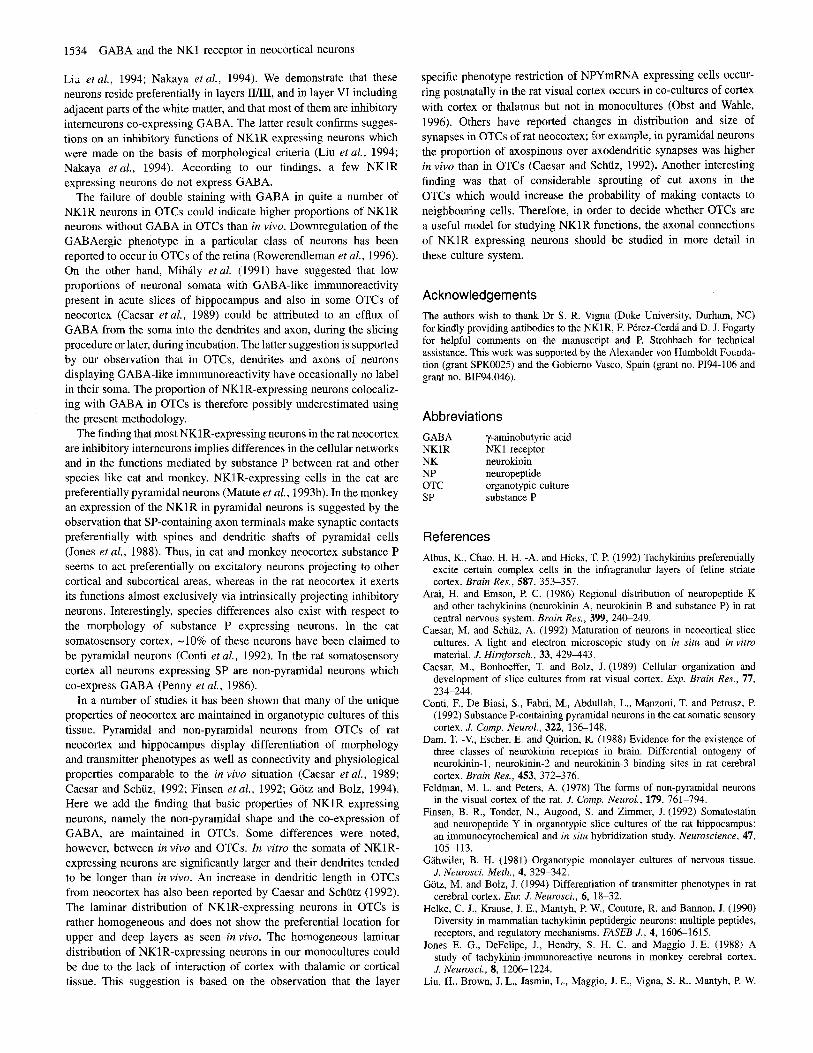
’* T FIG. 4. Double immunofluorescence labelling of OTCs from the rat occipital cortex with antibodies against GABA (A.C) and the NKlR (B,D). Each row shows the same field inlayer I11 (top) and layer VI (bottom) viewed under Texas Red (left column) or FITC fluorescence (right row). Arrows in D point to NKlR-positive somata which do not contain GABA. Note the small GABA- positive neuron in C which does not express the NKlR. Scale bar, 20 pm.
NKlR-specific immunoreactivity were intensely stained and had a non-pyramidal morphology either with multipolar or bitufted or a bipolar dendritic pattern (Fig. 2). Dendrites were either spine-free or carried fine hair-like appendages, which were seen occasionally also on the soma (Fig. 2D). Quite a number of dendrites were beaded (Figs. 2A, C, D). Hairlike processes on dendrites and soma and beaded dendrites were not observed in vivo. The most striking difference in the morphology of NKlR-positive neurons in vivo and in OTCs, however, was soma size which was smaller in vivo than in OTCs (in vivo: mean 5 SD = 13.5 5 4 pm; n = 225; upper and lower 95% confidence interval 15.3 pm, and 11.7 pm respectively; in vitro: mean i SD = 20 ? 4.3 Fm; n = 168; upper and lower 95% confidence interval 20.7 Fm, and 19.4 pm respectively). The difference was statistically significant (Mann-Whitney test, two-tailed P value < 0.0001). In addition, in OTCs dendrites were longer (at -16%) than those observed in vivo (in vitro: mean -f SD = 358.4 % 118.4 pm; n = 21; upper and lower 95% confidence interval: 412.3 and 304.5; in vivo: mean -C SD = 309 2 89.4pm; n = 21 upper and lower 95% confidence interval 349.9 and 268.2). The difference was not significant, however (Mann-Whitney test, two- tailed P value > 0.05).
Double immunofluorescence labelling revealed that NKl R-positive neurons express the inhibitory transmitter GABA: in vivo >90% of these neurons were stained with an antibody against GABA (Fig. 3), whereas in OTCs this proportion was less, -50% (Fig. 4). In order to determine the proportions of GABA-positive neurons containing the NKlR in vivo, a total of 426 GABA-positive cells was analysed (see above); 95 of these (22.3%) were NKlR-positive. The proportions of double labelled neurons were higher in lower parts (mean 2 SD: 38.7 2 24.1) as compared to upper parts (mean % SD: 19.3 ? 10.3) of occipital cortex (Mann-Whitney test, two-tailed P value < 0.05). Differences in proportions between lower and middle, and middle and upper parts of cortex were statistically not significant. Since in the rat visual cortex about -16% of the neurons express glutamic acid decarboxylase, the synthetic enzyme for GABA (Penny et aL,
1 2 3 4 5
FIG. 5. Representation of the laminar distribution of NKlR-expressing neurons in vivo and in OTC preparations of occipital cortex. (A) Laminar distribution in vivo. Layers I1 and I11 are presented as one lamina. Layer V was divided into an upper (Va) and lower (Vb) part. V W M : Layer VI including adjacent white matter. B, C: In order to facilitate comparison, the grey matter (from the pial surface to the layer VVwhite matter border) in vivo (B) and OTCs (C) was divided into five compartments of equal width and length; the number of neurons in each compartment was then calculated as a proportion (%) of all neurons counted. Cortical layers (roman numerals) relate to compartments (co) as follows: co 1 = I f I I ; co 2 = I11 + IV, co 3 = upper V, co 4 = lower V; co 5 = VI. The proportions in compartment 1 refer to neurons in layer 11; in layer I only very few neurons were stained. Fourteen in vivo sections were analysed and 2196 NKlR-expressing neurons counted; in OTCs the respective numbers were 3 and 520.
1986), one can estimate the population of neurons expressing both the NKlR and GABA to make up -3% of all neurons.
Pyramidal neurons containing NKlR specific immunoreactivity were detected neither in vivo nor in vitro. However, these negative findings do not definitely prove that the NKlR is absent in pyramidal neurons of the rat’s occipital cortex. The level of NKlR could be much lower in pyramidal neurons than in non-pyramidal neurons and thus escape detection by immunohistochemistry.
The laminar distributions were similar in Wistar and Sprague- Dawley rats, and between the primary visual area and surrounding visual areas (Swanson, 1992) and data therefore were pooled. As can be seen (Fig. 5), in vivo proportions of NKlR-expressing neurons in layers IVIII and VI, including adjacent white matter, are two to three times higher than in layer I, IV or V. In OTCs the NKlR-expressing neurons distribute throughout all layers in a rather homogenous manner.
Discussion
Our results confirm earlier findings that neurons expressing the NKlR distribute through all layers of the rat neocortex (Maeno et al., 1993;
1534 GABA and the NKl receptor in neocortical neurons
Liz etal., 1994; Nakaya etal., 1994). We demonstrate that these neurons reside preferentially in layers IUIII, and in layer VI including adjacent parts of the white matter, and that most of them are inhibitory interneurons co-expressing GABA. The latter result confirms sugges- tions on an inhibitory functions of NKlR expressing neurons which were made on the basis of morphological criteria (Liu et aZ., 1994; Nakaya etal., 1994). According to our findings, a few NKlR expressing neurons do not express GABA.
The failure of double staining with GABA in quite a number of NKlR neurons in OTCs could indicate higher proportions of NKlR neurons without GABA in OTCs than in vivo. Downregulation of the GABAergic phenotype in a particular class of neurons has been reported to occur in OTCs of the retina (Rowerendleman et al., 1996). On the other hand, Mihily et al. (1991) have suggested that low proportions of neuronal somata with GABA-like immunoreactivity present in acute slices of hippocampus and also in some OTCs of neocortex (Caesar et al., 1989) could be attributed to an efflux of GABA from the soma into the dendrites and axon, during the slicing procedure or later, during incubation. The latter suggestion is supported by our observation that in OTCs, dendrites and axons of neurons displaying GABA-like immmunoreactivity have occasionally no label in their soma. The proportion of NKIR-expressing neurons colocaliz- ing with GABA in OTCs is therefore possibly underestimated using the present methodology.
The finding that most NKlR-expressing neurons in the rat neocortex are inhibitory interneurons implies differences in the cellular networks and in the functions mediated by substance P between rat and other species like cat and monkey. NKlR-expressing cells in the cat are preferentially pyramidal neurons (Matute et al., 1993b). In the monkey an expression of the NKlR in pyramidal neurons is suggested by the observation that SP-containing axon terminals make synaptic contacts preferentially with spines and dendritic shafts of pyramidal cells (Jones et al., 1988). Thus, in cat and monkey neocortex substance P seems to act preferentially on excitatory neurons projecting to other cortical and subcortical areas, whereas in the rat neocortex it exerts its functions almost exclusively via intrinsically projecting inhibitory neurons. Interestingly, species differences also exist with respect to the morphology of substance P expressing neurons. In the cat somatosensory cortex, -10% of these neurons have been claimed to be pyramidal neurons (Conti et al., 1992). In the rat somatosensory cortex all neurons expressing SP are non-pyramidal neurons which co-express GABA (Penny et al., 1986).
In a number of studies it has been shown that many of the unique properties of neocortex are maintained in organotypic cultures of this tissue. Pyramidal and non-pyramidal neurons from OTCs of rat neocortex and hippocampus display differentiation of morphology and transmitter phenotypes as well as connectivity and physiological properties comparable to the in vivo situation (Caesar et al., 1989; Caesar and Schuz, 1992; Finsen et al., 1992; Gotz and Bolz, 1994). Here we add the finding that basic properties of NKlR expressing neurons, namely the non-pyramidal shape and the co-expression of GABA, are maintained in OTCs. Some differences were noted, however, between in vivo and OTCs. In vitro the somata of NKlR- expressing neurons are significantly larger and their dendrites tended to be longer than in vivo. An increase in dendritic length in OTCs from neocortex has also been reported by Caesar and Schutz (1992). The laminar distribution of NKlR-expressing neurons in OTCs is rather homogeneous and does not show the preferential location for upper and deep layers as seen invivo. The homogeneous laminar distribution of NKl R-expressing neurons in our monocultures could be due to the lack of interaction of cortex with thalamic or cortical tissue. This suggestion is based on the observation that the layer
specific phenotype restriction of NPYmRNA expressing cells occur- ring postnatally in the rat visual cortex occurs in co-cultures of cortex with cortex or thalamus but not in monocultures (Obst and Wahle, 1996). Others have reported changes in distribution and size of synapses in OTCs of rat neocortex; for example, in pyramidal neurons the proportion of axospinous over axodendritic synapses was higher in vivo than in OTCs (Caesar and Schuz, 1992). Another interesting finding was that of considerable sprouting of cut axons in the OTCs which would increase the probability of making contacts to neighbouring cells. Therefore, in order to decide whether OTCs are a useful model for studying NKlR functions, the axonal connections of NKlR expressing neurons should be studied in more detail in these culture system.
Acknowledgements The authors wish to thank Dr S. R. Vigna (Duke University, Durham, NC) for kindly providing antibodies to the NKlR, F. P6rez-CerdB and D. J. Fogarty for helpful comments on the manuscript and P. Strohbach for technical assistance. This work was supported by the Alexander von Humboldt Founda- tion (grant SPK0025) and the Gobiemo Vasco, Spain (grant no. PI94-106 and grant no. BIF94.046).
Abbreviations GABA y-aminobutyric acid NKlR NK1 receptor NK neurokinin NP neuropeptide OTC organotypic culture SP substance P
References Albus, K., Chao, H. H -A. and Hicks, T. P. (1992) Tachylunins preferentially
excite certain complex cells in the infragranular layers of feline striate cortex. Brain Res., 587, 353-357.
Arai, H. and Emson, P. C. (1986) Regional distribution of neuropeptide K and other tachykinins (neurokinin A, neurokinin B and substance P) in rat central nervous system. Brain Rex, 399, 240-249.
Caesar, M. and Schuz, A. (1992) Maturation of neurons in neocortical slice cultures. A light and electron microscopic study on in situ and in vitro material. J. Hirnforsch., 33, 429443.
Caesar, M., Bonhoeffer, T. and Bolz, J. (1989) Cellular organization and development of slice cultures from rat visual cortex. Exp. Brain Res., 77, 234-244.
Conti, F., De Biasi, S., Fabri, M., Abdullah, L., Manzoni, T. and Petrusz, P. (1992) Substance P-containing pyranudal neurons in the cat somatic sensory cortex. J . Comp. Neurol., 322, 136148.
Dam, T. -V., Escher, E. and Quirion, R. (1988) Evidence for the existence of three classes of neurokinin receptors in brain. Differential ontogeny of neurokinin-1, neurokinin-2 and neurokinin-3 binding sites in rat cerebral cortex. Brain Res., 453, 372-376.
Feldman, M. L. and Peters, A. (1978) The forms of non-pyramidal neurons in the visual cortex of the rat. J . Conzp. Neurol., 179, 761-794.
Finsen, B. R., Tonder, N., Augood, S. and Zimmer, J. (1992) Somatostatin and neuropeptide Y in organotypic slice cultures of the rat hippocampus: an immunocytochemical and in situ hybridization study. Neuroscience, 47,
Gtihwiler, B. H. (1981) Organotypic monolayer cultures of nervous tissue. J , Neurosci. Meth., 4, 329-342.
Gotz, M. and Bolz, J. (1994) Differentiation of transmitter phenotypes in rat cerebral cortex. Eul: J. Neurosci., 6, 18-32.
Helke, C. J., Krause, J. E., Mantyh, P. W., Couture, R. and Bannon, J. (1990) Diversity in mammalian tachykinin peptidergic neurons: multiple peptides, receptors, and regulatory mechanisms. FASEB J . , 4, 1606-1615.
Jones E. G., DeFelipe, J., Hendry, S. H. C. and Maggio J. E. (1988) A study of tachykinin-immunoreactive neurons in monkey cerebral cortex. J. Neurosci., 8, 1206-1224.
Liu, H., Brown, J. L., Jasmin, L., Maggio, J. E., Vigna, S. R., Mantyh, P. W.
105-1 13.
and Basbaum, A. I. (1994) Synaptic relationship between substance P and the substance P receptor: light and electron microscopic characterization of the mismatch between neuropeptides and their receptors. Proc. Nut1 Acud.
Maeno, H., Kiyama, H. and Toyhama, M. (1993) Distribution of the substance P receptor (NK1-receptor) in the central nervous system. Mol. Bruin Res., 18, 43-58.
Matute, C. and Streit, P. (1986) Monoclonal antibodies demonstrating GABA- like immunoreactivity. Histochemistry, 86, 147-157
Matute, C., Nguyen, Q. T. and Miledi, R. (1993a) mRNAs coding for neuro transmitter receptors in rabbit and rat visual areas. J. Neurosci. Res., 35, 652-663.
Matute, C., Wahle, P., Gutikez-Igarza, K. and Albus, K. (1993b) Distribution of neurons expressing substance P receptor messenger RNA in immature and adult visual cortex. Exp. Bruin Rex, 97, 295-300.
Mihhly, A,, Erdo S. L. and Kuhnt, U. (1991) Time dependent loss of tissue GABA content and immunoreactivity in hippocampal slices. Bruin Res. Bull., 26, 559-564.
Nakaya, Y. Kaneko, T., Shigemoto, R., Nakanishi, S. and Mizuno, N. (1994) Immunohistochemical localization of the substance P receptor in the central nervous system of the adult rat. J. Comp. Neurol., 347, 249-214.
Olpe, H. R., Heid, J., Bittiger, H. and Steinmann, M. W. (1987) Substance P
SC~. USA, 91, 1009-1113.
GABA and the NKl receptor in neocortical neurons 1535
depresses neuronal activity in the rat olfactory bulb in vitro and in vivo: possible mediation via y-aminobutyric acid release. Bruin Rex, 412, 269-274.
Obst, K. and Wahle, P. (1996) Shaping the molecular phenotype of cortical interneurons: role of axonal connectivity and diffusible factors. Proc. 24th Gottingen Neurobiology Conj , Voi. 11, p. 446.
Penny, G. R., Afsharpour S. and Kitai, S. T. (1986) Substance P- immunoreactive neurons in the neocortex of the rat: a subset of glutamic acid decarboxylase-immunoreactive neurons. Neurosci. Lett., 65, 53-59.
Rowerendlemau, C., Mitchell, C. K., Haberecht, M. and Redburn, D. A. (1996) Expression and downregulation of the GABAergic phenotype in explants of cultured rabbit retina. Invest. Ophthulmol. vis. Sci., 37, 1074-1083.
Swanson, L. W. (1992) Bruin Maps: Structure of the Rut Bruin. Elsevier, Amsterdam.
Takeda, Y., Takeda, J., Smart, B. M. and Krause, J. E. (1990) Regional distribution of neuropeptide gamma and other tachykinin peptides derived from the substance P gene in the rat. Regul. Peptides, 28, 323-333.
Vigna, S. R., Bowden, J. J., McDonald, D. M., Fisher, J., Okamoto, A., McVey, D. C., Payan, D. G. and Bunnett, N. W. (1994) Characterization of antibodies to the rat substance P (NK-1) receptor and to a chuneric substance P receptor expressed in mammalian cells. J. Neurosci., 14, 834-845.