Embed Size (px)
Citation preview

APPLIED AND ENVIRONMENTAL MICROBIOLOGY, Oct. 2005, p. 6292–6307 Vol. 71, No. 100099-2240/05/$08.00�0 doi:10.1128/AEM.71.10.6292–6307.2005
Speciation of Campylobacter coli, C. jejuni, C. helveticus, C. lari,C. sputorum, and C. upsaliensis by Matrix-Assisted Laser
Desorption Ionization–Time of FlightMass Spectrometry†
Robert E. Mandrell,* Leslie A. Harden, Anna Bates, William G. Miller,William F. Haddon, and Clifton K. Fagerquist
Produce Safety and Microbiology Research Unit, USDA Agricultural Research Service, Western RegionalResearch Center, 800 Buchanan St., Albany, California 94710
Received 6 October 2004/Accepted 18 May 2005
Multiple strains of Campylobacter coli, C. jejuni, C. helveticus, C. lari, C. sputorum, and C. upsaliensis isolatedfrom animal, clinical, or food samples have been analyzed by matrix-assisted laser desorption ionization–timeof flight mass spectrometry (MALDI-TOF MS). Whole bacterial cells were harvested from colonies or confluentgrowth on agar and transferred directly into solvent and then to a spot of dried 3-methoxy-4-hydroxycinnamicacid (matrix). Multiple ions in the 5,000- to 15,000-Da mass range were evident in spectra for each strain; oneor two ions in the 9,500- to 11,000-Da range were consistently high intensity. “Species-identifying” biomarkerions (SIBIs) were evident from analyses of multiple reference strains for each of the six species, including thegenome strains C. jejuni NCTC 11168 and C. jejuni RM1221. Strains grown on nine different combinations ofmedia and atmospheres yielded SIBI masses within �5 Da with external instrument calibration. The highest-intensity C. jejuni SIBIs were cytosolic proteins, including GroES, HU/HCj, and RplL. Multiple intraspeciesSIBIs, corresponding probably to nonsynonymous nucleotide polymorphisms, also provided some intraspeciesstrain differentiation. MALDI-TOF MS analysis of 75 additional Campylobacter strains isolated from humans,poultry, swine, dogs, and cats revealed (i) associations of SIBI type with source, (ii) strains previously speciatedincorrectly, and (iii) “strains” composed of more than one species. MALDI-TOF MS provides an accurate,sensitive, and rapid method for identification of multiple Campylobacter species relevant to public health andfood safety.
Thermophilic strains of Campylobacter account for a highpercentage of the estimated 76 million yearly incidents offood-borne illness in the United States (49). Although manyoutbreaks of Campylobacter disease have been reported inthe last 25 years, most illnesses occur sporadically and arecaused by C. jejuni (51). Of the 16 species of the genusCampylobacter identified to date, at least eight have beenidentified as potential human gastrointestinal pathogens:C. jejuni, C. coli, C. lari, C. fetus, C. upsaliensis, C. sputorum,C. concisus, and C. curvus (1, 7, 15, 22, 23, 29, 37, 38, 47, 48,53, 60, 62, 64, 66). Approximately 0.1% of C. jejuni cases areassociated with a serious paralytic disease, Guillain-Barresyndrome (55). Also, C. rectus and possibly C. showae areassociated with periodontal disease (44).
There are a variety of existing assays for confirmingCampylobacter by genus and species. These include bio-chemical (2), genetic (PCR) (3, 6, 20, 32, 71), immuno-chemical (34, 36, 45), chemotaxonomic fatty acid profiling(9), protein one-dimensional gel electrophoresis methods(67) and, recently, a microarray-based method (69). Some ofthe limitations in these methods are hippuricase-negative
C. jejuni strains (24, 30, 43), the necessity for multiple hy-bridization steps and/or primers with multispecies PCR-based assays (3, 6, 20, 25, 31, 43, 59, 68, 71), specificity issueswith 16S rRNA PCR (42, 43; unpublished observations),and chemotaxonomic methods (9, 63), and only limited in-formation (e.g., species) is obtained. Details of the finespecificities of immunochemical reagents and assay methodsand comparisons of results with multiple strains of C. jejuni,C. coli, and emerging Campylobacter species have been lim-ited (34, 36, 56).
Mass spectrometric methods for analyzing whole bacterialcells for intact proteins, including the identification of proteinbiomarker ions, have been reported previously (5, 11, 16, 28,35, 70). In a preliminary study most relevant to this work,Campylobacter and Helicobacter strains were analyzed by ma-trix-assisted laser desorption–time of flight mass spectrometry(MALDI-TOF MS), and biomarker ions in the 10- to 20-kDarange (presumably proteins) were reported to be the mostdiscriminatory of those observed (73).
The major advantages of MALDI-TOF MS for analyzingbacteria are (i) ease and speed of analysis, (ii) identification ofmixed cultures, and (iii) rapid identification of candidate bio-markers, even when minimal genetic data are available. Wehave developed an expanded MALDI-TOF MS method foranalyzing multiple Campylobacter species and applied thismethod to determine species, and limited subspecies classes, of
* Corresponding author. Mailing address: USDA, ARS, WRRC,800 Buchanan St., Albany, CA 94710. Phone: (510) 559-5829. Fax:(510) 559-6165. E-mail: [email protected].
† Supplemental material for this article may be found at http://aem.asm.org/.
6292
on August 19, 2019 by guest
http://aem.asm
.org/D
ownloaded from
suspected Campylobacter strains isolated from a variety of foodand animal sources.
MATERIALS AND METHODS
Bacterial strains. The RM strain number is the designation for strains in theProduce Safety and Microbiology Research Unit strain file. Sources and otherinformation about the strains are noted in Table 1.
Bacterial isolation and culture conditions. C. coli and C. jejuni strains weremaintained on brucella agar (BA) containing, per liter, 28 g brucella broth (BDBiosciences, San Jose, CA), supplements (0.25 g FeSO4, 0.25 g sodium meta-bisulfite [anhydrous], 0.25 g sodium pyruvate [anhydrous]) and 15 g of Bacto agar(BD Biosciences) as described previously (2, 50). Other Campylobacter strainswere grown on BA and/or brain heart infusion agar medium containing 37 g ofbrain heart infusion broth medium, 6 g yeast extract, 15 g Bacto agar (BDBiosciences), and 100 ml laked horse blood (Hema Resource and Supply,Aurora, OR) per liter. All Campylobacter strains were grown under microaero-philic conditions at 37°C or 42°C by placing plates in a sealable plastic bag witha gas mixture of 10% CO2, 5% H2, and 85% N2 (microaerophilic gas) or acommercial sachet (CampyPak; Oxoid, Inc.). In one experiment to compare theeffects of different growth conditions on species-identifying biomarker ions(SIBIs), C. jejuni strain RM1221 was grown also on BA with 5% laked horseblood (BAB) and GC agar medium, which contains GC medium base and 1%Bacto agar, and supplemented with final concentrations of 0.3% glucose,0.00084% ferric nitrate (nonahydrate), 0.01% L-glutamine, 0.001% thiaminepyrophosphate, and 0.005% L-cysteine, as described previously (46, 72). Themedia were incubated in a microaerophilic atmosphere as above or in 5% CO2,5% O2, 90% N2 at either 37°C or 42°C.
Some C. upsaliensis and C. helveticus strains analyzed in this study wereisolated by a membrane filtration method modified from one described previ-ously (41). An approximately 20% suspension of cat or dog feces in saline wasincubated at 37°C for 30 min, and then four 50-�l samples were applied atdifferent locations on a 0.6-�m membrane filter (ME26; 47 mm; Schleicher &Schuell) placed on top of a BAB plate. The sample was incubated on the filterat room temperature for 15 min, and then any solution remaining was removedand the filter was removed with sterile tweezers. The plate was placed in a plasticsealable bag (Ziploc Freezer; Johnson and Son), and the bag was filled with 10%H2, 10% CO2, and 80% N2 and evacuated at least twice before final filling withgas; the bag and plate were incubated at 37°C for 48 to 72 h. Suspect Campy-lobacter colonies were subcultured on BAB for further analysis. The majority ofthe colonies were Campylobacter species.
Materials. Acetonitrile, ferrous sulfate, sodium metabisulfite, sodium pyru-vate, brucella agar, and Bacto agar were purchased from Fisher Scientific(Pittsburgh, PA). Trifluoroacetic acid (TFA) and 3-methoxy-4-hydroxycinnamicacid (ferulic acid) were purchased from Aldrich Chemical (Milwaukee, WI).Adrenocorticotropic hormone clip (18-39), angiotensin II, substance P, bovineinsulin, and horse heart myoglobin were purchased from Sigma Chemical Com-pany (St. Louis, MO). Purified water (Milli-Q system; Millipore, Bedford, MA)was used for all experiments.
Preparation of bacterial cell lysates and MALDI sample targets. Cells weretransferred from the plate to the extraction tube with a 1.0-�l disposable inoc-ulating loop (NUNC Brand Products, Denmark); this volume corresponded toapproximately 109 cells. The extraction tube was a 2.0-ml conical screw capmicrotube (QSP; catalog no. 522; Porex Bio Products Inc.) with cap and O-ring.The extraction solution contained 0.5 ml of 33% HPLC-grade acetonitrile(Fisher Scientific, Fair Lawn, NJ), 67% HPLC-grade water (Burdick & Jackson,Muskegon, MI), 0.1% sequencing-grade TFA (Sigma-Aldrich, St. Louis, MO),and �40 mg of 0.1-mm zirconia-silica beads (catalog no. 11079101z; BioSpecProducts Inc., Bartlesville, OK). Extraction tubes were then tightly capped,placed into a 96-well aluminum holder, and agitated for 60 s on a beadbeater(Mini-beadbeater-96�; BioSpec Products, Bartlesville, OK). Bead beating forlonger than 60 s was avoided to minimize sample heating due to bead friction.The solution was frothy and opaque after this step. Tubes then were centrifugedfor 4 to 5 min at 10,600 � g (model 4515C; Eppendorf, Hamburg, Germany). Theundissolved cellular material and beads at the bottom of the tube were pelletedby centrifugation, after which the supernatant was clear (bacterial cell lysate).
A saturated solution of trans-4-hydroxy-3-methoxy-cinammic acid (Sigma-Aldrich, St. Louis, MO) also known as ferulic acid, was prepared in 0.5 ml of 33%acetonitrile, 67% water, and 0.1% TFA (saturated matrix solution). A workingmatrix solution was prepared by adding 100 �l of the saturated matrix solutionto 200 �l of 33% acetonitrile, 67% water, and 0.1% TFA. The solution was thenbriefly vortexed, 0.5 �l was deposited on a sample square of a 7 by 7 spothomemade stainless steel MALDI target plate, and the matrix solution was
allowed to air dry at room temperature. Finally, 0.5 �l of bacterial cell lysate-supernatant was deposited onto the dried matrix and allowed to dry. This wasrepeated for each bacterial lysate sample.
Mass spectrometry. All experiments were run in the positive ion mode on aBruker Daltonics (Billerica, MA) Reflex II instrument using the reflectron modewith delayed extraction; the delayed extraction setting was set to “medium.”Ionization of molecules in the samples was achieved with a nitrogen laser(337 nm). A minimum sum of 200 laser shots per sample was used to generateeach ion spectrum used for analysis. The instrument was calibrated externallybefore each batch run of test samples using the adrenocorticotropic hormone clip(peptide 18-39), angiotensin II, substance P, bovine insulin, and horse heartmyoglobin as standards.
Identification of SIBI proteins based on mass determined by MALDI-TOFMS. The masses of SIBIs determined by MALDI-TOF MS were compared to thepredicted molecular weights of proteins encoded by the annotated genes ofC. jejuni strain NCTC 11168 (58) and C. jejuni strain RM1221 (65) for a tentativeprotein identification. The C. jejuni SIBI type 1 was confirmed by tryptic massmapping (26). A tentative identification of other SIBIs was determined simi-larly from the partial genome sequences of C. coli strain RM2228, C. lari strainRM2100, and C. upsaliensis strain RM3195 (21).
MAbs. Two monoclonal antibodies (MAbs) shown previously to be specific forC. jejuni (anti-Cj) or both C. jejuni and C. coli (anti-Cc/Cj) were used in someexperiments in a whole-cell enzyme-linked immunosorbent assay to identifyC. coli and C. jejuni strains. Strains were cultured on BA or BAB, and thebacteria were harvested and then suspended in phosphate-buffered saline(10 mM sodium phosphate, 150 mM NaCl, pH 7.4) to an optical density at 620nm of 0.2 to 0.3. Seventy microliters of the suspension was added to polystyreneassay wells (Immulon II [Dynatech, Chantilly, Va.] or Maxisorp [Nunc, Roskilde,Denmark]). The plates were allowed to dry by incubating them for 18 to 24 h at37°C, and they were washed three times with Tris-HCl-buffered saline containing1% Tween 20 (TBS-Tween) or phosphate-buffered saline–Tween (diluents) andthen with a final rinse of pyrogen-free water. Nonspecific binding sites on thewells were blocked by adding 200 �l of 1% casein blocking buffer and thenincubating the plate at room temperature for 1 h. The plates were rinsed andwashed as described above and used immediately, or they were stored in adesiccator at 4°C for later use. Dilutions of MAbs (100 �l) diluted in 1% bovineserum albumin in TBS-Tween were added to wells, and the wells were incubatedwith gentle shaking for 1 to 2 h at room temperature. Wells were emptied andthen washed and rinsed with water. Bound MAb was detected by adding 100 �l(0.2 �g) of alkaline phosphatase-conjugated rabbit anti-mouse immunoglobulin(Zymed, South San Francisco, CA), incubating the wells for 1 h with shaking, andthen washing and rinsing the wells with water. One hundred microliters ofp-nitrophenylphosphate (1 mM in 10 mM diethanolamine, 0.5 mM MgCl2, pH9.5) was added, and the absorbance at 405 nm was determined on a Spectramax340 microplate reader (Molecular Devices, Sunnyvale, CA) (45).
Hippuricase activity. Hippuricase activity was determined by a method de-scribed previously (24).
MLST. Selected strains were analyzed by a multilocus sequence typing(MLST) method as described previously (52).
Fluorescent Campylobacter strains. C. jejuni strain RM1221 carrying a plasmidencoding cyan fluorescent protein (CFP) has been described previously (50).C. lari strain RM2100 was transformed with the same plasmid encoding greenfluorescent protein (GFP) by a similar method (50).
RESULTS
MALDI-TOF MS spectra obtained for Campylobacter spe-cies reference strains. Initial MALDI-TOF MS studies wereinitiated with well-characterized reference C. coli and C. jejunistrains, and then the analysis was expanded to C. lari, C. up-saliensis, C. helveticus, and C. sputorum strains. In a typicalanalysis of Campylobacter strains by MALDI-TOF MS, 10 to20 prominent ion peaks were noted in the spectra in the regionbetween 6,000 and 15,000 Da, with the highest-intensity peaksconsistently in the range of 9,000 to 10,300 Da. Figures 1 to 5,below, are representative generally of results obtained withmultiple strains of each of the six species tested. Comparisonsof spectra for multiple strains within a species revealed thatmultiple ions corresponded to a characteristic “fingerprint.”
VOL. 71, 2005 SPECIATION OF CAMPYLOBACTER BY MALDI-TOF MS 6293
on August 19, 2019 by guest
http://aem.asm
.org/D
ownloaded from

TABLE 1. Strains used in the study
PSMstrain no. Putative species Source Location Additional strain info.a Species by
MALDI-TOF MSb
RM1048 C. jejuni Human Canada ATCC 43432, HS4 C. jejuniRM1050 C. jejuni Human Canada ATCC 43449, HS23 C. jejuniRM1051 C. coli Human Canada ATCC 43479, HS30 C. coliRM1155 C. jejuni Human Canada HL1 C. jejuniRM1156 C. jejuni Human Canada HL2 C. jejuniRM1158 C. jejuni Human Canada HL5 C. jejuniRM1159 C. jejuni Human Canada HL6 C. jejuniRM1161 C. coli Human Canada HL8 C. coliRM1163 C. jejuni Human Canada HL11 C. jejuniRM1164 C. jejuni Human Canada HL15 C. jejuniRM1165 C. jejuni Chicken Canada HL17 C. jejuniRM1166 C. coli Chicken Canada HL21 C. coliRM1167 C. jejuni Human Canada HL28 C. jejuniRM1168 C. jejuni Human Canada HL36 C. jejuniRM1169 C. coli Human Canada HL55 C. coliRM1199 C. jejuni Chicken, processing
waterUnited States This study C. jejuni
RM1216 C. jejuni Chicken, retail United States This study C. jejuniRM1221 C. jejuni Chicken, retail United States This study and reference 50 C. jejuniRM1244 C. jejuni Human United States CDHS, 90A2737 C. jejuniRM1246 C. jejuni Human United States CDHS, 92A3120 C. jejuniRM1247 C. jejuni Human United States CDHS, 96A11074 C. jejuniRM1292 C. jejuni Turkey United States This study C. jejuniRM1327 C. jejuni Turkey United States This study C. jejuniRM1343 C. coli Turkey United States This study C. coliRM1352 C. jejuni Turkey United States This study C. jejuniRM1357 C. coli Turkey United States This study C. coliRM1367 C. jejuni Turkey United States This study C. jejuniRM1372 C. jejuni Turkey United States This study C. jejuniRM1373 C. coli Turkey United States This study C. coliRM1374 C. jejuni Turkey United States This study C. jejuniRM1376 C. jejuni Turkey United States This study C. jejuniRM1399 C. coli Turkey United States This study C. coliRM1420 C. jejuni Turkey United States This study C. jejuniRM1421 C. jejuni Turkey United States This study C. jejuniRM1438 C. jejuni Turkey United States This study C. jejuniRM1462 C. lari Human Not known ATCC 35223 C. lariRM1485 C. sputorum
biovar fecalisOvine Not known ATCC 33709 C. sputorum bv.
fecalisRM1488 C. upsaliensis Human United States ATCC 49815 C. upsaliensisRM1495 C. sputorum
biovar bulbulusNot determined Not known ATCC 33491 C. sputorum bv.
bulbulusRM1862 C. jejuni Human United Kingdom HL4, NCTC 11168 (58) C. jejuniRM1875 C. coli Swine Not known ATCC 33559 C. coliRM1876 C. coli Human Belgium ATCC 43473, HS14 C. coliRM1877 C. coli Human Canada ATCC 43482, HS46 C. coliRM1878 C. coli Human Canada ATCC 43474, HS20 C. coliRM1879 C. coli Human Canada ATCC 43479, HS30 C. coliRM1880 C. coli Human United States ATCC 43485, HS49 C. coliRM1881 C. jejuni Human United States ATCC 33291 C. jejuniRM1882 C. jejunic Human United States ATCC 43429, HS1 C. coli � C. jejuniRM1883 C. jejuni Calf Canada ATCC 43430, HS2 C. jejuniRM1885 C. jejuni Not known Not known ATCC 43463, HS44 C. jejuniRM1886 C. jejuni Bovine Not known ATCC 33560 C. jejuniRM1887 C. lari Human United Kingdom ATCC 35223, NCTC 11458 C. lariRM1888 C. lari Avian Northern Europe ATCC 35221, NCTC 11352 C. lariRM1889 C. lari Dog Northern Europe ATCC 35222, NCTC 11457 C. lariRM1890 C. lari Human Canada ATCC 43675 C. lari (� C.
jejuni?)RM1891 C. coli Chicken farm United States K20 C. coliRM1892 C. jejuni Chicken farm United States K21 C. jejuniRM1893 C. jejuni Chicken farm United States K22 C. jejuniRM1894 C. jejuni Chicken farm United States K23 C. jejuniRM1895 C. jejuni Chicken farm United States K24 C. jejuniRM1896 C. coli Swine farm United States 1921 C. coliRM1897 C. coli Swine farm United States 1926 C. coliRM1898 C. coli Swine farm United States 1931 C. coli
Continued on facing page�
6294 MANDRELL ET AL. APPL. ENVIRON. MICROBIOL.
on August 19, 2019 by guest
http://aem.asm
.org/D
ownloaded from

TABLE 1—Continued
PSMstrain no. Putative species Source Location Additional strain info.a Species by
MALDI-TOF MSb
RM1899 C. coli Swine farm United States 1936 C. coliRM1900 C. coli Swine farm United States 1941 C. coliRM1901 C. coli Swine farm United States 2151 C. coliRM1902 C. coli Swine farm United States 2156 C. coliRM1904 C. coli Swine farm United States 2171 C. coliRM1905 C. coli Swine farm United States 2176 C. coliRM1906 C. coli Swine farm United States 2181 C. coliRM1907 C. coli Swine farm United States 2191 C. coliRM1908 C. coli Swine farm United States 2196 C. coliRM1909 C. jejuni Human Canada ATCC 43431, TGH9011,
HS3C. jejuni
RM1910 C. jejuni Chicken United States This study C. jejuniRM2089 C. sputorum bv.
fecalisOvine Not known CDC D1137 C. upsaliensis
RM2090 C. sputorum bv.fecalis
Ovine United States CDC D1169, NCTC 11415 C. sputorum bv.fecalis
RM2091 C. sputorum bv.fecalis
Ovine Not known CDC D1138 C. sputorum bv.fecalis
RM2092 C. upsaliensis Human United States CDC D1673 C. upsaliensisRM2093 C. upsaliensis Human United States CDC D1178 C. upsaliensisRM2095 C. jejuni subsp.
doyleiHuman (blood) United States CDC 2722 C. jejuni subsp.
doyleiRM2096 C. jejuni subsp.
doyleiHuman United States CDC D2781 C. jejuni subsp.
doyleiRM2098 C. lari Human United States CDC D70 C. lariRM2099 C. lari Human United States CDC D71 C. lariRM2100 C. lari Human United States CDC D67 C. lariRM2225 C. jejuni Chicken United States MDR C. coliRM2228 C. jejuni Chicken United States MDR C. coliRM2230 C. coli Chicken United States MDR C. coliRM2231 C. coli Chicken United States MDR C. coliRM2236 C. coli Chicken United States MDR C. coliRM2241 C. jejuni Chicken United States MDR C. coliRM2243 C. jejuni Chicken United States MDR C. coliRM3195 C. upsaliensis Human South Africa 300.94 C. upsaliensisRM3228 C. helveticus Feline Switzerland ATCC 51209, NCTC 12470 C. helveticusRM3776 C. upsaliensis Human South Africa 365.96 C. upsaliensisRM3777 C. upsaliensis Human South Africa 3.97 C. upsaliensisRM3778 C. upsaliensis Human South Africa 5.97 C. upsaliensisRM3807 C. upsaliensis Feline United States This study C. helveticusRM3808 C. upsaliensis Canine United States This study C. upsaliensisRM3809 C. upsaliensis Canine United States This study C. upsaliensisRM3810 C. upsaliensis Feline United States This study C. upsaliensisRM3812 C. upsaliensis Canine United States This study C. upsaliensisRM3937 C. upsaliensis Human United States CDHS-LA #3 C. upsaliensisRM3939 C. upsaliensis Human United States CDHS-LA #21 C. upsaliensisRM3940 C. upsaliensis Human United States CDHS-LA #54 C. upsaliensisRM3941 C. upsaliensis Human United States CDHS-LA #58 C. upsaliensisRM3942 C. upsaliensis Human United States CDHS-LA #675 C. upsaliensisRM3943 C. upsaliensis Human United States CDHS-LA #1419 C. upsaliensisRM3945 C. upsaliensis Canine United States CDHS-LA #3846 C. upsaliensisRM3946 C. upsaliensis Canine United States CDHS-LA #1883 C. upsaliensisRM3947 C. upsaliensis Canine United States CDHS-LA #1884 C. upsaliensisRM3948 C. upsaliensis Canine United States CDHS-LA #1885 C. upsaliensisRM3949 C. upsaliensis Canine United States CDHS-LA #2041 C. upsaliensisRM3950 C. upsaliensis Canine United States CDHS-LA #2042A C. upsaliensisRM4039 C. upsaliensis Human South Africa SA 181.00 C. upsaliensisRM4040 C. upsaliensis Human South Africa 101.01 C. upsaliensisRM4042 C. upsaliensis Human South Africa 106.01 C. upsaliensisRM4087 C. helveticus Feline Switzerland CCUG 30566 C. helveticus �
C. upsaliensis?d
RM4088 C. helveticus Feline Sweden CCUG 34016 C. helveticusRM4120 C. sputorum bv.
paraureolyticusHuman Canada LMG11764 C. sputorum bv.
paraureolyticusRM4121 C. sputorum bv.
fecalisOvine United Kingdom CCUG 20703 C. sputorum bv.
fecalis
Continued on following page
VOL. 71, 2005 SPECIATION OF CAMPYLOBACTER BY MALDI-TOF MS 6295
on August 19, 2019 by guest
http://aem.asm
.org/D
ownloaded from
Samples analyzed at different times over the course of severalhours after calibration yielded results that were reproducibleto 0.03% (data not shown), indicating that the precision of themethod was sufficient for rapid determination of Campylo-bacter species. Mass abundance was reflected by the height ofa peak; however, peak height was not useful generally forspecies identification or accurate quantification of cells.Masses illustrated in Fig. 1 to 5, below, for C. jejuni, C. coli,C. lari, C. upsaliensis, C. helveticus and C. sputorum, respec-tively, are the values determined for an individual experiment.The molecular weights of putative SIBIs were determined byaveraging the results for multiple strains tested in multipleexperiments. The precision of the externally calibrated datawas approximately �10 Da.
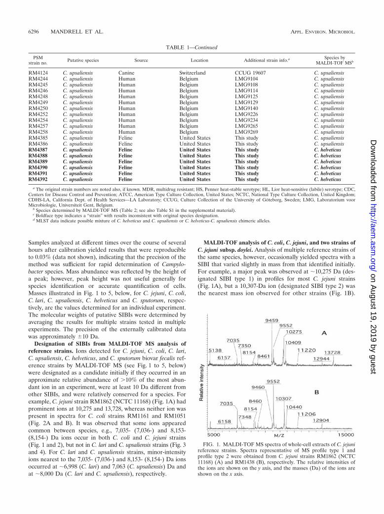
Designation of SIBIs from MALDI-TOF MS analysis ofreference strains. Ions detected for C. jejuni, C. coli, C. lari,C. upsaliensis, C. helveticus, and C. sputorum biovar fecalis ref-erence strains by MALDI-TOF MS (see Fig. 1 to 5, below)were designated as a candidate initially if they occurred in anapproximate relative abundance of �10% of the most abun-dant ion in an experiment, were at least 10 Da different fromother SIBIs, and were relatively conserved for a species. Forexample, C. jejuni strain RM1862 (NCTC 11168) (Fig. 1A) hadprominent ions at 10,275 and 13,728, whereas neither ion waspresent in spectra for C. coli strains RM1161 and RM1051(Fig. 2A and B). It was observed that some ions appearedcommon between species, e.g., 7,035- (7,036-) and 8,153-(8,154-) Da ions occur in both C. coli and C. jejuni strains(Fig. 1 and 2), but not in C. lari and C. upsaliensis strains (Fig. 3and 4). For C. lari and C. upsaliensis strains, minor-intensityions nearest to the 7,035- (7,036-) and 8,153- (8,154-) Da ionsoccurred at �6,998 (C. lari) and 7,063 (C. upsaliensis) Da andat �8,000 Da (C. lari and C. upsaliensis), respectively.
MALDI-TOF analysis of C. coli, C. jejuni, and two strains ofC. jejuni subsp. doylei. Analysis of multiple reference strains ofthe same species, however, occasionally yielded spectra with aSIBI that varied slightly in mass from that identified initially.For example, a major peak was observed at �10,275 Da (des-ignated SIBI type 1) in profiles for most C. jejuni strains(Fig. 1A), but a 10,307-Da ion (designated SIBI type 2) wasthe nearest mass ion observed for other strains (Fig. 1B).
FIG. 1. MALDI-TOF MS spectra of whole-cell extracts of C. jejunireference strains. Spectra representative of MS profile type 1 andprofile type 2 were obtained from C. jejuni strains RM1862 (NCTC11168) (A) and RM1438 (B), respectively. The relative intensities ofthe ions are shown on the y axis, and the masses (Da) of the ions areshown on the x axis.
TABLE 1—Continued
PSMstrain no. Putative species Source Location Additional strain info.a Species by
MALDI-TOF MSb
RM4124 C. upsaliensis Canine Switzerland CCUG 19607 C. upsaliensisRM4244 C. upsaliensis Human Belgium LMG9104 C. upsaliensisRM4245 C. upsaliensis Human Belgium LMG9108 C. upsaliensisRM4246 C. upsaliensis Human Belgium LMG9114 C. upsaliensisRM4248 C. upsaliensis Human Belgium LMG9125 C. upsaliensisRM4249 C. upsaliensis Human Belgium LMG9129 C. upsaliensisRM4250 C. upsaliensis Human Belgium LMG9140 C. upsaliensisRM4252 C. upsaliensis Human Belgium LMG9226 C. upsaliensisRM4254 C. upsaliensis Human Belgium LMG9234 C. upsaliensisRM4257 C. upsaliensis Human Belgium LMG9265 C. upsaliensisRM4258 C. upsaliensis Human Belgium LMG9269 C. upsaliensisRM4385 C. upsaliensis Feline United States This study C. upsaliensisRM4386 C. upsaliensis Feline United States This study C. upsaliensisRM4387 C. upsaliensis Feline United States This study C. helveticusRM4388 C. upsaliensis Feline United States This study C. helveticusRM4389 C. upsaliensis Feline United States This study C. helveticusRM4390 C. upsaliensis Feline United States This study C. helveticusRM4391 C. upsaliensis Feline United States This study C. helveticusRM4392 C. upsaliensis Feline United States This study C. helveticus
a The original strain numbers are noted also, if known. MDR, multidrug resistant; HS, Penner heat-stable serotype; HL, Lior heat-sensitive (labile) serotype; CDC,Centers for Disease Control and Prevention; ATCC, American Type Culture Collection, United States; NCTC, National Type Culture Collection, United Kingdom;CDHS-LA, California Dept. of Health Services—LA Laboratory; CCUG, Culture Collection of the University of Goteborg, Sweden; LMG, Laboratorium voorMicrobiologie, Universiteit Gent, Belgium.
b Species determined by MALDI-TOF MS (Table 2; see also Table S1 in the supplemental material).c Boldface type indicates a “strain” with results inconsistent with original species designation.d MLST data indicate possible mixture of C. helveticus and C. upsaliensis or C. helveticus-C. upsaliensis chimeric alleles.
6296 MANDRELL ET AL. APPL. ENVIRON. MICROBIOL.
on August 19, 2019 by guest
http://aem.asm
.org/D
ownloaded from

Similarly, a major ion of 10,031 Da (SIBI type 1), present inspectra for most C. coli strains (Fig. 2A), appeared at 10,061Da (SIBI type 2) for at least one C. coli strain analyzed(Fig. 2B). Similar differences in ions were noted for C. laristrains at masses of 9,617 (SIBI type 1) and 9,650 (SIBItype 2) Da (Fig. 3A and B).
An ion at 13,729 Da was present in profiles for the majorityof the C. jejuni reference strains and, thus, was designated aputative “common” ion (Table 2). However, this ion was miss-ing in a few strains, and an ion of 12,887 or 12,888 Da waspresent instead (Table 2, RM1050, RM1158, and RM1159). Itis not known whether these ions represent variant proteins orcompletely different proteins. However, subsequent analyses oftwo strains of C. jejuni subsp. doylei (RM2095 and RM2096)
identified the 10,276-Da C. jejuni type 1 SIBI, but they identi-fied a 12,888-Da ion in the absence of the 13,729-Da C. jejunicommon ion (data not shown). These results suggest that someof the C. jejuni reference strains (Table 2, RM1050, RM1158,and RM1159) and animal isolates (see Table S1 in the supple-mental material [RM1216, RM1420, and RM1886]) may beC. jejuni subsp. doylei. Additional well-characterized C. jejunisubsp. doylei strains would be required to clarify this subspeciesassignment.
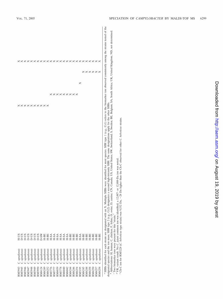
MALDI-TOF analysis of C. upsaliensis. Four SIBIs differingby �60 Da in mass were identified for C. upsaliensis strainsanalyzed in this study. These C. upsaliensis SIBI types (desig-nated 1 to 4) corresponded to ions at 10,171, 10,197, 10,272,and 10,229 Da, respectively (Fig. 4A to D). In addition, acommon ion at 9,504 Da was observed for most of theC. upsaliensis reference strains (Table 2), regardless of SIBItype. Although this appeared to be a “common” C. upsaliensisSIBI, subsequent work indicated it was present also inC. helveticus strains (see below).
MALDI-TOF analysis of C. helveticus. Three referencestrains of C. helveticus, including the type strain RM3228(ATCC 51209), were analyzed by MALDI. An ion at approx-imately 10,189 Da was designated the C. helveticus SIBI-1 ion,since it was identified in all 10 C. helveticus strains analyzed (a10,189-Da ion with a range of precision of �10 Da gives a10,199-Da ion as shown in Fig. 5A; this spectrum is an exampleof the �10 Da difference between the mass calculated withexternal standardization and the actual mass). A common ion
FIG. 2. MALDI-TOF MS spectra of whole-cell extracts of C. colireference strains. Spectra representative of MS profile type 1 andprofile type 2 were obtained from C. coli strains RM1161 (A) andRM1051 (B), respectively. The relative intensities of the ions areshown on the y axis, and the masses (in Da) of the ions are shown onthe x axis.
FIG. 3. MALDI-TOF MS spectra of whole-cell extracts of C. larireference strains. Spectra representative of MS profile type 1 andprofile type 2 were obtained from C. lari strains RM2100 (A) andRM2099 (B), respectively. The relative intensities of the ions areshown on the y axis, and the masses (in Da) of the ions are shown onthe x axis.
FIG. 4. MALDI-TOF MS spectra of whole-cell extracts ofC. upsaliensis reference strains. Spectra for reference strains represen-tative of SIBI types 1, 2, 3, and 4 were obtained with C. upsaliensisstrains RM2092 (A), RM3776 (B), RM3195 (C), and RM4245 (D),respectively. The relative intensities of the ions are shown on the y axis,and the masses (in Da) of the ions are shown on the x axis. The SIBItypes are designated above the singly charged ion for each strain.
VOL. 71, 2005 SPECIATION OF CAMPYLOBACTER BY MALDI-TOF MS 6297
on August 19, 2019 by guest
http://aem.asm
.org/D
ownloaded from
TA
BL
E2.
SIB
Isid
entifi
edfo
rre
fere
nce
stra
ins
ofC
.col
i,C
.jej
uni,
C.l
ari,
C.s
puto
rum
biov
ars
and
C.u
psal
iens
isby
MA
LD
I-T
OF
MSa
PSM
stra
inno
.Sp
ecie
sSo
urce
bC
c1(1
0,03
2D
a)
Cc2
(10,
060
Da)
Cc-
C(1
2,85
5D
a)
Cj1
(10,
276
Da)
Cj2
(10,
303
Da)
Cj-C
(13,
729
Da)
Cl1
(9,6
19D
a)
Cl2
(9,6
51D
a)
Cl-C
(12,
972
Da)
Ch1
(10,
189
Da)
Ch-
C(9
,504
Da)
Cu1
(10,
171
Da)
Cu2
(10,
197
Da)
Cu3
(10,
272
Da)
Cu4
(10,
229
Da)
Cu-
C(9
,504
Da)
Csp
1(9
,797
Da)
Csp
2(9
.817
Da)
Csp
-C(1
2,78
6D
a)
RM
1051
C.c
oli
H-C
NX
Xc
XR
M11
61C
.col
iH
-CN
XX
RM
1166
C.c
oli
C-C
NX
XR
M11
69C
.col
iH
-CN
XX
RM
2228
C.c
oli
C-U
SX
XR
M22
30C
.col
iC
-US
XX
RM
2231
C.c
oli
C-U
SX
XR
M22
36C
.col
iC
-US
XX
RM
1048
C.j
ejun
iH
-CN
XX
RM
1050
C.j
ejun
iH
-CN
XV
Rd
RM
1155
C.j
ejun
iH
-CN
XX
RM
1156
C.j
ejun
iH
-CN
XX
RM
1862
C.j
ejun
iH
-UK
XX
RM
1158
C.j
ejun
iH
-CN
XV
Rd
RM
1159
C.j
ejun
iH
-CN
XV
Rd
RM
1163
C.j
ejun
iH
-CN
XX
RM
1164
C.j
ejun
iH
-CN
XX
RM
1165
C.j
ejun
iC
-CN
XX
cX
RM
1167
C.j
ejun
iH
-CN
XX
RM
1168
C.j
ejun
iH
-CN
XX
RM
1462
C.l
ari
H-?
XX
XR
M20
98C
.lar
iH
-US
XX
RM
2099
C.l
ari
H-U
SX
RM
2100
C.l
ari
H-U
SX
X
RM
3228
C.h
elve
ticus
F-S
WX
Xe
RM
4087
C.h
elve
ticus
F-S
WX
XR
M40
88C
.hel
vetic
usF
-SX
X
RM
1485
C.s
puto
rum
bv.
feca
lisO
-?X
X
RM
1495
C.s
puto
rum
bv.
bulb
ulus
ND
XX
RM
2089
C.u
psal
iens
isO
-?X
XR
M20
90C
.spu
toru
mbv
.fe
calis
O-U
SX
X
RM
2091
C.s
puto
rum
bv.
feca
lisO
-?X
X
RM
4120
C.s
puto
rum
bv.
para
ureo
lytic
usH
-CN
XX
RM
4121
C.s
puto
rum
bv.
feca
lisO
-UK
XX
RM
1488
C.u
psal
iens
isH
-US
XX
RM
2092
C.u
psal
iens
isH
-US
XX
RM
2093
C.u
psal
iens
isH
-US
XX
RM
3937
C.u
psal
iens
isH
-US
XX
RM
3939
C.u
psal
iens
isH
-US
XX
RM
3940
C.u
psal
iens
isH
-US
XX
RM
3941
C.u
psal
iens
isH
-US
XX
RM
3942
C.u
psal
iens
isH
-US
XX
6298 MANDRELL ET AL. APPL. ENVIRON. MICROBIOL.
on August 19, 2019 by guest
http://aem.asm
.org/D
ownloaded from
RM
3943
C.u
psal
iens
isH
-US
XX
RM
3945
C.u
psal
iens
isD
-US
XX
RM
3946
C.u
psal
iens
isD
-US
XX
RM
3947
C.u
psal
iens
isD
-US
XX
RM
3948
C.u
psal
iens
isD
-US
XX
RM
3949
C.u
psal
iens
isD
-US
XX
RM
3950
C.u
psal
iens
isD
-US
XX
RM
4124
C.u
psal
iens
isD
-SW
XX
RM
4249
C.u
psal
iens
isH
-BE
XX
RM
4252
C.u
psal
iens
isH
-BE
XX
RM
3776
C.u
psal
iens
isH
-SA
XX
RM
3777
C.u
psal
iens
isH
-SA
XX
RM
4039
C.u
psal
iens
isH
-SA
XX
RM
3778
C.u
psal
iens
isH
-SA
XX
RM
4040
C.u
psal
iens
isH
-SA
XX
RM
4042
C.u
psal
iens
isH
-SA
XX
RM
4244
C.u
psal
iens
isH
-BE
XX
RM
4246
C.u
psal
iens
isH
-BE
XX
RM
4254
C.u
psal
iens
isH
-BE
XX
RM
3195
C.u
psal
iens
isH
-SA
XX
RM
4245
C.u
psal
iens
isH
-BE
XX
RM
4248
C.u
psal
iens
isH
-BE
XX
RM
4258
C.u
psal
iens
isH
-BE
XX
RM
4250
C.u
psal
iens
isH
-BE
XX
RM
4257
C.u
psal
iens
isH
-BE
XX
RM
4258
C.u
psal
iens
isH
-BE
XX
aSI
BIs
iden
tified
for
each
stra
inar
ede
sign
ated
with
anX
.Mul
tiple
SIB
Isw
ere
iden
tified
for
mos
tsp
ecie
s.SI
BI
type
1(e
.g.,
Cc1
)re
fers
toth
ebi
omar
ker
ion
obse
rved
cons
iste
ntly
amon
gth
est
rain
ste
sted
ofth
ede
sign
ated
spec
ies
and
the
ion
mas
s;SI
BI
type
2(e
.g.,
Cc2
)de
sign
ates
ase
cond
high
-inte
nsity
SIB
I.T
hesa
me
desi
gnat
ions
appl
yfo
rth
eot
her
SIB
Is.
bA
bbre
viat
ions
:H,h
uman
;C,c
hick
en;F
,fel
ine;
O,o
vine
;D,c
anin
e;C
N,C
anad
a;U
S,U
nite
dSt
ates
;SW
,Sw
itzer
land
;S,S
wed
en;B
E,B
elgi
um;S
A,S
outh
Afr
ica;
UK
,Uni
ted
Kin
gdom
;ND
,not
dete
rmin
ed.
cT
wo
biom
arke
rio
nsw
ere
pres
ent
for
this
“str
ain.
”d
VR
,var
iant
resu
lt.N
oC
.jej
unic
omm
onio
nw
asid
entifi
ed;a
12,8
87-
or12
,888
-Da
ion
was
note
d.e
Ch-
Cio
nfo
rR
M32
28(C
.hel
vetic
usty
pest
rain
)w
as9,
532
Da,
�28
Da
high
erth
anth
eC
h-C
obse
rved
for
othe
rC
.hel
vetic
usst
rain
s.
VOL. 71, 2005 SPECIATION OF CAMPYLOBACTER BY MALDI-TOF MS 6299
on August 19, 2019 by guest
http://aem.asm
.org/D
ownloaded from
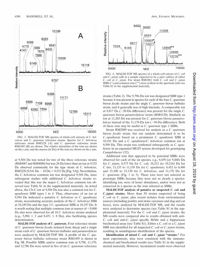
at 9,504 Da was noted for two of the three reference strains(RM4087 and RM4088) but was 28 Da lower than an ion at 9,532Da observed consistently for the type strain of C. helveticus,RM3228 (9,541 Da 10 Da 9,531 Da [Fig. 5A]). Nevertheless,the C. helveticus common ion was designated 9,504 Da, sincesubsequent studies with additional C. helveticus strains re-vealed that this was the major C. helveticus common ion ob-served (see Table S1 in the supplemental material). As notedabove, the Ch-C ion at 9,504 Da was also a common ion for C.upsaliensis SIBI types 1 to 4. Thus, observance of an ion at9,504 Da indicated a putative C. helveticus or C. upsaliensisstrain, necessitating accurate analysis of the C. helveticus SIBIat 10,189 Da and the type 2 C. upsaliensis SIBI at 10,197 Da. Itis worth noting that multiple common ions at masses of �9,000Da also were observed for all 10 C. helveticus strains analyzed(e.g., 5,094 � 2 and 8,451 � 8 Da), also facilitating speciesdetermination.
MALDI-TOF analysis of C. sputorum. Four reference strainsof C. sputorum biovar fecalis isolated from sheep and a singlestrain each of C. sputorum biovars bulbulus and paraureolyticuswere analyzed by MALDI-TOF MS. A profile of the C. spu-torum biovar bulbulus reference strain RM1495 is shown inFig. 5B. Possible SIBIs and/or common ions at 9,796, 11,170,and 12,786 Da were noted in five of six C. sputorum reference
strains (Table 2). The 9,796-Da ion was designated SIBI type 1because it was present in spectra for each of the four C. sputorumbiovar fecalis strains and the single C. sputorum biovar bulbulusstrain, and it generally was of high intensity. A comparable ionat 9,817 Da (�20-Da difference) was present for the single C.sputorum biovar paraureolyticus strain (RM4120). Similarly anion at 11,203 Da was present for C. sputorum biovar paraureo-lyticus instead of the 11,170-Da ion (�30-Da difference). Bothof these ions may be useful as C. sputorum type 1 SIBIs.
Strain RM2089 was received for analysis as a C. sputorumbiovar fecalis strain, but our analysis determined it to beC. upsaliensis based on a prominent C. upsaliensis SIBI at10,181 Da and a C. upsaliensis/C. helveticus common ion at9,509 Da. This strain was confirmed subsequently as C. upsa-liensis in an expanded MLST system developed for genotypingCampylobacter (52).
Additional ions that appeared to be potential SIBIs wereobserved for each of the six species, e.g., 9,459 (or 9,460) Dafor C. jejuni, 8,573 Da for C. coli, 10,212 (to 10,216) Da forC. lari, 11,135 to 11,139 Da for C. upsaliensis; 8,452 to 8,460and 15,100 to 15,130 for C. helveticus, and 11,170 Da forC. sputorum (Fig. 1 to 5). These ions were not selected asprototype SIBIs because they were not as clearly a species-identifying ion, were of lesser abundance, and/or were not asconserved in a species as the ions selected as SIBIs.
MALDI-TOF analysis of putative or suspected C. coli andC. jejuni strains. More than 50 strains suspected of beingC. coli or C. jejuni, plus recent isolates from environmentalsources (including poultry and swine carcasses and dog and catfeces), were analyzed by MALDI-TOF MS, and the resultswere analyzed to determine species (see Table S1 in the sup-plemental material). For the C. coli and C. jejuni strains, theMS results were compared also to results obtained with anti-C. coli and anti-C. jejuni specific MAbs and a hippuricasebiochemical assay (see Table S1). Either a C. coli or a C. jejuniSIBI was identified for all suspected C. coli or C. jejuni strains,resulting in unambiguous identification of the species.
Identification of mixed C. coli and C. jejuni “strains.” Formost experiments, data were consistent with the immuno-chemical and biochemical results (see Table S1 in the supple-mental material). However, inconsistent results were observed
FIG. 5. MALDI-TOF MS spectra of whole-cell extracts of C. hel-veticus and C. sputorum reference strains. Spectra for C. helveticusreference strain RM3228 (A) and C. sputorum reference strainRM1485 (B) are shown. The relative intensities of the ions are shownon the y axis, and the masses (in Da) of the ions are shown on the x axis.
FIG. 6. MALDI-TOF MS spectra of a whole-cell extract of C. coliand C. jejuni cells in a sample expected to be a pure culture of eitherC. coli or C. jejuni. For strain RM1882, both C. coli and C. jejuniSIBIs (*) and common ions (**) were evident in the spectrum (also seeTable S1 in the supplemental material).
6300 MANDRELL ET AL. APPL. ENVIRON. MICROBIOL.
on August 19, 2019 by guest
http://aem.asm
.org/D
ownloaded from
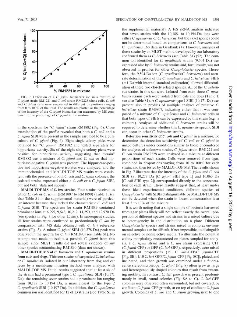
in the spectrum for “C. jejuni” strain RM1882 (Fig. 6). Closeexamination of the profile revealed that both a C. coli and aC. jejuni SIBI were present in the sample assumed to be a pureculture of C. jejuni (Fig. 6). Eight single-colony picks wereobtained for “C. jejuni” RM1882 and tested separately forhippuricase activity. Six of the eight single-colony picks werepositive for hippuricase activity, suggesting that “strain”RM1882 was a mixture of C. jejuni and C. coli or that hip-puricase-negative C. jejuni was present. The hippuricase-posi-tive and hippuricase-negative isolates were analyzed, and theimmunochemical and MALDI-TOF MS results were consis-tent with the presence of both C. coli and C. jejuni colonies: theisolated strains expressed either a C. coli or a C. jejuni SIBI,but not both (data not shown).
MALDI-TOF MS of C. lari strains. Four strains received aseither C. coli or C. jejuni (RM1887 to RM1890) (Table 1; seealso Table S1 in the supplemental material) were of particu-lar interest because they lacked the characteristic C. coli andC. jejuni SIBI. The spectrum for strain RM1889 containedprominent ions at 6,995, 9,648, 10,212, 11,250, and 12,970 Da(see spectra in Fig. 3 for other C. lari). In subsequent studies,all four strains were confirmed as predominately C. lari bycomparison with MS data obtained with C. lari referencestrains (Fig. 3). A minor C. jejuni SIBI (10,278-Da) peak wasobserved in the spectra for C. lari RM1890 (see Table S1). Noattempt was made to isolate a possible C. jejuni from thissample, since MLST results did not reveal evidence of anyother species contaminating RM1890 (data not shown).
MALDI-TOF MS of C. helveticus and C. upsaliensis strainsfrom cats and dogs. Thirteen strains of suspected C. helveticusor C. upsaliensis isolated in our laboratory from dog and catfeces by a membrane filtration method were analyzed withMALDI-TOF MS. Initial results suggested that at least six ofthe strains had a prominent type 1 C. upsaliensis SIBI (10,171Da); the remaining seven strains had a prominent ion rangingfrom 10,188 to 10,194 Da, a mass closest to the type 2C. upsaliensis SIBI (10,197 Da). In addition, the C. upsaliensiscommon ion was identified for 12 of 13 strains (see Table S1 in
the supplemental material). A 16S rRNA analysis indicatedthat seven strains with the 10,188- to 10,194-Da ions wereeither C. upsaliensis or C. helveticus, but the exact species couldnot be determined based on comparisons to C. helveticus andC. upsaliensis 16S data in GenBank (4). However, analyses ofthese strains by an MLST method developed by our laboratoryconfirmed them as C. helveticus (see Table S1) (52). The com-mon ion identified for C. upsaliensis strains (9,504 Da) wasexpressed also by C. helveticus strains and, fortuitously, was notobserved in profiles for other Campylobacter species. There-fore, the 9,504-Da ion (C. upsaliensis/C. helveticus) and accu-rate determination of the C. upsaliensis and C. helveticus SIBIs(�1 Da with internal standard calibration) allowed differenti-ation of these two closely related species. All of the C. helveti-cus strains in this set were isolated from cats; three C. upsa-liensis strains each were isolated from cats and dogs (Table 1;see also Table S1). A C. upsaliensis type 1 SIBI (10,171 Da) waspresent also in profiles of multiple analyses of putative C.helveticus strain RM3807, indicating either that it was com-posed of a mixture of C. upsaliensis and C. helveticus cells orthat both types of SIBIs can be expressed by this strain (e.g., achimera). Analyses of additional C. helveticus strains will berequired to determine whether this C. upsaliensis-specific SIBIcan occur in other C. helveticus strains.
Detection sensitivity of C. coli and C. jejuni in a mixture. Todetermine the detection sensitivity of C. coli or C. jejuni inmixed cultures under conditions similar to those encounteredfor analyses of unknown strains, C. jejuni strain RM1221 andC. coli strain RM2228 were analyzed as a mixture at differentproportions of each strain. Cells were removed from agar,combined in proportions varying from 10 to 100% for eachstrain, and then tested by MALDI-TOF MS. The results shownin Fig. 7 illustrate that the intensity of the C. jejuni and C. coliSIBI (at 10,277 Da [C. jejuni SIBI type 1] and 10,063 Da[C. coli SIBI type 2]) corresponded directly to the concentra-tion of each strain. These results suggest that, at least underthese ideal experimental conditions, different species ofCampylobacter with SIBIs distinguishable by MALDI-TOF MScan be detected when the strain in lowest concentration is atleast 5 to 10% of the mixture.
It is worth noting that a single sample of bacteria harvestedfrom agar plates likely will not reflect exactly the overall pro-portion of different species and strains in a mixed culture dueto heterogeneity in the distribution on a plate. DifferentCampylobacter species and strains mixed in complex environ-mental samples can be difficult, if not impossible, to distinguishon selective or nonselective media. To illustrate the potentialcolony morphology encountered on plates sampled for analy-sis, a C. jejuni strain and a C. lari strain expressing CFP(C. jejuni-CFP) or GFP (C. lari-GFP), respectively, were mixedin different proportions (1:1 C. lari-GFP/C. jejuni-CFP[Fig. 8B]; 1:10 C. lari-GFP/C. jejuni-CFP [Fig. 8C]), plated, andincubated, and then growth was examined under a fluores-cence stereomicroscope. C. jejuni (Fig. 8) often grew as largeand heterogeneously shaped colonies that result from swarm-ing motility. In contrast, C. lari growth was present predomi-nantly in small, round colonies (Fig. 8A to C). C. lari-GFPcolonies were observed often surrounded, but not covered, byconfluent C. jejuni-CFP growth, or on top of confluent C. jejunigrowth. Colonies of C. lari and C. jejuni growing next to one
FIG. 7. Detection of a C. jejuni biomarker ion in a mixture ofC. jejuni strain RM1221 and C. coli strain RM2228 whole cells. C. coliand C. jejuni cells were suspended in different proportions rangingfrom 0 to 100% of the total. The results are plotted as the percentageof the intensity of the C. jejuni biomarker ion measured by MS com-pared to the percentage of C. jejuni in the mixture.
VOL. 71, 2005 SPECIATION OF CAMPYLOBACTER BY MALDI-TOF MS 6301
on August 19, 2019 by guest
http://aem.asm
.org/D
ownloaded from

another and indistinguishable under regular light were some-times observed (Fig. 8C, top). No sectored colonies corre-sponding to a mixed strain “CFU” were observed in this ex-periment. These results reflect the importance of propersampling and analysis of multiple samples to obtain a thoroughassessment of the diversity of Campylobacter strains and spe-cies that may be present in either clinical or environmentalsamples.
Consistency of spectra of C. jejuni strain RM1221 aftergrowth on different media and under different atmosphericconditions. To determine whether the SIBI expression wasstable under different culture conditions, C. jejuni strainRM1221 was subcultured on three different media under twoatmospheric conditions at two different temperatures, andsamples of bacteria were analyzed by MALDI-TOF MS. Themasses determined for the C. jejuni SIBI type 1 (10,276 Da),the C. jejuni common ion (13,729 Da), and a third ion at�9,551 Da (probable 30S ribosomal protein S17) were veryreproducible for samples of C. jejuni RM1221 grown undereach of nine combinations of conditions (see Table S2 in thesupplemental material). No significant differences were notedin the mass of high-intensity ions, regardless of the media (e.g.,blood), gas mixture (5% or 10% CO2 and 5% H2 or 5% O2),or incubation temperature (37°C versus 42°C), as shown in thefollowing results: C. jejuni SIBI type 1 10,276.0 � 1.3;C. jejuni common SIBI 13,729.0 � 1.6; “ion 2” 9,551.0 �1.4. In addition, eight replicate samples from a plate of C. coliRM2228, C. lari RM2100, and C. upsaliensis RM3195 grownunder a single condition (BAB medium, 10% CO2–5% H2,37°C) were analyzed (see Table S2). The standard deviationsfor the replicates for the three ions for the three strains rangedfrom 0.9 (C. coli SIBI type 1) to 3.1 (C. upsaliensis ion 2),reflecting good intraexperimental reproducibility. A similar ex-periment with three different strains of C. helveticus (RM3228,RM4087, and RM4088) grown under the same conditions re-sulted in average values of 10,188.3 � 1.4, 9,522.1, and9,504.0 � 0.7 for the C. helveticus SIBI, C. helveticus commonSIBI for RM4087 and 4088, and C. helveticus common SIBI forRM3228, respectively, for six samples total (two for eachstrain) for each ion (data not shown). Two strains of C. spu-torum biovar fecalis (RM1495 and RM4121) and C. sputorum
biovar paraureolyticus strain RM4120 were analyzed in a sec-ond experiment with replicate samples. The average values forthe SIBI and common SIBI for the two C. sputorum biovarfecalis strains were 9,797.1 � 1.26 and 11,167 � 2.61, respec-tively; for the single C. sputorum biovar paraureolyticus strainthe values were 9,813.7 � 0.1 and 11,193 � 0.4 (data notshown). These values reflect the potential difference betweenC. sputorum biovar fecalis/C. sputorum biovar bulbulus andC. sputorum biovar paraureolyticus described above (Table 2),although additional strains of each biovar would need to betested to confirm this biovar-specific difference.
These results indicate that conventional growth conditionsdo not influence markedly the mass of ions detected. It is worthnoting, however, that horse blood-related proteins apparentlyincorporated by the cells or into CFU from the underlyingblood agar medium were identified in some profiles. This wasconfirmed in control experiments with samples of BAB me-dium extracted directly from plates without bacteria (e.g.,14,890, 15,050, 15,100, and 16,075 Da). We suspected that the16,075-Da ion corresponded to the �-chain protein of horsehemoglobin (61). This was exploited in later experiments as auseful internal protein standard of an appropriate mass forcalibration, thus facilitating extremely accurate mass assign-ments of SIBIs (�1 Da) for identification of C. helveticus,C. upsaliensis, and C. sputorum biovar fecalis strains (seeTable S1 in the supplemental material).
DISCUSSION
We have presented data that multiple Campylobacter speciesgrown on solid medium under a variety of conditions can beanalyzed by MALDI-TOF MS to yield high-intensity and, inmost cases, intact protein ions in the 9- to 14-kDa range thatare diagnostic of Campylobacter species. Pure cultures ofCampylobacter can be analyzed rapidly with less ambiguousresults, compared to many methods used currently for confirm-ing species. An added advantage of MALDI-TOF MS is thatmultiple species of Campylobacter in mixed cultures can beidentified more easily by MS than by conventional methods;reproducibility of the MALDI-TOF MS protein profiles for
FIG. 8. Mixed culture of C. jejuni-CFP and C. lari-GFP on BAB medium. C. jejuni-CFP and C. lari-GFP cells were harvested from BABmedium and suspended in broth in proportions of 1:1 (B) or 10:1 (C), respectively, mixed, and then plated on BAB. The plates were incubatedfor 30 h, and growth was observed under a fluorescence stereomicroscope (Leica MZIII). (A) No filters; the image shows growth of a 1:1proportion of C. jejuni and C. lari. Note the larger colonies of C. jejuni-CFP (blue) compared to C. lari-GFP (green) (B and C). The red arrowpoints to a C. lari colony next to a C. jejuni colony (A); the white arrow in panel B points to a C. lari-GFP colony on top of a C. jejuni-CFP colony;the violet arrow in panel C points to a small C. lari-GFP colony on top of C. jejuni-CFP growth.
6302 MANDRELL ET AL. APPL. ENVIRON. MICROBIOL.
on August 19, 2019 by guest
http://aem.asm
.org/D
ownloaded from
C. coli and C. jejuni strains was sufficient to allow species iden-tification of isolates.
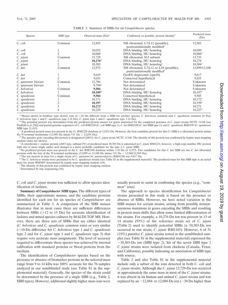
Summary of Campylobacter SIBI types. The different types ofSIBIs, their approximate masses, and the candidate proteinsidentified for each ion for six species of Campylobacter aresummarized in Table 3. A comparison of the SIBI massesillustrates that in most cases there are sufficient differencesbetween SIBIs (�12 to 15 Da) for accurate identification ofisolates and mixed species cultures by MALDI-TOF MS. How-ever, there are three sets of SIBIs that are either identical(C. helveticus and C. upsaliensis common ions) or very similar(�10-Da difference for C. helveticus type 1 and C. upsaliensistype 2 and for C. jejuni type 1 and C. upsaliensis type 3) thatrequire very accurate mass assignments. The level of accuracyrequired to differentiate these species was achieved by internalcalibration with standard proteins or blood proteins from themedium.
The identification of Campylobacter species based on thepresence or absence of biomarker proteins in the selected massrange from 9 to 14 kDa was 100% accurate for the 76 samplesanalyzed in our semiblinded study (see Table S1 in the sup-plemental material). Generally, the species of the strain couldbe determined by the presence of a single SIBI (e.g., Table 2,SIBI types). However, additional slightly higher mass ions were
usually present to assist in confirming the species (e.g., “com-mon” ions).
The approach to species identification for Campylobacterstrains presented in this study is based on the presence orabsence of SIBIs. However, we have noted variation in theSIBI masses for certain strains, arising from possibly nonsyn-onymous mutations in genes encoding the SIBIs and resultingin protein mass shifts that allow some limited differentiation ofthe strains. For example, a 10,276-Da ion was present in 13 of14 strains (93%) of the reference strain set of C. jejuni(Table 2) used to identify potential SIBIs (a 10,303-Da ionoccurred in one strain, C. jejuni RM1165). However, 6 of 31(19%) putative C. jejuni strains tested in the semiblinded sam-ples (see Table S1 in the supplemental material) expressed the�10,303-Da ion (SIBI type 2). Six of the seven SIBI type 2C. jejuni strains were isolated from chickens (Canada, Texas,and California), possibly indicating an association of SIBI typewith source.
Table 2 and also Table S1 in the supplemental materialinclude only a subset of the ions detected in both C. coli andC. jejuni strains. Although the C. jejuni 13,729-Da ion occurredat approximately the same mass in most of the C. jejuni strains,it was absent in six human and animal C. jejuni strains and wasreplaced by an �12,884- to 12,888-Da ion (�30 Da higher than
TABLE 3. Summary of SIBIs for six Campylobacter species
Species SIBI type Observed mass (Da)a Confirmed, or possible, protein identityb Predicted mass(Da)
C. coli Common 12,855 50S ribosomal, L7/L12 (possible),posttranslationally modifiedc
12,985
C. coli 1 10,032 DNA binding, HU homolog 10,030C. coli 2 10,060 DNA binding, HU homolog 10,060 j
C. jejuni Common 13,729 30S ribosomal, S13 subunit 13,735C. jejuni 1 10,276d DNA binding, HU homolog 10,274C. jejuni 2 10,303 DNA binding, HU homologe 10,304 j
C. lari Common 12,972 50S ribosomal, L7/L12 or L18 (possible),posttranslationally modifiedf
13,099/13,100
C. lari 1 9,619 GroES chaperonin (cpn10) 9,617C. lari 2 9,651 Conserved hypothetical 9,655C. sputorum biovars Common 12,786 Not determined UnknownC. sputorum biovars 1 9,796g Not determined UnknownC. helveticus Common 9,504 Not determined UnknownC. helveticus 1 10,189h DNA binding, HU homolog 10,187 j
C. upsaliensis Common 9,504 Conserved hypothetical 9,503C. upsaliensis 1 10,171 DNA binding, HU homolog 10,172 j
C. upsaliensis 2 10,197 DNA binding, HU homolog 10,199 j
C. upsaliensis 3 10,272i DNA binding, HU homolog 10,271C. upsaliensis 4 10,229 DNA binding, HU homolog 10,227 j
a Masses shown in boldface type denote ions of �10 Da different from a SIBI for another species: C. helveticus common and C. upsaliensis common (0 Da),C. helveticus type 1 and C. upsaliensis type 2 (8 Da), C. jejuni type 1 and C. upsaliensis type 3 (4 Da).
b The potential protein was determined from the predicted protein masses for genes annotated for the completed genomes of C. jejuni strains NCTC 11168 (seeParkhill et al. [58]) and partial genome sequences of C. coli RM2228 (C. coli SIBI type 1), C. lari RM2100 (C. lari SIBI type 1), and C. upsaliensis RM3195 (C. upsaliensisSIBI type 3) (65).
c A predicted protein mass was present in the Cc RM2228 database at 12,851 Da. However, the best candidate protein for this Cc SIBI is a ribosomal protein minusthe N-terminal methionine (12,985 Da minus 131 Da 12,854 Da).
d The putative gene encoding this protein is designated Cj0913c for C. jejuni strain NCTC 11168. The identity of this protein was confirmed by tryptic mass mappinganalysis (data not shown).
e A cytochrome c oxidase protein (cbb3 type, subunit IV) of predicted mass 10,303 Da is annotated in C. jejuni RM1221; however, a high-copy-number HU proteinwith one or more single amino acid changes is a more probable candidate for the type 2 C. jejuni SIBI.
f No predicted protein mass was present in the C. lari RM2100 database within �10 Da of 12,972. Two candidates for this C. lari SIBI are two C. lari ribosomalproteins with the loss of the N-terminal methionine (13,099 or 13,100 Da minus 131 Da 12,968/12,969 Da).
g The ion nearest in mass for a single strain of C. sputorum bv. paraureolyticus (RM4120) was 9,817 Da.h The C. helveticus strains were presumed to be C. upsaliensis strains (see Table S1 in the supplemental material). The predicted mass for this SIBI type is an actual
mass for strain RM3807 determined by tryptic mass mapping analysis (18).i The identity of this protein was confirmed by tryptic mass mapping analysis.j Determined by hup sequencing (18).
VOL. 71, 2005 SPECIATION OF CAMPYLOBACTER BY MALDI-TOF MS 6303
on August 19, 2019 by guest
http://aem.asm
.org/D
ownloaded from
the C. coli common ion) (Table 2; see also Table S1 in thesupplemental material). The “12,888 ion” was identified also inthe only C. jejuni subsp. doylei strains we had available foranalysis (both human clinical strains). Similarly, the C. coliSIBI at 12,857 Da was absent in a few strains tested, and an ionat 12,914 Da was observed (57-Da difference). These resultsindicate the importance of identifying multiple SIBIs for accu-rate analysis and identification and the potential value ofMALDI-TOF analysis for identifying subspecies/biovar differ-ences in Campylobacter.
In a previous study of a few C. coli and C. jejuni strains byMALDI-TOF MS, biomarker ions were identified at 10,074 Dafor C. coli strain ATCC 43474 and at 10,285 Da for C. jejunistrain ATCC 43464 (73). It is probable that these ions corre-spond to the C. coli type 2 (10,060 Da) and C. jejuni type 1(10,276 Da) SIBIs identified in our study. The 9- and 14-Damass differences for these ions in the two studies may reflect anactual difference (e.g., amino acid polymorphism or posttrans-lational modification) or differences in measurement precision.However, our analysis of the same C. coli strain as the previousstudy (RM1878) (Table 1; see also Table S1 in the supplemen-tal material) suggests the difference is due to precision.
Identities or putative identities of some SIBIs. Many of theSIBIs identified in this study are putatively abundant, cytosolicproteins, of relatively low mass compared to the majority ofproteins, and apparently conducive to ionization in MALDI-TOF MS (Table 3, HU-homolog, ribosomal subunit, and chap-eronin proteins). A C. jejuni HU homolog encoded by the hupgene was characterized previously by Konkel et al. and desig-nated HCj (33). The calculated molecular mass of HCj was10,267 Da, with a pI of 10.1. Presumably, this small, highlybasic, histone-like protein has properties similar to other HUproteins with regards to abundance (30,000 to 50,000 HUdimers per cell in E. coli) and DNA binding and condensingactivity (14). It is probable that the high intensity of HU/HCjand possible HCj-homologs in other Campylobacter species isdue to a combination of similar high abundance plus efficientionization of these highly basic proteins. In addition, we spec-ulate that cytosolic proteins whose functions and sequenceshave been highly conserved through evolution are good can-didates for species identification, although of limited utility forstrain differentiation. It will be interesting to determinewhether these same high-intensity ions identified by MALDI-TOF MS (e.g., the HU protein) are good predictors of otherCampylobacter species (18) and bacterial species of other genera.
The possible identities for some of the SIBIs shown inTable 3 must be considered speculative until confirmed bytryptic mass mapping experiments. For example, the C. coliand C. lari common SIBIs have been designated as 50S ribo-somal L7/L12 proteins (RplL), even though the predicted pro-tein is �130 Da larger than that observed by MALDI-TOFMS. This is because no predicted protein mass closer than 10Da was present in the genome database, suggesting that post- orcotranslational modification had occurred, resulting in removal ofthe N-terminal methionine (131 Da) by a specific aminopeptidase(13, 27).
Polymorphisms and variation of SIBIs. Ongoing genetic andmass mapping studies in our laboratory indicate that the massdifferences between SIBI types, in fact, are due to nonsynony-mous single or multiple nucleotide polymorphisms, rather than
differences due to sensitivities of methods (18). These SIBItypes representing authentic nonsynonymous mutations will beuseful for population analyses of strains. Strains related epi-demiologically (e.g., from an outbreak or prevalent in a singlesource) can be identified based on a pattern of small massdifferences reflecting single or multiple amino acid changesthat are different from other strains. For example, all of theC. upsaliensis strains from the United States were C. upsaliensisSIBI type 1 (�10,171 Da), in contrast to six of seven C. upsa-liensis strains from South Africa, which were C. upsaliensisSIBI type 2 (�10,197 Da). However, of the 11 C. upsaliensisreference strains from Belgium, two, three, and six strainsrepresented SIBI types 1, 2, and 4, respectively. The commonion at 9,504 Da was observed in all of the C. upsaliensisMALDI-TOF MS profiles. The four C. upsaliensis SIBI typesare consistent with the genomic heterogeneity reported previ-ously for animal and human (8, 40, 57) and, specifically, dogand cat C. upsaliensis strains (40, 54). Twelve of 13 dog and catC. upsaliensis strains in our study were from the United States,and all were C. upsaliensis SIBI type 1. Although SIBI typesprovide probably only a minor discrimination of strains, it willbe interesting eventually to compare SIBI types with sequencetypes determined by a new MLST method for C. coli, C. lari,C. helveticus, and C. upsaliensis (52).
As we expand our MALDI-TOF MS analyses to includeother species of Campylobacter, it is anticipated that proteinhomologs to the SIBIs described in this study will be identifiedas biomarkers. For example, we have identified recently a10.5-kDa protein as the HU protein, and a potential SIBI, forC. concisus strains (18). The probability will increase that SIBIsin different species with similar masses (�10 Da) will be iden-tified, again emphasizing the need for accurate mass assign-ments. As noted above, the identification of medium-derivedhorse hemoglobin �-chain in some experiments permittedrecalibration of data, yielding �1 Da accuracy that proved tobe essential for differentiating the C. helveticus SIBI (�10,189Da) from a C. upsaliensis type 2 SIBI (�10,197 Da). Thus,media proteins distinct from bacterial proteins, or addition ofa reference protein to each sample, can be exploited for inter-nal calibration to improve precision and accuracy of mass mea-surement.
The correct species designations for �130 strains analyzedin this study were identified usually by identifying only twoSIBIs: a single SIBI among multiple variant SIBIs (e.g.,C. upsaliensis), plus a SIBI common among all or most strainsof a species. However, it is worth emphasizing that additionalSIBIs are observed in the mass range of 7 to 15 kDa thatpermit confirmation of species when SIBIs for different speciesare of similar mass (�10 Da) or are variable among environ-mental strains of the same species. Therefore, identifying a setof SIBIs would be advantageous for unambiguous assignmentsof species and important intraspecies strain differences.
As more organisms are sequenced and the data are enteredinto public databases (e.g., NCBI GenBank), the accuracy inidentification and analysis of unknown isolates by proteomicsapproaches also will increase (12, 13, 19). In a single studyreporting species identification by querying ion masses againstprotein databases (19), organisms could be identified only tothe genus level. In our study, and other studies, isolates areclassified tentatively as members of a particular genus and/or
6304 MANDRELL ET AL. APPL. ENVIRON. MICROBIOL.
on August 19, 2019 by guest
http://aem.asm
.org/D
ownloaded from
species through both sample source and the special selectionand enrichment conditions involved in obtaining them.MALDI-TOF MS has proved to be useful for rapid confirma-tion of species.
Environmental samples and mixed species cultures. Selec-tive media for isolation of C. coli and C. jejuni from clinical orenvironmental (animals, food, and water) samples commonlyinclude antibiotics for minimizing other microbial flora. Theunintended consequence of this approach is that antibiotic-sensitive strains of C. coli and C. jejuni and other Cam-pylobacter species are not culturable. MALDI-TOF MS spe-ciation is very efficient with pure cultures of Campylobacter.However, we have shown also that multiple Campylobacterspecies can be identified with a preparation of Campylobactercells growing confluently, or as single colonies, on commonagar media (Fig. 8). We are developing an approach currentlyto analyze multiple colonies of suspected Campylobacter spe-cies obtained following passage through 0.6-�m filters andgrowth on nonselective culture medium in the appropriateatmosphere (22, 39). Since multiple strains of a majority ofCampylobacter, Helicobacter, and other spiral-form bacteriacould be present and difficult to distinguish, MALDI-TOF MSanalysis provides a simple method for identifying potentialCampylobacter species, and also potential mixed-species colo-nies (Fig. 8), as has been reported previously (17, 50). Weconfirmed numerous times during this study that “isolates”received from other sources were either speciated incorrectlyor contained multiple species.
Intraspecies variability of the Campylobacter SIBIs, poten-tially due to single or multiple amino acid sequence changes,has been identified; these data are not attainable rapidly inmost other assays. MALDI-TOF MS may be especially usefulfor characterizing non-C. coli and non-C. jejuni strains becauseof the minimal genetic and biochemical data available foremerging Campylobacter species (10). The recent increase ingenomic sequence data available for Campylobacter specieswill enhance MALDI-TOF MS as a method of analysis forCampylobacter (21, 65), since accurate protein masses yieldgene and protein identifications for strain differentiation andfurther analysis.
ACKNOWLEDGMENTS
Strains used in this study were provided generously by SharonAbbott, Paula Cray, Mark Englen, Patricia Guerry, Roger Harvey, AlLastovica, Rick Meinersmann, Mabel Nicholson, Stephen L. On, LarryStanker, Peter Vandamme, Irene Wesley, David Woodward, and theCalifornia Dept. of Health Services—Los Angeles Microbiology Lab-oratory. Andrew Lieberman wrote the software for comparing andcorrelating data from multiple spectra (BACMASS). We thank Bran-don Garbus for creating a database of MS data acquired by thePSMRU and software for web-based access and data analysis. Wethank William Vensel for assistance in MS, Feli Bautista for help inpreparing samples of bacteria, and David Brandon for collaboration inproduction of MAbs.
This work was supported by the USDA Agricultural Research Ser-vice CRIS project 5325-42000-041 and supports a U.S. collaboration inthe European Commission Fifth Framework Project QLK1-CT-2002-02201, “CAMPYCHECK.”
ADDENDUM IN PROOF
We have recently created a database of all of the MALDI-TOF MS data acquired in our laboratory for Campylobacter
species and other species of bacteria, as well as a program(M2MASS) for comparing all of the ions detected for a strainwith ions obtained for every other strain represented in thedatabase. This analysis yields a score for determining the sim-ilarities between mass data among strains and assigning spe-cies. Within the program, the masses of selected ions can becompared to predicted masses for genes in public and PSMRUsequence databases, facilitating comparisons of large amountsof data and, thus, increasing the accuracy of the conclusions.
REFERENCES
1. Abbott, S. L., M. Waddington, D. Lindquist, J. Ware, W. Cheung, J. Ely, andJ. M. Janda. 2005. Description of Campylobacter curvus and C. curvus-likestrains associated with sporadic episodes of bloody gastroenteritis and Brai-nerd’s diarrhea. J. Clin. Microbiol. 43:585–588.
2. Acuff, G. R. 1992. Media, reagents, and stains, p. 1093–1208. In C. Vander-zant and D. F. Splittstoesser (ed.), Compendium of methods for themicrobiological examination of foods. American Public Health Association,Washington, D.C.
3. Al Rashid, S. T., I. Dakuna, H. Louie, D. Ng, P. Vandamme, W. Johnson, andV. L. Chan. 2000. Identification of Campylobacter jejuni, C. coli, C. lari,C. upsaliensis, Arcobacter butzleri, and A. butzleri-like species based on theglyA gene. J. Clin. Microbiol. 38:1488–1494.
4. Altschul, S. F., W. Gish, W. Miller, E. W. Myers, and D. J. Lipman. 1990.Basic local alignment search tool. J. Mol. Biol. 215:403–410.
5. Arnold, R. J., J. A. Karty, A. D. Ellington, and J. P. Reilly. 1999. Monitoringthe growth of a bacteria culture by MALDI-MS of whole cells. Anal. Chem.71:1990–1996.
6. Bang, D. D., A. Wedderkopp, K. Pedersen, and M. Madsen. 2002. RapidPCR using nested primers of the 16S rRNA and the hippuricase (hipO)genes to detect Campylobacter jejuni and Campylobacter coli in environmen-tal samples. Mol. Cell. Probes 16:359–369.
7. Bourke, B., V. L. Chan, and P. Sherman. 1998. Campylobacter upsaliensis:waiting in the wings. Clin. Microbiol. Rev. 11:440–449.
8. Bourke, B., P. M. Sherman, D. Woodward, H. Lior, and V. L. Chan. 1996.Pulsed-field gel electrophoresis indicates genotypic heterogeneity amongCampylobacter upsaliensis strains. FEMS Microbiol. Lett. 143:57–61.
9. Brondz, I., and I. Olsen. 1991. Multivariate analyses of cellular fatty acids inBacteroides, Prevotella, Porphyromonas, Wolinella, and Campylobacter spp.J. Clin. Microbiol. 29:183–189.
10. CAMPYCHECK. 2003. Improved physiological, immunological and molec-ular tools for the recovery and identification of emerging Campylobacter-aceae (CAMPYCHECK): a European Commission research project (QLK1CT 2002 02201). [Online.] www.campycheck.org.
11. Claydon, M. A., S. N. Davey, V. Edwards-Jones, and D. B. Gordon. 1996. Therapid identification of intact microorganisms using mass spectrometry. Nat.Biotechnol. 14:1584–1586.
12. Demirev, P. A., Y. P. Ho, V. Ryzhov, and C. Fenselau. 1999. Microorganismidentification by mass spectrometry and protein database searches. Anal.Chem. 71:2732–2738.
13. Demirev, P. A., J. S. Lin, F. J. Pineda, and C. Fenselau. 2001. Bioinformaticsand mass spectrometry for microorganism identification: proteome-widepost-translational modifications and database search algorithms for charac-terization of intact H. pylori. Anal. Chem. 73:4566–4573.
14. Drlica, K., and J. Rouviere-Yaniv. 1987. Histone-like proteins of bacteria.Microbiol. Rev. 51:301–319.
15. Dronda, F., I. Garcia-Arata, E. Navas, and L. de Rafael. 1998. Meningitis inadults due to Campylobacter fetus subspecies fetus. Clin. Infect. Dis. 27:906–907.
16. Easterling, M. L., C. M. Colangelo, R. A. Scott, and I. J. Amster. 1998.Monitoring protein expression in whole bacterial cells with MALDI time-of-flight mass spectrometry. Anal. Chem. 70:2704–2709.
17. Englen, M. D., and P. J. Fedorka-Cray. 2002. Evaluation of a commercialdiagnostic PCR for the identification of Campylobacter jejuni and Campy-lobacter coli. Lett. Appl. Microbiol. 35:353–356.
18. Fagerquist, C. K., W. G. Miller, L. A. Harden, A. H. Bates, W. H. Vensel,G. Wang, and R. E. Mandrell. 2005. Genomic and proteomic identifica-tion of a DNA-binding protein used in the “fingerprinting” of Campy-lobacter species and strains by MALDI-TOF-MS protein biomarker anal-ysis. Anal. Chem. 77:4897–4907.
19. Fenselau, C., and P. A. Demirev. 2001. Characterization of intact microor-ganisms by MALDI mass spectrometry. Mass Spectrom. Rev. 20:157–171.
20. Fermer, C., and E. O. Engvall. 1999. Specific PCR identification and differ-entiation of the thermophilic campylobacters, Campylobacter jejuni, C. coli,C. lari, and C. upsaliensis.J. Clin. Microbiol. 37:3370–3373.
21. Fouts, D. E., E. F. Mongodin, R. E. Mandrell, W. G. Miller, D. A. Rasko, J.Ravel, L. M. Brinkac, R. T. DeBoy, C. T. Parker, S. C. Daugherty, R. J.Dodson, A. S. Durkin, R. Madupu, S. A. Sullivan, J. U. Shetty, M. A. Ayodeji,A. Shvartsbeyn, M. C. Schatz, J. H. Badger, C. M. Fraser, and K. E. Nelson.
VOL. 71, 2005 SPECIATION OF CAMPYLOBACTER BY MALDI-TOF MS 6305
on August 19, 2019 by guest
http://aem.asm
.org/D
ownloaded from
2005. Major structural differences and novel potential virulence mechanismsfrom the genomes of multiple Campylobacter species. PLoS Biol. 3:72–85.
22. Goossens, H., B. Pot, L. Vlaes, C. Van den Borre, R. Van den Abbeele, C. VanNaelten, J. Levy, H. Cogniau, P. Marbehant, J. Verhoef, et al. 1990. Char-acterization and description of “Campylobacter upsaliensis” isolated fromhuman feces. J. Clin. Microbiol. 28:1039–1046.
23. Goossens, H., L. Vlaes, M. De Boeck, B. Pot, K. Kersters, J. Levy, P. De Mol,J. P. Butzler, and P. Vandamme. 1990. Is “Campylobacter upsaliensis” anunrecognised cause of human diarrhoea? Lancet 335:584–586.
24. Hani, E. K., and V. L. Chan. 1995. Expression and characterization ofCampylobacter jejuni benzoylglycine amidohydrolase (hippuricase) gene inEscherichia coli. J. Bacteriol. 177:2396–2402.
25. Harmon, K. M., G. M. Ransom, and I. V. Wesley. 1997. Differentiation ofCampylobacter jejuni and Campylobacter coli by multiplex polymerase chainreaction. Mol. Cell. Probes 11:195–200.
26. Henzel, W. J., T. M. Billeci, J. T. Stults, S. C. Wong, C. Grimley, and C.Watanabe. 1993. Identifying proteins from two-dimensional gels by molec-ular mass searching of peptide fragments in protein sequence databases.Proc. Natl. Acad. Sci. USA 90:5011–5015.
27. Hirel, P. H., M. J. Schmitter, P. Dessen, G. Fayat, and S. Blanquet. 1989.Extent of N-terminal methionine excision from Escherichia coli proteins isgoverned by the side-chain length of the penultimate amino acid. Proc. Natl.Acad. Sci. USA 86:8247–8251.
28. Holland, R. D., C. R. Duffy, F. Rafii, J. B. Sutherland, T. M. Heinze, C. L.Holder, K. J. Voorhees, and J. O. Lay, Jr. 1999. Identification of bacterialproteins observed in MALDI TOF mass spectra from whole cells. Anal.Chem. 71:3226–3230.
29. Ichiyama, S., S. Hirai, T. Minami, Y. Nishiyama, S. Shimizu, K. Shimokata,and M. Ohta. 1998. Campylobacter fetus subspecies fetus cellulitis associatedwith bacteremia in debilitated hosts. Clin. Infect. Dis. 27:252–255.
30. Jauk, V. 2003. Phenotypic and genotypic differentiation of Campylobacterspp. isolated from Austrian broiler farms: a comparison. Avian Pathol. 32:33–37.
31. Kirk, R., and M. T. Rowe. 1994. A PCR assay for the detection of Cam-pylobacter jejuni and Campylobacter coli in water. Lett. Appl. Microbiol.19:301–303.
32. Klena, J. D., C. T. Parker, K. Knibb, J. C. Ibbitt, P. M. L. Devane, S. T. Horn,W. G. Miller, and M. E. Konkel. 2004. Differentiation of Campylobacter coli,Campylobacter jejuni, Campylobacter lari, and Campylobacter upsaliensis us-ing a multiplex PCR developed from the nucleotide sequence of the lipid Agene lpxA. J. Clin. Microbiol. 42:5549–5557.
33. Konkel, M. E., R. T. Marconi, D. J. Mead, and W. Cieplak, Jr. 1994. Cloningand expression of the hup encoding a histone-like protein of Campylobacterjejuni. Gene 146:83–86.
34. Kosunen, T. U., B. E. Bang, and M. Hurme. 1984. Analysis of Cam-pylobacter jejuni antigens with monoclonal antibodies. J. Clin. Microbiol.19:129–133.
35. Krishnamurthy, T., P. L. Ross, and U. Rajamani. 1996. Detection of patho-genic and non-pathogenic bacteria by matrix-assisted laser desorption/ion-ization time-of-flight mass spectrometry. Rapid Commun. Mass Spectrom.10:883–888.
36. Lamoureux, M., A. MacKay, S. Messier, I. Fliss, B. W. Blais, R. A. Holley,and R. E. Simard. 1997. Detection of Campylobacter jejuni in food andpoultry viscera using immunomagnetic separation and microtitre hybridiza-tion. J. Appl. Microbiol. 83:641–651.
37. La Scola, B., S. Chambourlier, and P. Bouillot. 1998. Campylobacter fetusssp. fetus brain abscess. J. Infect. 37:309–310.
38. Lastovica, A., E. Le Roux, R. Warren, and H. Klump. 1993. Clinical isolatesof Campylobacter mucosalis. J. Clin. Microbiol. 31:2835–2836.
39. Lastovica, A. J., and E. le Roux. 2000. Efficient isolation of campylobacteriafrom stools. J. Clin. Microbiol. 38:2798–2799.
40. Lentzsch, P., B. Rieksneuwohner, L. H. Wieler, H. Hotzel, and I. Moser.2004. High-resolution genotyping of Campylobacter upsaliensis strains origi-nating from three continents. J. Clin. Microbiol. 42:3441–3448.
41. le Roux, E., and A. J. Lastovica. 1998. The Cape Town Protocol: how toisolate the most campylobacters for your dollar, pound, franc, yen, etc.,p. 30–33. In A. J. Lastovica, D. G. Newell, and E. E. Lastovica (ed.),Campylobacter, Helicobacter and related organisms, 9th international work-shop. Institute of Child Health, University of Cape Town, Cape Town, SouthAfrica.
42. Linton, D., F. E. Dewhirst, J. P. Clewley, R. J. Owen, A. P. Burnens, and J.Stanley. 1994. Two types of 16S rRNA gene are found in Campylobacterhelveticus: analysis, applications and characterization of the intervening se-quence found in some strains. Microbiology 140:847–855.
43. Linton, D., A. J. Lawson, R. J. Owen, and J. Stanley. 1997. PCR detection,identification to species level, and fingerprinting of Campylobacter jejuni andCampylobacter coli direct from diarrheic samples. J. Clin. Microbiol. 35:2568–2572.
44. Macuch, P. J., and A. C. Tanner. 2000. Campylobacter species in health,gingivitis, and periodontitis. J. Dent. Res. 79:785–792.
45. Mandrell, R. E., A. H. Bates, and D. L. Brandon. May 2002. Monoclonal
antibodies against Campylobacter jejuni and Campylobacter coli outer mem-brane antigens. U.S. patent 6,395,879 B1.
46. Mandrell, R. E., A. J. Lesse, J. V. Sugai, M. Shero, J. M. Griffiss, J. A. Cole,N. J. Parsons, H. Smith, S. A. Morse, and M. A. Apicella. 1990. In vitro andin vivo modification of Neisseria gonorrhoeae lipooligosaccharide epitopestructure by sialylation. J. Exp. Med. 171:1649–1664.
47. Martinot, M., B. Jaulhac, R. Moog, S. De Martino, P. Kehrli, H. Monteil,and Y. Piemont. 2001. Campylobacter lari bacteremia. Clin. Microbiol. Infect.7:96–97.
48. Matsheka, M. I., A. J. Lastovica, and B. G. Elisha. 2001. Molecular identi-fication of Campylobacter concisus. J. Clin. Microbiol. 39:3684–3689.
49. Mead, P. S., L. Slutsker, V. Dietz, L. F. McCaig, J. S. Bresee, C. Shapiro,P. M. Griffin, and R. V. Tauxe. 1999. Food-related illness and death in theUnited States. Emerg. Infect. Dis. 5:607–625.
50. Miller, W. G., A. H. Bates, S. T. Horn, M. T. Brandl, M. R. Wachtel, andR. E. Mandrell. 2000. Detection on surfaces and in Caco-2 cells of Cam-pylobacter jejuni cells transformed with new gfp, yfp, and cfp marker plasmids.Appl. Environ. Microbiol. 66:5426–5436.
51. Miller, W. G., and R. E. Mandrell. 2005. Campylobacter in the food andwater supply: prevalence, outbreaks, isolation, and detection, p. 101–163. InJ. Ketley and M. E. Konkel (ed.), Campylobacter jejuni: new perspectives inmolecular and cellular biology. Horizon Scientific Press, Norfolk, UnitedKingdom.
52. Miller, W. G., S. L. On, G. Wang, S. Fontanoz, A. J. Lastovica, and R. E.Mandrell. 2005. Extended multilocus sequence typing system for Campy-lobacter coli, C. lari, C. upsaliensis, and C. helveticus. J. Clin. Microbiol.43:2315–2329.
53. Montero, A., X. Corbella, J. A. Lopez, M. Santin, and I. H. Ballon. 1997.Campylobacter fetus-associated aneurysms: report of a case involving thepopliteal artery and review of the literature. Clin. Infect. Dis. 24:1019–1021.
54. Moser, I., B. Rieksneuwohner, P. Lentzsch, P. Schwerk, and L. H. Wieler. 2001.Genomic heterogeneity and O-antigenic diversity of Campylobacter upsaliensisand Campylobacter helveticus strains isolated from dogs and cats in Germany.J. Clin. Microbiol. 39:2548–2557.
55. Nachamkin, I., B. M. Allos, and T. Ho. 1998. Campylobacter species andGuillain-Barre syndrome. Clin. Microbiol. Rev. 11:555–567.
56. Newell, D. G. 1986. Monoclonal antibodies directed against the flagella ofCampylobacter jejuni: cross-reacting and serotypic specificity and potentialuse in diagnosis. J. Hyg. (London) 96:377–384.
57. Owen, R. J., and J. Hernandez. 1990. Genotypic variation in “Campylobacterupsaliensis” from blood and faeces of patients in different countries. FEMSMicrobiol. Lett. 60:5–10.
58. Parkhill, J., B. W. Wren, K. Mungall, J. M. Ketley, C. Churcher, D. Basham,T. Chillingworth, R. M. Davies, T. Feltwell, S. Holroyd, K. Jagels, A. V.Karlyshev, S. Moule, M. J. Pallen, C. W. Penn, M. A. Quail, M. A.Rajandream, K. M. Rutherford, A. H. van Vliet, S. Whitehead, and B. G.Barrell. 2000. The genome sequence of the food-borne pathogen Cam-pylobacter jejuni reveals hypervariable sequences. Nature 403:665–668.
59. Rasmussen, H. N., J. E. Olsen, K. Jorgensen, and O. F. Rasmussen. 1996.Detection of Campylobacter jejuni and Camp. coli in chicken faecal samplesby PCR. Lett. Appl. Microbiol. 23:363–366.
60. Simor, A. E., and L. Wilcox. 1987. Enteritis associated with Campylobacterlaridis.J. Clin. Microbiol. 25:10–12.
61. Smith, D. B. 1968. Amino acid sequences of some tryptic peptides from thebeta-chain of horse hemoglobin. Can. J. Biochem. 46:825–843.
62. Soderstrom, C., C. Schalen, and M. Walder. 1991. Septicaemia caused byunusual Campylobacter species (C. laridis and C. mucosalis). Scand. J. Infect.Dis. 23:369–371.
63. Steinbrueckner, B., G. Haerter, K. Pelz, and M. Kist. 1999. Routine identi-fication of Campylobacter jejuni and Campylobacter coli from human stoolsamples. FEMS Microbiol. Lett. 179:227–232.
64. Tauxe, R. V., C. M. Patton, P. Edmonds, T. J. Barrett, D. J. Brenner, andP. A. Blake. 1985. Illness associated with Campylobacter laridis, a newlyrecognized Campylobacter species. J. Clin. Microbiol. 21:222–225.
65. The Institute for Genomic Research. 2004. TIGR microbial database, un-finished microbial genomes. [Online.] http://www.tigr.org.
66. Vandamme, P., E. Falsen, B. Pot, B. Hoste, K. Kersters, and J. De Ley. 1989.Identification of EF group 22 campylobacters from gastroenteritis cases asCampylobacter concisus. J. Clin. Microbiol. 27:1775–1781.
67. Vandamme, P., B. Pot, and K. Kersters. 1991. Differentiation of cam-pylobacters and Campylobacter-like organisms by numerical analysis of one-dimensional electrophoretic protein patterns. Syst. Appl. Microbiol. 14:57–66.
68. van Doorn, L. J., A. Verschuuren-van Haperen, A. Burnens, M. Huysmans,P. Vandamme, B. A. Giesendorf, M. J. Blaser, and W. G. Quint. 1999. Rapididentification of thermotolerant Campylobacter jejuni, Campylobacter coli,Campylobacter lari, and Campylobacter upsaliensis from various geographiclocations by a GTPase-based PCR-reverse hybridization assay. J. Clin. Mi-crobiol. 37:1790–1796.
69. Volokhov, D., V. Chizhikov, K. Chumakov, and A. Rasooly. 2003. Microar-
6306 MANDRELL ET AL. APPL. ENVIRON. MICROBIOL.
on August 19, 2019 by guest
http://aem.asm
.org/D
ownloaded from
ray-based identification of thermophilic Campylobacter jejuni, C. coli, C. lari,and C. upsaliensis.J. Clin. Microbiol. 41:4071–4080.
70. Wall, D. B., D. M. Lubman, and S. J. Flynn. 1999. Rapid profiling of inducedproteins in bacteria using MALDI-TOF mass spectrometric detection of non-porous RP HPLC-separated whole cell lysates. Anal. Chem. 71:3894–3900.
71. Wang, G., C. G. Clark, T. M. Taylor, C. Pucknell, C. Barton, L. Price,D. L. Woodward, and F. G. Rodgers. 2002. Colony multiplex PCR assayfor identification and differentiation of Campylobacter jejuni, C. coli, C.
lari, C. upsaliensis, and C. fetus subsp. fetus. J. Clin. Microbiol. 40:4744–4747.
72. White, L. A., and D. S. Kellogg. 1965. Neisseria gonorrhoeae identification indirect smears by a fluorescent antibody-counterstain method. Appl. Micro-biol. 13:171.
73. Winkler, M. A., J. Uher, and S. Cepa. 1999. Direct analysis and identificationof Helicobacter and Campylobacter species by MALDI-TOF mass spectrom-etry. Anal. Chem. 71:3416–3419.
VOL. 71, 2005 SPECIATION OF CAMPYLOBACTER BY MALDI-TOF MS 6307
on August 19, 2019 by guest
http://aem.asm
.org/D
ownloaded from