Embed Size (px)
Citation preview

CLINICAL MICROBIOLOGY REVIEWS,0893-8512/98/$04.0010
July 1998, p. 555–567 Vol. 11, No. 3
Copyright © 1998, American Society for Microbiology. All Rights Reserved.
Campylobacter Species and Guillain-Barre SyndromeIRVING NACHAMKIN,1* BAN MISHU ALLOS,2 AND TONY HO3
Department of Pathology & Laboratory Medicine, University of Pennsylvania School of Medicine,Philadelphia, Pennsylvania,1 Department of Medicine, Division of Infectious Diseases,
Vanderbilt University School of Medicine, Nashville, Tennessee,2 and Departmentof Neurology, Johns Hopkins University School of Medicine,
Baltimore, Maryland3
INTRODUCTION .......................................................................................................................................................555GBS...............................................................................................................................................................................555
Incidence and Seasonality .....................................................................................................................................556Demographic Characteristics ................................................................................................................................556Evidence of a Link between C. jejuni Infection and GBS..................................................................................556Serologic Studies.....................................................................................................................................................556Culture Surveys.......................................................................................................................................................557Campylobacter Serotypes Associated with GBS...................................................................................................557Risk of Developing GBS after C. jejuni Infection...............................................................................................558
PATHOGENESIS........................................................................................................................................................558Mechanisms of Immune Injury to Nerve Fibers in GBS ..................................................................................558
AIDP .....................................................................................................................................................................558Axonal forms of GBS..........................................................................................................................................559
(i) AMSAN .......................................................................................................................................................559(ii) AMAN ........................................................................................................................................................559(iii) Pathology of AMAN................................................................................................................................560
Miller-Fisher syndrome......................................................................................................................................560GLYCOCONJUGATES, CAMPYLOBACTER, AND GBS......................................................................................560
Antiglycoconjugate Antibodies ..............................................................................................................................561MOLECULAR MIMICRY AND GBS......................................................................................................................561HOST SUSCEPTIBILITY TO DEVELOPING GBS .............................................................................................562ANIMAL MODELS OF DISEASE...........................................................................................................................563DIAGNOSTIC CONSIDERATIONS........................................................................................................................563THERAPY ....................................................................................................................................................................563CONCLUSIONS AND FUTURE DIRECTIONS....................................................................................................563REFERENCES ............................................................................................................................................................564
INTRODUCTION
Over the past 2 decades, our understanding of the role ofCampylobacter jejuni subsp. jejuni (referred to simply as C.jejuni in this review) as well as other Campylobacter species incausing human infection has greatly increased. We now knowthat C. jejuni is the most common cause of bacterial gastroen-teritis in the United States, surpassing Salmonella in moststudies. It is estimated that over 2.5 million cases occur eachyear in the United States (156). With the development ofbetter culture and serologic techniques, it has been possible todefine new associations of campylobacter infection with newdiseases.
Since laboratories began to isolate Campylobacter from stoolspecimens some 20 years ago, there have been many reports ofGuillain-Barre syndrome (GBS) following Campylobacter in-fection. Only during the past few years has strong evidencesupporting this association developed (103). The purpose ofthis review is to summarize our current knowledge about theclinical, epidemiological, pathogenetic, and laboratory aspectsof campylobacter-associated GBS.
GBS
Since the eradication of polio in most parts of the world,GBS has become the most common cause of acute flaccidparalysis. GBS is an autoimmune disorder of the peripheralnervous system (PNS) characterized by weakness, usually sym-metrical, evolving over a period of several days or more (2).Affected persons rapidly develop weakness of the limbs, weak-ness of the respiratory muscles, and areflexia (loss of reflexes).The disease is self-limited, with muscle strength usually reach-ing a nadir within 2 to 3 weeks, followed by partial or completerecovery taking place over weeks to months. Up to 20% ofpatients may require mechanical ventilation (83, 127, 171).Although most people have an uneventful recovery, 15 to 20%of GBS patients are left with severe neurologic deficits (8, 22,30, 53, 134, 170). Mortality rates of GBS have been reduced to2 to 3% in the developed world but remain higher in much ofthe developing world (34, 171). Because C. jejuni-associatedGBS may be more severe than GBS occurring after anotherinciting event, the proportion of patients who die, requiremechanical ventilation, and have severe residual neurologicdeficits may be higher in this group. Despite two beneficialtreatments, plasmapheresis and intravenous human immuno-globulin (IVIG) administration, that have lowered the patientfatality rate of GBS (40, 53, 122, 165), GBS remains a majorpublic health burden.
* Corresponding author. Mailing address: Department of Pathology& Laboratory Medicine, University of Pennsylvania, 4th Floor, GatesBuilding, 3400 Spruce St., Philadelphia, PA 19104-4283. Phone: (215)662-6651. Fax: (215) 662-6655. E-mail: [email protected].
555
on May 29, 2020 by guest
http://cmr.asm
.org/D
ownloaded from
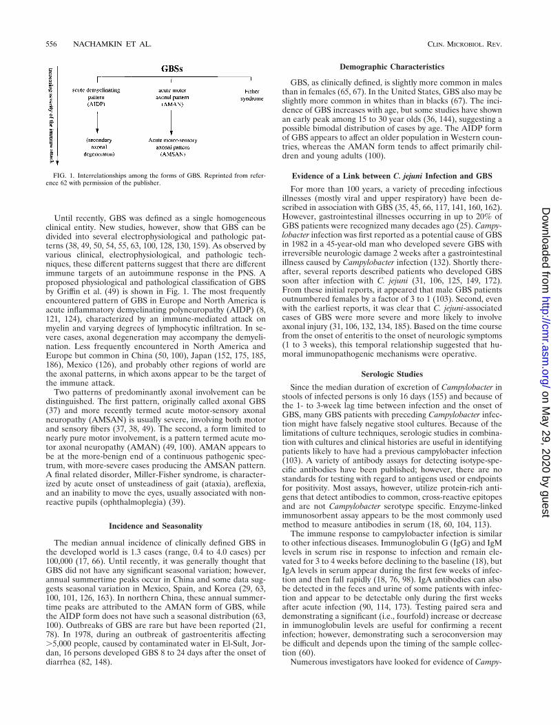
Until recently, GBS was defined as a single homogeneousclinical entity. New studies, however, show that GBS can bedivided into several electrophysiological and pathologic pat-terns (38, 49, 50, 54, 55, 63, 100, 128, 130, 159). As observed byvarious clinical, electrophysiological, and pathologic tech-niques, these different patterns suggest that there are differentimmune targets of an autoimmune response in the PNS. Aproposed physiological and pathological classification of GBSby Griffin et al. (49) is shown in Fig. 1. The most frequentlyencountered pattern of GBS in Europe and North America isacute inflammatory demyelinating polyneuropathy (AIDP) (8,121, 124), characterized by an immune-mediated attack onmyelin and varying degrees of lymphocytic infiltration. In se-vere cases, axonal degeneration may accompany the demyeli-nation. Less frequently encountered in North America andEurope but common in China (50, 100), Japan (152, 175, 185,186), Mexico (126), and probably other regions of world arethe axonal patterns, in which axons appear to be the target ofthe immune attack.
Two patterns of predominantly axonal involvement can bedistinguished. The first pattern, originally called axonal GBS(37) and more recently termed acute motor-sensory axonalneuropathy (AMSAN) is usually severe, involving both motorand sensory fibers (37, 38, 49). The second, a form limited tonearly pure motor involvement, is a pattern termed acute mo-tor axonal neuropathy (AMAN) (49, 100). AMAN appears tobe at the more-benign end of a continuous pathogenic spec-trum, with more-severe cases producing the AMSAN pattern.A final related disorder, Miller-Fisher syndrome, is character-ized by acute onset of unsteadiness of gait (ataxia), areflexia,and an inability to move the eyes, usually associated with non-reactive pupils (ophthalmoplegia) (39).
Incidence and Seasonality
The median annual incidence of clinically defined GBS inthe developed world is 1.3 cases (range, 0.4 to 4.0 cases) per100,000 (17, 66). Until recently, it was generally thought thatGBS did not have any significant seasonal variation; however,annual summertime peaks occur in China and some data sug-gests seasonal variation in Mexico, Spain, and Korea (29, 63,100, 101, 126, 163). In northern China, these annual summer-time peaks are attributed to the AMAN form of GBS, whilethe AIDP form does not have such a seasonal distribution (63,100). Outbreaks of GBS are rare but have been reported (21,78). In 1978, during an outbreak of gastroenteritis affecting.5,000 people, caused by contaminated water in El-Sult, Jor-dan, 16 persons developed GBS 8 to 24 days after the onset ofdiarrhea (82, 148).
Demographic Characteristics
GBS, as clinically defined, is slightly more common in malesthan in females (65, 67). In the United States, GBS also may beslightly more common in whites than in blacks (67). The inci-dence of GBS increases with age, but some studies have shownan early peak among 15 to 30 year olds (36, 144), suggesting apossible bimodal distribution of cases by age. The AIDP formof GBS appears to affect an older population in Western coun-tries, whereas the AMAN form tends to affect primarily chil-dren and young adults (100).
Evidence of a Link between C. jejuni Infection and GBS
For more than 100 years, a variety of preceding infectiousillnesses (mostly viral and upper respiratory) have been de-scribed in association with GBS (35, 45, 66, 117, 141, 160, 162).However, gastrointestinal illnesses occurring in up to 20% ofGBS patients were recognized many decades ago (25). Campy-lobacter infection was first reported as a potential cause of GBSin 1982 in a 45-year-old man who developed severe GBS withirreversible neurologic damage 2 weeks after a gastrointestinalillness caused by Campylobacter infection (132). Shortly there-after, several reports described patients who developed GBSsoon after infection with C. jejuni (31, 106, 125, 149, 172).From these initial reports, it appeared that male GBS patientsoutnumbered females by a factor of 3 to 1 (103). Second, evenwith the earliest reports, it was clear that C. jejuni-associatedcases of GBS were more severe and more likely to involveaxonal injury (31, 106, 132, 134, 185). Based on the time coursefrom the onset of enteritis to the onset of neurologic symptoms(1 to 3 weeks), this temporal relationship suggested that hu-moral immunopathogenic mechanisms were operative.
Serologic Studies
Since the median duration of excretion of Campylobacter instools of infected persons is only 16 days (155) and because ofthe 1- to 3-week lag time between infection and the onset ofGBS, many GBS patients with preceding Campylobacter infec-tion might have falsely negative stool cultures. Because of thelimitations of culture techniques, serologic studies in combina-tion with cultures and clinical histories are useful in identifyingpatients likely to have had a previous campylobacter infection(103). A variety of antibody assays for detecting isotype-spe-cific antibodies have been published; however, there are nostandards for testing with regard to antigens used or endpointsfor positivity. Most assays, however, utilize protein-rich anti-gens that detect antibodies to common, cross-reactive epitopesand are not Campylobacter serotype specific. Enzyme-linkedimmunosorbent assay appears to be the most commonly usedmethod to measure antibodies in serum (18, 60, 104, 113).
The immune response to campylobacter infection is similarto other infectious diseases. Immunoglobulin G (IgG) and IgMlevels in serum rise in response to infection and remain ele-vated for 3 to 4 weeks before declining to the baseline (18), butIgA levels in serum appear during the first few weeks of infec-tion and then fall rapidly (18, 76, 98). IgA antibodies can alsobe detected in the feces and urine of some patients with infec-tion and appear to be detectable only during the first weeksafter acute infection (90, 114, 173). Testing paired sera anddemonstrating a significant (i.e., fourfold) increase or decreasein immunoglobulin levels are useful for confirming a recentinfection; however, demonstrating such a seroconversion maybe difficult and depends upon the timing of the sample collec-tion (60).
Numerous investigators have looked for evidence of Campy-
FIG. 1. Interrelationships among the forms of GBS. Reprinted from refer-ence 62 with permission of the publisher.
556 NACHAMKIN ET AL. CLIN. MICROBIOL. REV.
on May 29, 2020 by guest
http://cmr.asm
.org/D
ownloaded from
lobacter infection in series of GBS patients, using serologicmethods. Not surprisingly, these serologic studies documenteda high prevalence of antibodies to C. jejuni in the serum ofpatients with GBS (52, 63, 77, 87, 135, 150, 170). Using immu-nodot assays to determine the frequency of C. jejuni antibod-ies, Gruenewald and colleagues found that 3 (18%) of 17patients with GBS in an uncontrolled population had elevatedtiters in two or more immunoglobulin classes (52). Similarly,using a complement fixation technique, Winer and colleaguesfound that 14% of 99 patients with GBS had positive C. jejuniserologic tests, compared to only 2% of controls (170). Kaldorand Speed, in a nonblinded serologic study of 56 patients withGBS and 57 controls found 38% of the patients and none ofthe controls met their criteria for positive serologic responses(77). In a large, blinded, case-control study, Mishu et al. eval-uated 118 GBS patients in the United States and 113 controls;36% of the GBS patients were seropositive for Campylobacter.GBS patients were more than five times likelier than controlsto have serologic evidence of recent Campylobacter infection(104). Serologic tests were done as a part of a Japanese studyof GBS patients, and 36% of the patients were seropositive forCampylobacter (87). In a prospective study, Ho et al. showedthat Campylobacter infections are common in both AMAN andAIDP patients from China and that, depending upon the def-inition of seropositivity, rates of infection ranged from 24 to76% for AMAN patients (63).
Culture Surveys
Although serologic studies suggested that some patients withGBS had preceding Campylobacter infection, culture studieswere needed to make a stronger case for this intriguing asso-ciation. Culture-based studies could underestimate the occur-rence of infection because of variation in culture techniques,the delayed onset of GBS after infection, and the low likeli-hood of positive cultures several weeks after infection.
As reported previously (111), “the ability to recover Campy-lobacter from patients with GBS is related to the duration ofexcretion of the organism following the patient’s acute diar-rheal illness. The convalescent excretion of Campylobacter or-ganisms after acute infection has been studied. In a study ofchildren from Thailand, Taylor et al. (157) found that theduration of excretion was 14 6 2 days for children ,1 year oldand 8 6 2 days for children 1–5 years old. In a cohort study ofMexican children ,5 years of age, Calva et al. (24) found thatthe duration of excretion of the organism was 7 days (range,7–26). Early studies by Karmali and Fleming (80) showed thatthe duration of excretion in 4% of untreated children contin-ued up to 6 weeks after the onset of symptoms. At 2 weeks,about two-thirds of the patients were positive for Campy-lobacter infection by culture, and at 4 weeks, one-third of pa-tients were still positive. About 50% of the patients withCampylobacter infection had negative stool cultures 2 weeksafter the onset of diarrheal illness in a Swedish series (155).Convalescent carriage of Campylobacter organisms averaged37.6 days (range, 15–69) in a Norwegian study (79).”
Nevertheless, several investigators have succeeded in isolat-ing C. jejuni from the stools of patients with GBS at the onsetof their neurologic symptoms. As reported previously (111), “Itis difficult to determine the frequency of Campylobacter infec-tion among patients with GBS because few studies have sys-tematically done cultures for Campylobacter species. Kuroki etal. (87) recovered C. jejuni from 30% of patients with GBS,whereas Rees et al. (129) had a recovery rate of only 8%.Ropper (135) recovered Campylobacter organisms from 4(44.4%) of 9 patients with diarrhea preceding GBS.”
Overall, Campylobacter was cultured from the stools of 8 to50% of GBS patients very soon after the onset of neurologicsymptoms (52, 87, 129, 135, 149). From both serologic andculture studies, it is estimated that at least 30 to 40% of GBSpatients are infected with Campylobacter 10 days to 2 weeksprior to the onset of their neurologic symptoms. Because of thelag time between C. jejuni infection and the onset of neurologicsymptoms, these numbers likely underestimate the associationbetween C. jejuni infection and GBS.
The vast majority of isolates obtained from patients withGBS have been reported as C. jejuni. It is not known whetherC. coli, which causes diarrheal illness that is indistinguishableclinically from C. jejuni infection and is difficult to differentiatefrom C. jejuni by phenotypic methods, is also associated withGBS (110). More recently, C. upsaliensis was recovered from aU.S. patient with AMAN (61), suggesting that other Campy-lobacter species may well be important in GBS. This has im-portant implications for the culture methods used to investi-gate Campylobacter and GBS.
Campylobacter Serotypes Associated with GBS
As previously discussed (111), “typing studies are critical toour understanding of the epidemiology and pathogenesis ofCampylobacter-associated GBS. In particular, serotyping stud-ies have led to the identification of potentially unique strainsinvolved in the pathogenesis of GBS. Two major serotypingschemes are used worldwide and detect heat-labile (HL) (96)and O (120) antigens. The HL serotyping scheme originallydescribed by Lior (96) detects over 100 serotypes of C. jejuni,C. coli, and C. lari. Uncharacterized bacterial surface antigensand, in some serotypes, flagella are the serodeterminants forthis serotyping system (4). The Penner O serotyping scheme(120) detects 60 types of C. jejuni and C. coli (118) and is basedon detection of LPS antigens.”
Although serologic and culture studies showed that somepatients with GBS had evidence of infection, the landmarkstudy of Kuroki and colleagues (87) solidified the associationof Campylobacter and GBS. In that study conducted in Japan,Kuroki et al. isolated C. jejuni from 14 of 46 GBS patients(30.4%) compared with only 6 (1.2%) of 503 subjects in ahealthy control population. By O serotyping to characterize theisolates, 10 of 12 available isolates had the same O serotype,O:19. This serotype, however, occurred in only 1.7% of 1,150C. jejuni isolates from patients with uncomplicated gastroin-testinal infection. Lectin typing of the 10 O:19 strains showedthat all belonged to lectin type 8, whereas only 1 of 14 O:19strains from uncomplicated enteritis cases belonged to thistype. A recent analysis of 31 strains of C. jejuni from GBSpatients by Yuki et al. (180) showed that 52% of strains wereO:19 strains but that these strains occurred in 5% of 215 strainsfrom patients with uncomplicated gastroenteritis. In isolatesfrom patients with Miller-Fisher syndrome, O:2 strains wereoverrepresented compared to control isolates (71 and 38%,respectively) although only seven patients were studied (180).Thus, this study provided clear evidence that certain types of C.jejuni were associated with the development of GBS.
Further studies, however, have shown that other O serotypesthat occur more frequently in uncomplicated infections werebeing isolated from patients with GBS. In particular, O:41strains have been recently found to be highly associated withGBS patients in South Africa (46, 92). O serotypes from GBSpatients that have been reported include O:1, O:2, O:4, O:4complex, O:5, O:10, O:16, O:23, O:37, O:44, and O:64 (9, 73,87, 129, 140, 145, 180, 182, 183). In contrast, an association ofspecific HL (Lior) serotypes in GBS has not been found at this
VOL. 11, 1998 CAMPYLOBACTER SPECIES AND GUILLAIN-BARRE SYNDROME 557
on May 29, 2020 by guest
http://cmr.asm
.org/D
ownloaded from

time. For example, among eight strains of C. jejuni O:19 thatwere studied by flagellin gene typing, there were four differentHL serotypes represented, including HL7, HL23, HL70, andHL84 (112). Yuki et al. found that among 16 O:19 strains fromGBS patients, 12 (75%) were serotype HL7 whereas only 3 of11 (27%) O:19 strains from non-GBS patients were serotypeHL7 (180). Thus, these studies suggest that there may besubtypes of C. jejuni involved in eliciting GBS and/or that hostsusceptibility plays an important role in determining the out-come of uncomplicated Campylobacter infection.
In various studies, the occurrence of O:19 strains causinguncomplicated gastrointestinal infection has been estimated at1 to 6% (3, 44, 75, 102). Serotype O:19 has also been found inlaboratory animals, including dogs, cats, and primates (158).Several outbreaks of campylobacter infection in which O:19serotypes were implicated have been reported (75, 119); how-ever, only in one case associated with O:19 did the patientdevelop GBS (138).
Risk of Developing GBS after C. jejuni Infection
Although C. jejuni infections appear to commonly precedeGBS, C. jejuni infections occur far more commonly than GBS;therefore, the risk of developing GBS after infection withCampylobacter is actually quite low. The U.S. Centers for Dis-ease Control and Prevention estimates there are 1,000 cases ofC. jejuni infection per 100,000 per year (156). The NationalCenter for Health Statistics Hospital Discharge data docu-mented 7,874 GBS cases in the United States in 1995. There-fore, assuming that 30% of GBS cases are preceded by C. jejuniinfection and that the U.S. population is 250 million, it can beestimated that 1 of every 1,058 cases of C. jejuni infection isfollowed by GBS.
The risk of developing GBS may be higher after infectionwith C. jejuni type O:19. Of 12 C. jejuni isolates from JapaneseGBS patients, 10 were serotype O:19 (87). This O:19 typerepresents less than 2% of C. jejuni isolates from patients withuncomplicated enteritis in Japan. The association betweenO:19 and GBS is not as strong outside of Japan. For example,in the United States, two of seven GBS-associated Campy-lobacter isolates were serotype O:19 (105); this is still signifi-cant, since O:19 accounts for only 3% of isolates from patientswith uncomplicated enteritis. In a British study (129), fourCampylobacter isolates from GBS patients were serotyped; twowere nontypeable, and the other two were not type O:19.Assuming that 20% of GBS-associated C. jejuni isolates areserotype O:19, then the risk of developing GBS after infectionwith C. jejuni type O:19 is estimated to be 1 in 158.
Although not well defined, some investigators have reportedthat GBS following Campylobacter infection may be more se-vere and result in more irreversible neurologic damage thanGBS following other putative infections. Of 58 GBS patientsstudied by Vriesendorp and colleagues (167), 10 had serologicevidence of recent C. jejuni infection, and of these, 3 (30%)had severe disease. Severe disease was defined as fulminatingdisease with quadriplegia and ventilatory dependence within24 to 48 h of onset. None of the 48 patients without recent C.jejuni infection had severe disease. In a British study of 101GBS patients, 23% of GBS patients with Campylobacter infec-tion were unable to walk unassisted 1 year after the onset ofsymptoms, compared with only 9% of uninfected GBS patients(129). Similarly, in The Netherlands 14 of 24 C. jejuni-infectedGBS patients treated with plasma exchange were unable towalk unassisted 6 months after the onset of their symptoms,compared with only 12% of similarly treated GBS patientswithout evidence of preceding Campylobacter infection (74).
Additional prospective studies on a larger number of patientswith and without Campylobacter infection and defined accord-ing to clinical and electrophysiological criteria are needed tosubstantiate these reports.
PATHOGENESIS
Peripheral nerves are composed of numerous motor andsensory fibers. The motor fibers originate from motor neuronsin the ventral horns of the spinal cord and carry nerve impulsesto the muscles. The sensory fibers carry nerve impulses fromthe specialized sensory receptors in the periphery to the spinalcord. Their cell body resides in the dorsal root ganglia next tothe spinal cord. In order to speed the conduction of thesenerve impulses, some of these fibers are wrapped by insulatinglayers of myelin formed by Schwann cells. Between two adja-cent myelin sheaths is a gap called the node of Ranvier, wheresodium channels are concentrated. This specialized structureallows nerve impulses to regenerate. The myelin sheaths pre-vent impulses from leaking away and allow impulses to jumpfrom one node to the next. The nerve impulses can be effi-ciently conducted at up to 75 m/s by the myelinated nerves.
Access to the PNS by the immune system requires that theblood-nerve barrier be altered. Specialized endothelial cellsline the blood vessels inside the endoneurium (the connectivetissue enveloping individual nerve fibers within a peripheralnerve). Part of the blood-nerve barrier is due to the presenceof negatively charged sialic acid containing glycoconjugates inthe lumen that repel negatively charged molecules (94, 123).Tight junctions between endothelial cells contribute to thisbarrier. Entry of molecules around the nerve is also limited bythe perineurium (the connective tissue sheath surrounding afascicle of nerve fibers in a peripheral nerve). This structureconsists of layers of specialized fibroblasts, each layer of whichis bounded by a basal lamina with tight junctions betweenadjacent perineural cells. However, the blood-nerve barrier isnot as tight as the blood-brain barrier, so that small amounts ofcirculating proteins such as albumin, IgG, and exogenouslyadministered horseradish peroxidase (none of which can enterthe central nervous system [CNS]) (99) can gain entrance tothe endoneurial space (7). This relative leakiness may renderthe PNS more vulnerable than the CNS to antibody-mediateddisorders. The blood-nerve barrier is particularly leaky withinthe dorsal root ganglia and is altogether absent at nerve ter-minals in the periphery (for example, at the neuromuscularjunction), making these areas especially vulnerable to immune-mediated attacks.
Mechanisms of Immune Injury to Nerve Fibers in GBS
AIDP. On physical examination, patients with AIDP presentwith flaccid paralysis, areflexia, and usually some sensory loss.Electrophysiological testing typically reveals findings sugges-tive of demyelination in both motor and sensory nerves (43,63). Pathologically, macrophage-mediated demyelination andlymphocytic infiltrates are evident (8, 50). As previously dis-cussed (62), “AIDP has long been presumed to be a T-cell-mediated disorder based on the lymphocytic inflammationfound in many cases (8) and on the analogy to experimentalallergic neuritis (EAN) (for reviews, see references 6 and 57 to59). Many markers of T-cell activation can be detected in theserum of AIDP patients, including soluble interleukin-2 recep-tor and gamma interferon (14). However, several lines of evi-dence have suggested the importance of antibody-mediatednerve fiber damage in AIDP, including the response to plas-mapheresis (40, 53), the presence of antimyelin antibodies as
558 NACHAMKIN ET AL. CLIN. MICROBIOL. REV.
on May 29, 2020 by guest
http://cmr.asm
.org/D
ownloaded from
detected in complement activation assays (85, 86), the frequentpresence of antiglycoconjugate antibodies, and the demonstra-tion of demyelinating immunoglobulins in sera by either inject-ing the sera intraneurally (153) or incubating the sera withnerve or Schwann cells in vitro (16, 85, 142, 143).”
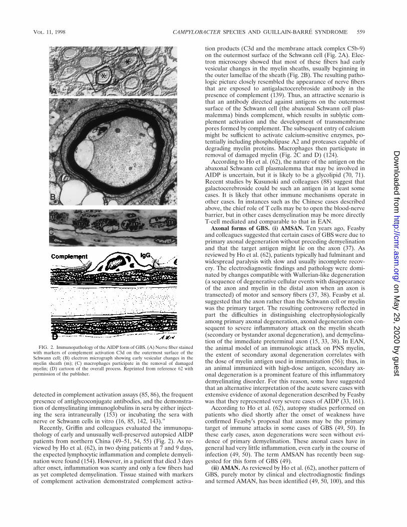
Recently, Griffin and colleagues evaluated the immunopa-thology of early and unusually well-preserved autopsied AIDPpatients from northern China (49–51, 54, 55) (Fig. 2). As re-viewed by Ho et al. (62), in two dying patients at 7 and 9 days,the expected lymphocytic inflammation and complete demyeli-nation were found (154). However, in a patient that died 3 daysafter onset, inflammation was scanty and only a few fibers hadas yet completed demyelination. Tissue stained with markersof complement activation demonstrated complement activa-
tion products (C3d and the membrane attack complex C5b-9)on the outermost surface of the Schwann cell (Fig. 2A). Elec-tron microscopy showed that most of these fibers had earlyvesicular changes in the myelin sheaths, usually beginning inthe outer lamellae of the sheath (Fig. 2B). The resulting patho-logic picture closely resembled the appearance of nerve fibersthat are exposed to antigalactocerebroside antibody in thepresence of complement (139). Thus, an attractive scenario isthat an antibody directed against antigens on the outermostsurface of the Schwann cell (the abaxonal Schwann cell plas-malemma) binds complement, which results in sublytic com-plement activation and the development of transmembranepores formed by complement. The subsequent entry of calciummight be sufficient to activate calcium-sensitive enzymes, po-tentially including phospholipase A2 and proteases capable ofdegrading myelin proteins. Macrophages then participate inremoval of damaged myelin (Fig. 2C and D) (124).
According to Ho et al. (62), the nature of the antigen on theabaxonal Schwann cell plasmalemma that may be involved inAIDP is uncertain, but it is likely to be a glycolipid (70, 71).Recent studies by Kusunoki and colleagues (88) suggest thatgalactocerebroside could be such an antigen in at least somecases. It is likely that other immune mechanisms operate inother cases. In instances such as the Chinese cases describedabove, the chief role of T cells may be to open the blood-nervebarrier, but in other cases demyelination may be more directlyT-cell mediated and comparable to that in EAN.
Axonal forms of GBS. (i) AMSAN. Ten years ago, Feasbyand colleagues suggested that certain cases of GBS were due toprimary axonal degeneration without preceding demyelinationand that the target antigen might lie on the axon (37). Asreviewed by Ho et al. (62), patients typically had fulminant andwidespread paralysis with slow and usually incomplete recov-ery. The electrodiagnostic findings and pathology were domi-nated by changes compatible with Wallerian-like degeneration(a sequence of degenerative cellular events with disappearanceof the axon and myelin in the distal axon when an axon istransected) of motor and sensory fibers (37, 38). Feasby et al.suggested that the axon rather than the Schwann cell or myelinwas the primary target. The resulting controversy reflected inpart the difficulties in distinguishing electrophysiologicallyamong primary axonal degeneration, axonal degeneration con-sequent to severe inflammatory attack on the myelin sheath(secondary or bystander axonal degeneration), and demyelina-tion of the immediate preterminal axon (15, 33, 38). In EAN,the animal model of an immunologic attack on PNS myelin,the extent of secondary axonal degeneration correlates withthe dose of myelin antigen used in immunization (56); thus, inan animal immunized with high-dose antigen, secondary ax-onal degeneration is a prominent feature of this inflammatorydemyelinating disorder. For this reason, some have suggestedthat an alternative interpretation of the acute severe cases withextensive evidence of axonal degeneration described by Feasbywas that they represented very severe cases of AIDP (33, 161).
According to Ho et al. (62), autopsy studies performed onpatients who died shortly after the onset of weakness haveconfirmed Feasby’s proposal that axons may be the primarytarget of immune attacks in some cases of GBS (49, 50). Inthese early cases, axon degenerations were seen without evi-dence of primary demyelination. These axonal cases have ingeneral had very little inflammation, even early in the course ofinfection (49, 50). The term AMSAN has recently been sug-gested for this form of GBS (49).
(ii) AMAN. As reviewed by Ho et al. (62), another pattern ofGBS, purely motor by clinical and electrodiagnostic findingsand termed AMAN, has been identified (49, 50, 100), and this
FIG. 2. Immunopathology of the AIDP form of GBS. (A) Nerve fiber stainedwith markers of complement activation C3d on the outermost surface of theSchwann cell; (B) electron micrograph showing early vesicular changes in themyelin sheath (m); (C) macrophages participate in the removal of damagedmyelin; (D) cartoon of the overall process. Reprinted from reference 62 withpermission of the publisher.
VOL. 11, 1998 CAMPYLOBACTER SPECIES AND GUILLAIN-BARRE SYNDROME 559
on May 29, 2020 by guest
http://cmr.asm
.org/D
ownloaded from
pattern of GBS can usually be distinguished from other formsof GBS (63). The clinical features of the AMAN pattern havelargely been established by studies in northern China. Everysummer, hundreds of children and young adults with GBSinundate the hospitals of northern China. Over 70% of GBSpatients studied at one hospital, the Second Teaching Hospitalin Shijiazhuang, showed the clinical and electrodiagnostic pic-ture termed AMAN (63, 100). AMAN is characterized byweakness or paralysis without sensory loss. Electrodiagnosticdata suggest that motor fibers can be lost selectively, whilesensory nerve fibers are preserved and features of demyelina-tion are absent (63, 100). It is now clear that the AMANpattern of GBS occurs frequently in other parts of Asia (29)and less often in North America (61, 72), Europe (97, 128,129), and Latin America (126). The AMAN pattern is closelyassociated with antecedent Campylobacter infection (63, 129,130, 177).
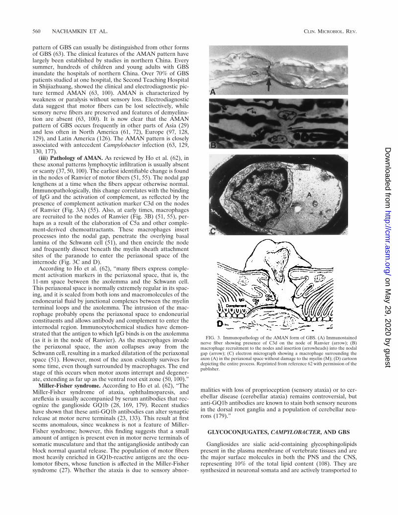
(iii) Pathology of AMAN. As reviewed by Ho et al. (62), inthese axonal patterns lymphocytic infiltration is usually absentor scanty (37, 50, 100). The earliest identifiable change is foundin the nodes of Ranvier of motor fibers (51, 55). The nodal gaplengthens at a time when the fibers appear otherwise normal.Immunopathologically, this change correlates with the bindingof IgG and the activation of complement, as reflected by thepresence of complement activation marker C3d on the nodesof Ranvier (Fig. 3A) (55). Also, at early times, macrophagesare recruited to the nodes of Ranvier (Fig. 3B) (51, 55), per-haps as a result of the elaboration of C5a and other comple-ment-derived chemoattractants. These macrophages insertprocesses into the nodal gap, penetrate the overlying basallamina of the Schwann cell (51), and then encircle the nodeand frequently dissect beneath the myelin sheath attachmentsites of the paranode to enter the periaxonal space of theinternode (Fig. 3C and D).
According to Ho et al. (62), “many fibers express comple-ment activation markers in the periaxonal space, that is, the11-nm space between the axolemma and the Schwann cell.This periaxonal space is normally extremely regular in its spac-ing, and it is sealed from both ions and macromolecules of theendoneurial fluid by junctional complexes between the myelinterminal loops and the axolemma. The intrusion of the mac-rophage probably opens the periaxonal space to endoneurialconstituents and allows antibody and complement to enter theinternodal region. Immunocytochemical studies have demon-strated that the antigen to which IgG binds is on the axolemma(as it is in the node of Ranvier). As the macrophages invadethe periaxonal space, the axon collapses away from theSchwann cell, resulting in a marked dilatation of the periaxonalspace (51). However, most of the axon evidently survives forsome time, even though surrounded by macrophages. The endstage of this occurs when motor axons interrupt and degener-ate, extending as far up as the ventral root exit zone (50, 100).”
Miller-Fisher syndrome. According to Ho et al. (62), “TheMiller-Fisher syndrome of ataxia, ophthalmoparesis, andareflexia is usually accompanied by serum antibodies that rec-ognize the ganglioside GQ1b (28, 169, 179). Recent studieshave shown that these anti-GQ1b antibodies can alter synapticrelease at motor nerve terminals (23, 133). This result at firstseems anomalous, since weakness is not a feature of Miller-Fisher syndrome; however, this finding suggests that a smallamount of antigen is present even in motor nerve terminals ofsomatic musculature and that the antiganglioside antibody canblock normal quantal release. The population of motor fibersmost heavily enriched in GQ1b-reactive antigens are the ocu-lomotor fibers, whose function is affected in the Miller-Fishersyndrome (27). Whether the ataxia is due to sensory abnor-
malities with loss of proprioception (sensory ataxia) or to cer-ebellar disease (cerebellar ataxia) remains controversial, butanti-GQ1b antibodies are known to stain both sensory neuronsin the dorsal root ganglia and a population of cerebellar neu-rons (179).”
GLYCOCONJUGATES, CAMPYLOBACTER, AND GBS
Gangliosides are sialic acid-containing glycosphingolipidspresent in the plasma membrane of vertebrate tissues and arethe major surface molecules in both the PNS and the CNS,representing 10% of the total lipid content (108). They aresynthesized in neuronal somata and are actively transported to
FIG. 3. Immunopathology of the AMAN form of GBS. (A) Immunostainednerve fiber showing presence of C3d on the node of Ranvier (arrow); (B)macrophage recruitment to the nodes and insertion (arrowheads) into the nodalgap (arrow); (C) electron micrograph showing a macrophage surrounding theaxon (A) in the periaxonal space without damage to the myelin (M); (D) cartoondepicting the entire process. Reprinted from reference 62 with permission of thepublisher.
560 NACHAMKIN ET AL. CLIN. MICROBIOL. REV.
on May 29, 2020 by guest
http://cmr.asm
.org/D
ownloaded from
specific sites of enrichment, such as synapses and nodes ofRanvier. Where different gangliosides are enriched in the ner-vous system can be found by using toxins and lectins that havehigh specificities of different oligosaccharides epitopes. Chol-era toxin, which has high affinity to the GM1 epitope, binds tothe nodes of Ranvier and paranodes, including the paranodalSchwann cell (32, 145). Peanut agglutinin, which has high af-finity to asialo-GM1, binds to the nodes of Ranvier only (5,145). Tetanus toxin, which binds to the B series gangliosides(disialosyl gangliosides, e.g., GT1b and GD1b), shows bindingto both nodal and internodal axons (145).
Antiglycoconjugate Antibodies
Antiglycoconjugate antibodies, often referred to as antigan-glioside antibodies, are frequently found in both AMAN andAIDP patients (63, 69, 71, 84, 168, 178, 187). Some immuno-chemical evidence supports the hypothesis that glycolipids(rather than glycoproteins) are the target antigen in GBS(168). Yuki et al. found that the proportion of patients withIgG and IgM antibodies against GM1 were higher for patientsinfected with O:19 strains than for patients infected with non-O:19 strains (86 versus 45% for IgG and 79 versus 40% forIgM) (180). These antiglycoconjugate antibodies are more fre-quently detected in the sera of AMAN patients than in those ofAIDP patients. In particular, IgG anti-GM1 antibodies arereported to be relatively specific for AMAN and are rarelyfound in other disorders (84). However, this assay has notproved to be sensitive; less than 50% of Chinese AMAN pa-tients are positive for this antibody (63). IgG anti-GD1a anti-bodies appear to be a more specific marker for the axonal formof GBS (64, 97, 184, 186).
An important issue in evaluating the possible role of antig-lycoconjugate antibodies in GBS is whether appropriate anti-gens are at the sites of known antibody binding in nerve fibers.Studies of ganglioside localization indicate that GM1-likeepitopes are concentrated at the nodal region as well as at theparanodal myelin loops (32, 145), the same sites where IgGand complement have been shown to bind in autopsied AMANpatients (51, 55). With regard to the predominantly motorinvolvement in AMAN cases, it is noteworthy that when hu-man motor and sensory roots were compared, Ogawa-Goto etal. found that motor nerve myelin contained abundant GM1(15% of total gangliosides) whereas sensory nerve myelin con-tained only trace amounts (115). GT1b and possibly GD1a alsoappear to be concentrated on the axolemma and may act asligands for the myelin-associated glycoprotein. Myelin-associ-ated glycoprotein is concentrated on the Schwann cell adax-onal surface (174) and has been postulated to bind to axonalGT1b or GD1a in the periaxonal space.
MOLECULAR MIMICRY AND GBS
While the proof that Campylobacter causes GBS still awaitsdefinitive evidence, several lines of evidence support the hy-pothesis that structural features of Campylobacter elicit an au-toimmune-mediated attack against host nerve tissue. SomeGBS patients have antiganglioside antibodies, some lipopoly-saccharides (LPS) of C. jejuni organisms isolated from GBSpatients have ganglioside-like structures, and ganglioside-likemoieties are present on relevant sites on nerve fibers.
Some post-Campylobacter infection GBS patients have anti-bodies against the sugar antigen Gal(b1-3)GalNAc, a sequenceof sugars present in the ganglioside GM1 (9, 185). These an-tibodies may be overrepresented in axonal cases but occur indemyelinating cases as well (185). The correlation of antigan-
glioside antibodies in some patients with GBS but not in othersmay be in large part due to technical factors for assaying theseantibodies (168). A variety of models, including intraneuralinjection of antibodies and the mouse hemidiaphragm model,have shown physiologic effects of sera containing anti-GM1and anti-GQ1b, supporting the role of these targets in thepathogenesis of GBS (168).
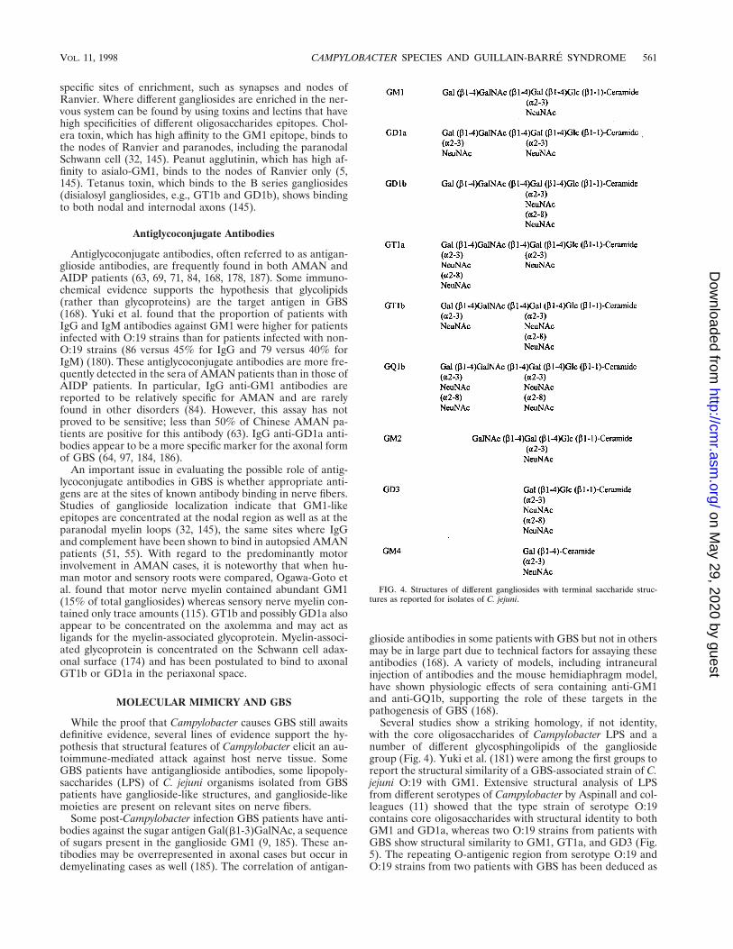
Several studies show a striking homology, if not identity,with the core oligosaccharides of Campylobacter LPS and anumber of different glycosphingolipids of the gangliosidegroup (Fig. 4). Yuki et al. (181) were among the first groups toreport the structural similarity of a GBS-associated strain of C.jejuni O:19 with GM1. Extensive structural analysis of LPSfrom different serotypes of Campylobacter by Aspinall and col-leagues (11) showed that the type strain of serotype O:19contains core oligosaccharides with structural identity to bothGM1 and GD1a, whereas two O:19 strains from patients withGBS show structural similarity to GM1, GT1a, and GD3 (Fig.5). The repeating O-antigenic region from serotype O:19 andO:19 strains from two patients with GBS has been deduced as
FIG. 4. Structures of different gangliosides with terminal saccharide struc-tures as reported for isolates of C. jejuni.
VOL. 11, 1998 CAMPYLOBACTER SPECIES AND GUILLAIN-BARRE SYNDROME 561
on May 29, 2020 by guest
http://cmr.asm
.org/D
ownloaded from
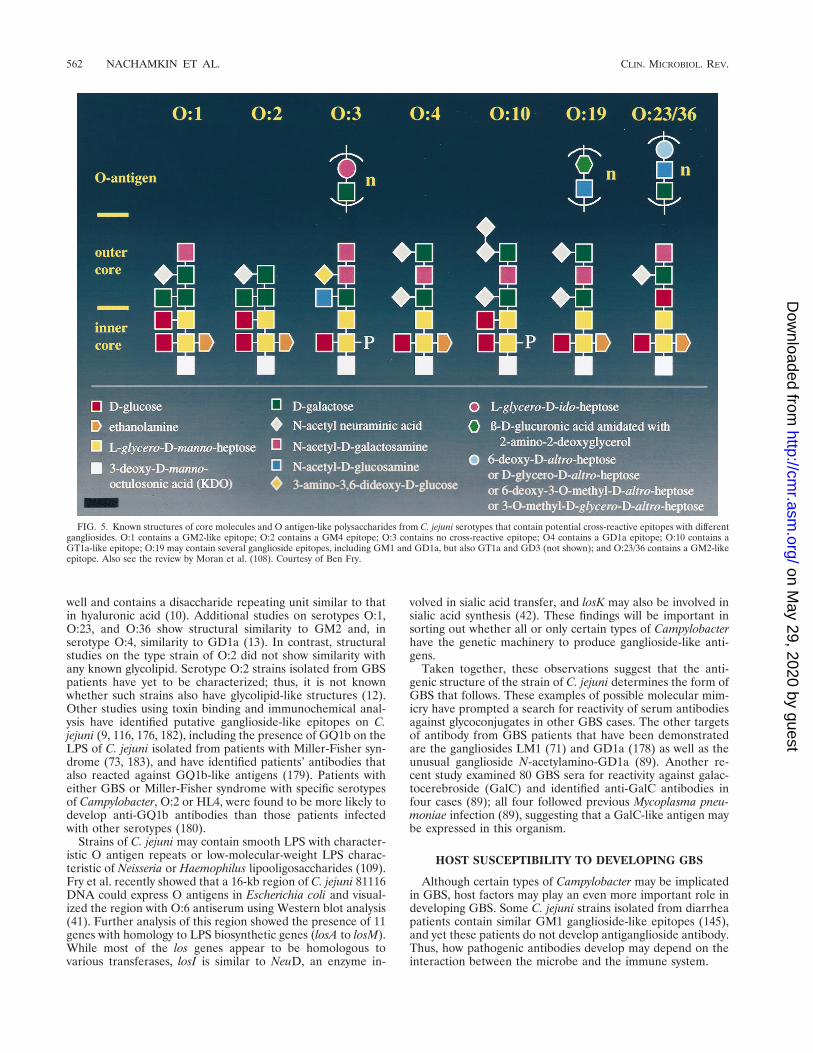
well and contains a disaccharide repeating unit similar to thatin hyaluronic acid (10). Additional studies on serotypes O:1,O:23, and O:36 show structural similarity to GM2 and, inserotype O:4, similarity to GD1a (13). In contrast, structuralstudies on the type strain of O:2 did not show similarity withany known glycolipid. Serotype O:2 strains isolated from GBSpatients have yet to be characterized; thus, it is not knownwhether such strains also have glycolipid-like structures (12).Other studies using toxin binding and immunochemical anal-ysis have identified putative ganglioside-like epitopes on C.jejuni (9, 116, 176, 182), including the presence of GQ1b on theLPS of C. jejuni isolated from patients with Miller-Fisher syn-drome (73, 183), and have identified patients’ antibodies thatalso reacted against GQ1b-like antigens (179). Patients witheither GBS or Miller-Fisher syndrome with specific serotypesof Campylobacter, O:2 or HL4, were found to be more likely todevelop anti-GQ1b antibodies than those patients infectedwith other serotypes (180).
Strains of C. jejuni may contain smooth LPS with character-istic O antigen repeats or low-molecular-weight LPS charac-teristic of Neisseria or Haemophilus lipooligosaccharides (109).Fry et al. recently showed that a 16-kb region of C. jejuni 81116DNA could express O antigens in Escherichia coli and visual-ized the region with O:6 antiserum using Western blot analysis(41). Further analysis of this region showed the presence of 11genes with homology to LPS biosynthetic genes (losA to losM).While most of the los genes appear to be homologous tovarious transferases, losI is similar to NeuD, an enzyme in-
volved in sialic acid transfer, and losK may also be involved insialic acid synthesis (42). These findings will be important insorting out whether all or only certain types of Campylobacterhave the genetic machinery to produce ganglioside-like anti-gens.
Taken together, these observations suggest that the anti-genic structure of the strain of C. jejuni determines the form ofGBS that follows. These examples of possible molecular mim-icry have prompted a search for reactivity of serum antibodiesagainst glycoconjugates in other GBS cases. The other targetsof antibody from GBS patients that have been demonstratedare the gangliosides LM1 (71) and GD1a (178) as well as theunusual ganglioside N-acetylamino-GD1a (89). Another re-cent study examined 80 GBS sera for reactivity against galac-tocerebroside (GalC) and identified anti-GalC antibodies infour cases (89); all four followed previous Mycoplasma pneu-moniae infection (89), suggesting that a GalC-like antigen maybe expressed in this organism.
HOST SUSCEPTIBILITY TO DEVELOPING GBS
Although certain types of Campylobacter may be implicatedin GBS, host factors may play an even more important role indeveloping GBS. Some C. jejuni strains isolated from diarrheapatients contain similar GM1 ganglioside-like epitopes (145),and yet these patients do not develop antiganglioside antibody.Thus, how pathogenic antibodies develop may depend on theinteraction between the microbe and the immune system.
FIG. 5. Known structures of core molecules and O antigen-like polysaccharides from C. jejuni serotypes that contain potential cross-reactive epitopes with differentgangliosides. O:1 contains a GM2-like epitope; O:2 contains a GM4 epitope; O:3 contains no cross-reactive epitope; O4 contains a GD1a epitope; O:10 contains aGT1a-like epitope; O:19 may contain several ganglioside epitopes, including GM1 and GD1a, but also GT1a and GD3 (not shown); and O:23/36 contains a GM2-likeepitope. Also see the review by Moran et al. (108). Courtesy of Ben Fry.
562 NACHAMKIN ET AL. CLIN. MICROBIOL. REV.
on May 29, 2020 by guest
http://cmr.asm
.org/D
ownloaded from
It is unclear whether host genetic factors are important inGBS. Several investigators have studied HLA molecules inpatients with GBS (1, 91, 93, 130, 151). Chiba et al. (26) wereunable to find HLA class I associations in patients with GBS orMiller-Fisher syndrome; however, insensitive serologic analysisfor class I (A, B, and C) and class II (DR and DQ) wereperformed. Yuki et al. (178) demonstrated an increased fre-quency of HLA B35 in patients with GBS following Campy-lobacter infection; only five patients were studied. In a morerecent study by Yuki et al. (180), B35 was only slightly in-creased in GBS patients (21%) versus controls (13.9%). Goro-dezky et al. (48) suggested a possible association of GBS withDR3 in Mexican patients. In a well-controlled study by Rees etal., 83% of C. jejuni-positive GBS patients had HLADQB1*03, compared with 49% of the C. jejuni-negative GBSpatients (P 5 0.05) (130). However, Yuki et al. did not findDQB1*03 to be associated with GBS (180). Preliminary studieson patients in China with either the AMAN or AIDP form ofGBS indicate that certain HLA alleles are overrepresented inthe different forms of AMAN as compared to controls (107).This raises the interesting issue of whether solely host factorsdetermine the outcome of GBS.
ANIMAL MODELS OF DISEASE
EAN is a T-cell-mediated disease in Lewis rats and is con-sidered to be the in vivo model for GBS (136). Injection ofLewis rats with proteins or peptides derived from myelin of thePNS induces a primarily T-cell-mediated disease with patho-logic features of GBS (demyelination). The model has not,however, been found to be an animal model for Campy-lobacter-induced GBS (137, 166). Only recently has there beensome suggestion that animal models of Campylobacter-inducedGBS can be developed and used to study pathogenic mecha-nisms. Based on a case of GBS in a human following exposureto paralyzed chickens with pathology similar to that of humanAMAN, Li et al. (95) used a C. jejuni isolate from a patientwith AMAN to develop an animal model of AMAN. Numer-ous chickens infected with C. jejuni isolated from an AMANpatient developed paralysis, and examination of their nervesshowed Wallerian-like degeneration similar to that seen in thehuman form of the disease (95). These preliminary studiessuggest that an animal model of AMAN can be developed byusing the specific strain of C. jejuni.
DIAGNOSTIC CONSIDERATIONS
As discussed by Nachamkin (111), “the isolation of Campy-lobacter organisms from patients with GBS will greatly dependupon the methods used and upon whether the patient has beengiven antimicrobial therapy for a previous illness. Antimicro-bial agents, including the fluoroquinolones and macrolides,commonly used for treating diarrheal disease have excellentactivity against Campylobacter species. Such agents quicklyclear Campylobacter organisms from the gastrointestinal tract,making the isolation of the organisms nearly impossible. . . .Other antimicrobial agents used to treat seemingly unrelatedillnesses may also affect the recovery of Campylobacter species.It is important, therefore, to elicit a history of antimicrobial usefrom patients with GBS, as their use will have a marked impacton culture results.”
For optimal isolation of Campylobacter from patients withGBS, multiple stool samples should be obtained for increasedsensitivity (87, 146, 164). Only one study of GBS patients hasexamined this issue. As reported previously (111), “Kuroki etal. (87) found that of 14 GBS patients with Campylobacter-
positive stool cultures, 57% were detected with 1 sample, 93%were detected with 2 samples, and all were detected with 3samples.” Thus, multiple stool samples (or rectal swabs)should be obtained from GBS patients immediately upon ad-mission to the hospital, preferably 3 over a 3-day period. Sam-ples should be transported to the laboratory in a suitable trans-port medium such as Cary-Blair medium. Both direct platingand enrichment methods should be used (110, 111).
A number of primary selective media can be used for iso-lating C. jejuni and C. coli, including blood-containing (131,147) and blood-free media (19, 47, 68, 81; reviewed in refer-ence 110). As previously described (111), “enrichment culturemethods are designed to isolate Campylobacter organisms fromsamples containing low numbers of organisms. In cases ofCampylobacter associated GBS, we presume that the level ofCampylobacter in the stool, if present, is likely to be lower thanthe concentration during the acute diarrheal illness. Thus, en-richment cultures should be included among the laboratorytests for patients with GBS. A study by Taylor et al. (157)clearly showed that enrichment cultures dramatically improvedthe yield of Campylobacter organisms when most GBS patientswould be seen. An increase of as much as 31% over conven-tional plating techniques was seen when patients were cultured.20 days after the onset of their diarrheal illness.” A numberof enrichment culture media can be used. In our studies inNorthern China and elsewhere, we have had good success withPreston enrichment broth (20).
THERAPY
The major reduction of mortality in GBS has been due toadvances in supportive care of critically ill patients. However,increased understanding of the immunologic basis of this dis-ease over the past 15 years has allowed us to change the naturalcourse of this disease. Plasmapheresis was the first therapyshown to be effective in speeding up the course of recovery (40,53). In this procedure, a patient’s blood is removed and cen-trifuged to separate the cellular and plasma components andthe cellular components, diluted with artificial plasminate, arereinfused to the patient. The therapeutic effect is presumablydue to the removal of inciting circulating factors such as anti-bodies. Another effective therapy is administration of IVIG.There have been two controlled studies of the use of IVIG inGBS (122, 165). Both indicated that IVIG is as effective asplasmapheresis in the treatment of GBS (122, 165). The mech-anism of action of infused immunoglobulin is not clear, but onepossibility is that pooled immunoglobulins contain anti-idio-typic antibodies that inactivate the disease-specific antibodies.
CONCLUSIONS AND FUTURE DIRECTIONS
Based on serologic and culture evidence, the association ofCampylobacter with the development of GBS now appearsfirmly established. However, much work remains to determinehow Campylobacter can induce this disease. A recent consensusmeeting on Campylobacter and GBS was conducted by theNational Institutes of Health and outlined several areas ofresearch (91) for the future as follows. (i) Conduct additionalsurveillance studies to provide a better estimate of infectionrates and incidence of GBS and to obtain a much better pictureof the epidemiology of Campylobacter-induced GBS. (ii) En-courage studies to assess the host susceptibility to GBS. (iii)Standardized microbiological laboratory procedures areneeded to ensure isolation of Campylobacter strains associatedwith GBS. (iv) Serologic assays for diagnosis of C. jejuni infec-tions need to be standardized and validated. (v) A diagnostic
VOL. 11, 1998 CAMPYLOBACTER SPECIES AND GUILLAIN-BARRE SYNDROME 563
on May 29, 2020 by guest
http://cmr.asm
.org/D
ownloaded from
test specific for various Campylobacter species is needed, sincea large number of Campylobacter infections likely goes unrec-ognized. (vi) An animal model for Campylobacter enteritis withensuing GBS is urgently needed. (vii) There should be aCampylobacter strain bank established in which bacterialstrains isolated from patients who develop GBS (or variants)can be deposited. These strains should be available to re-searchers in the field. (viii) There is a need for standardizedreagents, particularly monoclonal antibodies, that can be usedto identify bacterial epitopes and which can be tested for theirability to react with different neural targets. (ix) Better typingsystems (phenotypic and molecular) are needed. (x) Additionalstudies on the pathogenesis of the different forms of GBS areneeded.
REFERENCES1. Adams, D., J. D. Gibson, P. K. Thomas, J. R. Batchelor, R. A. Hughes, L.
Kennedy, H. Festenstein, and J. Sachs. 1977. HLA antigens in Guillain-Barre syndrome. Lancet ii:504–505.
2. Adams, R. D., and M. Victor. 1993. Diseases of the peripheral nerves, p.1117–1169. In R. D. Adams and M. Victor (ed.), Principles of neurology.McGraw-Hill, Inc., New York, N.Y.
3. Albert, M. J., A. Leach, V. Asche, J. Hennessy, and J. L. Penner. 1992.Serotype distribution of Campylobacter jejuni and Campylobacter coli iso-lated from hospitalized patients with diarrhea in central Australia. J. Clin.Microbiol. 30:207–210.
4. Alm, R. A., P. Guerry, M. E. Power, H. Lior, and T. J. Trust. 1991. Analysisof the role of flagella in the heat-labile Lior serotyping scheme of thermo-philic campylobacters by mutant allele exchange. J. Clin. Microbiol. 29:2438–2445.
5. Apostolski, S., S. A. Sadiq, A. Hays, M. Corbo, L. Suturkova-Milosevic, P.Chaliff, K. Stefansson, R. G. LeBaron, E. Ruoslahti, A. P. Hays, and N.Latov. 1994. Identification of Gal(b1-3)GalNAc bearing glycoproteins atthe nodes of Ranvier in peripheral nerve. J. Neurosci. Res. 38:134–141.
6. Arnason, B. G. W., and B. Soliven. 1993. Acute inflammatory demyelinatingpolyradiculopathy, p. 1437–1497. In P. J. Dyck, P. K. Thomas, J. W. Griffin,P. A. Low, and J. F. Poduslo (ed.), Peripheral neuropathy. W. B. Saunders,Philadelphia, Pa.
7. Arvidson, B. 1977. Cellular uptake of exogenous horseradish peroxidase inmouse peripheral nerve. Acta Neuropathol. 37:35–41.
8. Asbury, A. K., B. G. Arnason, and R. D. Adams. 1969. The inflammatorylesion in idiopathic polyneuritis. Medicine 48:173–215.
9. Aspinall, G. O., S. Fujimoto, A. G. McDonald, H. Pang, L. A. Kurjanczyk,and J. L. Penner. 1994. Lipopolysaccharides from Campylobacter jejuniassociated with Guillain-Barre syndrome patients mimic human ganglio-sides in structure. Infect. Immun. 62:2122–2125.
10. Aspinall, G. O., A. G. McDonald, and H. Pang. 1994. Lipopolysaccharidesof Campylobacter jejuni serotype O:19: structures of O antigen chains fromthe serostrain and two bacterial isolates from patients with Guillain-Barresyndrome. Biochemistry 33:250–255.
11. Aspinall, G. O., A. G. McDonald, H. Pang, L. A. Kurjanczyk, and J. L.Penner. 1994. Lipopolysaccharides of Campylobacter jejuni serotype O:19:structures of core oligosaccharide regions from the serostrain and twobacterial isolates from patients with the Guillain-Barre syndrome. Bio-chemistry 33:241–249.
12. Aspinall, G. O., A. G. McDonald, T. S. Raju, H. Pang, L. A. Kurjanczyk,J. L. Penner, and A. P. Moran. 1993. Chemical structure of the core regionof Campylobacter jejuni serotype O:2 lipopolysaccharide. Eur. J. Biochem.213:1029–1037.
13. Aspinall, G. O., A. G. McDonald, T. S. Raju, H. Pang, A. P. Molan, and J. L.Penner. 1993. Chemical structures of the core regions of Campylobacterjejuni serotypes O:1, O:4, O:23, and O:36 lipopolysaccharides. Eur. J. Bio-chem. 213:1017–1027.
14. Bansil, S., F. A. Mithen, S. D. Cook, A. Sheffet, and C. Rohowsky-Kochan.1991. Clinical correlation with serum-soluble interleukin-2 receptor levelsin Guillain-Barre syndrome. Neurology 41:1302–1305.
15. Berciano, J., F. Coria, F. Monton, J. Calleja, J. Figols, and M. Lafarga.1993. Axonal form of Guillain-Barre syndrome: evidence for macrophage-associated demyelination. Muscle Nerve 16:744–751.
16. Birchem, R., F. A. Mithen, K. M. L’Empereur, and M. M. Wessels. 1987.Ultrastructural effects of Guillain-Barre serum in cultures containing onlyrat Schwann cells and dorsal root ganglion neurons. Brain Res. 421:173–185.
17. Black, R. E., M. M. Levine, M. L. Clements, T. P. Hughs, and M. J. Blaser.1988. Experimental Campylobacter jejuni infections in humans. J. Infect.Dis. 157:472–480.
18. Blaser, M. J., and D. J. Duncan. 1984. Human serum antibody response toCampylobacter jejuni infection as measured in an enzyme-linked immu-nosorbent assay. Infect. Immun. 44:292–298.
19. Bolton, F. J., D. M. Hutchinson, and D. Coates. 1984. Blood-free selectivemedium for isolation of Campylobacter jejuni from feces. J. Clin. Microbiol.19:169–171.
20. Bolton, F. J., and L. Robertson. 1982. A selective medium for isolatingCampylobacter jejuni/coli. J. Clin. Pathol. 35:462–467.
21. Breman, J. G., and J. S. Hayner. 1984. Guillain-Barre syndrome and itsrelationship to swine influenza vaccination in Michigan, 1976–1977. Am. J.Epidemiol. 119:880–889.
22. Briscoe, D. M., J. B. McMenamin, and N. V. O’Donahue. 1987. Prognosisin Guillain-Barre syndrome. Arch. Dis. Child. 62:733–735.
23. Buchwald, B., A. Weishaupt, K. V. Toyka, and J. Dudel. 1995. Immuno-globulin G from a patient with Miller-Fisher syndrome rapidly and revers-ibly depresses evoked quantal release at the neuromuscular junction ofmice. Neurosci. Lett. 201:163–166.
24. Calva, J. J., G. M. Ruiz-Palacios, A. B. Lopez-Vidal, A. Ramos, and R.Bojalil. 1988. Cohort study of intestinal infection with Campylobacter inMexican children. Lancet i:503–505.
25. Campbell, A. M. G. 1958. The aetiology of polyneuritis. Proc. R. Soc. Med.51:157–159.
26. Chiba, A., S. Kusinoki, S. Kuwata, T. Juji, Y. Sibata, and I. Kanazawa.1995. HLA and anti-GQ1b IgG antibody in Miller Fisher syndrome andGuillain-Barre syndrome. J. Neuroimmunol. 61:85–88.
27. Chiba, A., S. Kusunoki, H. Obata, R. Machinami, and I. Kanazawa. 1993.Serum anti-GQ1b antibody is associated with ophthalmoplegia in MillerFisher syndrome and Guillain-Barre syndrome: clinical and immunohisto-chemical studies. Neurology 43:1911–1917.
28. Chiba, A., S. Kusunoki, T. Shimizu, and I. Kanazawa. 1992. Serum IgGantibody to ganglioside GQ1b is a possible marker of Miller Fisher syn-drome. Ann. Neurol. 31:677–679.
29. Coe, C. J. 1989. Guillain-Barre syndrome in Korean children. Yonsei Med.J. 30:81–87.
30. Cole, G. F., and D. J. Matthew. 1987. Progress in severe Guillain-Barresyndrome. Arch. Dis. Child. 62:288–291.
31. Constant, O. C., C. C. Bentley, A. M. Denman, J. R. Lehane, and H. E.Larson. 1983. The Guillain-Barre syndrome following Campylobacter en-teritis with recovery after plasmapheresis. J. Infect. 6:89–91.
32. Corbo, M., A. Quattrini, N. Latov, and A. P. Hays. 1993. Localization ofGM1 and Gal(b1-3)GalNAc antigenic determinants in peripheral nerve.Neurology 43:809–814.
33. Cros, D., and W. J. Triggs. 1994. There are no neurophysiologic featurescharacteristic of “axonal” Guillain-Barre syndrome. Muscle Nerve 17:675–677.
34. de Jager, A. E., and H. J. Sluiter. 1991. Clinical signs in severe Guillain-Barre syndrome: analysis of 63 patients. J. Neurol. Sci. 104:143–150.
35. Dowling, P. C. 1981. Role of infection in Guillain-Barre syndrome: labo-ratory confirmation of herpesviruses in 41 cases. Ann. Neurol. 9:44–55.
36. Dowling, P. C., J. P. Menonna, and S. D. Cook. 1977. Guillain-Barresyndrome in greater New York-New Jersey. JAMA 238:317–318.
37. Feasby, T. E., J. J. Gilbert, W. F. Brown, C. F. Bolton, A. F. Hahn, W. F.Koopman, and D. W. Zochodne. 1986. An acute axonal form of Guillain-Barre polyneuropathy. Brain 109:1115–1126.
38. Feasby, T. E., A. F. Hahn, W. F. Brown, C. F. Bolton, J. J. Gilbert, and W. J.Koopman. 1993. Severe axonal degeneration in acute Guillain-Barre syn-drome: evidence of two different mechanisms? J. Neurol. Sci. 116:185–192.
39. Fisher, M. 1956. An unusual variant of acute idiopathic polyneuritis (syn-drome of ophthalmoplegia ataxia and areflexia). N. Engl. J. Med. 255:57–65.
40. French Cooperative Group on Plasma Exchange in Guillain-Barre Syn-drome. 1987. Efficacy of plasma exchange in Guillain-Barre syndrome: roleof replacement fluids. Ann. Neurol. 22:753–761.
41. Fry, B. N., V. Korolik, B. J. J. Teunis, J. A. ten Brinke, M. T. T. Pennings,and B. A. M. Van Der Zeijst. 1997. Identification of the locus encodingCampylobacter O-antigens and its expression in Escherichia coli, p. 78–85. InMolecular biology of Campylobacter: natural transformation and lipopoly-saccharides. Thesis. University of Utrecht, Utrecht, The Netherlands.
42. Fry, B. N., J. A. ten Brinke, B. J. J. Teunis, R. Zalm, V. Korolik, andB. A. M. Van Der Zeijst. 1997. Molecular characterization of the lipopoly-saccharide biosynthesis locus of Campylobacter jejuni 81116, p. 88–116. InMolecular biology of Campylobacter: natural transformation and lipopoly-saccharides. Thesis. University of Utrecht, Utrecht, The Netherlands.
43. Gao, C. Y., T. W. Ho, G. L. Wang, G. H. Zhang, J. X. Mao, C. Y. Li, J. W.Griffin, A. K. Asbury, G. M. McKhann, and D. R. Cornblath. 1997. Elect-rodiagnostic studies of Guillain-Barre syndrome in northern China, p. 119–128. In J. Kimura and R. Kaji (ed.), Physiology of ALS and related disor-ders. Elsevier, Amsterdam, The Netherlands.
44. Georges-Courbot, M. C., C. Baya, A. M. Beraud, D. M. Y. Meunier, andA. J. Georges. 1986. Distribution and serotypes of Campylobacter jejuni andCampylobacter coli in enteric Campylobacter strains isolated from childrenin the Central African Republic. J. Clin. Microbiol. 23:592–594.
45. Glaze, D. G. 1992. Guillain-Barre syndrome, p. 470–482. In R. D. Feigenand J. D. Cherry (ed.), Pediatric infectious diseases, 4th ed. W. B. Saunders,Philadelphia, Pa.
564 NACHAMKIN ET AL. CLIN. MICROBIOL. REV.
on May 29, 2020 by guest
http://cmr.asm
.org/D
ownloaded from
46. Goddard, E. A., A. J. Lastovica, and A. C. Argent. 1997. Campylobacter O:41isolation in Guillain-Barre syndrome. Arch. Dis. Child. 76:526–528.
47. Goossens, H., L. Vlaes, I. Galand, C. Van den Borre, and J.-P. Butzler.1989. Semisolid blood-free selective-motility medium for the isolation ofcampylobacters from stool specimens. J. Clin. Microbiol. 27:1077–1080.
48. Gorodezky, C., B. Varela, L. E. Castro-Escobar, A. Chavez-Negrete, A.Escobar-Gutierrez, and J. Martinez-Mata. 1983. HLA-DR antigens inMexican patients with Guillain-Barre sydrome. J. Neuroimmunol. 4:1–7.
49. Griffin, J. W., C. Y. Li, T. W. Ho, M. Tian, C. Y. Gao, P. Xue, B. Mishu, D. R.Cornblath, C. Macko, G. M. McKhann, and A. K. Asbury. 1996. Pathologyof the motor-sensory axonal Guillain-Barre syndrome. Ann. Neurol. 39:17–28.
50. Griffin, J. W., C. Y. Li, T. W. Ho, P. Xue, C. Macko, D. R. Cornblath, C. Y.Gao, C. Yang, M. Tian, B. Mishu, G. M. McKhann, and A. K. Asbury. 1995.Guillain-Barre syndrome in northern China: the spectrum of neuropatho-logic changes in clinically defined cases. Brain 118:577–595.
51. Griffin, J. W., C. Y. Li, C. Macko, T. W. Ho, S.-T. Hsieh, P. Xue, F. A. Wang,D. R. Cornblath, G. M. McKhann, and A. K. Asbury. 1996. Early nodalchanges in the acute motor axonal neuropathy pattern of the Guillain-Barresyndrome. J. Neurocytol. 25:33–51.
52. Gruenewald, R., A. H. Ropper, H. Lior, J. Chan, R. Lee, and V. S. Molinaro.1991. Serologic evidence of Campylobacter jejuni/coli enteritis in patientswith Guillain-Barre syndrome. Arch. Neurol. 48:1080–1082.
53. Guillain-Barre Study Group. 1985. Plasmapheresis and acute Guillain-Barre syndrome. Neurology 35:1096–1104.
54. Hafer-Macko, C., S.-T. Hsieh, C. Y. Li, T. W. Ho, K. A. Sheikh, D. R.Cornblath, G. M. McKhann, A. K. Asbury, and J. W. Griffin. 1996. Acutemotor axonal neuropathy: an antibody-mediated attack on axolemma. Ann.Neurol. 40:635–644.
55. Hafer-Macko, C., K. A. Sheikh, C. Y. Li, T. W. Ho, D. R. Cornblath, G. M.McKhann, A. K. Asbury, and J. W. Griffin. 1996. Immune attack on theSchwann cell surface in acute inflammatory demyelinating polyneuropathy.Ann. Neurol. 39:625–635.
56. Hahn, A. F., T. E. Feasby, A. Steele, D. S. Lovgren, and J. Berry. 1988.Demyelination and axonal degeneration in Lewis rat experimental allergicneuritis depend on myelin dosage. Lab. Invest. 59:115–126.
57. Hartung, H.-P., J. D. Pollard, G. K. Harvey, and K. V. Toyka. 1995. Im-munopathogenesis and treatment of the Guillain-Barre syndrome—part I.Muscle Nerve 18:137–153.
58. Hartung, H.-P., J. D. Pollard, G. K. Harvey, and K. V. Toyka. 1995. Im-munopathogenesis and treatment of the Guillain-Barre syndrome—part II.Muscle Nerve 18:154–164.
59. Hartung, H.-P., G. Stoll, and K. V. Toyka. 1993. Immune reactions in theperipheral nervous system, p. 418–444. In P. J. Dyck, P. K. Thomas, J. W.Griffin, P. A. Low, and J. F. Poduslo (ed.), Peripheral neuropathy. W. B.Saunders, Philadelphia, Pa.
60. Herbrink, P., H. A. M. Van den Munckhof, M. Bumkens, J. Lindeman, andW. C. Van Dijk. 1988. Human serum antibody response in Campylobacterjejuni enteritis as measured by enzyme-linked immunosorbent assay. Eur.J. Clin. Microbiol. Infect. Dis. 7:388–393.
61. Ho, T. W., S.-T. Hsieh, I. Nachamkin, H. J. Willison, K. Sheikh, J. Kiehl-bauch, K. Flanigan, J. C. McArthur, D. R. Cornblath, G. M. McKhann, andJ. W. Griffin. 1997. Motor nerve terminal degeneration provides a potentialmechanism for rapid recovery in acute motor axonal neuropathy afterCampylobacter infection. Neurology 48:717–724.
62. Ho, T. W., G. M. McKhann, and J. W. Griffin. 1998. Human autoimmuneneuropathies. Annu. Rev. Neurosci. 21:187–226.
63. Ho, T. W., B. Mishu, C. Y. Li, C. Y. Gao, D. R. Cornblath, J. W. Griffin,A. K. Asbury, M. J. Blaser, and G. M. McKhann. 1995. Guillain-Barresyndrome in northern China: relationship to Campylobacter jejuni infectionand anti-glycolipid antibodies. Brain 118:597–605.
64. Ho, T. W., H. Willison, I. Nachamkin, C. Y. Li, D. R. Cornblath, A. K.Asbury, J. W. Griffin, and G. M. McKhann. Anti-GD1a antibody distin-guishes axonal from demyelinating forms of Guillain-Barre syndrome. Sub-mitted for publication.
65. Hughes, R. A. C. 1991. Guillain-Barre syndrome. Springer-Verlag, London,United Kingdom.
66. Hughes, R. A. C., and J. H. Rees. 1997. Clinical and epidemiologic featuresof Guillain-Barre syndrome. Clin. Infect. Dis. 176(Suppl. 2):S92–S98.
67. Hurwitz, E. S., R. C. Holman, D. B. Nelson, and L. B. Schonberger. 1983.National surveillance for Guillain-Barre syndrome: January 1978–March1979. Neurology 33:150–157.
68. Hutchinson, D. N., and F. J. Bolton. 1984. Improved blood free selectivemedium for the isolation of Campylobacter jejuni from faecal specimens.J. Clin. Pathol. 37:956–957.
69. Ilyas, A. A., M. C. Dalakas, R. O. Brady, and R. H. Quarles. 1986. Sulfatedglucuronyl glycolipids reacting with anti-myelin-associated-glycoproteinmonoclonal antibodies including IgM paraproteins in neuropathy: speciesdistribution and partial characterization of epitopes. Brain Res. 385:1–9.
70. Ilyas, A. A., F. A. Mithen, M. C. Dalakas, Z.-W. Chen, and S. D. Cook. 1992.Antibodies to acidic glycolipids in Guillain-Barre syndrome and chronicinflammatory demyelinating polyneuropathy. J. Neurol. Sci. 107:111–121.
71. Ilyas, A. A., H. J. Willison, R. H. Quarles, F. B. Jungawala, D. R. Cornblath,B. D. Trapp, D. E. Griffin, J. W. Griffin, and G. M. McKhann. 1988. Serumantibodies to gangliosides in Guillain-Barre syndrome. Ann. Neurol. 23:440–447.
72. Jackson, C. E., R. J. Barohn, and J. R. Mendell. 1993. Acute paralyticsyndrome in three American men. Comparison with Chinese cases. Arch.Neurol. 50:732–735.
73. Jacobs, B. C., H. P. Endtz, F. G. A. van der Meche, M. P. Hazenberg,H. A. M. Actereekte, and P. A. van Doorn. 1995. Serum anti-GQ1b IgGantibodies recognize surface epitopes on Campylobacter jejuni from patientswith Miller Fisher syndrome. Ann. Neurol. 37:260–264.
74. Jacobs, B. C., P. I. M. Schmitz, and F. G. A. van der Meche. 1996. Campy-lobacter jejuni infection and treatment for Guillain-Barre syndrome.N. Engl. J. Med. 335:208–209.
75. Jones, D. M., E. M. Sutcliffe, and J. D. Abbott. 1985. Serotyping of Campy-lobacter species by combined use of two methods. Eur. J. Clin. Microbiol.4:562–565.
76. Kaldor, J., H. Pritchard, A. Serpell, and W. Metcalf. 1983. Serum antibod-ies in Campylobacter enteritis. J. Clin. Microbiol. 18:1–4.
77. Kaldor, J., and B. R. Speed. 1984. Guillain-Barre syndrome and Campy-lobacter jejuni: a serologic study. Br. Med. J. 288:1867–1870.
78. Kaplan, J. E., P. J. Poduska, G. C. McIntosh, R. S. Hopkins, S. W. Fergu-son, and L. B. Schonberger. 1985. Guillain-Barre syndrome in LarimerCounty, Colorado: a high incidence area. Neurology 35:581–584.
79. Kapperud, G., J. Lassen, S. M. Ostroff, and S. Aasen. 1992. Clinical fea-tures of sporadic campylobacter infections in Norway. Scand. J. Infect. Dis.24:741–749.
80. Karmali, M. A., and P. C. Fleming. 1979. Campylobacter enteritis in chil-dren. J. Pediatr. 94:527–533.
81. Karmali, M. A., A. E. Simor, M. Roscoe, P. C. Flemming, S. S. Smith, andJ. Lane. 1986. Evaluation of a blood-free, charcoal-based, selective mediumfor the isolation of Campylobacter organisms from feces. J. Clin. Microbiol.23:456–459.
82. Khoury, S. H. 1978. Guillain-Barre syndrome: epidemiology of an outbreak.Am. J. Epidemiol. 107:433–438.
83. Koobatian, T. J., G. S. Birkhead, M. M. Schramm, and R. L. Vogt. 1991.The use of hospital discharge data for public health surveillance of Guillain-Barre syndrome. Ann. Neurol. 30:618–621.
84. Kornberg, A. J., A. Pestronk, K. Bieser, T. W. Ho, G. M. McKhann, H. S.Wu, and Z. Jiang. 1994. The clinical correlates of high-titer IgG anti-GM1antibodies. Ann. Neurol. 35:234–237.
85. Koski, C. L., D. K. H. Chou, and F. B. Jungalwala. 1989. Anti-peripheralnerve myelin antibodies in Guillain-Barre syndrome bind a neutral glyco-lipid of peripheral myelin and cross-react with Forssman antigen. J. Clin.Invest. 84:280–287.
86. Koski, C. L., R. Humphrey, and M. L. Shin. 1985. Anti-peripheral myelinantibodies in patients with demyelinating neuropathy: quantitative and ki-netic determination of serum antibody by complement component 1 fixa-tion. Proc. Natl. Acad. Sci. USA 82:905–909.
87. Kuroki, S., T. Saida, M. Nukina, T. Haruta, M. Yoshioka, Y. Kobayashi,and H. Nakanishi. 1993. Campylobacter jejuni strains from patients withGuillain-Barre syndrome belong mostly to Penner serogroup 19 and con-tain B-N-acetylglucosamine residues. Ann. Neurol. 33:243–247.
88. Kusunoki, S., A. Chiba, S. Hitoshi, H. Takizawa, and I. Kanazawa. 1995.Anti-Gal-C antibody in autoimmune neuropathies subsequent to myco-plasma infection. Muscle Nerve 18:409–413.
89. Kusunoki, S., A. Chiba, K. Kon, S. Ando, K. Arisawa, A. Tate, and I.Kanazawa. 1994. N-acetylgalactosaminyl GD1a is a target molecule forserum antibody in Guillain-Barre syndrome. Ann. Neurol. 35:570–576.
90. Lane, E. M., R. Batchelor, A. L. Bourgeois, D. H. Burr, and J. G. Olson.1987. Urine and fecal IgA responses during naturally acquired infectionwith Campylobacter jejuni. Lancet i:1141. (Letter.)
91. Lang, D. R., B. M. Allos, and M. J. Blaser. 1997. Workshop summary andrecommendations regarding the development of Guillain-Barre syndromefollowing Campylobacter infection. J. Infect. Dis. 176(Suppl. 2):S198–S200.
92. Lastovica, A. J., E. A. Goddard, and A. C. Argent. 1997. Guillain-Barresyndrome in South Africa associated with Campylobacter jejuni O:41 strains.Clin. Infect. Dis. 176(Suppl. 2):S139–S143.
93. Latovitzki, N., N. Suciu-Foca, A. S. Penn, M. R. Olarte, and A. M. Chuto-rian. 1979. HLA typing and Guillain-Barre syndrome. Neurology 29:743–745.
94. Lawrenson, J. G., A. R. Reid, and G. Allt. 1994. Molecular characterizationof anionic sites on the luminal front of endoneurial capillaries in sciaticnerve. J. Neurocytol. 23:29–37.
95. Li, C. Y., P. Xue, C. Y. Gao, W. Q. Tian, R. C. Liu, and C. Yang. 1996.Experimental Campylobacter jejuni infection in the chicken: an animalmodel of axonal Guillain-Barre syndrome. J. Neurol. Neurosurg. Psychiatry61:279–284.
96. Lior, H., D. L. Woodward, J. A. Edgar, L. J. Laroche, and P. Gill. 1982.Serotyping of Campylobacter jejuni by slide agglutination based on heat-labile antigenic factors. J. Clin. Microbiol. 15:761–768.
97. Lugaresi, A., M. Ragno, F. Torrieri, G. DiGuglielmo, P. Fermani, and A.
VOL. 11, 1998 CAMPYLOBACTER SPECIES AND GUILLAIN-BARRE SYNDROME 565
on May 29, 2020 by guest
http://cmr.asm
.org/D
ownloaded from
Uncini. 1997. Acute motor axonal neuropathy with high titer IgG and IgAanti-GD1a antibodies following Campylobacter enteritis. J. Neurol. Sci.147:193–200.
98. Mascart-Lemone, F. O., J. R. Duchateau, J. Oosterom, J.-P. Butzler, andD. L. Delacroix. 1987. Kinetics of anti-Campylobacter jejuni monomeric andpolymeric immunoglobulin A1 and A2 response in serum during acuteenteritis. J. Clin. Microbiol. 25:1253–1257.
99. Mato, M., S. Ookawara, M. Sugamata, and E. Aikawa. 1984. Evidences forthe possible function of the fluorescent granular perithelial cells in brain asscavengers of high molecular-weight waste products. Experientia 40:399–402.
100. McKhann, G. M., D. R. Cornblath, J. W. Griffin, T. W. Ho, C. Y. Li, Z.Jiang, H. S. Wu, G. Zhaori, Y. Liu, L. P. Jou, T. C. Liu, C. Y. Gao, J. Y. Mao,M. J. Blaser, B. Mishu, and A. K. Asbury. 1993. Acute motor axonalneuropathy: a frequent cause of acute flaccid paralysis in China. Ann.Neurol. 33:333–342.
101. McKhann, G. M., D. R. Cornblath, T. W. Ho, C. Y. Li, A. Y. Bai, H. S. Wu,Q. F. Yei, W. C. Zhang, Z. Zhaori, Z. Jiang, J. W. Griffin, and A. K. Asbury.1991. Clinical and electrophysiologic aspects of acute paralytic disease ofchildren and young adults in northern China. Lancet 338:593–597.
102. McMyne, P. M. S., J. L. Penner, R. G. Mathias, W. A. Black, and J. N.Hennessy. 1982. Serotyping of Campylobacter jejuni isolated from sporadiccases and outbreaks in British Columbia. J. Clin. Microbiol. 16:281–285.
103. Mishu, B., and M. J. Blaser. 1993. Role of infection due to Campylobacterjejuni in the initiation of Guillain-Barre syndrome. Clin. Infect. Dis. 17:104–108.
104. Mishu, B., A. A. Ilyas, C. L. Koski, F. Vriesendorp, S. D. Cook, F. A.Mithen, and M. J. Blaser. 1993. Serologic evidence of previous Campy-lobacter jejuni infection in patients with the Guillain-Barre syndrome. Ann.Intern. Med. 118:947–953.
105. Mishu, B., C. M. Patton, and M. J. Blaser. 1993. Microbiologic character-istics of Campylobacter jejuni strains isolated from patients with Guillain-Barre syndrome. Clin. Infect. Dis. 17:538.
106. Molnar, G. K., J. Mertsola, and M. Erkko. 1982. Guillain-Barre syndromeassociated with Campylobacter infection. Br. Med. J. 285:652. (Letter.)
107. Monos, D., M. Papaioakim, E. Argyris, E. Iordanidou, I. Nachamkin, A. K.Asbury, T. W. Ho, C. Y. Li, J. W. Griffin, D. R. Cornblath, and G. M.McKhann. 1996. Differential distribution of HLA alleles in two forms ofGuillain-Barre syndrome. Ann. Neurol. 40:M121. (Abstract.)
108. Moran, A. P., B. J. Appelmelk, and G. O. Aspinall. 1996. Molecular mimicryof host structures by lipopolysaccharides of Campylobacter and Helicobacterspp.: implications in pathogenesis. J. Endotoxin Res. 3:521–531.
109. Moran, A. P., M. M. Prendergast, and B. J. Appelmelk. 1996. Molecularmimicry of host structures by bacterial lipopolysaccharides and its contri-bution to disease. FEMS Immunol. Med. Microbiol. 16:105–115.
110. Nachamkin, I. 1995. Campylobacter and Arcobacter, p. 483–491. In P. R.Murray, E. J. Baron, M. A. Pfaller, F. C. Tenover, and R. H. Yolken (ed.),Manual of clinical microbiology. ASM Press, Washington, D.C.
111. Nachamkin, I. 1997. Microbiologic approaches for studying Campylobacterin patients with Guillain-Barre syndrome. J. Infect. Dis. 176(Suppl. 2):S106–S114.
112. Nachamkin, I., H. Ung, and C. M. Patton. 1996. Analysis of HL and Oserotypes of Campylobacter strains by the flagellin gene typing system.J. Clin. Microbiol. 34:277–281.
113. Nachamkin, I., and X.-H. Yang. 1989. Human antibody response to Campy-lobacter jejuni flagellin protein and a synthetic N-terminal flagellin peptide.J. Clin. Microbiol. 27:2195–2198.
114. Nachamkin, I., and X.-H. Yang. 1992. Local immune responses to theCampylobacter flagellin in acute Campylobacter gastrointestinal infection.J. Clin. Microbiol. 30:509–511.
115. Ogawa-Goto, K., N. Funamoto, T. Abe, and K. Nagashima. 1992. Myelingangliosides of human peripheral nervous system: an enrichment of GM1 inthe motor nerve myelin isolated from cauda equina. J. Neurochem. 59:1844–1849.
116. Oomes, P. G., B. C. Jacobs, M. P. H. Hazenberg, J. R. J. Banffer, andF. G. A. van der Meche. 1995. Anti-GM1 IgG antibodies and Campylobacterbacteria in Guillain-Barre syndrome, evidence of molecular mimicry. Ann.Neurol. 38:170–175.
117. Osler, W. 1892. Principles and practice of medicine. Appleton, New York,N.Y.
118. Patton, C. M., and I. K. Wachsmuth. 1992. Typing schemes: are currentmethods useful?, p. 110–128. In I. Nachamkin, M. J. Blaser, and L. S.Tompkins (ed.), Campylobacter jejuni: current status and future trends.American Society for Microbiology, Washington, D.C.
119. Patton, C. M., I. K. Wachsmuth, G. M. Evins, J. A. Kiehlbauch, B. D.Plikaytis, N. Troup, L. Tompkins, and H. Lior. 1991. Evaluation of 10methods to distinguish epidemic-associated Campylobacter strains. J. Clin.Microbiol. 29:680–688.
120. Penner, J. L., and J. N. Hennessy. 1980. Passive hemagglutination tech-nique for serotyping Campylobacter fetus subsp. jejuni on the basis of solubleheat-stable antigens. J. Clin. Microbiol. 12:732–737.
121. Peterman, A. F., D. D. Daly, F. R. Dion, and H. M. Keith. 1959. Infectious
neuronitis (Guillain-Barre syndrome) in children. Neurology 9:533–538.122. Plasma Exchange/Sandoglobulin Guillain-Barre Syndrome Trial Group.
1997. Randomised trial of plasma exchange, intravenous immunoglobulin,and combined treatments in Guillain-Barre syndrome. Lancet 349:225–230.
123. Poduslo, J. F., and G. L. Curran. 1994. Macromolecular permeability acrossthe blood-nerve and blood-brain barrier. Proc. Natl. Acad. Sci. USA 91:5705–5709.
124. Prineas, J. W. 1972. Acute idiopathic polyneuritis. An electron microscopestudy. Lab. Invest. 26:133–147.
125. Pryor, W. M., J. S. Freiman, M. A. Gillies, and R. R. Tuck. 1984. Guillain-Barre syndrome associated with Campylobacter infection. Aust. N. Z. J.Med. 14:687–688.
126. Ramos-Alvarez, M., L. Bessudo, and A. Sabin. 1969. Paralytic syndromesassociated with noninflammatory cytoplasmic or nuclear neuronopathy:acute paralytic disease in Mexican children, neuropathologically distin-guishable from Landry-Guillain-Barre syndrome. JAMA 207:1481–1492.
127. Rantala, H., M. Uhari, and M. Niemela. 1991. Occurrence, clinical mani-festations, and prognosis of Guillain-Barre syndrome. Arch. Dis. Child.66:706–708.
128. Rees, J. H., N. A. Gregson, and R. A. C. Hughes. 1995. Anti-gangliosideGM1 antibodies in Guillain-Barre syndrome and their relationship toCampylobacter jejuni infection. Ann. Neurol. 38:809–816.
129. Rees, J. H., S. E. Soudain, N. A. Gregson, and R. A. Hughes. 1995. Campy-lobacter jejuni infection and Guillain-Barre syndrome. N. Engl. J. Med.333:1374–1379.
130. Rees, J. H., R. W. Vaughan, E. Kondeatis, and R. A. C. Hughes. 1995.HLA-class II alleles in Guillain-Barre syndrome and Miller Fisher syn-drome and their association with preceding Campylobacter jejuni infection.J. Neuroimmunol. 62:53–57.
131. Reller, L. B., S. Mirrett, and L. G. Reimer. 1983. Controlled evaluation ofan improved selective medium for isolation of Campylobacter jejuni, abstr.C-274, p. 357. In Abstracts of the Annual Meeting of the American Societyfor Microbiology. American Society for Microbiology, Washington, D.C.
132. Rhodes, K. M., and A. E. Tattersfield. 1982. Guillain-Barre syndromeassociated with Campylobacter infection. Br. Med. J. 285:173–174.
133. Roberts, M., H. Willison, A. Vincent, and J. Newsom-Davis. 1994. Serumfactor in Miller-Fisher variant of Guillain-Barre syndrome and neurotrans-mitter release. Lancet 343:454–455.
134. Ropper, A. H. 1986. Severe acute Guillain-Barre syndrome. Neurology36:429–432.
135. Ropper, A. H. 1988. Campylobacter diarrhea and Guillain-Barre syndrome.Arch. Neurol. 45:655–656.
136. Rostami, A. M. 1995. Guillain Barre syndrome: clinical and immunologicalaspects. Springer Semin. Immunopathol. 17:29–42.
137. Rostami, A. M. 1997. P2-reactive T cells in inflammatory demyelination ofthe peripheral nerve. Clin. Infect. Dis. 176(Suppl. 2):S160–S163.
138. Sacks, J. J., S. Lieb, L. M. Baldy, S. Berta, C. M. Patton, M. C. White, W. J.Bigler, and J. J. Witte. 1986. Epidemic campylobacteriosis associated witha community water supply. Am. J. Public Health 76:424–428.
139. Saida, K., T. Saida, M. J. Brown, and D. H. Silberberg. 1979. In vivodemyelination induced by intraneural injection of antigalactocerebrosideserum. Am. J. Pathol. 95:99–116.
140. Salloway, S., L. A. Mermel, M. Seamans, G. O. Aspinall, J. E. N. Shin, A.Kurjanczyk, and J. L. Penner. 1996. Miller-Fisher syndrome associatedwith Campylobacter jejuni bearing lipopolysaccharide molecules that mimichuman ganglioside GD3. Infect. Immun. 64:2945–2949.
141. Sanders, E. A., A. C. Peters, J. W. Gratana, and R. A. C. Hughes. 1987.Guillain-Barre syndrome after varicella-zoster infection. Report of twocases. J. Neurol. 234:437–439.
142. Sawant-Mane, S., M. B. Clark, and C. L. Koski. 1991. In vitro demyelina-tion by serum antibody from patients with Guillain-Barre syndrome re-quires terminal complement complexes. Ann. Neurol. 29:397–404.
143. Sawant-Mane, S., A. Estep, 3rd, and C. L. Koski. 1994. Antibody of patientswith Guillain-Barre syndrome mediates complement-dependent cytolysis ofrat Schwann cells: susceptibility to cytolysis reflects Schwann cell pheno-type. J. Neuroimmunol. 49:145–152.
144. Schonberger, L. B., E. S. Hurwitz, P. Katona, R. C. Holman, and D. J.Bregman. 1981. Guillain-Barre syndrome: its epidemiology and associa-tions with influenza vaccination. Ann. Neurol. 9:31–38.
145. Sheikh, K. A., I. Nachamkin, T. W. Ho, H. J. Willison, J. Veitch, B. S. Ung,C. Y. Li, B.-G. Shen, D. R. Cornblath, A. K. Asbury, G. M. McKhann, andJ. W. Griffin. Campylobacter jejuni lipopolysaccharides in Guillain-Barresyndrome: molecular mimicry and host susceptibility. Neurology, in press.
146. Sjogren, E., G. B. Lindblom, and B. Kaijser. 1987. Comparison of differentprocedures, transport media, and enrichment media for isolation of Campy-lobacter species from healthy laying hens and humans with diarrhea. J. Clin.Microbiol. 25:1966–1968.
147. Skirrow, M. B. 1977. Campylobacter enteritis: a “new” disease. Br. Med. J.ii:9–11.
148. Sliman, N. A. 1978. Outbreak of Guillain-Barre syndrome associated withwater pollution. Br. Med. J. 1:751–752.
149. Speed, B., J. Kaldor, and P. Cavanagh. 1984. Guillain-Barre syndrome
566 NACHAMKIN ET AL. CLIN. MICROBIOL. REV.
on May 29, 2020 by guest
http://cmr.asm
.org/D
ownloaded from

associated with Campylobacter jejuni enteritis. J. Infect. Dis. 8:85–86.150. Speed, B. R., J. Kaldor, J. Watson, H. Newton-John, W. Tee, D. Noonan,
and B. W. Dwyer. 1987. Campylobacter jejuni/Campylobacter coli associatedGuillain-Barre syndrome. Med. J. Aust. 147:13–16.
151. Stewart, G. J., J. D. Pollard, J. G. McLeod, and C. M. Wolnizer. 1978. HLAantigens in the Landry-Guillain-Barre syndrome and chronic relapsingpolyneuritis. Ann. Neurol. 4:285–289.
152. Sugita, K., M. Ishii, J. Takanashi, H. Niimi, and N. Yuki. 1994. Guillain-Barre syndrome associated with IgM anti-GM1 antibody following Campy-lobacter jejuni enteritis. Eur. J. Pediatr. 153:181–183.
153. Sumner, A., G. Said, I. Idy, and S. Metral. 1982. Syndrome de Guillain-Barre. Rev. Neurol. 138:17–24.
154. Sumner, A. J. 1980. The physiology of peripheral nerve disease. W. B.Saunders, Philadelphia, Pa.
155. Svedhem, A., and B. Kaijser. 1980. Campylobacter fetus subspecies jejuni: acommon cause of diarrhea in Sweden. J. Infect. Dis. 142:353–359.
156. Tauxe, R. V. 1992. Epidemiology of Campylobacter jejuni infections in theUnited States and other industrialized nations, p. 9–19. In I. Nachamkin,M. J. Blaser, and L. S. Tompkins (ed.), Campylobacter jejuni: current statusand future trends. American Society for Microbiology, Washington, D.C.
157. Taylor, D. N., P. Echeverria, C. Pitarangsi, J. Seriwatana, L. Bodhidatta,and M. J. Blaser. 1988. Influence of strain characteristics and immunity onthe epidemiology of Campylobacter infections in Thailand. J. Clin. Micro-biol. 26:863–868.
158. Taylor, N. S., M. A. Elenberger, P. Y. Wu, and J. G. Fox. 1989. Diversity ofserotypes of Campylobacter jejuni and Campylobacter coli isolated in labo-ratory animals. Lab. Anim. Sci. 39:219–221.
159. Thomas, P. K. 1992. The Guillain-Barre syndrome: no longer a simpleconcept. J. Neurol. 239:361–362.
160. Thornton, C. A., A. S. Latif, and J. C. Emmanuel. 1991. Guillain-Barresyndrome associated with human immunodeficiency virus infection in Zim-babwe. Neurology 41:812–815.
161. Triggs, W. J., D. Cros, S. C. Gominak, G. Zuniga, A. I. Beric, B. T. Shahani,A. H. Ropper, and S. M. Roongta. 1992. Motor nerve inexcitability inGuillain-Barre syndrome. Brain 115:1291–1302.
162. Tsukada, N., C. S. Koh, A. Inoue, and N. Yanagisawa. 1987. Demyelinatingneuropathy associated with hepatitis B virus infection. J. Neurol. Sci. 77:203–216.
163. Valenciano, E. N., F. Perez-Gallardo, A. Gonzales, and J. Enrech. 1971.Outbreak of paralytic illness of unknown etiology in Albecete, Spain. Am. J.Epidemiol. 94:450–456.
164. Valenstein, P., M. Pfaller, and M. Yungbluth. 1996. The use and abuse ofroutine stool microbiology: a College of American Pathologists Q-probesstudy of 601 institutions. Arch. Pathol. Lab. Med. 120:206–211.
165. van der Meche, F. G. A., P. I. M. Schmitz, and Dutch Guillain-Barre StudyGroup. 1992. A randomized trial comparing intravenous immune globulinand plasma exchange in Guillain-Barre syndrome. N. Engl. J. Med. 326:1123–1129.
166. Vriesendorp, F. J. 1997. Insights into Campylobacter jejuni induced Guil-lain-Barre syndrome from the Lewis rat model of experimental allergicneuritis. Clin. Infect. Dis. 176(Suppl. 2):S164–S168.
167. Vriesendorp, F. J., B. Mishu, M. Blaser, and C. L. Koski. 1993. Serumantibodies to GM1, peripheral nerve myelin, and Campylobacter jejuni inpatients with Guillain-Barre syndrome and controls: correlation and prog-nosis. Ann. Neurol. 34:130–135.
168. Willison, H., G. O’Hanlon, G. Paterson, C. P. O’Leary, J. Veitch, G. Wilson,M. Roberts, T. Tang, and A. Vincent. 1997. Mechanisms of action of anti-GM1 and anti-GQ1b ganglioside antibodies in Guillain-Barre syndrome.Clin. Infect. Dis. 176(Suppl. 2):S144–S149.
169. Willison, H. J., J. Veitch, G. Patterson, and P. G. E. Kennedy. 1993. MillerFisher syndrome is associated with serum antibodies to GQ1b ganglioside.J. Neurol. Neurosurg. Psychiatry 56:204–206.
170. Winer, J. B., R. A. C. Hughes, and C. Osmond. 1988. A prospective studyof acute idiopathic neuropathy. 2. Antecedent events. J. Neurol. Neurosurg.Psychiatry 51:613–618.
171. Winer, S. J., and J. G. Evans. 1993. Guillain-Barre syndrome in Oxford-shire: clinical features in relation to age. Age Ageing 22:164–170.
172. Wroe, S. J., and L. D. Blumhardt. 1985. Polyneuritis with cranial nerveinvolvement following Campylobacter jejuni infection. J. Neurol. Neurosurg.Psych. 48:593.
173. Wu, S.-J., G. Pazzaglia, R. L. Haberberger, J. J. Oprandy, D. G. Sieckmann,D. M. Watts, and C. Hayes. 1993. Detection of immunoglobulin A in urinespecimens from children with Campylobacter-associated diarrhea by achemiluminescent indicator-based Western immunodot assay. J. Clin. Mi-crobiol. 31:1394–1396.
174. Yang, L. J. S., C. B. Zeller, N. L. Shaper, M. Kiso, A. Hasegawa, R. E.Shapiro, and R. L. Schnaar. 1996. Gangliosides are neuronal ligands formyelin-associated glycoprotein. Proc. Natl. Acad. Sci. USA 93:814–818.
175. Yuki, N. 1994. Pathogenesis of axonal Guillain-Barre syndrome: hypothesis.Muscle Nerve 17:680–682.
176. Yuki, N., S. Handa, T. Tai, M. Takahashi, K. Saito, Y. Tsujino, and T. Taki.1995. Ganglioside-like epitopes of lipopolysaccharides from Campylobacterjejuni (PEN 19) in three isolates from patients with Guillain-Barre syn-drome. J. Neurol. Sci. 130:112–116.
177. Yuki, N., S. Sato, S. Fujimoto, Y. Yamada, A. Kinoshita, and T. Itoh. 1992.Serotype of Campylobacter jejuni, HLA, and the Guillain-Barre syndrome.Muscle Nerve 16:968–969.
178. Yuki, N., S. Sato, T. Itoh, and T. Miyatake. 1991. HLA-B34 and acuteaxonal polyneuropathy following Campylobacter infection. Neurology 41:1561–1563.
179. Yuki, N., S. Sato, S. Tsuji, T. Ohsawa, and T. Miyatake. 1993. Frequentpresence of anti-GQ1b antibody in Fisher’s syndrome. Neurology 43:414–417.
180. Yuki, N., M. Takahashi, Y. Tagawa, K. Kashiwase, K. Tadokoro, and K.Saito. 1997. Association of Campylobacter jejuni serotype and antiganglio-side antibody in Guillain-Barre syndrome and Fisher’s syndrome. Ann.Neurol. 42:28–33.
181. Yuki, N., T. Taki, F. Inagaki, T. Kasama, M. Takahashi, K. Saito, S. Handa,and T. Miyatake. 1993. A bacterium lipopolysaccharide that elicits Guillain-Barre syndrome has a GM1 ganglioside structure. J. Exp. Med. 178:1771–1775.
182. Yuki, N., T. Taki, M. Takahashi, K. Saito, T. Tai, T. Miyatake, and S.Handa. 1994. Penner’s serogroup 4 of Campylobacter jejuni has a lipopoly-saccharide that bears a GM1 ganglioside epitope as well as one that bearsa GD1a epitope. Infect. Immun. 62:2101–2103.
183. Yuki, N., T. Taki, M. Takahashi, K. Saito, H. Yoshino, T. Tai, S. Handa,and T. Miyatake. 1994. Molecular mimicry between GQ1b ganglioside andlipopolysaccharides of Campylobacter jejuni isolated from patients withFishers syndrome. Ann. Neurol. 36:791–793.
184. Yuki, N., M. Yamada, S. Sato, E. Ohama, Y. Kawase, F. Ikuta, and T.Miyatake. 1993. Association of IgG anti-GD1a antibody with severe guillainbarre syndrome. Muscle Nerve 16:642–647.
185. Yuki, N., H. Yoshino, S. Sato, and T. Miyatake. 1990. Acute axonal poly-neuropathy associated with anti-GM1 antibodies following Campylobacterenteritis. Neurology 40:1900–1902.
186. Yuki, N., H. Yoshino, S. Sato, K. Shinozawa, and T. Miyatake. 1992. Severeacute axonal form of Guillain-Barre syndrome associated with IgG anti-GD1a antibodies. Muscle Nerve 15:899–903.
VOL. 11, 1998 CAMPYLOBACTER SPECIES AND GUILLAIN-BARRE SYNDROME 567
on May 29, 2020 by guest
http://cmr.asm
.org/D
ownloaded from