Embed Size (px)
Citation preview
C. jejuni-Mediated Enteritis 55Curr. Issues Intest. Microbiol. (2001) 2(2): 55-71.
© 2001 Horizon Scientific Press
The Pathogenesis ofCampylobacter jejuni-Mediated Enteritis
*For correspondence. Email [email protected]; Tel. (509)-335-5039;Fax. (509)-335-1907.
Michael E. Konkel,1* Marshall R. Monteville,1 VanessaRivera-Amill,1 and Lynn A. Joens2
1School of Molecular Biosciences, Washington StateUniversity, Pullman, Washington 99164-4233, USA2Departments of Veterinary Science and Microbiology,University of Arizona, Tucson, AZ 85721, USA
Abstract
Campylobacter jejuni, a gram-negative spiral shapedbacterium, is a frequent cause of gastrointestinal food-borne illness in humans throughout the world. Illnesswith C. jejuni ranges from mild to severe diarrhealdisease. This article focuses on Campylobactervirulence determinants and their potential role in thedevelopment of C. jejuni-mediated enteritis. A modelis presented that diagrams the interactions of C. jejuniwith the intestinal epithelium. Additional work toidentify and characterize C. jejuni virulencedeterminants is certain to provide novel insights intothe diversity of strategies employed by bacterialpathogens to cause disease.
Introduction
The genus Campylobacter contains 14 species of whichC. jejuni, C. coli and C. fetus are the most frequently isolatedfrom humans. Campylobacter species are gram-negativerods that have curved or spiral morphology and are motileby means of unipolar or bipolar flagella. They grow best inmicroaerophilic atmosphere at temperatures ranging from37 to 42°C. The genome of Campylobacter jejuni is roughly1.6 to 1.7 Mbp with a GC ratio of approximately 30% (Owenand Leaper, 1981). Extrachromosomal elements includingplasmids and bacteriophages have also been detected inCampylobacter sp. (Bradbury et al., 1983; Bacon et al.,2000).
The illness associated with Campylobacter infectionsranges from mild to severe diarrheal disease, with stoolspecimens often containing blood and leukocytes. Othersymptoms include fever, nausea and abdominal pain. Theincubation period of Campylobacter enteritis ranges fromtwo to seven days and the illness is often self-limiting. Insevere cases, patients are treated with erythromycin.Experimental C. jejuni infections in humans have revealedthat a low dose of organisms (500-800) is sufficient to causediarrhea and that the numbers of individuals who incur thedisease increases with higher doses (Black et al., 1988).The most notable complication of C. jejuni infections is thedevelopment of Guillain-Barré syndrome (GBS), an acute
demyelinating polyneuropathy. Development of GBSfollows gastrointestinal disease and is characterized byflaccid paralysis. O-side chain serotyping studies haverevealed that certain Campylobacter serotypes are linkedto GBS. Most GBS cases in the United States occur afteran infection with serotype O:19. However, other C. jejuniserotypes have also been identified in association with GBSincluding O:1, O:2, O:2/44, O:4 complex, O:5, O:10, O:16,O:23, O:37, O:41, O:44 and O:64 (Reviewed in (Nachamkinet al., 1998); (Prendergast et al., 1998)).
The ecologic cycle of C. jejuni involves water, animalsand food (Figure 1). Infection with C. jejuni is mostfrequently acquired from the consumption and handling ofchicken. Infections also occur from drinking unpasteurizedmilk and contaminated water. The majority of C. jejuniinfections are sporadic in nature (Friedman et al., 2000).Despite this, C. jejuni is the leading cause of bacterialgastroenteritis in the United States with an estimated 2.4million cases per year (Allos and Blaser, 1995; Altekruseet al., 1999; Allos, 2001). The incidence of C. jejuniinfections in the United States is higher during the latesummer and early fall with few cases occurring throughoutthe year (Allos and Blaser, 1995). C. jejuni affects all agegroups but infants and young adults have the highestreported rates of infection (Allos and Blaser, 1995).
In this article, we focus on C. jejuni virulencedeterminants and their potential role in the developmentof C. jejuni-mediated enteritis. In the past decade, there
Figure 1. C. jejuni infections are commonly acquired by handling andconsuming undercooked chicken, and drinking unpasteurized milk andpolluted water. Human illness with C. jejuni ranges from mild to severediarrheal disease, the latter of which is characterized by the presence ofblood and leukocytes in stool specimens.

• MALDI-TOF Mass Spectrometry in Microbiology
Edited by: M Kostrzewa, S Schubert (2016) www.caister.com/malditof
• Aspergillus and Penicillium in the Post-genomic Era
Edited by: RP Vries, IB Gelber, MR Andersen (2016) www.caister.com/aspergillus2
• The Bacteriocins: Current Knowledge and Future Prospects
Edited by: RL Dorit, SM Roy, MA Riley (2016) www.caister.com/bacteriocins
• Omics in Plant Disease Resistance
Edited by: V Bhadauria (2016) www.caister.com/opdr
• Acidophiles: Life in Extremely Acidic Environments
Edited by: R Quatrini, DB Johnson (2016) www.caister.com/acidophiles
• Climate Change and Microbial Ecology: Current Research and Future Trends
Edited by: J Marxsen (2016) www.caister.com/climate
• Biofilms in Bioremediation: Current Research and Emerging Technologies
Edited by: G Lear (2016) www.caister.com/biorem
• Microalgae: Current Research and Applications
Edited by: MN Tsaloglou (2016) www.caister.com/microalgae
• Gas Plasma Sterilization in Microbiology: Theory, Applications, Pitfalls and New Perspectives
Edited by: H Shintani, A Sakudo (2016) www.caister.com/gasplasma
• Virus Evolution: Current Research and Future Directions
Edited by: SC Weaver, M Denison, M Roossinck, et al. (2016) www.caister.com/virusevol
• Arboviruses: Molecular Biology, Evolution and Control
Edited by: N Vasilakis, DJ Gubler (2016) www.caister.com/arbo
• Shigella: Molecular and Cellular Biology
Edited by: WD Picking, WL Picking (2016) www.caister.com/shigella
• Aquatic Biofilms: Ecology, Water Quality and Wastewater Treatment
Edited by: AM Romaní, H Guasch, MD Balaguer (2016) www.caister.com/aquaticbiofilms
• Alphaviruses: Current Biology
Edited by: S Mahalingam, L Herrero, B Herring (2016) www.caister.com/alpha
• Thermophilic Microorganisms
Edited by: F Li (2015) www.caister.com/thermophile
• Flow Cytometry in Microbiology: Technology and Applications
Edited by: MG Wilkinson (2015) www.caister.com/flow
• Probiotics and Prebiotics: Current Research and Future Trends
Edited by: K Venema, AP Carmo (2015) www.caister.com/probiotics
• Epigenetics: Current Research and Emerging Trends
Edited by: BP Chadwick (2015) www.caister.com/epigenetics2015
• Corynebacterium glutamicum: From Systems Biology to Biotechnological Applications
Edited by: A Burkovski (2015) www.caister.com/cory2
• Advanced Vaccine Research Methods for the Decade of Vaccines
Edited by: F Bagnoli, R Rappuoli (2015) www.caister.com/vaccines
• Antifungals: From Genomics to Resistance and the Development of Novel Agents
Edited by: AT Coste, P Vandeputte (2015) www.caister.com/antifungals
• Bacteria-Plant Interactions: Advanced Research and Future Trends
Edited by: J Murillo, BA Vinatzer, RW Jackson, et al. (2015) www.caister.com/bacteria-plant
• Aeromonas
Edited by: J Graf (2015) www.caister.com/aeromonas
• Antibiotics: Current Innovations and Future Trends
Edited by: S Sánchez, AL Demain (2015) www.caister.com/antibiotics
• Leishmania: Current Biology and Control
Edited by: S Adak, R Datta (2015) www.caister.com/leish2
• Acanthamoeba: Biology and Pathogenesis (2nd edition)
Author: NA Khan (2015) www.caister.com/acanthamoeba2
• Microarrays: Current Technology, Innovations and Applications
Edited by: Z He (2014) www.caister.com/microarrays2
• Metagenomics of the Microbial Nitrogen Cycle: Theory, Methods and Applications
Edited by: D Marco (2014) www.caister.com/n2
Caister Academic Press is a leading academic publisher of advanced texts in microbiology, molecular biology and medical research. Full details of all our publications at caister.com
Further Reading
Order from caister.com/order
56 Konkel et al.
has been an explosion of new information as researchershave begun to study the molecular basis of C. jejunipathogenesis. While a clear picture of C. jejuni virulencedeterminants and their role in disease has yet to emerge,research in this area is intensifying with the developmentof new tools to genetically manipulate the organism (Goldenet al., 2000) and with the availability of the genomesequence (Parkhill et al., 2000). Perhaps one of the mostinteresting questions that researchers are just beginningto address is whether C. jejuni isolates possess a repertoireof virulence genes that can be expressed discordantly orare comprised of a mosaic of virulence-associated genes.Surely a picture is likely to evolve that involves both ofthese possibilities. In this article, we attempt to present aglobal view of C. jejuni pathogenesis. In most instances,the contribution of a given gene to the organism’s virulenceis tenuous due to the lack of availability of a simple andinexpensive animal model to researchers. We also presenta working model that diagrams the interactions of C. jejuniwith the intestinal epithelium. While our current workingmodel is based on speculation, we hope that it will serveto stimulate additional discussion and research in this area.Finally, many fine review articles have been published thatfocus on only one of the topics discussed below in muchgreater detail (Ketley, 1997; Wassenaar, 1997; Wooldridgeand Ketley, 1997; Nachamkin et al., 1998; Altekruse et al.,1999; Pickett and Whitehouse, 1999; Allos, 2001). Weencourage you to revisit the articles as we only brieflydiscuss some of this organism’s virulence attributes dueto space constraints.
Motility and Flagella
The role of motility in C. jejuni colonization and subsequentdisease production has been intensely studied. Researchin this area has been greatly aided by availability of simpleand inexpensive models to study C. jejuni colonization andthe ease of isolating C. jejuni nonmotile mutants by severalmethods including treatment of the bacteria with chemicalmutagens. The flagellum of C. jejuni is composed of a basalbody, hook, and filament. In C. jejuni and Campylobactercoli, the flagellar filament is comprised of two proteinstermed FlaA and FlaB. Both C. jejuni flagellin proteins aresynthesized concomitantly, but flaA, which is regulated byσ28 (Nuijten et al., 1991), is expressed at much greaterlevels than flaB, which is regulated by σ54 (Hendrixson etal., 2001). In contrast to C. jejuni flaA+flaB- mutants in whichfull-length filaments are produced, C. jejuni flaA-flaB+
mutants produce filaments that are truncated (Guerry etal., 1991; Wassenaar et al., 1991). In Campylobacterorganisms, motility correlates with the synthesis of the FlaAprotein. However, C. jejuni flaA-flaB+ mutants have beenisolated that produce full-length flagella and are motile(Wassenaar et al., 1994).
In 1985, Morooka et al. (Morooka et al., 1985) treatedC. jejuni with N-methyl-N’-nitro-N-nitrosoguanidine andmethyl methane sulphonate and isolated bacteria withmotility defects as judged by the hanging drop method.Each of the C. jejuni nonmotile isolates tested, of whichone was flagellated and two were nonflagellated, wereunable to colonize suckling mice and were cleared from
the intestinal tract 2 d post-challenge. The investigatorshypothesized that motility was required for C. jejuni to swimthrough the viscous mucus (Morooka et al., 1985).Subsequently, Newell (Newell, 1986) reported that a C.jejuni nonflagellated isolate termed SF-2 poorly colonizedmice and was cleared from the intestinal tract 7 d post-challenge. In the same study, Newell (Newell, 1986) foundthat a C. jejuni nonmotile, flagellated isolate termed SF-1colonized the intestinal tract of mice as successfully asthe wild-type strain. It was latter established that the SF-1isolate was indeed motile, although not to the same extentas that of the C. jejuni wild-type isolate, thus clarifying thediscrepancy between their findings and those publishedby Morooka et al. (Morooka et al., 1985; Wassenaar et al.,1993). Wassenaar et al. (Wassenaar et al., 1993) usedgenetically defined C. jejuni flaA and flaB mutants todetermine the importance of each of the flagellin proteinsin the colonization of 1-d-old chicks. The investigators foundthat neither flagellin expression nor motility were essentialfor colonization in this model, but that a C. jejuni flaA+flaB-
mutant colonized chicks at a level 1000-fold greater thanthe wild-type isolate. Based on their results, Wassenaar etal. (Wassenaar et al., 1993) concluded that bacteriaexpressing the flaA+ gene promotes maximal colonization.Consistent with these findings, others have reported thatmotility is important in promoting the colonization of animalsby C. jejuni (Pavlovskis et al., 1991; Nachamkin et al.,1993).
In the early 1990s, several studies were undertakento further dissect the importance of motility versus the actualflagellum in the interaction of C. jejuni with cultured epithelialcells (Wassenaar et al., 1991; Grant et al., 1993; Yao etal., 1994). Motility, conferred by the expression of the flaA+
gene, was found necessary for the maximal invasion ofeukaryotic cells and for the translocation of polarized cellmonolayers by C. jejuni (Wassenaar et al., 1991; Grant etal., 1993). However, differences were noted in the invasivepotential of the C. jejuni flaA- flaB+ and C. jejuni flaA- flaB-
isolates, with the former being more invasive (Wassenaaret al., 1991; Grant et al., 1993). Given the differencesobserved in invasive potential of the C. jejuni mutants, Grantet al. (Grant et al., 1993) concluded that the flagellarstructure played a role in the internalization process of C.jejuni that was independent of motility.
Of interest is the finding that C. jejuni flagella undergophase variation (Caldwell et al., 1985). In a study in whichhuman volunteers were challenged with a mixture of a C.jejuni motile isolate and a non-motile phase-variant of thesame isolate, only the motile phase variant was recoveredfrom stool samples (Black et al., 1988). Consistent withthis study and the importance of motility and flagella inbacteria-host cell interactions, Wassenaar et al.(Wassenaar et al., 1994) isolated a motile variant of a C.jejuni flaA-flaB+ mutant after performing an invasion assay.As discussed above, flaA is normally expressed at greaterlevels than flaB. Given this fact, Wassenaar et al.(Wassenaar et al., 1994) hypothesized that the co-cultivation of C. jejuni with intestinal cells leads to theexpression of flaB and the shutdown of flaA transcription.Collectively, previous work indicates that motility contributessignificantly to the colonization of animals by C. jejuni and
C. jejuni-Mediated Enteritis 57
subsequently in the development of disease in susceptiblehosts.
Chemotaxis
Chemotaxis is the movement of an organism towards oraway from a chemical stimulus. To determine whether aparticular chemical acts as an attractant or repellent,investigators have commonly used a plate assay in whichthe bacteria are mixed in a PBS-solution containing 0.35to 0.4% agar (Hugdahl et al., 1988). After the addition oftest chemicals to plates by either hard-agar plugs (HAP)or filter discs, the plates can be incubated under a varietyof environmental conditions and chemotactic responseassessed. A zone of turbidity in an agar plate reflects anorganism’s migration toward the substrate, and is indicativeof a positive chemoattractant response. Hazeleger et al.(Hazeleger et al., 1998) examined the chemotactic behaviorof C. jejuni to a variety of chemical stimuli. In their study,C. jejuni was found to exhibit a positive chemotacticresponse to the carbohydrate L-fucose, the amino acidsL-aspartate, L-cysteine, L-glutamate, and L-serine, and theorganic acids pyruvate, succinate, fumarate, citrate, malate,and α-ketoglutarate (Hugdahl et al., 1988). Theinvestigators also found that mucin, a glycoprotein of highmolecular weight that contains L-fucose as its terminalsugar, acts as chemoattractant for C. jejuni.
The contribution of chemotaxis in colonization hasbeen noted to be important for other pathogenic bacteriaincluding V. cholerae, S. typhimurium, and E. coli (Allweisset al., 1977). Several reports also suggest that chemotaxisis an important C. jejuni virulence determinant. In 1992,Takata et al. (Takata et al., 1992) found that mice werecolonized when challenged orally with 110 CFU of a C.jejuni wild-type isolate, but not when challenged with asmany as 5 x 10⁷ CFU of two-independently isolated C.jejuni chemotaxis mutants. The C. jejuni nonchemotacticmutants used in the study were isolated using the HAPassay, and judged to possess flagella of the same lengthand width as that of the wild-type isolate. Yao et al. (Yao etal., 1997) explored the in vitro and in vivo role of chemotaxisusing a set of defined C. jejuni mutants. Here, a C. jejunicheY null mutant was generated, and found to display anonchemotactic but motile phenotype. This C. jejuni mutantexhibited a threefold increase in adherence and invasionof INT 407 cells when compared to the wild-type isolate,but was unable to colonize mice or cause symptoms ininfected ferrets. A possible explanation for these findingsis that the motility of a cheY mutant is altered such that theorganism makes longer runs, resulting in increased hostcell contacts that promote irreversible cell adherence andinvasion. However, the increase in the lengths of the runsin vivo, without chemotaxis providing appropriatedirectionality towards mucus, could lead to the organism’sexpulsion from the host by fluid flow and peristaltic activity.In the same study, Yao et al. (Yao et al., 1997) performedsimilar experiments with a C. jejuni isolate containing twocopies of cheY. Of interest was the finding that the C. jejunicheY diploid isolate displayed remarkably different behaviorthan the cheY null mutant. While the C. jejuni cheY diploidisolate demonstrated chemotactic behavior as expected,
it also exhibited a decrease in its in vitro adherence andinvasion capabilities and colonized mice. Similar to thecheY null mutant, the cheY diploid isolate was unable tocause disease in the ferret model. In the in vivoexperiments, it is possible that the organism migratedtowards the mucus within the crypts, but was unable toproduce runs of sufficient lengths to penetrate the mucusdue to its viscosity. Thus, the chemotactic response of C.jejuni appears important in directing the organism to specificsites in the host’s intestinal tract.
Translocation
Investigators have utilized a unique cell culture system toassess the ability of pathogens to translocate across cellbarriers. Briefly, the cells are cultured on a permeablemembrane, and fresh media is added to the apical andbasolateral chambers to promote cell growth anddifferentiation. Cell differentiation results in theestablishment of distinct apical and basolateral cell surfaceswith their own set of surface markers. The apical cellsurfaces are further characterized by well-developedmicrovilli and brush borders. Monolayer integrity can bemonitored throughout the course of the assay by measuringtransepithelial electrical resistance (TER), and a decrease,or loss of TER, indicates disruption of the tight junctions.
The HT29, Caco-2, and T84 human adenocarcinomacell lines have been most commonly used to examine theability of enteric pathogens to translocate across a cellbarrier. This capability is considered an important virulenceattribute for some pathogens as it permits them access tounderlying tissues and could promote their disseminationthroughout the host. Nevertheless, the degree to which apathogen translocates across a cell barrier and theorganism’s fate beyond the local environment differs greatlyamong pathogens. For example, S. typhi rapidlytranslocates across a polarized monolayer, causing cellulardestruction and extrusion that leads to a complete loss ofmonolayer integrity. In contrast, the translocation of S.typhimurium across polarized cells causes minimal damageto the cell monolayer early in the process (Kops et al.,1996). Presumably these data are reflective of in vivodisease presentations where an infection with S. typhi iscommonly septic in nature and an infection with S.typhimurium is generally localized to the intestinal mucosa.
In 1992, Everest et al. (Everest et al., 1992) notedthat 86% of Campylobacter isolates from individuals withcolitis were able to translocate across polarized Caco-2cells versus 48% of strains isolated from individual withnoninflammatory disease. The translocation of C. jejuniacross polarized Caco-2 cell monolayers was determinedby plating serial dilutions of the basolateral chamber mediaon agar plates. Interestingly, six C. jejuni isolatescharacterized as being “non-invasive” were able totranslocate across the polarized monolayers. To furtherdefine the interactions of C. jejuni with polarized cells,Harvey et al. (Harvey et al., 1999) compared the ability of4 clinical C. jejuni isolates to translocate across polarizedCaco-2 cells with their ability to invade both polarized andnon-polarized cells. The authors found that an organism’sinvasiveness does not quantitatively correlate with its ability
58 Konkel et al.
to translocate across a cell monolayer. While theinvestigators also detected fluctuations in the measurableTER with the different C. jejuni isolates over the course ofthe assay (6 hr), monolayer integrity was maintained andfinal TER values were comparable to starting baselinevalues. Maintenance of monolayer integrity, at least overa relatively short period of time (8 h), has also been reportedby others (Konkel et al., 1992c; Brás and Ketley, 1999).Noteworthy is that Bras et al. (Brás and Ketley, 1999)detected a loss in TER of Caco-2 cells inoculated with C.jejuni after 24 h, indicating an eventual disruption of cellulartight junctions. The investigators proposed that the loss inmonolayer integrity was either a result of long-term effectsof translocation and/or invasion or the accumulation of abacterial toxin(s). The aforementioned studies suggest thatthe genes that encode the products responsible for invasionin C. jejuni are distinct from those that confer translocationability.
The mechanism by which C. jejuni translocates acrosspolarized cells is presently unclear but could beaccomplished by either a transcellular (through a cell) or aparacellular (between cells) route. Evidence supporting thetranscellular route of passage is the presence ofintracellular bacteria and the fact that C. jejuni-cellulartranslocation is reduced at 20°C (Konkel et al., 1992c).Temperatures of 18-22°C preferentially inhibit eukaryoticendocytic and phagocytic processes (Silverstein et al.,1977). Evidence supporting the paracellular route ofpassage is that the kinetics of C. jejuni translocation andinternalization significantly differ (Konkel et al., 1992c;Konkel et al., 1993). In addition, the invasiveness of C.jejuni isolates does not quantitatively correlate withtranslocation efficiency (Harvey et al., 1999). It is possiblethat C. jejuni organisms utilize the paracellular route ofpassage based on work indicating that cellular tightjunctions can reseal following bacterial penetration(Takeuchi, 1967). Previous studies have also demonstratedthat tight junctions temporally relax to allow regulatedpassage of both solutes and neutrophils (Madara, 1998).
The consensus among investigators is that C. jejuniinitially colonizes the jejunum and ileum, and then the colon(Allos and Blaser, 1995; Skirrow and Blaser, 2000).However, histological examination of C. jejuni-infectedhumans and animals has revealed pathology primarily inthe colon (Black et al., 1988; Babakhani et al., 1993; Russellet al., 1993). Advantages for C. jejuni reaching theunderlying tissue and submucosa include access to adifferent set of cellular molecules that serve as receptorsand the fact that the organisms are no longer subject tothe peristaltic action of the intestine. If cellular translocationis associated with the development of C. jejuni-mediatedenteritis, then several mechanisms of translocation mayoccur depending on the target site. In the intestinal tract,access to the submucosa could be achieved viatranslocation of the intestinal epithelia by either thetranscellular or paracellular routes discussed above.Alternatively, C. jejuni may gain access to the submucosavia uptake by M cells (Walker et al., 1988). Not known iswhether C. jejuni can translocate across the cells in thecolon. Noteworthy is that the incidence of C. jejunisepticemia is low (0.4% cases) (Allos and Blaser, 1995),
suggesting that C. jejuni organisms are not well equippedto survive and proliferate following dissemination from theintestine.
Adhesins and the Role of Adherence
Adhesins are surface-exposed molecules that facilitate apathogen’s attachment to host cell receptor molecules. Invitro adherence assays have been used extensively tocharacterize the interactions of C. jejuni with host cells andto attempt to identify the bacterial proteins that mediatebinding. Although C. jejuni are capable of binding to cellsof human (INT 407, HEp-2, HeLa, and 293) and non-humanorigin (Vero, CHO-K1, and MDCK) with equal efficiency,the binding of C. jejuni to INT 407 (Henle, a human intestinalepithelial cell line) and Caco-2 (a human colonic cell line)cells has been most extensively studied as these cells arethought to be more reflective of those that C. jejuniencounters in vivo. To date, the molecules proposed to actas adhesins have been found to be synthesizedconstitutively by C. jejuni. This fact is consistent with earlystudies, in which metabolically inactive (heat-killed andsodium azide-killed) C. jejuni were found to bind to culturedcells at levels equivalent to metabolically active organisms(Konkel and Cieplak, 1992). In addition, treatment of C.jejuni with chloramphenicol, a specific inhibitor of bacterialprotein synthesis, has no effect on adherence (Konkel andCieplak, 1992).
Prior to the identification of C. jejuni adhesins, theimportance of C. jejuni binding to host target cells wasquestionable. Lee et al. (Lee et al., 1986) observed C. jejunispecifically associated with the intestinal mucus-blanketand mucus-filled crypts of BALB/c mice. This associationinvolved highly motile organisms with no apparent adhesionto epithelial cells of the gut mucosa. However, mucusassociation was only studied over the course of severaldays due to experimental difficulties in maintainingdepletion of normal surface-associated bacteria. In addition,the relevance of the model is debatable because C. jejuni-infected mice do not develop disease. The investigatorshypothesized that the lack of pathology in the mouse modelwas a result of the host cells lacking the appropriatereceptors for bacterial products. The interaction of C. jejuniwith mucin was later investigated by Szymanski et al.(Szymanski et al., 1995) using non-polarized Caco-2 cellsand carboxymethylcellulose. The addition ofcarboxymethylcellulose to the cells was used to mimic anin vivo mucus layer. The investigators observed increasesin the binding and entry of C. jejuni to thecarboxymethylcellulose-treated Caco-2 cells. While it isunclear whether the increase in binding was a result of C.jejuni specifically binding to host cell receptors or tocomponents of the carboxymethylcellulose, an increasein C. jejuni entry into the Caco-2 cells was also noted. Theinvestigators also found that the increased viscosityimparted by the carboxymethylcellulose resulted in longerruns by C. jejuni. The longer runs were proposed to resultin an increase in the frequency of contacts between C.jejuni and host cells, thus leading to increases in host celladherence and invasion. Based on these observations, theassociation of C. jejuni with the mucus in the crypts was

C. jejuni-Mediated Enteritis 59
proposed to be essential for cell invasion.De Melo and Pechère (De Melo and Pechère, 1990)
identified four outer membrane proteins (omps) withapparent molecular masses of 28, 32, 36 and 42 kDa thatmay play a role in mediating C. jejuni binding to host cellsusing a ligand-binding assay. By screening a C. jejunigenomic - λgt11 library with a hyperimmune antibody raisedagainst the 28 kDa protein, Pei and Blaser (Pei and Blaser,1993) cloned a gene encoding a protein with a calculatedmolecular mass of 28,181 Da. More recent evidencesuggests that the 28 kDa protein, termed PEB1, mediatesthe binding of C. jejuni to epithelial cells (Pei et al., 1998).PEB1 is homologous with membrane proteins from othergram-negative bacteria that function in amino acidtransport. We have cloned and partially characterized a37 kDa omp from C. jejuni, termed CadF, that mediatesthe binding of C. jejuni to fibronectin (Fn) (Konkel et al.,1997). CadF is conserved among all C. jejuni and C. coliisolates tested to date (Konkel et al., 1999a). Whether the36 kDa protein identified by De Melo and Pechère (DeMelo and Pechère, 1990) is the CadF protein is not known.Jin et al. (Jin et al., 2001) identified a 42.3 kDa lipoprotein(JlpA = jejuni lipoprotein A) that mediates the binding of C.jejuni to HEp-2 cells. A mutation in the jlpA gene resultedin an 18 to 19.4% reduction in adherence when comparedto the C. jejuni wild-type isolate, but had no effect on C.jejuni invasion. In addition, pretreatment of Hep-2 cells withrecombinant JlpA reduced the binding of C. jejuni to thecells in a dose-dependent fashion. It is not known whetherJlpA is the same protein as that identified by De Melo andPechère (De Melo and Pechère, 1990). Investigators haveyet to follow up on the adhesive properties of the 32 kDaprotein.
Other molecules proposed to act as adhesins includethe flagellum, lipopolysaccharide (McSweegan and Walker,1986; Moser et al., 1992), major outer membrane protein(MOMP, also called OmpE) (Moser et al., 1997; Schröderand Moser, 1997), and P95 (Kelle et al., 1998). Thesemolecules are listed as putative adhesins because theiradhesive properties are less well characterized. WhileMcSweegan and Walker (McSweegan and Walker, 1986)and Moser et al. (Moser et al., 1992) both reported thatpurified flagellin is capable of binding to host cells and INT407 cell membrane fractions, Wassenaar et al. (Wassenaaret al., 1991) found that the addition of purified flagellin didnot competitively inhibit the binding of C. jejuni to culturedcells. Thus, the role of the flagellum as an adhesin remainsill-defined. Noteworthy is that the examination of C. jejuni-infected INT 407 cells by scanning electron microscopyhas shown the flagella in contact with host cells (Konkel etal., 1992a). In 1986, McSweegan and Walker (McSweeganand Walker, 1986) proposed that the binding of C. jejuni toINT 407 cells was mediated by LPS. This proposal wasbased on observations that radioactively labeled LPSbound to INT 407 cells and that pretreatment of INT 407cells with LPS concentrations of 250 µg per well and greaterreduced the binding of C. jejuni to cultured cells. Alterationsin LPS have been shown to affect binding, and in certaininstances internalization, of a number of enteric bacteriaincluding E. coli (Bradley et al., 1991), S. typhi (Mroczenski-Wildey et al., 1989), and N. gonorrhoeae (Schwan et al.,
1995) to host cells. These investigators hypothesized thatLPS structural changes could affect the pathogen’s bindingpotential by altering the organism’s surface charge,masking the specific adhesins, and changing the integrityof the outer membrane. Because the extraction and labelingprotocol used by McSweegan and Walker (McSweeganand Walker, 1986) is likely to have resulted in the use ofmaterial that is rich in both LPS and capsularpolysaccharide, their data with respect to the role of C.jejuni LPS as an adhesive molecule is difficult to interpret.In 1997, Schroder et al. (Schröder and Moser, 1997)proposed that the MOMP of C. jejuni serves as an adhesinbecause it was found to bind to INT 407 cell membranes.In their study, the MOMP was prepared from crude outermembrane preparations using sarcosyl extraction andfurther purified using SDS-polyacrylamide gelelectrophoresis (PAGE) or native gel electrophoresis. Incontrast to the MOMP that was purified by SDS-PAGE,the MOMP purified by native gel electrophoresis was foundto bind to INT 407 membranes as determined by enzyme-linked immunosorbant assays. The authors concluded thatin addition to its role as a porin, the MOMP may also serveas an adhesin. However, the specificity of the MOMPbinding to the INT 407 cell membranes was not determined.The P95 protein was identified by screening C. jejuni clinicalisolates with a degenerative oligonucleotide probe bySouthern hybridization analysis (Kelle et al., 1998). Thesequence of the probe was based on the nucleotidesequences of adhesins identified in other gram-negativebacteria (Kelle et al., 1998). A hybridizing band wasdetected in six of thirteen C. jejuni clinical isolates as judgedby Southern blot analysis. After cloning and sequencingthe hybridizing C. jejuni chromosomal DNA fragment, anORF was subsequently identified that was capable ofencoding a polypeptide of 869 amino acids with an Mr of95 kDa. The investigators reported that the deduced aminoacid sequence of the C. jejuni P95 protein shared similaritywith adhesins found in other gram-negative organismsincluding Bordetella pertussis and Haemophilus influenzae.Not determined was whether the P95 gene was expressedin the C. jejuni isolates examined and the phenotype of aC. jejuni P95 mutant. The role of each of the C. jejunimolecules discussed above in adherence requiresadditional study.
Adhesive pili or fimbriae have been identified in entericpathogens including E. coli, Salmonella and Vibrio cholerae(Hultgren et al., 1991; Hultgren et al., 1996; Low et al.,1996). In 1996, Doig et al. (Doig et al., 1996) reported thatboth C. jejuni and C. coli produced environmentallyregulated peritrichous pilus-like appendages. Theinvestigators also reported that a mutation in a gene termedpspA (pilus-synthesis protease) resulted in an isolate thatwas incapable of synthesizing pili when cultured in thepresence of the bile salt deoxycholate as evidenced byexamination of the bacteria by transmission electronmicroscopy. While in vitro assays demonstrated that piliplayed no role in promoting the organism’s adherence orinvasion of epithelial cells, in vivo studies revealed thatthe C. jejuni pspA mutant exhibited reduced pathology inferrets when compared to animals infected with the C. jejuniwild-type isolate. Subsequent work by Gaynor et al.
60 Konkel et al.
(Gaynor et al., 2001) found that the pilus-like appendagesin these Campylobacter isolates were bacteria-independentartifacts induced by the culture conditions. WhetherCampylobacter organisms produce fimbriae that assist incolonization remains uncertain. Also not known is the roleof the pspA gene from C. jejuni.
Though several adhesins have been characterized,little is known regarding the host cell surface receptors towhich C. jejuni bind. CadF, a 37kDa outer membraneprotein, binds to Fn. Fn is a component of the extracellularmatrix (ECM). The host cell receptor molecules that JlpAand PEB1 bind remain to be elucidated. In addition tobroadening of host range, the advantage of possessingmultiple adhesins is that these molecules could actindividually or in concert or at different stages of theinfection. Certain adhesins might be involved initially inpromoting the adherence of C. jejuni to the apical surfaceof intestinal epithelial cells or M cells, while other adhesinscould be involved in promoting the organism’s binding tomolecules or receptors found on basolateral cell surfacesfollowing translocation. Additional characterization of theseinteractions may provide a basis to help determine thesequential steps in Campylobacter pathogenesis.
In summary, several reports suggest that the adhesinssynthesized by C. jejuni are important in colonization. Acorrelation has been observed between the clinicalsymptoms of C. jejuni-infected individuals and the degreeto which C. jejuni isolates adhered to cultured cells.Fauchere et al. (Fauchere et al., 1986) found that C. jejunistrains isolated from patients with fever and diarrheaadhered to cultured cells at a greater efficiency than thosestrains isolated from asymptomatic individuals. The role ofthe adhesins in enabling successful colonization issupported by the experimental inoculation of animals withC. jejuni mutants. For example, the C. jejuni peb1A nullmutant exhibits a reduction in the duration of mouseintestinal colonization when compared to the C. jejuni wild-type isolate (Pei et al., 1998). In addition, a C. jejuni cadFmutant lacks the ability to colonize the cecum of newlyhatched leghorn chickens (Ziprin et al., 1999). However,studies are lacking to demonstrate that the colonization-impaired phenotype displayed by the peb1 and cadFmutants is due to the specific mutation introduced. In thecase of the CadF protein, we have not been able toconstruct a Campylobacter shuttle vector harboring thecadF gene because expression of the gene from itsendogenous promoter appears toxic in a heterologous hostsuch as E. coli.
Invasion
The ability of C. jejuni to enter, survive, and replicate inmammalian cells has been studied extensively using tissueculture models (Newell et al., 1985; De Melo et al., 1989;Konkel and Joens, 1989; Konkel et al., 1990; Wassenaaret al., 1991; Everest et al., 1992; Konkel et al., 1992a;Konkel et al., 1992b; Grant et al., 1993; Oelschlaeger etal., 1993; Russell and Blake, 1994; Yao et al., 1994; Doiget al., 1996; Pei et al., 1998; Konkel et al., 1999b; Rivera-Amill et al., 2001). Typically such model systems involvedetermination of the number of intracellular C. jejuni by
assaying bacterial protection from an antibiotic, such asgentamicin, that does not penetrate eukaryotic cellmembranes (Hale et al., 1979). The relative ability of C.jejuni to invade cultured cells appears to be strain-dependent (Newell et al., 1985; Konkel and Joens, 1989;Everest et al., 1992). Newell et al. (Newell et al., 1985)found that environmental isolates were much less invasivefor HeLa cells than clinical isolates as determined byimmunofluorescence and electron microscopy examinationof C. jejuni-infected cells. Everest et al. (Everest et al.,1992) observed a statistically significant difference in thelevel of invasion between C. jejuni strains isolated fromindividuals with colitis versus those isolated from individualswith noninflammatory diarrhea. Also of interest is that theability of C. jejuni to invade cells has been noted todecrease after extensive in vitro passage (Konkel et al.,1990). The percent of the inoculum internalized for C. jejuni81-176, a strain isolated from a milk-borne outbreak ofdiarrheal illness, has been reported to range between 0.8to 1.8% (Yao et al., 1994; Doig et al., 1996; Yao et al.,1997). Biswas et al. (Biswas et al., 2000) and Hu andKopecko (Hu and Kopecko, 1999) found that C. jejuniinvasion is optimal when mammalian cells are inoculatedat low MOIs. However, Biswas et al. (Biswas et al., 2000)also noted that the maximal number of internalized bacteriaoccurs at higher MOIs. Once internalized, C. jejuniorganisms can survive for extended periods of time withinepithelial cells and ultimately induce a cytotoxic response(Konkel et al., 1992b).
Parasite-directed endocytosis is a process in which amicroorganism synthesizes the proteins required topromote their internalization by host cells. In addition tothis basic requirement, investigators also discovered thatthe maximal uptake into cells occurs with metabolicallyactive Haemophilus influenzae (St. Geme III and Falkow,1990), Neisseria gonorrhoeae (Richardson and Sadoff,1988), Rickettsia prowazekii (Walker and Winkler, 1978),Salmonella typhimurium (Finlay et al., 1989; Lee andFalkow, 1990), and Shigella flexneri (Hale and Bonventre,1979; Headley and Payne, 1990). In the early 1990s, theinternalization of C. jejuni was found to be significantlyreduced in the presence of chloramphenicol, a specificinhibitor of bacterial protein synthesis (Konkel and Cieplak,1992). This finding, coupled with the fact that metabolicallyinactive (heat-killed and sodium azide-killed) C. jejuni arenot internalized, suggested that C. jejuni synthesize entry-promoting proteins (Konkel and Cieplak, 1992). One andtwo-dimensional electrophoretic analyses of metabolicallylabeled C. jejuni cultured in the presence and absence ofepithelial cells revealed that a number of proteins weresynthesized exclusively, or preferentially, in the presenceof epithelial cells while others were selectively repressed(Konkel and Cieplak, 1992; Konkel et al., 1993). In supportof these findings, Panigrahi et al. (Panigrahi et al., 1992)demonstrated that C. jejuni synthesized a number ofproteins during growth in rabbit ileal loops that were notsynthesized under standard laboratory conditions. Two ofthe newly synthesized proteins, with apparent molecularmasses of 84 and 47 kDa, were detectable usingconvalescent sera from C. jejuni-infected individuals.Additional work revealed that the de novo proteins
C. jejuni-Mediated Enteritis 61
synthesized by C. jejuni upon co-cultivation with INT 407cells were unique from those proteins induced by thermalstress of C. jejuni (Konkel et al., 1998). These findingssuggest a coordinated response, whereby C. jejuniexpresses certain genes after encountering the epithelialcell microenvironment.
The challenge of Macaca mulatta primates with C.jejuni has provided the most convincing experimentalevidence that the primary mechanism of colonic damageand diarrheal disease is related to the organism’s ability toinvade colonic epithelial cells (Russell et al., 1993).Examination of colonic biopsy specimens from C. jejuni-infected primates revealed organisms, in association withdense concentrations of microfilaments, penetratingepithelial cells. C. jejuni were also observed withinmembrane bound vacuoles and within the cytoplasm ofdamaged cells. The investigators concluded that earlymucosal damage, occurring prior to any inflammatoryresponse, resulted from C. jejuni penetrating the colonicepithelial cells. Infection of a number of animals (i.e.:newborn piglets (Babakhani et al., 1993), chicken embryos(Field et al., 1986b), newly-hatched chicks (Welkos, 1984),hamsters (Humphrey et al., 1985), and gamma-irradiatedmice (Sosula et al., 1988)) has supported the hypothesisthat the ability of C. jejuni to cause illness is related to itsability to invade the epithelial cells lining the intestinal tract.
Secretion
Campylobacter jejuni secretes a set of proteins termed theCampylobacter invasion antigens (Cia proteins). The Mrof the Cia proteins range from 12.8 to 108 kDa (Konkel etal., 1999b). In the laboratory, the synthesis of the Ciaproteins can be induced by culturing C. jejuni on mediumsupplemented with bile salts whereas both Cia proteinsynthesis and secretion are induced by culturing C. jejuniwith eukaryotic cells or in serum-supplemented medium(Rivera-Amill et al., 2001). Thus, it appears that C. jejuniCia protein synthesis and secretion are induced in thelaboratory when the organism is cultured using conditionsthat mimic the in vivo environment.
To date, only one secreted protein termed CiaB hasbeen identified (Konkel et al., 1999b). The C. jejuni ciaBgene encodes a protein of 610 aa with a calculatedmolecular mass of 73,154 Da. While confocal microscopicexamination of C. jejuni-infected cells suggests that CiaBis translocated into the cytoplasm of the host cells, thespecific function of CiaB is not known. Perplexing is thatC. jejuni ciaB null mutants are deficient in the secretion ofthe total pool of Cia proteins. One possible explanation forthis observation is that the synthesis of a 3’-truncatedversion of CiaB may obstruct the secretory apparatus. Datasuggest that the Cia proteins promote C. jejuni uptake byhost cells. C. jejuni ciaB null mutants bind to culturedepithelial cells at levels equal to or slightly greater than C.jejuni wild-type isolates, but exhibit a reduction in INT 407cell invasion when compared to the wild-type isolates. Inaddition, preculturing C. jejuni wild-type isolates on platessupplemented with the bile salt deoxycholate retards theinhibitory effect of chloramphenicol on C. jejuni invasionas judged by the gentamicin-protection assay (Rivera-Amill
et al., 2001). This finding supports the notion that the Ciaproteins promote the organism’s uptake because thesynthesis of the Cia proteins is induced by deoxycholateprior to incubating the bacteria in chloramphenicolcontaining media. Infection of newborn piglets with C. jejuniCia secretion-competent and secretion-deficient isolateshas revealed that the secreted Cia proteins contribute tothe pathology of C. jejuni-mediated enteritis. Pigletsinfected with the C. jejuni wild-type and complemented ciaBisolate developed diarrhea 24 h post-infection, whereasdiarrhea was not observed in piglets infected with the C.jejuni ciaB mutant until 3 days post-infection. More severehistological lesions were also observed in piglets infectedwith the C. jejuni complemented ciaB isolate whencompared to the C. jejuni ciaB mutant. Additional studiesare necessary to determine the roles of the Cia proteins.While some of the Cia proteins may serve as functionalcomponents of the secretory apparatus, others likelydirectly interact with host cell molecules.
Preliminary data has been generated in our laboratorysuggesting that the Cia proteins are secreted via theflagellar type III secretion apparatus. A precedent for proteinsecretion via the flagellar apparatus does exist in thatYersinia secrete proteins termed Fops for flagellar outerproteins from this apparatus (Young et al., 1999). Whilerecent data indicates that the Fops contribute to thepathogenesis of Yersinia (Schmiel et al., 1998), the preciserole of these proteins in infection has yet to be defined. Ifthe Cia proteins are indeed secreted via the flagellarapparatus in C. jejuni, it would indicate that motility andvirulence are linked in a novel fashion in this organism asYersinia are nonmotile when cultured at 37°C.
Cytolethal Distending Toxin
A number of Campylobacter strains, including C. jejuni, C.coli, C. lari, C. fetus, and C. upsaliensis, produce cytolethaldistending toxin (CDT) (Johnson and Lior, 1988; Mooneyet al., 2001). The production of CDT by Campylobacterisolates was first reported by Johnson and Lior in 1988(Johnson and Lior, 1988). The investigators reported that41% of the 718 isolates examined produced CDT. Typically,the production of CDT by Campylobacter isolates isassessed by the addition of serially-diluted bacterial wholecell lysates (sonicates) to actively proliferating cells. Insusceptible cells, toxin activity is evident from cell distensioncharacterized by both elongation and swelling to nearly 5times its normal size. Enlargement of nuclei is also commonin the distended cells. Ultimately, CDT-treated cells die ordisintegrate. Cell lines found to be susceptible to CDTinclude CHO, Vero, HeLa, HEp-2, Caco-2, COS-1, REF52,and INT 407 cells (Johnson and Lior, 1988; Whitehouse etal., 1998; Pickett and Whitehouse, 1999; Lara-Tejero andGalán, 2000). The sensitivity of different cell lines to CDTis variable, which may be due to differences in their surfacereceptors (Pickett and Whitehouse, 1999).
Pickett et al. (Pickett et al., 1996) was the first to clonethe cdt toxin genes. The CDT toxin was found to beencoded by three adjacent genes termed cdtA, cdtB, andcdtC (Pickett et al., 1996). The cdtA, cdtB, and cdtC genesencode proteins of approximately 30, 29 and 21 kDa,
62 Konkel et al.
respectively. Not known is whether the expression of thetoxin genes is environmentally regulated. Moreover, nodifferences have been detected in CDT production by C.jejuni in response to modified environmental conditionsincluding alterations in iron, growth phase, and growthtemperature (Pickett, 2000). With respect to its biologicalproperties, CDT is both heat-labile and trypsin-sensitive(Johnson and Lior, 1988). All three components of the toxin,which are associated with the bacterial outer membrane,are required for toxin delivery and activity (Hickey et al.,2000; Lara-Tejero and Galán, 2001). Microinjection studieshave revealed that CdtB is the active subunit of the toxin(Lara-Tejero and Galán, 2000). The structure of the matureholotoxin is not known, and attempts to purify the holotoxinfrom membrane fractions have proved inherently difficultdue to association of the toxin with outer membranecomponents (Pickett, 2000). However, Lara-Tejero andGalán (Lara-Tejero and Galán, 2001) recently reported thatthe purified toxin components can be reconstituted bymixing the recombinant CdtA, CdtB, and CdtC proteins.The reconstituted toxin exhibits biological activity asevidenced by cytoplasmic distension and cell cycle arrest(see below). The investigators concluded that CdtA andCdtC most likely comprise the heterodimeric B subunit ofthe toxin, and are required for CdtB delivery into a cell.Based on their work, Lara-Tejero and Galán (Lara-Tejeroand Galán, 2001) propose that CDT is an AB2 heterodimerictoxin.
CDT causes progressive cell distention by causingcells to irreversibly arrest in the G2/M transition phase ofthe cell cycle (Whitehouse et al., 1998; Lara-Tejero andGalán, 2000). CDT prevents dephosphorylation of CDC2.CDC2 is the catalytic subunit of the cyclin-dependent kinaseand must be activated (dephosphorylated) for cells to entermitosis. Thus, CDT prevents CDC2 dephosphorylation,which in turn causes cells to arrest in the G2 phase. Howthis occurs is not yet understood. CDT may cause the G2block by directing the cell into a DNA damage/incompletereplication checkpoint pathway (Pickett and Whitehouse,1999). Lara-Tejero and Galán (Lara-Tejero and Galán,2000) reported that the deduced amino acid sequence ofCdtB shares similarity with members of the type Ideoxyribonuclease protein family and speculated that DNAdamage could occur during the S phase of the cell cycle.In a recent report, Mooney et al. (Mooney et al., 2001)found that extracts prepared from CDT-producing isolatesof C. upsaliensis induced cells to undergo programmedcell death (apoptosis) as judged by TUNEL and flowcytometric analyses. However, it is not yet known whetherCDT from C. upsaliensis, or C. jejuni, is in itself capable ofinducing apoptosis as purified toxin or cdt mutants werenot included in their study (Mooney et al., 2001).
The effects of CDT on cultured cells are profound, butlittle is known regarding the functional role of the toxin inbacterial pathogenesis. To begin to address the contributionof CDT in C. jejuni pathogenesis, Purdy et al. (Purdy et al.,2000) intragastrically challenged severe combinedimmunodeficient (SCID) mice with 109 cfu of a C. jejuniwild-type isolate and an isogenic cdtB mutant. Blood, liverand spleen samples were acquired at 2, 6, and 24 h post-challenge to assay for the presence of invasive
Campylobacter organisms. A total of 30 mice were infectedand 5 mice per C. jejuni isolate sacrificed at each timepoint.Wild-type bacteria were readily present in 8 of 15 samples(3 spleens, 4 livers, and 1 blood) at 2 h. However, the C.jejuni cdtB mutant was recovered in only 4 of 15 samples(1 spleen and 3 livers). Later timepoints showedapproximately identical results for both the C. jejuni wild-type and cdtB mutant. Colonization levels were alsomonitored over the course of the assay and found to beidentical between the two isolates. Based on these data,the authors suggested a possible role for CDT in invasion(Purdy et al., 2000). The investigators also noted that thesonicates of a cdtB mutant still exhibited some cytopathiceffects on HeLa cells and suggested that there may be asecond active toxin. Nevertheless, CDT appeared to bethe principal toxin that is active in C. jejuni sonicates. Futurework involving the characterization of CDT and its role inbacterial invasion or alternative functions in Campylobacterpathogenesis will surely prove interesting.
Lipopolysaccharide and Capsular Polysaccharide
Lipopolysaccharide (LPS) is a major component of theouter membrane in gram-negative bacteria. LPS has threedistinct structural components: lipid A, which serves as themembrane anchor; a core composed of heterogeneousglycoses; and the somatic O antigen (O Ag) composed ofa repeating unit of one or more glycosyl residues attachedcovalently to the core. LPS molecules without O-side chainsare referred to as lipooligosaccharides. Early reportsindicated that the LPS of C. jejuni is similar to that ofHaemophilus and Neisseria spp. More specifically, C. jejuniLPS was characterized as being of low molecular weight(Mr) and lacking detectable amounts of O polysaccharidechains (Logan and Trust, 1984). Interestingly, theinvestigators remarked that it was unusual for the low MrLPS of C. jejuni to confer such an extensive number ofserotypes as identified by the Penner serotyping scheme.The Penner serotyping scheme, based on the presence ofsoluble heat-stable (HS) antigens that were presumed tobe LPS in nature, was introduced in 1980 (Penner andHennessy, 1980). In 1987, Preston and Penner (Prestonand Penner, 1987) reported that approximately one-thirdof C. jejuni isolates produced a high Mr LPS, characteristicof LPS molecules with O side chains, that could beobserved by immunoblotting with serotyping antisera.
An impressive amount of work has been done todetermine the structures of LPS molecules from variousC. jejuni isolates. Three different lipid A backbones havebeen identified in serostrain HS:2. Approximately 73% ofthe LPS molecules in this strain have a disaccharidebackbone composed of diaminoglucose and D-glucosamine, 15% contain a backbone with twodiaminoglucose residues, and the remaining 12% containa backbone consisting of two D-glucosamines (Moran etal., 1991; Moran and Penner, 1999). All three backbonesare acylated and phosphorylated in a similar manner(Moran et al., 1991; Moran, 1997; Moran and Penner,1999). Identical backbones are present in other C. jejuniserostrains, but the molar ratios of the disaccharide unitsvary (Moran, 1997). The C. jejuni core oligosaccharide
C. jejuni-Mediated Enteritis 63
contains two distinct regions. The inner core is invariablycomprised of a trisaccharide of 2-keto-3-deoxy-octulosonicacid (Kdo) and two heptoses (Aspinall et al., 1993). Asecond common feature is that the heptose adjacent toKdo is substituted by D- glucose (beta 1-4 linkage) (Moranand Penner, 1999). Variation has been observed in thecore region of the C. jejuni HS:1 and HS:2 reference strains,where the second heptose is substituted by glucose.Another variation observed among strains is that theheptose adjacent to Kdo is substituted at position 6 witheither a phosphate or a phosphoethanolamine. In contrastto the conserved nature of the inner core, the outer core ismore variable consisting of two or three hexoses that aresubstituted laterally or terminally with sialic acid orquinovosamine residues. Structural variations within thisregion were proposed to provide the diversity seen amongserostrains containing only low Mr components (Aspinallet al., 1992; Aspinall et al., 1993; Moran and Penner, 1999).
Karlyshev et al. (Karlyshev et al., 2000) recentlyreported that all C. jejuni isolates, including those isolatespreviously thought to contain only low Mr polysaccharide,produce high Mr polysaccharide. As previously proposedby Chart et al. (Chart et al., 1996), this high Mr componentwas found to be biochemically similar to group II capsularpolysaccharide and not O-antigen (Karlyshev et al., 2000).The investigators identified kps-like genes showingsignificant sequence similarity and overall organization tocapsular polysaccharide genes of E. coli. Mutagenesis ofthe kps-like genes in C. jejuni resulted in production lossof capsular material and attributed serotypic determinants.The high Mr LPS material was also demonstrated byimmunoblotting to be susceptible to phospholipasetreatment presumably due to cleavage of a phospholipidmoiety, instead of lipid A, from the polysaccharide. Removalof the lipid moiety hinders the polysaccharide frommigrating in SDS-polyacrylamide gels (Tsai and Frasch,1982). The investigators concluded that the basis of Pennerserotyping is the capsular material and not the LOS or O-antigen. Bacon et al. (Bacon et al., 2001) reached similarconclusions, but noted the presence of a possible secondglycan structure that could be visualized in a kpsM mutant.These data are intriguing and provide the foundation forfuture studies to investigate the importance of capsularpolysaccharide in the development of GBS and in evasionof the pathogen from the host immune response. Ofadditional interest will be dissecting the roles of C. jejuniLPS and the capsular polysaccharide in the pathogenesisof C. jejuni.
Iron Acquisition
In the host, free iron is complexed with transferrin andlactoferrin at binding constants of approximately 1020
(Ratledge and Dover, 2000), making it a limiting nutrientfor bacterial growth. Therefore, most pathogenic bacteriahave developed mechanisms to scavenge iron that enablesthem to successfully colonize and survive within a host.Siderophore mediated iron uptake (1022 to 1050 dissociationconstant) is a common method used by bacterial pathogensto acquire iron in the extracellular environment (Drechseland Winkelmann, 1997). Iron loaded siderophores are
bound via specific outer membrane receptors and aretransported to the cytosol. The synthesis of siderophoresand corresponding uptake systems is often iron repressed.Field et al. (Field et al., 1986a) found that only 7 of 26 C.jejuni isolates produced siderophores when grown in irondepleted media. However, each of three C. jejuni isolatestested were also found to utilize exogenously suppliedenterochelin and ferrichrome siderophores to satisfy theiriron requirement (Field et al., 1986a). Genes encoding boththe enterochelin (ceuBCDE) and ferrichome (fhuABD)uptake systems have been characterized in Campylobacterisolates (Richardson and Park, 1995; Galindo et al., 2001).
The cfrA gene of C. jejuni encodes a protein that sharessimilarity with the siderophore receptor BfrA from Bordetellabronchiseptica (Guerry et al., 1997). The siderophore thatbinds to the CfrA putative receptor is not known. The cfrAgene was identified in 27 of 33 isolates as judged bySouthern hybridization analysis (Guerry et al., 1997). Asecond iron uptake system, the fhuABD operon, wasrecently found within a C. jejuni putative pathogenicityisland. A C. jejuni fhuA putative mutant failed to grow iniron depleted media, was readily killed followinginternalization in cultured epithelial cells, and elicitedincreased sensitivity to peroxide killing when compared tothe wild-type isolate (L.A. Joens, unpublished).Interestingly, fhuABD was found in only 6 of 11 isolatestested (Galindo et al., 2001). Hence, cfrA and fhuABD arenot uniformly present in C. jejuni isolates and it is not knownwhether C. jejuni contain only one or both of these systems.Noteworthy is the absence of an outer membrane receptorin the ceuBCDE operon and a cytoplasmic ATPase in thefhuABD operon. It is possible that individual componentsof various iron-uptake systems may complement eachother.
Analysis of the C. jejuni NCTC 11168 genome hasrevealed the presence of additional iron acquisition systemsincluding a hemin uptake operon consisting of the fourproteins ChuABCD (Van Vliet et al., 1998). The NCTC11168 genome also contains genes that encode for: aputative siderophore receptor; a periplasmic binding proteindependent system (Parkhill et al., 2000); and a ferrousuptake protein that shares similarity with the FeoB from E.coli (Van Vliet and Ketley, 2001). The importance of theFeoB protein in the pathogenesis of C. jejuni awaitsinvestigation, however, the synthesis of FeoB affects theability of H. pylori to colonize the stomach of mice(Velayudhan et al., 2000).
Transport of siderophores and certain host-ironsequestering molecules across the outer membranerequires the activity of the TonB system (Ratledge andDover, 2000). The system is encoded by the tonB-exbB-exbD genes. TonB couples the proton motive force toactively drive the movement of molecules across the outermembrane and into the periplasm (Larsen et al., 1999).Analysis of the C. jejuni genome has revealed three setsof genes whose deduced amino acid sequences sharesimilarity with TonB, ExbB, and ExbD. Two sets of thesegenes are located in the proximity of a putative siderophorereceptor and cfrA (Parkhill et al., 2000). While mostEnterobacteriaceae, including E. coli, only possess onecopy of the TonB system, Vibrio cholerae encodes two
64 Konkel et al.
complete systems (Seliger et al., 2001). Studies are neededto determine whether each of the three sets of genesidentified in the C. jejuni genome encode for a functionalsystem, and whether there are particular environmentalconditions under which each system operates. Of additionalinterest will be to determine whether any of the threeputative systems displays specificity for a specificsiderophore/heme.
Fur is a repressor of iron regulated genes which usesferrous ion (Fe2+) as a cofactor. In a C. jejuni fur mutant,the transcription of various iron uptake genes including cfrAbecame derepressed under high iron conditions (Van Vlietet al., 1998). However, oxidative stress response genessuch as ahpC and katA maintain iron regulation. AnotherFur homolog, PerR, was shown to repress the activity ofoxidative stress genes in the presence of iron (Van Vliet etal., 1999). PerR had no effect on the transcription of otheriron-regulated genes. Hence, C. jejuni contains two furhomologs that repress the activity of separate iron-regulated genes.
Ferritin, which stores iron intracellularly and preventsoxidative damage, has been purified from C. jejuni (Wai etal., 1995). Ferritin, encoded by cft, prevents oxidativedamage by lowering the intracellular concentration of iron
which may react to form various oxygen radicals (Wai etal., 1996). A C. jejuni cft mutant not only grows poorly iniron depleted media but is also more sensitive to peroxidekilling than a wild-type isolate (Wai et al., 1996). Thus, thepresence of the iron storage protein ferritin not only protectsagainst oxidative stress but might also allow the organismto weather a varied range of iron concentrations.
In summary, C. jejuni has a number of iron-uptakesystems for both ferric (Fe3+) and ferrous ions (Fe2+). Therole of these systems in C. jejuni pathogenesis is not yetclear, however, the redundancy in function suggests thatnone will be singularly required for virulence.
Intracellular Survival in Mononuclear Phagocytes
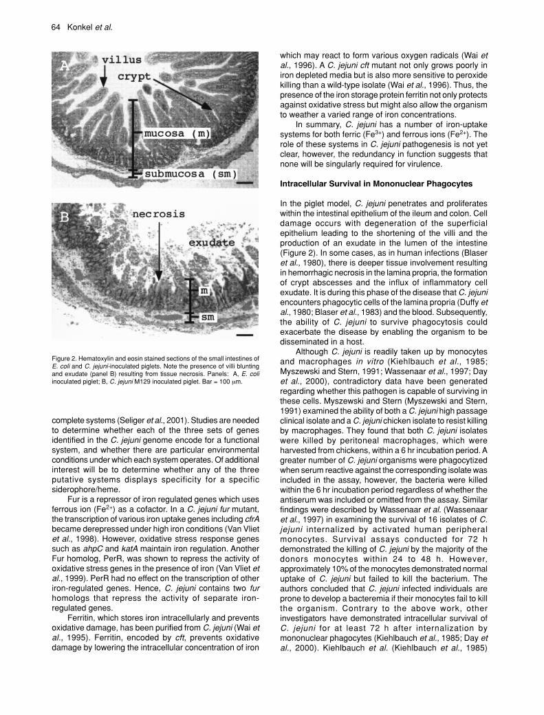
In the piglet model, C. jejuni penetrates and proliferateswithin the intestinal epithelium of the ileum and colon. Celldamage occurs with degeneration of the superficialepithelium leading to the shortening of the villi and theproduction of an exudate in the lumen of the intestine(Figure 2). In some cases, as in human infections (Blaseret al., 1980), there is deeper tissue involvement resultingin hemorrhagic necrosis in the lamina propria, the formationof crypt abscesses and the influx of inflammatory cellexudate. It is during this phase of the disease that C. jejuniencounters phagocytic cells of the lamina propria (Duffy etal., 1980; Blaser et al., 1983) and the blood. Subsequently,the ability of C. jejuni to survive phagocytosis couldexacerbate the disease by enabling the organism to bedisseminated in a host.
Although C. jejuni is readily taken up by monocytesand macrophages in vitro (Kiehlbauch et al., 1985;Myszewski and Stern, 1991; Wassenaar et al., 1997; Dayet al., 2000), contradictory data have been generatedregarding whether this pathogen is capable of surviving inthese cells. Myszewski and Stern (Myszewski and Stern,1991) examined the ability of both a C. jejuni high passageclinical isolate and a C. jejuni chicken isolate to resist killingby macrophages. They found that both C. jejuni isolateswere killed by peritoneal macrophages, which wereharvested from chickens, within a 6 hr incubation period. Agreater number of C. jejuni organisms were phagocytizedwhen serum reactive against the corresponding isolate wasincluded in the assay, however, the bacteria were killedwithin the 6 hr incubation period regardless of whether theantiserum was included or omitted from the assay. Similarfindings were described by Wassenaar et al. (Wassenaaret al., 1997) in examining the survival of 16 isolates of C.jejuni internalized by activated human peripheralmonocytes. Survival assays conducted for 72 hdemonstrated the killing of C. jejuni by the majority of thedonors monocytes within 24 to 48 h. However,approximately 10% of the monocytes demonstrated normaluptake of C. jejuni but failed to kill the bacterium. Theauthors concluded that C. jejuni infected individuals areprone to develop a bacteremia if their monocytes fail to killthe organism. Contrary to the above work, otherinvestigators have demonstrated intracellular survival ofC. jejuni for at least 72 h after internalization bymononuclear phagocytes (Kiehlbauch et al., 1985; Day etal., 2000). Kiehlbauch et al. (Kiehlbauch et al., 1985)
Figure 2. Hematoxylin and eosin stained sections of the small intestines ofE. coli and C. jejuni-inoculated piglets. Note the presence of villi bluntingand exudate (panel B) resulting from tissue necrosis. Panels: A, E. coliinoculated piglet; B, C. jejuni M129 inoculated piglet. Bar = 100 µm.
C. jejuni-Mediated Enteritis 65
examined the survival of a C. jejuni clinical isolate usingthe J774G8 BALB/c mouse macrophage cell line, BALB/cmacrophages, and human monocytes using acridineorange as a vital stain. The researchers were able torecover C. jejuni from all three cell types over a 6 d period.Day et al. (Day et al., 2000) reported similar findings inthat a clinical isolate of C. jejuni (M129) was able to survivefollowing phagocytosis by porcine peritoneal macrophages,murine peritoneal macrophages and J774A.1 cells.However, there was a noticeable reduction in the numberof C. jejuni recovered from the three phagocytic cell typesat 72 h post-inoculation; the greatest survival of C. jejuniwas noted with the porcine peritoneal macrophages. It isour belief that the differences noted between laboratorieswith respect to the ability of C. jejuni to survive withinphagocytes reflects the use of phagocyte cells of differentorigins and various bacterial isolates.
Role of Oxidative Radicals in Phagolysosome Survival
Although intracellular existence provides bacteria anunoccupied niche and shelter from immune surveillance,internalized bacteria must be able to survive a variety ofreactive oxygen species, especially in the phagolysosomeof professional phagocytes. Bacterial factors such assuperoxide dismutase and catalase, which inactivate theseproducts, allow invasive bacteria to persist in host cellsand tissues. Experiments have been conducted to examinethe effect of oxygen radicals on the survival of Salmonellatyphimurium (DeGroote et al., 1997). It was found that asodC mutant of S. typhimurium was more susceptible tokilling by superoxide and nitric oxide than the wild-typeisolate. Moreover, greater numbers of the S. typhimuriumsodC mutant were recovered when the respiratory burstinhibitor acetovanillone or nitric oxide synthase inhibitorNG-L-monomethyl arginine was added to the culturemedium. To address the role of superoxide dismutase inC. jejuni survival in macrophages, assays were performedwith a C. jejuni sodB mutant and the J774A.1 murinemacrophage-like cell line. No difference was noted in thesurvival of the C. jejuni sodB mutant in J774A.1 cells whencompared to the C. jejuni 81-176 wild-type isolate (L.A.Joens, unpublished data). However, a C. jejuni katA mutantwas not recovered from J774A.1 cells 24 h post-inoculation(Day et al., 2000); the katA gene from C. jejuni encodesthe enzyme catalase. Also noteworthy is that the C. jejunikatA mutant was recovered when the respiratory burst orproduction of nitric oxide was inhibited. This findingdemonstrates that C. jejuni possesses certain virulenceattributes that enable it to survive intracellularly withinmononuclear phagocytes. Additional work is required todetermine whether C. jejuni is able to alter its intracellulartrafficking and the precise role of macrophages in thedevelopment of campylobacteriosis.
A Model of C. jejuni Pathogenesis
We conclude this review by presenting a diagram thatrepresents our current perspective of C. jejuni-virulencedeterminants and their potential role in the developmentof the histological lesions observed in C. jejuni-infected
individuals (Figure 3). Our model is based on articlesdiscussed above and examination of biopsy specimensfrom piglets infected with C. jejuni clinical isolates. Webelieve that the infection of piglets with C. jejuni representsa relevant model for C. jejuni-mediated enteritis as theseanimals are anatomically similar to humans. Moreover, C.jejuni-infected piglets develop the clinical symptoms (e.g.,bloody diarrhea) and histopathological lesions (e.g.,epithelial cell degeneration, exudation of fibrin andinflammatory cells in both the small intestine and colon)similar to that of C. jejuni-infected humans.
C. jejuni is proposed to initially colonize the jejunumand ileum, and then the colon, of humans (Allos and Blaser,1995; Skirrow and Blaser, 2000). However, the precise invivo target site of C. jejuni is not known because autopsyand surgical material is rare. Motility and chemotaxis likelyplay critical roles in disease. Upon passage into the smallintestine and migration of bacteria toward the mucus-filledcrypts, we propose that C. jejuni engage in an adaptiveresponse to the intestinal microenvironment where theysynthesize a novel set of proteins that promote theirsubsequent interaction with host target cells. In vitro datasupports the notion that C. jejuni has the ability to migrateacross the enterocytes via a paracellular or transcellularroute (Everest et al., 1992; Konkel et al., 1992c; Grant etal., 1993; Brás and Ketley, 1999; Harvey et al., 1999).However, not known is the significance of either route oftranslocation versus the role of M cells in the organism’sability to breach the intestinal barrier. We speculate thatadherence plays an early role in the infectious processand that C. jejuni binds specifically to host cell receptors.If studies continue to support the necessity of adhesins inestablishing disease, it will prove interesting to determinewhether C. jejuni has a predilection for receptors on theapical or basolateral surfaces of the host cells. Followingthe intimate binding of C. jejuni to host cells, in vivoevidence indicates that C. jejuni is internalized by a hostcell. Not clear is the contribution of invasion, versus Ciaprotein secretion, to the severity of Campylobacter-mediated enteritis. One of the hallmarks of infection in C.jejuni-inoculated piglets is villous atrophy (Babakhani etal., 1993). More specifically, C. jejuni appears to destroythe cells at the tips of the villi that are fully differentiatedrather than the undifferentiated cells in the crypt. Wepropose that the necrosis of the villi is primarily caused byone or more bacterial toxins. We speculate that CDTcontributes to villous atrophy by targeting the activelyproliferating cells within the crypt. Thus, replacement ofdifferentiated cells at the tips of the villi could be retardedby inhibiting crypt cell hyperplasia. How CDT is deliveredto the cells within the crypt is not known. We would beremiss without mentioning that C. jejuni infection isaccompanied by an intense inflammatory response thatno doubt results from the heightened production of cellularcytokines. While this aspect of C. jejuni infection warrantsa review article all to itself, space constraints have notpermitted us to discuss this area. We hypothesize that theinflammatory response is responsible for intensifying thesymptoms exhibited by C. jejuni-infected individuals, butis not responsible for causing the villous atrophy observedearly in infection. We base this statement on the apparent

66 Konkel et al.
lack of significant numbers of neutrophils and otherinflammatory cells at tissue damaged sites. However,detailed studies are required to more closely examine thepresence of inflammatory cell infiltrates at sites of C. jejuniinfection.
Concluding Comments
A more accurate and comprehensive understanding of C.jejuni-mediated enteritis will emerge as researchersfunctionally characterize putative virulence genes anddiscover virulence attributes that are unique amongparticular C. jejuni isolates. There is little doubt that
additional work will unveil that C. jejuni organisms have arepertoire of unique virulence strategies. Moreover,elucidation of C. jejuni virulence determinants and thestages at which they contribute in infection will yield newinsights into the diverse mechanisms by which bacteriacause disease.
Acknowledgements
We thank Brian Raphael for assistance in preparation ofthis manuscript. Work in MEK’s laboratory is supported bya grant from the NIH (DK58911) and USDA NationalResearch Initiative Competitive Grants Program (99-
Figure 3. Diagram depicting C. jejuni-virulence determinants and their potential role in the development of C. jejuni-mediated enteritis. The top panelillustrates the interactions of C. jejuni with enterocytes and sites where certain virulence determinants might contribute in disease. The bottom panel illustratesthe gross morphological changes that occur in the intestinal tract of an infected host. Not highlighted is the potential interaction of C. jejuni with M cells orprofessional phagocytic cells, as well as the contribution of the inflammatory response in infection.

C. jejuni-Mediated Enteritis 67
35201-8579). Work in LAJ’s laboratory is supported byUSDA-NRICGP grants (98-35201-6195 and 01-35201-9948).
References
Allos, B.M. 2001. Campylobacter jejuni infections: Updateon emerging issues and trends. Clin. Infect. Dis. 32: 1201-1206.
Allos, B.M. and Blaser, M.J. 1995. Campylobacter jejuniand the expanding spectrum of related infections. Clin.Infect. Dis. 20: 1092-1099.
Allweiss, B., Dostal, J., Carey, K.E., Edwards, T.F. andFreter, R. 1977. The role of chemotaxis in the ecology ofbacterial pathogens of mucosal surfaces. Nature 266:448-450.
Altekruse, S.F., Stern, N.J., Fields, P.I. and Swerdlow, D.L.1999. Campylobacter jejuni - an emerging foodbornepathogen. Emerg. Infect. Dis. 5: 28-35.
Aspinall, G.O., McDonald, A.G., Raju, T.S., Pang, H.,Kurjanczyk, L.A., Penner, J.L. and Moran, A.P. 1993.Chemical structure of the core region of Campylobacterjejuni serotype 0:2 lipopolysaccharide. Eur. J. Biochem.213: 1029-1037.
Aspinall, G.O., McDonald, A.G., Raju, T.S., Pang, H., Mills,S.D., Kurjanczyk, L.A. and Penner, J.L. 1992. Serologicaldiversity and chemical structures of Campylobacter jejunilow-molecular weight lipopolysaccharides. J. Bacteriol.174: 1324-1332.
Babakhani, F.K., Bradley, G.A. and Joens, L.A. 1993.Newborn piglet model for campylobacteriosis. Infect.Immun. 61: 3466-3475.
Bacon, D.J., Alm, R.A., Burr, D.H., Hu, L., Kopecko, D.J.,Ewing, C.P., Trust, T.J. and Guerry, P. 2000. Involvementof a plasmid in virulence of Campylobacter jejuni 81-176.Infect. Immun. 68: 4384-4390.
Bacon, D.J., Szymanski, C.M., Burr, D.H., Silver, R.P., Alm,R.A. and Guerry, P. 2001. A phase-variable capsule isinvolved in virulence of Campylobacter jejuni 81-176. Mol.Microbiol. 40: 769-777.
Biswas, D., Itoh, K. and Sasakawa, C. 2000. Uptakepathways of clinical and healthy animal isolates ofCampylobacter jejuni into INT-407 cells. FEMS Immunol.Med. Microbiol. 29: 203-211.
Black, R.E., Levine, M.M., Clements, M.L., Hughes, T.P.and Blaser, M.J. 1988. Experimental Campylobacter jejuniinfection in humans. J. Infect. Dis. 157: 472-479.
Blaser, M.J., Hardesty, H.L., Powers, B. and Wang, W.L.1980. Survival of Campylobacter fetus subsp. jejuni inbiological milieus. J. Clin. Microbiol. 11: 309-313.
Blaser, M.J., Wells, J.G., Feldman, R.A., Pollard, R.A. andAllen, J.R. 1983. Campylobacter enteritis in the UnitedStates. A multicenter study. Ann. Intern. Med. 98: 360-365.
Bradbury, W.C., Marko, M.A., Hennessy, J.N. and Penner,J.L. 1983. Occurrence of plasmid DNA in serologicallydefined strains of Campylobacter jejuni andCampylobacter coli. Infect. Immun. 40: 460-463.
Bradley, D.E., Anderson, A.N. and Perry, M.B. 1991.Differences between the LPS cores in adherent and non-adherent strains of enteropathogenic Escherichia coli
0119. FEMS Microbiol. Lett. 64: 13-17.Brás, A.M. and Ketley, J.M. 1999. Transcellular
translocation of Campylobacter jejuni across humanpolarised epithelial monolayers. FEMS Microbiol. Lett.179: 209-215.
Caldwell, M.B., Guerry, P., Lee, E.C., Burans, J.P. andWalker, R.I. 1985. Reversible expression of flagella inCampylobacter jejuni. Infect. Immun. 50: 941-943.
Chart, H., Frost, J.A., Oza, A., Thwaites, R., Gillanders, S.and Rowe, B. 1996. Heat-stable serotyping antigensexpressed by strains of Campylobacter jejuni are probablycapsular and not long-chain polysaccharides. J. Appl.Bacteriol. 81: 635-640.
Day, W.A., Sajecki, J.L., Pitts, T.M. and Joens, L.A. 2000.Role of catalase in Campylobacter jejuni intracellularsurvival. Infect. Immun. 68: 6337-6345.
De Melo, M.A., Gabbiani, G. and Pechère, J.-C. 1989.Cellular events and intracellular survival of Campylobacterjejuni during infection of HEp-2 cells. Infect. Immun. 57:2214-2222.
De Melo, M.A. and Pechère, J.-C. 1990. Identification ofCampylobacter jejuni surface proteins that bind toeucaryotic cells in vitro. Infect. Immun. 58: 1749-1756.
DeGroote, M.A., Ochsner, U.A., Shiloh, M.U., Nathan, C.,McCord, J.M., Dinaure, M.C., Libby, S.J., Vasquez-Torres,A., Xu, Y. and Fang, F.C. 1997. Periplasmic superoxidedismutase protects Salmonella from products ofphagocyte NADPH-oxidase and nitric oxidase synthase.Proc. Natl. Acad. Sci. USA. 94: 13997-14001.
Doig, P., Yao, R., Burr, D.H., Guerry, P. and Trust, T.J. 1996.An environmentally regulated pilus-like appendageinvolved in Campylobacter pathogenesis. Mol. Microbiol.20: 885-894.
Drechsel, H. and Winkelmann, G.: Iron chelation andsiderophores. In: Winkelmann, G. and Carrano, C.J.(Eds.), Transition metals in microbial metabolism.Harwood Acad, Amsterdam, Netherlands, 1997, pp. 1-9.
Duffy, M.C., Benson, J.B. and Rubin, S.J. 1980. Mucosalinvasion in Campylobacter enteritis. Amer. J. Clin. Path.73: 706-708.
Everest, P.H., Goossens, H., Butzler, J.P., Lloyd, D.,Knutton, S., Ketley, J.M. and Williams, P.H. 1992.Differentiated Caco-2 cells as a model for enteric invasionby Campylobacter jejuni and C. coli. J Med Microbiol 37:319-325.
Fauchere, J.L., Rosenau, A., Veron, M., Moyen, E.N.,Richard, S. and Pfister, A. 1986. Association with HeLacells of Campylobacter jejuni and Campylobacter coliisolated from human feces. Infect. Immun. 54: 283-287.
Field, L.H., Headley, V.L., Payne, S.M. and Berry, L.J.1986a. Influence of iron on growth, morphology, outermembrane protein composition, and synthesis ofsiderophores in Campylobacter jejuni. Infect. Immun. 54:126-132.
Field, L.H., Headley, V.L., Underwood, J.L., Payne, S.M.and Berry, L.J. 1986b. The chicken embryo as a modelfor Campylobacter invasion: comparative virulence ofhuman isolates of Campylobacter jejuni andCampylobacter coli. Infect. Immun. 54: 118-125.
Finlay, B.B., Heffron, F. and Falkow, S. 1989. Epithelialcell surfaces induce Salmonella proteins required for
68 Konkel et al.
bacterial adherence and invasion. Science 243: 940-943.Friedman, C.R., Neimann, J., Wegener, H.C. and Tauxe,
R.V.: Epidemiology of Campylobacter jejuni infections inthe United States and other industrialzed nations. In:Nachamkin, I. and Blaser, M.J. (Eds.), Campylobacter.American Society for Microbiology, Washington, D.C.,2000, pp. 121-138.
Galindo, M.A., Day, W.A., Raphael, B.H. and Joens, L.A.2001. Cloning and characterization of a Campylobacterjejuni iron-uptake operon. Curr. Microbiol. 42: 139-143.
Gaynor, E.C., Ghori, N. and Falkow, S. 2001. Bile-induced‘pili’ in Campylobacter jejuni are bacteria-independentartifacts of the culture medium. Mol. Microbiol. 39: 1546-1549.
Golden, N.J., Camilli, A., Acheson, D.W. 2000. Randomtransposon mutagenesis of Campylobacter jejuni. Infect.Immun. 68: 5450-5453.
Grant, C.C.R., Konkel, M.E., Cieplak, W., Jr. and Tompkins,L.S. 1993. Role of flagella in adherence, internalization,and translocation of Campylobacter jejuni in nonpolarizedand polarized epithelial cell cultures. Infect. Immun. 61:1764-1771.
Guerry, P., Alm, R.A., Power, M.E., Logan, S.M. and Trust,T.J. 1991. Role of two flagellin genes in Campylobactermotility. J. Bacteriol. 173: 4757-4764.
Guerry, P., Perez-Casal, J., Yao, R., McVeigh, A. and Trust,T.J. 1997. A genetic locus involved in iron utilization uniqueto some Campylobacter strains. J. Bacteriol. 179: 3997-4002.
Hale, T.L. and Bonventre, P.F. 1979. Shigella infection ofHenle intestinal epithelial cells: role of the bacterium.Infect. Immun. 24: 879-886.
Hale, T.L., Morris, R.E. and Bonventre, P.F. 1979. Shigellainfection of Henle intestinal epithelial cells: role of thehost cell. Infect. Immun. 24: 887-894.
Harvey, P., Battle, T. and Leach, S. 1999. Different invasionphenotypes of Campylobacter isolates in Caco-2 cellmonolayers. J. Med. Microbiol. 48: 461-469.
Hazeleger, W.C., Wouters, J.A., Rombouts, F.M. and Abee,T. 1998. Physiological activity of Campylobacter jejunifar below the minimal growth temperature. Appl. Environ.Microbiol. 64: 3917-3922.
Headley, V.L. and Payne, S.M. 1990. Differential proteinexpression by Shigella flexneri in intracellular andextracellular environments. Proc. Natl. Acad. Sci. USA87: 4179-4183.
Hendrixson, D.R., Akerley, B.J. and Dirita, V.J. 2001.Transposon mutagenesis of Campylobacter jejuniidentifies a bipartite energy taxis system required formotility. Mol. Microbiol. 40: 214-224.
Hickey, T.E., McVeigh, A.L., Scott, D.A., Michielutti, R.E.,Bixby, A., Carroll, S.A., Bourgeois, A.L. and Guerry, P.2000. Campylobacter jejuni cytolethal distending toxinmediates release of interleukin-8 from intestinal epithelialcells. Infect. Immun. 68: 6535-6541.
Hu, L. and Kopecko, D.J. 1999. Campylobacter jejuni 81-176 associates with microtubules and dynein duringinvasion of human intestinal cells. Infect. Immun. 67:4171-4182.
Hugdahl, M.B., Beery, J.T. and Doyle, M.P. 1988.Chemotactic behavior of Campylobacter jejuni. Infect.
Immun. 56: 1560-1566.Hultgren, S.J., Jones, C.H. and Normark, S.: Bacterial
adhesins and their assembly. In: Neidhardt, F.C. (Ed.),Escherichia coli and Salmonella. American Society forMicrobiology, Washington, D.C., 1996, pp. 2730-56.
Hultgren, S.J., Normark, S. and Abraham, S.N. 1991.Chaperone-assisted assembly and molecular architectureof adhesive pili. Annu. Rev. Microbiol. 45: 383-415.
Humphrey, C.D., Montag, D.M. and Pittman, F.E. 1985.Experimental infection of hamsters with Campylobacterjejuni. J. Infect. Dis. 151: 485-493.
Jin, S., Joe, A., Lynett, J., Hani, E.K., Sherman, P. andChan, V.L. 2001. JlpA, a novel surface-exposedlipoprotein specific to Campylobacter jejuni, mediatesadherence to host epithelial cells. Mol. Microbiol. 39:1225-1236.
Johnson, W.M. and Lior, H. 1988. A new heat-labilecytolethal distending toxin (CLDT) produced byCampylobacter spp. Microb. Pathog. 4: 115-126.
Karlyshev, A.V., Linton, D., Gregson, N.A., Lastovica, A.J.and Wren, B.W. 2000. Genetic and biochemical evidenceof a Campylobacter jejuni capsular polysaccharide thataccounts for Penner serotype specificity. Mol. Microbiol.35: 529-541.
Kelle, K., Pages, J.M. and Bolla, J.M. 1998. A putativeadhesin gene cloned from Campylobacter jejuni. Res.Microbiol. 149: 723-733.
Ketley, J.M. 1997. Pathogenesis of enteric infection byCampylobacter. Microbiol. 143: 5-21.
Kiehlbauch, J.A., Albach, R.A., Baum, L.L. and Chang, K.-P. 1985. Phagocytosis of Campylobacter jejuni and itsintracellular survival in mononuclear phagocytes. Infect.Immun. 48: 446-451.
Konkel, M.E., Babakhani, F. and Joens, L.A. 1990.Invasion-related antigens of Campylobacter jejuni. J.Infect. Dis. 162: 888-895.
Konkel, M.E. and Cieplak, W., Jr. 1992. Altered syntheticresponse of Campylobacter jejuni to cocultivation withhuman epithelial cells is associated with enhancedinternalization. Infect. Immun. 60: 4945-4949.
Konkel, M.E., Corwin, M.D., Joens, L.A. and Cieplak, W.,Jr. 1992a. Factors that influence the interaction ofCampylobacter jejuni with cultured mammalian cells. J.Med. Microbiol. 37: 30-37.
Konkel, M.E., Garvis, S.G., Tipton, S.L., Anderson, D.E.,Jr. and Cieplak, W., Jr. 1997. Identification and molecularcloning of a gene encoding a fibronectin-binding protein(CadF) from Campylobacter jejuni. Mol. Microbiol. 24:953-963.
Konkel, M.E., Gray, S.A., Kim, B.J., Garvis, S.G. and Yoon,J. 1999a. Identification of the enteropathogensCampylobacter jejuni and Campylobacter coli based onthe cadF virulence gene and its product. J. Clin. Microbiol.37: 510-517.
Konkel, M.E., Hayes, S.F., Joens, L.A. and Cieplak, W., Jr.1992b. Characteristics of the internalization andintracellular survival of Campylobacter jejuni in humanepithelial cell cultures. Microbial. Pathog. 13: 357-370.
Konkel, M.E. and Joens, L.A. 1989. Adhesion to andinvasion of HEp-2 cells by Campylobacter spp. Infect.Immun. 57: 2984-2990.

C. jejuni-Mediated Enteritis 69
Konkel, M.E., Kim, B.J., Klena, J.D., Young, C.R. and Ziprin,R. 1998. Characterization of the thermal stress responseof Campylobacter jejuni. Infect. Immun. 66: 3666-3672.
Konkel, M.E., Kim, B.J., Rivera-Amill, V. and Garvis, S.G.1999b. Bacterial secreted proteins are required for theinternalization of Campylobacter jejuni into culturedmammalian cells. Mol. Microbiol. 32: 691-701.
Konkel, M.E., Mead, D.J. and Cieplak, W., Jr. 1993. Kineticand antigenic characterization of altered protein synthesisby Campylobacter jejuni during cultivation with humanepithelial cells. J. Infect. Dis. 168: 948-954.
Konkel, M.E., Mead, D.J., Hayes, S.F. and Cieplak, W., Jr.1992c. Translocation of Campylobacter jejuni acrosshuman polarized epithelial cell monolayer cultures. J.Infect. Dis. 166: 308-315.
Kops, S.K., Lowe, D.K., Bement, W.M. and West, A.B.1996. Migration of Salmonella typhi through intestinalepithelial monolayers: an in vitro study. Microbiol Immunol40: 799-811.
Lara-Tejero, M. and Galán, J.E. 2000. A bacterial toxin thatcontrols cell cycle progression as a deoxyribonuclease I-like protein. Science 290:354-357.
Lara-Tejero, M. and Galán, J.E. 2001. CdtA, CdtB, andCdtC form a tripartate complex that is required forcytolethal distending activity. Infect. Immun. 69: 4358-4365.
Larsen, R.A., Thomas, M.G., Postle, K. 1999. Protonmotiveforce, ExbB and ligand-bound FepA drive conformationalchanges in TonB. Mol. Microbiol. 31:1809-1824.
Lee, A., O’Rouke, J.L., Barrington, P.J. and Trust, T.J. 1986.Mucus colonization as a determinant of pathogenicity inintestinal infection by Campylobacter jejuni: a mousececal model. Infect. Immun. 51: 536-546.
Lee, C.A. and Falkow, S. 1990. The ability of Salmonellato enter mammalian cells is affected by bacterial growthstate. Proc. Natl. Acad. Sci., USA 87: 4304-4308.
Logan, S.M. and Trust, T.J. 1984. Structural and antigenicheterogeneity of lipopolysaccharides of Campylobacterjejuni and Campylobacter coli. Infect. Immun. 45: 210-216.
Low, D., Braaten, B. and Van der Woude, M.: Fimbriae. In:Neidhardt, F.C. (Ed.), Escherichia coli and Salmonella.American Society for Microbiology, Washington, D.C.,1996, pp. 146-57.
Madara, J.L. 1998. Regulation of the movement of solutesacross tight junctions. Annu. Rev. Physiol. 60: 143-159.
McSweegan, E. and Walker, R.I. 1986. Identification andcharacterization of two Campylobacter jejuni adhesinsfor cellular and mucous substrates. Infect. Immun. 53:141-148.
Mooney, A., Clyne, M., Curran, T., Doherty, D., Kilmartin,B. and Bourke, B. 2001. Campylobacter upsaliensisexcerts a cytolethal distending toxin effect on HeLa cellsand T lymphocytes. Microbiol. 147: 735-743.
Moran, A.P. 1997. Structure and conserved characteristicsof Campylobacter jejuni lipopolysaccharides. J. Infect. Dis.176 (Suppl 2): S115-121.
Moran, A.P. and Penner, J.L. 1999. Serotyping ofCampylobacter jejuni based on heat-stable antigens:relevance, molecular basis and implications inpathogenesis. J. Appl. Microbiol. 86: 361-377.
Moran, A.P., Zahringer, U., Seydel, U., Scholz, D., Stutz,P. and Rietschel, E.T. 1991. Structural analysis of the lipidA component of Campylobacter jejuni CCUG 10936(serotype O:2) lipopolysaccharide. Description of a lipidA containing a hybrid backbone of 2-amino-2-deoxy-D-glucose and 2,3-diamino-2,3-dideoxy-D-glucose. Eur. J.Biochem. 198: 459-469.
Morooka, T., Umeda, A. and Amako, K. 1985. Motility asan intestinal colonization factor for Campylobacter jejuni.J. Gen. Microbiol. 131: 1973-1980.
Moser, I., Schröder, W.F.K.J. and Hellmann, E. 1992. Invitro binding of Campylobacter jejuni/coli outer membranepreparations to INT 407 cell membranes. Med. Micrbiol.Immunol. 180: 289-303.
Moser, I., Schroeder, W. and Salnikow, J. 1997.Campylobacter jejuni major outer membrane protein anda 59-kDa protein are involved in binding to fibronectinand INT 407 cell membranes. FEMS Mirobiol. Lett. 157:233-8.
Mroczenski-Wildey, M.J., Di Fabio, J.L. and Cabello, F.C.1989. Invasion and lysis of HeLa cell monolayers bySalmonella typhi: role of lipopolysaccharide. Microb.Pathog. 6: 143-152.
Myszewski, M.A. and Stern, N.J. 1991. Phagocytosis andintracellular killing of Campylobacter jejuni by elicitedchicken peritoneal macrophages. Avian Dis. 35: 750-755.
Nachamkin, I., Allos, B.M. and Ho, T. 1998. Campylobacterspecies and Guillian-Barré syndrome. Clin. Microbiol. Rev.11: 555-567.
Nachamkin, I., Yang, X.H. and Stern, N.J. 1993. Role ofCampylobacter jejuni flagella as colonization factors forthree-day-old chicks: analysis with flagellar mutants. Appl.Environ. Microbiol. 59: 1269-1273.
Newell, D.G. 1986 . Monoclonal antibodies directed againstthe flagella of Campylobacter jejuni: production,characterization and lack of effect on the colonization ofinfant mice. J. Hyg. (London) 96: 131-141.
Newell, D.G., McBride, H., Saunders, F., Dehele, Y. andPearson, A.D. 1985. The virulence of clinical andenvironmental isolates of Campylobacter jejuni. J. Hyg.(London) 94: 45-54.
Nuijten, P.J., van der Zeijst, B.A. and Newell, D.G. 1991.Localization of immunogenic regions on the flagellinproteins of Campylobacter jejuni 81116. Infect. Immun.59: 1100-1105.
Oelschlaeger, T.A., Guerry, P. and Kopecko, D.J. 1993.Unusual microtubule-dependent endocytosismechanisms triggered by Campylobacter jejuni andCitrobacter freundii. Proc. Natl. Acad. Sci. USA 90: 6884-6888.
Owen, R.J. and Leaper, S. 1981. Base composition, sizeand nucleotide sequence similarities of genomedeoxyribonucleic acids from species of the genusCampylobacter. FEMS Microbiol. Lett. 12: 395-400.
Panigrahi, P., Losonky, G., DeTolla, L.J. and Morris, J.G.1992. Human immune response to Campylobacter jejuniproteins expressed in vitro. Infect. Immun. 60: 4938-4944.
Parkhill, J., Wren, B.W., Mungall, K., Ketley, J.M., Churcher,C., Basham, D., Chillingworth, T., Davies, R.M., Feltwell,T., Holroyd, S., Jagels, K., Karlshev, A.V., Moule, S.,Pallen, M.J., Penn, C.W., Quall, M.A., Rajandrean, M.A.,
70 Konkel et al.
Rutherford, K.M., Van Vliet, A.H.M., Whitehead, S. andBarrell, B.G. 2000. The genome sequence of the food-borne pathogen Campylobacter jejuni revealshypervariable sequences. Nature 403: 665-668.
Pavlovskis, O.R., Rollins, D.M., Haberberger, R.L. Jr.,Green, A.E., Habash, L., Strocko, S. and Walker, R.I.1991. Significance of flagella in colonization resistanceof rabbits immunized with Campylobacter spp. Infect.Immun. 59: 2259-2264.
Pei, Z. and Blaser, M.J. 1993. PEB1, the major cell-bindingfactor of Campylobacter jejuni, is a homolog of the bindingcomponent in Gram-negative nutrient transport systems.J. Biol. Chem. 268: 18717-18725.
Pei, Z., Burucoa, C., Grignon, B., Baqar, S., Huang, X.-Z.,Kopecko, D.J., Bourgeois, A.L., Fauchere, J.-L. andBlaser, M.J. 1998. Mutation in the peb1A locus ofCampylobacter jejuni reduces interactions with epithelialcells and intestinal colonization of mice. Infect. Immun.66: 938-943.
Penner, J.L. and Hennessy, J.N. 1980. Passivehemagglutination technique for serotyping Campylobacterfetus subsp. jejuni on the basis of soluble heat-stableantigens. J. Clin. Microbiol. 12: 732-737.
Pickett, C.L.: Campylobacter toxins and their role inpathogenesis. In: Nachamkin, I. and Blaser, M.J. (Eds.),Campylobacter. American Society for Microbiology,Washington, D.C., 2000, pp. 179-190.
Pickett, C.L., Pesci, E.C., Cottle, D.L., Russell, G., Erdem,A.N. and Zeytin, H. 1996. Prevalence of cytolethaldistending toxin production in Campylobacter jejuni andrelatedness of Campylobacter sp. cdtB genes. Infect.Immun. 64: 2070-2078.
Pickett, C.L. and Whitehouse, C.A. 1999. The cytolethaldistending toxin family. Trends in Microbiol. 7: 292-297.
Prendergast, M.M., Lastovica, A.J. and Moran, A.P. 1998.Lipopolysaccharides from Campylobacter jejuni O:41strains associated with Guillain-Barré syndrome exhibitmimicry of GM1 ganglioside. Infect. Immun. 66: 3649-3655.
Preston, M.A. and Penner, J.L. 1987. Structural andantigenic properties of lipopolysaccharides from serotypereference strains of Campylobacter jejuni. Infect. Immun.55: 1806-1812.
Purdy, D., Buswell, C.M., Hodgson, A.E., McAlpine, K.,Henderson, I. and Leach, S.A. 2000. Characterisation ofcytolethal distending toxin (CDT) mutants ofCampylobacter jejuni. J. Med. Microbiol. 49: 473-479.
Ratledge, C. and Dover, L.G. 2000. Iron metabolism inpathogenic bacteria. Annu. Rev. Microbiol. 54: 881-941.
Richardson, P.T. and Park, S.F. 1995. Enterochelinacquisition in Campylobacter coli: characterization ofcomponents of a binding-protein-dependent transportsystem. Microbiol. 141: 3181-3191.
Richardson, W.P. and Sadoff, J.C. 1988. Inducedengulfment of Neisseria gonorrhoeae by tissue culturecells. Infect. Immun. 56: 2512-2514.
Rivera-Amill, V., Kim, B.J., Seshu, J. and Konkel, M.E.2001. Secretion of the virulence associatedCampylobacter invasion antigens from Campylobacterjejuni requires a stimulatory signal. J. Infect. Dis. 183:1607-1616.
Russell, R.G. and Blake, D.C., Jr. 1994. Cell associationand invasion of Caco-2 cells by Campylobacter jejuni.Infect. Immun. 62: 3773-3779.
Russell, R.G., O’Donnoghue, M., Blake, D.C., Jr., Zulty, J.and DeTolla, L.J. 1993. Early colonic damage andinvasion of Campylobacter jejuni in experimentallychallenged infant Macaca mulatta. J. Infect. Dis. 168: 210-215.
Schmiel, D.H., Wagar, E., Karamanou, L., Weeks, D. andMiller, V.L. 1998. Phospholipase A of Yersiniaenterocolitica contributes to pathogenesis in a mousemodel. Infect. Immun. 66: 3941-3951.
Schröder, W. and Moser, I. 1997. Primary structure analysisand adhesion studies on the major outer membraneprotein of Campylobacter jejuni. FEMS Microbiol. Lett.150: 141-147.
Schwan, E.T., Robertson, B.D., Brade, H. and van Putten,J.P. 1995. Gonococcal rfaF mutants express Rd2chemotype LPS and do not enter epithelial host cells.Mol. Microbiol. 15: 267-275.
Seliger, S.S., Mey, A.R., Valle, A.-M., and Payne, S.M. 2001.The two TonB systems of Vibrio cholerae: redundant andspecific functions. Mol. Microbiol. 39:801-812.
Silverstein, S.C., Steinman, R.M. and Cohn, Z.A. 1977.Endocytosis. Annu. Rev. Biochem. 46: 669-722.
Skirrow, M.B. and Blaser, M.J.: Clinical aspects ofCampylobacter infection. In: Nachamkin, I. and Blaser,M.J. (Eds.), Campylobacter, 2nd Edition. ASM Press,Washington, DC, 2000, pp. 69-88.
Sosula, L., Nicholls, E.M. and Skeen, M. 1988.Ultrastructure of Campylobacter jejuni in gamma-irradiated mouse jejunum. Am. J. Pathol. 131: 125-131.
St. Geme III, J.W. and Falkow, S. 1990. Haemophilusinfluenzae adheres to and enters cultured humanepithelial cells. Infect. Immun. 58: 4036-4044.
Szymanski, C.M., King, M., Haardt, M. and Armstrong, G.D.1995. Campylobacter jejuni motility and invasion of Caco-2 cells. Infect. Immun. 63: 4295-4300.
Takata, T., Fujimoto, S. and Amako, K. 1992. Isolation ofnonchemotactic mutants of Campylobacter jejuni and theircolonization of the mouse intestinal tract. Infect. Immun.60: 3596-3600.
Takeuchi, A. 1967. Electron microscopic studies ofexperimental Salmonella infection. Penetration into theintestinal epithelium by Salmonella typhimurium. Am JPathol 50: 109-36.
Tsai, C.-M. and Frasch, C.E. 1982. A sensitive silver stainfor detecting lipopolysaccharides in polyacrylamide gels.Anal. Biochem. 119: 115-119.
Van Vliet, A.H., Baillon, M.L., Penn, C.W. and Ketley, J.M.1999. Campylobacter jejuni contains two fur homologs:characterization of iron-responsive regulation of peroxidestress defense genes by the PerR repressor. J. Bacteriol.181: 6371-6376.
Van Vliet, A.H. and Ketley, J.M. 2001. Pathogenesis ofenteric Campylobacter infection. J. Appl. Microbiol. 90:45S-56S.
Van Vliet, A.H., Wooldridge, K.G. and Ketley, J.M. 1998.Iron-responsive gene regulation in a Campylobacter jejunifur mutant. J. Bacteriol. 180: 5291-5298.
Velayudhan, J., Hughes, N.J., McColm, A.A., Bagshaw,
C. jejuni-Mediated Enteritis 71
J., Clayton, C.L., Andrews, S.C. and Kelly, D.J. 2000. Ironacquisition and virulence in Helicobacter pylori: a majorrole for FeoB, a high-affinity ferrous iron transporter. Mol.Microbiol. 37: 274-286.
Wai, S.N., Nakayama, K., Umene, K., Moriya, T. andAmako, K. 1996. Construction of a ferritin-deficient mutantof Campylobacter jejuni: contribution of ferritin to ironstorage and protection against oxidative stress. MolMicrobiol 20: 1127-1134.
Wai, S.N., Takata, T., Takade, A., Hamasaki, N. and Amako,K. 1995. Purification and characterization of ferritin fromCampylobacter jejuni. Arch. Microbiol. 164: 1-6.
Walker, R.I., Schmauder-Chock, E.A., Parker, J.L. and Burr,D. 1988. Selective association and transport ofCampylobacter jejuni through M cells of rabbit Peyer’spatches. Can. J. Microbiol. 34: 1142-1147.
Walker, T.S. and Winkler, H.H. 1978. Penetration of culturedmouse fibroblasts (L cells) by Rickettsia prowazeki. Infect.Immun. 22: 200-208.
Wassenaar, T.M. 1997. Toxin production by Campylobacterspp. Clin. Microbiol. Rev. 10: 466-476.
Wassenaar, T.M., Bleumink-Pluym, N.M., Newell, D.G.,Nuijten, P.J.M. and van der Zeijst, B.A.M. 1994.Differential flagellin expression in a flaA flaB+ mutant ofCampylobacter jejuni. Infect. Immun. 62: 3901-3906.
Wassenaar, T.M., Bleumink-Pluym, N.M. and van der Zeijst,B.A. 1991. Inactivation of Campylobacter jejuni flagellingenes by homologous recombination demonstrates thatflaA but not flaB is required for invasion. EMBO J. 10:2055-2061.
Wassenaar, T.M., Engelskirchen, M., Park, S. andLastovica, A. 1997. Differential uptake and killing potentialof Campylobacter jejuni by human peripheral monocytes/macrophages. Med. Micrbiol. Immunol. 186: 139-144.
Wassenaar, T.M., van der Zeijst, B.A., Ayling, R. and Newell,D.G. 1993. Colonization of chicks by motility mutants ofCampylobacter jejuni demonstrates the importance offlagellin A expression. J. Gen. Microbiol. 139: 1171-1175.
Welkos, S.L. 1984. Experimental gastroenteritis in newly-hatched chicks infected with Campylobacter jejuni. J.Med. Microbiol. 18: 233-248.
Whitehouse, C.A., Balbo, P.B., Pesci, E.C., Cottle, D.L.,Mirabito, P.M. and Pickett, C.L. 1998. Campylobacterjejuni cytolethal distending toxin causes a G2-phase cellcycle block. Infect. Immun. 66: 1934-1940.
Wooldridge, K.G. and Ketley, J.M. 1997. Campylobacter-host cell interactions. Trends Microbiol. 5: 96-102.
Yao, R., Burr, D.H., Doig, P., Trust, T.J., Niu, H. and Guerry,P. 1994. Isolation of motile and non-motile insertionalmutants of Campylobacter jejuni: the role of motility inadherence and invasion of eukaryotic cells. Mol. Microbiol.14: 883-893.
Yao, R., Burr, D.H. and Guerry, P. 1997. CheY-mediatedmodulation of Campylobacter jejuni virulence. Mol.Microbiol. 23: 1021-1031.
Young, G.M., Schmiel, D.H. and Miller, V.L. 1999. A newpathway for the secretion of virulence factors by bacteria:The flagellar export apparatus functions as a protein-secretion system. Proc. Natl. Acad. Sci. USA 96: 6456-6461.
Ziprin, R.L., Young, C.R., Hume, M.E. and Konkel, M.E.
1999. The absence of cecal colonization of chicks by amutant of Campylobacter jejuni not expressing bacterialfibronectin-binding protein. Avian Dis. 43: 586-589.

72 Konkel et al.