Embed Size (px)
Citation preview

1005ANALYTICAL SCIENCES AUGUST 2008, VOL. 24 1005
2008 © The Japan Society for Analytical Chemistry
Introduction
Aluminum(III) is a trivalent cation unable to undergo redox reactions; it has been linked to many diseases, such as dialysis dementia and microcytic anemia without iron deficiency. It has also been implicated in Alzheimer’s disease. Parkinson’s disease (PD) is the second-most common neurodegenerative disease, affecting approximately 1% of the human population aged 65 and above, which is also associated with Al(III).1 Many studies have been carried out in order to study the molecular mechanism responsible for the toxic effects of this metal ion on both humans and animals. The results indicate that aluminum has been found inside cells in many illnesses.2 Therefore, studying the binding of metal ion and its complexes by peptides and proteins is important for understanding many biological systems. It has been proposed that, in some cases, aluminum binds to certain natural carriers. The interaction of aluminum with amino acids, peptides and proteins is a subject of current interest.3 Transferring at the sites left vacant by iron mainly transports aluminum in blood circulation. As a highly charged typical hard metal ion, Al3+ can interact with a large number of peptides, which contain O and N donors. Negatively charged carboxylate and phosphate function groups are the strongest Al(III) binders. It is well known that Al(III) has a great affinity
to those groups, and that the interaction of this metal ion with biomolecules bearing such groups may play an important role in its intake and circulation in biological media, which is connected with the transport mechanism of aluminum.
Oxidized glutathione (GSSG, Fig. 1) exists in animal cells and in the form of phytochelatins in plant cells. It is, therefore, widely available in all living things. It is formed by enzymatic oxidation catalyzed by glutathione peroxidase (GPx), generating a disulfide bridge between two molecules of reduced glutathione (GSH). GSSG is a physiological indicator of intracellular defense system activity against reactive oxygen species (ROS).4 Furthermore, glutathione exhibits several functions in the brain, and chiefly acts as an antioxidant and redox regulator. Oxidative
Speciation of Aluminum(III) Complexes with Oxidized Glutathione in Acidic Aqueous Solutions
Xiao Di YANG,*,** Qian Qian ZHANG,** Rong Fu CHEN,* and Ren Fang SHEN*†
* State Key Laboratory of Soil and Sustainable Agriculture, Institute of Soil Science, Chinese Academy of Sciences, Nanjing 210008, P. R. China
** College of Chemistry and Environment Science, Department of Environment Science, Nanjing Normal University, Nanjing 210097, P. R. China
The structural speciation aspects, including the binding sites, species, complexation abilities and effects of the oxidized glutathione (GSSG) with aluminum(III) in aqueous solutions, have been studied by means of many analytical techniques: pH-potentiometry (25˚C, 0.1 M KCl and 37˚C, 0.15 M NaCl medium) was used to characterize the stoichiometry and stability of the species formed in the interactions of the Al(III) ion and the peptide GSSG, while multinuclear (1H, 13C, 27Al) nuclear magnetic resonance (NMR) and electrospray mass spectroscopy (ESI-MS) were applied to characterize the binding sites and species of the metal ion in the complexes. Two-dimensional (1H, 1H-NOESY) was also employed to reveal the difference in the conformational behavior of the peptide and its complexes. The following results were obtained: (1) Aluminum(III) can coordinate with the important biomolecule GSSG through the following binding sites: glycyl and glutamyl carboxyl groups to form various mononuclear 1:1 (AlLH4, AlLH3, AlLH2, AlLH, AlL, AlLH–1, AlLH–2) and several binuclear 2:1 (Al2LH4, Al2LH2, Al2L) species (where H6L2+ denotes the totally protonated oxidized glutathione) in acidic aqueous solutions. (2) It indicates that the COO– groups at low level of preorganization in such small peptide are not sufficient to keep the Al(III) ion in solution and to prevent the precipitation of Al(OH)3 in the physiological pH range. (3) It also suggests that the occurrence of an Al-linked complexation, the conformation of the peptide GSSG in aqueous solutions appeared to change a little, relative to the initial structure.
(Received March 27, 2007; Accepted September 28, 2007; Published August 10, 2008)
† To whom correspondence should be addressed.E-mail: [email protected]
Fig. 1 Structure and labeling of oxidized glutathione (H2L species in GSSG).

1006 ANALYTICAL SCIENCES AUGUST 2008, VOL. 24
damage via GSH depletion leads to the death of neuronal cells during PD.5 It is, therefore, the effect of glutathione on various life processes and the importance of the phytochelatins for metal detoxification that triggered metal-binding studies of GSSG early on. GSSG is present at a concentration suitable for convenient NMR observations in vivo (for example, at approximately 2 mM in the human erythrocyte).5 In the past, Kiss and some other researchers conducted many studies on metal ions: VO2+, As(III), Cu(II), La(III), Cr(V), Zn(II), Pb(II) complexation with GSH and GSSG.6–14 However, to our knowledge, no systematic study on the complexation of Al(III) with GSSG has so far been reported in the literature. The coordination chemistry of Al(III) with glutathione is of vital importance, since it serves as a model system for the binding of this metal ion by larger peptide and protein molecules.15 Due to the number and variety of coordination sites in GSSG, its coordination chemistry may be complicated, and the most definitive information has been obtained by using results from a combination of various techniques. The development of novel methods for characterizing unstable metal complexes in aqueous solution, including nuclear magatic spectroscopy (NMR) and electrospray mass spectrometry (ESI-MS), has promoted us to investigate the Al(III) species formed in the Al(III) + GSSG interaction systems.
Experimental
Chemicals and reagentsOxidized glutathione (GSSG, >98%) was purchased from
Sigma Chemical Co. (St. Louis, MO). All GSSG solutions were freshly prepared daily with double-distilled water, and all samples were flushed with high-purity nitrogen or purified argon. Al(III) solutions were prepared by dissolving high-purity metallic Al powder (99.99%) in hydrochloric acid. More dilute solutions were prepared by diluting these solutions with double-distilled water. D2O was obtained form Beijing Chemical Company. Other chemicals were of analytical reagent grade. Multinuclear (1H, 13C, 27Al) and two-dimensional (1H, 1H-NOESY) NMR measurements were prepared by dissolving appropriate amounts of GSSG and AlCl3·6H2O in D2O. Necessary polyethylene vessels were used. All glass-wares were soaked in 10% HNO3 for at least 24 h, then carefully washed with double-distilled water. The pH values in D2O solutions were corrected for the deuterium isotope effect by adding 0.45 to the meter reading.16
Potentiometric titrationsThe stability constants of the proton and Al complexes of the
ligand (GSSG) were determined by pH-metric titrations of 50.0 mL samples. The concentration of the ligand was 0.001 M, and the metal ion to ligand ratios were 0:1, 1:1, 1:2, 1:5 for the binary system, respectively. The titrations were performed over the pH range 3.45 – 10.87 for GSSG and the pH range 3.26 – 4.82 for Al-GSSG (beyond pH 5.0 precipitation occurred), with a KOH solution of known concentration (ca. 0.1 M) under a pure nitrogen atmosphere. The potentiometric measurements were conducted at 25˚C in a 0.1 M KCl ionic medium in order to allow a direct comparison with stability constants varying with the physiological conditions at 37˚C in a 0.15 M NaCl ionic medium. The temperatures 25.0 ± 0.1˚C and 37.0 ± 0.1˚C were maintained by circulating thermostated water through the jacket. Duplicate titrations were carried out, and the reproducibility of the titration curves was within 0.01-pH unit throughout the whole pH range. Because of the rather sluggish
ligand-exchange kinetics of Al and the precipitation reactions, when equilibration could not be reached within 10 min, the corresponding titration points were omitted from calculations.16 The pH was measured with a pH213 Microprocessor pH meter (HANNA Instruments, Portugal) with a glass combination electrode, which was firstly calibrated for the hydrogen-ion concentration according to Irving et al.17 The stability constants of the main metal species were calculated with the aid of the computer program SPE & BEST.18 The pKw value used in the 0.1 M KCl ionic system (25˚C) is 13.76, while in a 0.15 M NaCl (37˚C) ionic medium it is 13.20.19 The formation of hydroxo-complexes of Al(III) was taken into account in the calculations, which are shown in Tables 1 and 2.
NMR and ESI-MS measurementsThe 27Al NMR spectra were operated at 130.3 MHz on a
Bruker DRX 500 spectrometer (Swiss). Chemical shifts were referenced to an external coaxial insert containing 0.1 M Al (H2O)6
3+ (0 ppm), included with every sample for 27Al NMR experiments. The 1H, 13C NMR spectra were performed on a
Table 1 Proton and aluminum(III) complex-formation constants (log b) of the hydroxo-Al(III) and the oxidized glutathione (H6L2+) at 25.0 ± 0.1˚C and 0.10 M KCl
Ligandspecies
Hydroxospecies
Complexspecies
log b a log b b log b a
This work AlH–1 –5.33 AlLH4 26.82 ± 0.03H6L 28.95 ± 0.02 AlH–2 –10.91 AlLH3 23.56 ± 0.03H5L 27.35 ± 0.01 AlH–3 –16.64 AlLH2 20.32 ± 0.03H4L 25.32 ± 0.02 AlH–4 –23.46 AlLH 15.83 ± 0.03H3L 21.99 ± 0.02 Al2H–2 –7.15 AlL 11.92 ± 0.04H2L 18.23 ± 0.02 Al3H–4 –13.13 AlLH–1 4.96 ± 0.01HL 9.54 ± 0.02 Al13H–32 –107.41 AlLH–2 –5.06 ± 0.01Ref. 7 Al2L 14.07 ± 0.04H6L 29.30 Al2LH2 21.93 ± 0.03H5L 27.70 Al2LH4 28.86 ± 0.05H4L 25.34H3L 22.18H2L 18.37HL 9.55
a. Averages (± standard deviations) for three and �ve titrations.b. Ref. 39.
Table 2 Proton and aluminum(III) complex-formation constants (log b) of the hydroxo-Al(III) and oxidized glutathione (H6L2+) at 37.0 ± 0.1˚C and 0.15 M NaCl
Ligandspecies
Hydroxospecies
Complexspecies
log b a log b b log b a
This work AlH–1 –4.67 AlLH4 27.02 ± 0.03H6L 30.60 ± 0.03 AlH–2 –10.22 AlLH3 23.96 ± 0.02H5L 28.10 ± 0.02 AlH–3 –13.60 AlLH2 20.31 ± 0.03H4L 25.60 ± 0.03 AlH–4 –23.87 AlLH 16.23 ± 0.02H3L 22.96 ± 0.01 Al2H–2 –7.10 AlL 12.32 ± 0.03H2L 19.06 ± 0.02 Al3H–4 –12.23 AlLH–1 5.16 ± 0.04HL 10.65 ± 0.02 Al13H–32 –99.62 AlLH–2 –4.76 ± 0.01 Al2L 15.03 ± 0.03 Al2LH2 22.63 ± 0.02 Al2LH4 29.86 ± 0.02
a. Averages (± standard deviations) for three and �ve titrations.b. Ref. 19.

1007ANALYTICAL SCIENCES AUGUST 2008, VOL. 24
Bruker AVANCE 300 spectrometer at 300.1, 75.5 MHz, respectively, and 10000 scans were accumulated per 13C-NMR spectra. Chemical shifts were referenced to D2O (4.70 ppm) or TMS (0 ppm) for 1H, 13C-NMR experiments. Two-dimensional (1H, 1H-NOESY) NMR experiments were carried out with a Bruker AVANCE 300 apparatus at 300.1 MHz. By means of the standard Bruker microprograms and quadrature detection in both dimensions, the sequence 90˚-t1-90˚-tm-90˚-acquisition (t2) was performed with a mixing time (tm) of 500 ms for D2O. The ESI-MS analyses were performed by using a Finnagn Mat LCQ mass spectrometer and in the CH3OH and H2O mixture solvent. Samples were analyzed by flow-injection ESI-MS using the sample solvent as the carrier. The optimized geometry of the GSSG was performed using the AMBER force field in the Gaussian 03 software program package.20
Results and Discussion
Binding species and abilitiesGSSG contains four carboxylic functions, which have a high
affinity for Al(III). For GSSG, six pKa values could be determined: two glutamyl –COOH and –NH3
+ and two terminal –COOH, totally protonated GSSG being therefore represented as H6L2+. In order to investigate the Al(III)-GSSG equilibrium through glass electrode potentiometry under physiological conditions, the measurements were performed not only in a 25˚C 0.10 M KCl ionic system, but also in a 37˚C 0.15 M NaCl ionic medium. The titration points of the Al(III)-GSSG system measured at different metal ion-to-ligand ratios and the recorded titration curves are presented in Figs. 2 and 3. The calculated formation constants (log b) are listed in Tables 1 and 2. In these tables, pKa1, pKa2 (HL, H2L) correspond predominantly to two glutamyl –COOH groups, pKa3, pKa4 (H3L, H4L) to two glycyl –COOH groups and pKa5, pKa6 (H5L, H6L) to two –NH3
+ groups. Our values agree well with earlier reports.7 The best fit of the experimental data was obtained by considering the formation of various mononuclear 1:1 (AlLH4, AlLH3, AlLH2, AlLH, AlL, AlLH–1, AlLH–2) and binuclear 2:1 (Al2LH4, Al2LH2, Al2L) species in acidic aqueous solutions. Species distribution curves are shown in Figs. 4 and 5. In Fig. 4, in a 25˚C, 0.10 M KCl
ionic medium, the pH-metric speciation curves obtained for the Al(III)-GSSG system indicated that complex formation started before pH 2, with the presence of four-protonated species, AlLH4 and Al2LH4. In these species, monodentate coordination two-Al(III) with the two-glutamyl deprotonated carboxyl groups (COO–)(COOH, NH3
+)free occurs, while probably two glycyl carboxylic groups and the terminal NH2 groups remain protonated in a more acidic solution. At about pH 3.5, mononuclear (AlLH3, AlLH2) and binuclear (Al2LH2) species are predominant in the aqueous solution. In the species AlLH2 and Al2LH2, monodentate coordination one or two Al(III) with the two-glycyl deprotonated carboxyl groups (COO–)(COO–, NH3
+)free existed, while the terminal NH2 groups remained protonated in the aqueous solutions. Accordingly, the 37˚C 0.15 M NaCl ionic system is shown in Fig. 5, which is very similar to Fig. 4. At about pH 2, only the Al2LH4 species is predominant. Increasing the pH to 3.5, the species AlLH2 existed. Above pH 5, there was a hydrolytic species Al13
7+ in the solution. The binding modes are mostly the same as those of the 25˚C 0.10 M KCl ionic system.
Fig. 2 Titration data measured in the Al(III)-GSSG system at various metal ion-to-ligand ratios, at 25˚C, 0.10 M KCl, CAl = 0.001 M, CKOH = 0.1026 M. The full curves were calculated using the set of stability constants listed in Table 1.
Fig. 3 Titration data measured in the Al(III)-GSSG system at various metal ion-to-ligand ratios, at 37˚C, 0.15 M NaCl, CAl = 0.001 M, CKOH = 0.1026 M. The full curves were calculated using the set of stability constants listed in Table 2.
Fig. 4 Distribution curves of complexes formed in the Al(III)-GSSG (0.02 M:0.02 M) system at 25˚C, 0.10 M KCl, calculated using the stability constants listed in Table 1.

1008 ANALYTICAL SCIENCES AUGUST 2008, VOL. 24
Recently, ESI-MS has been used for metal speciation analysis with the development and application of this technique. It was proven to be a powerful tool to identify individual species in aqueous solutions.19 The positive ion ESI-MS spectrum of the Al(III)-GSSG (0.02 M:0.02 M) acidic solution (pH 3.3) showed the presence of significant amounts of free ligand GSSG (MW: C20H32N6O12S2, 612.6). A signal with m/z = 613.1 was attributed to the species H4LH+, which is in agreement with the potentiometric results. In Fig. 6, there are many other signals assigned to the species of Al-GSSG products: species AlLH2 and AlLH3 or AlLH4 (the signals with m/z = 637.1), Al2LH2 (the signals with m/z = 668.6) and Al2LH4 (the signals with m/z = 670.3) in the spectrum. They are the most salient m/z values corresponding to positively charged structures relative to solutions of Al(III)-GSSG mixtures under the same pH conditions. Other ESI-MS signals can be attributed to either unknown species or impurities in the GSSG solution under the ESI-MS conditions.
Binding sitesIn order to examine complexation and conformation of the Al-
GSSG systems, extensive 13C, 1H and 27Al-NMR measurements were carried out in acidic aqueous solutions. Complete assignments of the NMR signals of the protons and carbons of the peptide GSSG were made with the aid of ChemNMR 1H and
13C Estimation and some references.11,12 The 1H-NMR spectra obtained at 25˚C with GSSG (0.02 M) at pD = 2.5 (about pH 3.0) are shown in Fig. 7A. It has more than five signals. A singlet at 3.67 ppm is for the a-CH group in g-Glu or a a-CH2 group of Gly in GSSG, which are overlapping (Fig. 1). Because of the presence of a chiral center in the GSSG, cysteine methylene protons generally showed two different signals:21 a doublet at 3.18 and 2.86 ppm for the b-CH2 group of Cys in GSSG. A doublet at 2.42 ppm is the b-CH2 group signal of g-Glu in GSSG, and the other at 2.05 ppm is the g-CH2 group signal of g-Glu in GSSG. The a-CH group signal of Cys around 4.70 ppm was overlapped by D2O. The proton signal of the g-Glu terminal NH2 group around 7.0 ppm could not be detected because of a rapid proton exchange with the solvent. The chemical shift of the Gly backbone amide NH proton appears at 8.30 ppm with a triplet, owing to coupling with the Gly a-CH2 proton. At pH 3.0, the Cys backbone amide NH proton signal is a doublet at d = 8.60 ppm, due to coupling with the Cys a-CH proton. In the presence of Al(III), there is a considerable broadening of the signals (Fig. 7B), as compared with the spectrum of the free peptide (Fig. 7A). The resonance at 3.67 ppm of the a-CH group in the g-Glu or a-CH2 group of Gly undergoes a slight downfield shift (3.75 ppm), which is indicative of the formation of the Al-GSSG complex. Therefore, it demonstrated that Al(III) could coordinate with GSSG through the glutamyl or glycyl carboxyl group to form various mononuclear or binuclear species in acidic solutions. Since the F– ion exists in an acidic environmental and biological system, and it may bind with amino acids or peptides through hydrogen bonds. Therefore, we added F– ions to the acidic GSSG solution (Fig. 7C). There were no significant changes observed, which suggests that these groups are not obviously affected by the F– ion. However, in the presence of the Al(III) ion (Fig. 7D), most resonance signals undergo a slight downfield shift, except for the Cys backbone amide NH proton signal, which means the Al(III) ion can influence most protons of GSSG with the help of hydrogen bonding with the Al-F species and Al(III) ternary complexes of Al-F-GSSG. This point was further confirmed by the following 13C-NMR and 1H, 1H- NOESY-NMR spectra.
To obtain more information of Al(III) binding with GSSG, the 13C-NMR investigations were then undertaken in acidic aqueous
Fig. 6 Positive ESI mass spectrum of an aqueous solution of aluminum (0.02 M) and oxidized glutathione (0.02 M) at pH 3.3.
Fig. 7 1H-NMR spectra of the Al(III) (0.02 M)-GSSG (0.02 M) D2O solution. (A) GSSG at pD 2.5, (B) Al-GSSG at pD 2.5, (C) F-GSSG at pD 2.5, (D) Al-F-GSSG at pD 2.5.
Fig. 5 Distribution curves of complexes formed in the Al(III)-GSSG (0.02 M:0.02 M) system at 37˚C, 0.15 M NaCl, calculated using the stability constants listed in Table 2.

1009ANALYTICAL SCIENCES AUGUST 2008, VOL. 24
solutions. The 13C-NMR spectra of the free ligand GSSG (0.02 M) in D2O aqueous solution at pD = 2.5 (about pH 3.0) is shown in Fig. 8A. It consists of ten peaks at 26.2, 31.3, 38.8, 43.5, 52.6, 54.1, 171.8, 174.0, 174.9 and 176.2 ppm, being assigned to the Cys b-CH2 group, the g-Glu b-CH2 group, the Gly a-CH2 group, the g-Glu g-CH2 group, the Cys a-CH group, the g-Glu a-CH group, the Cys CO group, the g-Glu COO– group, the g-Glu CO group and the Gly COO– group, respectively. Upon adding aluminum to the GSSG solution (Fig. 8B), these peaks shifted to 26.1, 31.3, 38.7, 42.4, 52.6, 53.9, 172.2, 173.9 and 174.8 ppm. The Gly in GSSG carboxyl group peak signal shifted upfield, and might be overlapped by other signals. It indicated the aluminum binding with GSSG through the Gly COO– groups at pH 3. Two carbonyl signals did not exhibit a chemical shift or any significant change in intensity upon complexation. This suggests that they do not coordinate with Al(III). In the presence of the F– ion in a GSSG solution (Fig. 8C), all of the peaks were still at 26.2, 31.4, 38.8, 43.3, 52.7, 54.1, 172.0, 174.1, 174.9 and 175.9 ppm, which suggests that the GSSG carbon groups are not significantly affected by the F– ion in the spectra. Meanwhile, in the spectra of ternary complexes, Al-F-GSSG (Fig. 8D), only a little broadening was shown for some carbon signals of GSSG after mixing Al(III) and a F– ion in the solution. These peaks were at 26.2, 31.4, 38.8, 43.3, 52.7, 54.1, 172.0, 174.1, 174.9 and 176.0 ppm. It demonstrated that the Al(III) ion does not influence the GSSG carbon groups obviously in this case. However, the ternary Al-F-GSSG complexes are formed in the solution. These results are in good agreement with the 1H-NMR spectra study.
The complexation was also studied as a function of the pH value by 27Al-NMR experiments. Figure 9, from A to C, depicts the 27Al-NMR spectra of the peptide GSSG (0.02 M) in the presence of equimolar Al(III) and in the pD value from 2.5 to 3.5 in aqueous solutions. There was only one peak near to the reference aqua complex Al(H2O)6
3+ around 0 ppm in each pD 2.5 and pD 3.0 spectrum. At pD 3.5 (pH 4.0), two peaks appear at –0.128 ppm and –0.226 ppm in Fig. 9D. They may contribute to the Al(D2O)6
3+ signals.22 The precipitation occurring at pH > 4.0 did not allow us to carry out further 27Al-NMR measurements. On this basis, we may assume that the coordination of the Al3+ ion is mainly through a monodenate manner, and it does not form a ring. Since chelation between the two COO– functions is hardly conceivable, because of their
large distance from each other, the formation of a binuclear monodenate species is a more feasible assumption. The changes of the above-mentioned signals in the 1H, 27Al and 13C-NMR spectra suggest the involvement of a corresponding peptide in the coordination of Al(III). Since Al(III) is most probably bound at the C-terminus of the peptide through the terminal carboxylic function of Gly or Glu in GSSG, the GSSG in the interaction is potentially multi-dentate, and a variety of stoichiometries are possible; many kinds of different Al-GSSG species may exist in aqueous solutions. The exact structures of these species and isomers need to be further studied.
Binding effectsNOE data carry the most significant spectral information for
deducing the three-dimensional overall shape of biomolecules. In order to investigate the effects of Al(III) on the binding of the oxidized glutathione in aqueous solution, two-dimensional 1H, 1H-NOESY NMR experiments of GSSG and Al-GSSG in D2O aqueous solutions were carried out to estimate the possible GSSG conformation changes induced by the addition of Al(III). As shown in Figs. 10 to 12, the 300 MHz phase-sensitive NOESY spectra of GSSG (0.02 M, pH 3.0), Al-GSSG (0.02 M:0.02 M, pH 3.0) and Al-F-GSSG (0.02 M:0.02 M:0.02 M, pH 3.0) in D2O aqueous solutions have a mixing time of 500 ms. The NOESY cross-peaks are readily detected, and some intraresidue and interresidue NOE cross-peaks are also shown, and are changed with the different Al-GSSG binding species. These studies concerning the 1H, 1H-NOESY NMR spectra of ligand GSSG and complexes Al-GSSG and ternary complexes Al-F-GSSG in D2O aqueous solutions, not only substantiated the previous binding sites and species finding, but also led to a better insight into the influence of complexation on the conformation change of the ligand GSSG and its Al complexes, which is a requisite for the expression of Al(III) in biological systems.
Fig. 8 13C-NMR spectra of the Al(III) (0.02 M)-GSSG (0.02 M) D2O solution. (A) GSSG at pD 2.5, (B) Al-GSSG at pD 2.5, (C) F-GSSG at pD 2.5, (D) Al-F-GSSG at pD 2.5.
Fig. 9 27Al-NMR spectra of the Al(III) (0.02 M)-GSSG (0.02 M) D2O solution. (A) GSSG at pD 2.5, (B) Al-GSSG at pD 2.5, (C) Al-GSSG at pD 3.0, (D) Al-GSSG at pD 3.5.

1010 ANALYTICAL SCIENCES AUGUST 2008, VOL. 24
As shown in Fig. 10, there are some cross-peaks. It is shown that the a-CH group in g-Glu exhibits a weak NOE with the b-CH2 group of g-Glu protons in the same GSSG molecule. Meanwhile, there are NOE signials between the two b-CH2 groups of Cys of GSSG in the same molecule. Therefore, those intraresidue NOE signals demonstrate the extended form of the GSSG conformation in the D2O solution. As shown in Fig. 11, the addition of AlCl3·6H2O to the free ligand GSSG in the D2O solution gradually established a process of slow exchange between the protons of GSSG with the residual solvent water. Those signals cause the “noisy” NOE effects in the spectrum. The coordination of Al(III) with GSSG results in slight upfield shifts and a broadening of the signals of the a-CH group in the g-Glu or a-CH2 group of Gly. The NOE signals between the g-CH2 protons of g-Glu and the juxtaposed b-CH2 protons of g-Glu were enhanced in the presence of Al(III). Meanwhile, the a-CH group in Cys caused the NOE effects with b-CH2 in the Cys in the same GSSG molecule. In these cases, it suggested that they are close to the metal center, and that two terminal carboxyl groups of GSSG are close and readily coordinate with Al(III) to form various species in acidic solutions. These small changes in spectra implied that the conformation of GSSG does not show an obvious change in the Al(III)-GSSG solution, which is further confirmed by molecular-mechanic calculations using the
AMBER force field in the Gaussian 03 package. In the spectrum of ternary complexes, Al-F-GSSG, as shown in Fig. 12, there is an enhanced NOE effect between the a-CH group in g-Glu and the b-CH2 group signal of g-Glu of GSSG. It can be inferred that Al(III) induces the formation of some Al-F-GSSG species by the binding process in the g-Glu moiety of GSSG.
In order to verify the proposed binding modes of Al(III) with GSSG, and to examine whether Al(III) coordination results in any conformational change in the peptide backbone, the optimal conformations of the free peptide GSSG (H4L, H2L) and one of complexes (Al2LH2) were calculated. The optimized geometries of the GSSG and its Al(III) species were obtained by a molecular mechanic calculation using the AMBER force field in Gaussian 03, and depicted in Figs. 13 to 15. Figures 13 and 14 present the optimized structures of the free peptide GSSG (H4L, H2L) and display the extended form. After two glycyl-carboxyl groups are deprotonated by two protons, and then coordinate with two Al(III) ions, the conformation of GSSG (Al2LH2 species) changes a little, as shown in Fig. 15. The extended form conformation of the peptide still keeps its shape. The results are consistent with an above two-dimensional 1H, 1H-NOESY NMR spectra. It is well known that the chelation of metals by either glutathione (GSH) or its oxidized form (GSSG) is a process that may allow their transport in a controlled manner.7 Therefore, the results indicate that, in more acidic solutions, GSSG might be capable of participating in the stabilization and transport of the Al(III) ion in living systems.
Conclusions
The aqueous chemistry of aluminium(III) is important in diverse areas, such as catalysis, cosmetic science, environmental science,23 biology,24–36 and material chemistry, agro-chemistry, geochemistry and water treatment. Meanwhile, the coordination chemistry of disulfide glutathione (GSSG) is of vital importance, because it serves as a model system for the binding of metal ions by larger peptide and protein molecules. More recently, biological findings35–37 have stimulated our efforts in the development of coordination chemistry and the biochemistry of aluminum with GSSG and GSH. The interactions between Al(III) and GSSG were unambiguously detected by multinuclear (1H, 13C, 27Al) NMR measurements and pH-potentiometric studies in acidic solutions. Summarizing the results, it can be
Fig. 11 1H, 1H-NOESY NMR spectra of the Al-GSSG (0.02 M:0.02 M) system at pD 2.5.
Fig. 12 1H, 1H-NOESY NMR spectra of the Al-F-GSSG (0.02 M:0.02 M:0.02 M) system at pD 2.5.
Fig. 10 1H, 1H-NOESY NMR spectra of the GSSG (0.02 M) system at pD 2.5.
1011ANALYTICAL SCIENCES AUGUST 2008, VOL. 24
stated that GSSG coordinates with Al(III) with an average efficiency, like simple monodentate amino acids. The carboxylate groups are effective binding sites for Al(III). The possible binding sites are the negatively charged C-terminal Gly-COO– and Glu-COO– groups. Unfortunately, this small peptide GSSG can not prevent the precipitation of Al(III) complexes around the physiological pH range; it only keeps Al(III) ions in aqueous solution in an acidic pH range. The coordination of Al(III) induces small conformational changes of peptide GSSG, as confirmed by both 1H, 1H-NOESY NMR spectra and the optimized geometry by molecular mechanics calculations. Therefore, the present results suggest that Al(III) complexation would not alter the GSSG structure significantly in aqueous solutions, and more research must be conducted to fully understand its biological effects.
We also compared several related elements, which coordinate with GSSG in biological systems. As shown in Table 3, VO2+ and Cu2+ have high affinity to GSSG compared to Al(III) ion. This is because the residues of COO– and NH2 of GSSG are the binding sites for pH values between 6 and 11, which included the metal ion Zn(II).41 More recently, the concept of speciation analysis has found application in a wider range of fields, which include the exact structures and chemical forms of the analyte
(e.g. NMR-derived structure).38 Some analytical methodologies, which were used in this paper, can provide more structural information. It allows the identification of the bioligand and its complexes, not only in the molecular binding sites, species, abilities and effects, but also in the molecular constitution, configuration and conformation. These structural aspects will yield a better understanding of the role of trace elements in biological and environmental systems.
Acknowledgements
This project is supported by National Science Foundation of China (Nos. 20675039, 30571114) and CAS International
Fig. 13 Optimized structure of the H4L species of GSSG. Carbon, oxygen, nitrogen and sulfur atoms were colored gray, red, blue and yellow, respectively. Hydrogen atoms were omitted for clarity.
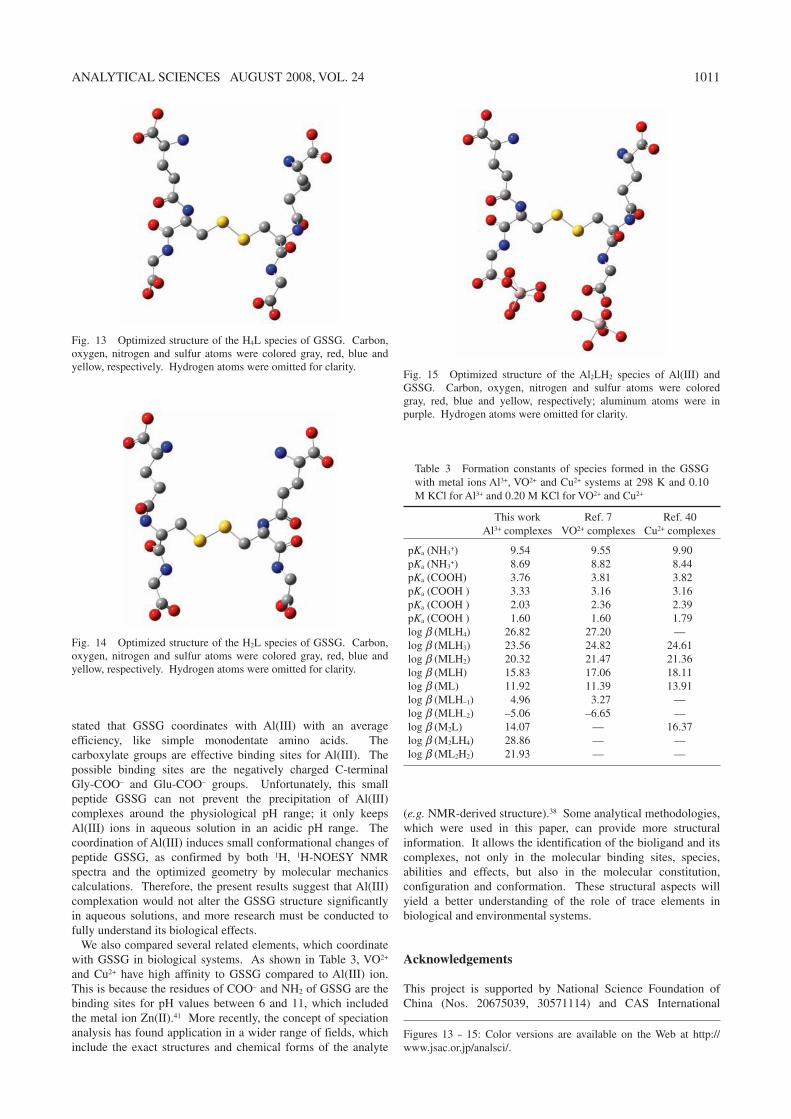
Fig. 14 Optimized structure of the H2L species of GSSG. Carbon, oxygen, nitrogen and sulfur atoms were colored gray, red, blue and yellow, respectively. Hydrogen atoms were omitted for clarity.
Fig. 15 Optimized structure of the Al2LH2 species of Al(III) and GSSG. Carbon, oxygen, nitrogen and sulfur atoms were colored gray, red, blue and yellow, respectively; aluminum atoms were in purple. Hydrogen atoms were omitted for clarity.
Table 3 Formation constants of species formed in the GSSG with metal ions Al3+, VO2+ and Cu2+ systems at 298 K and 0.10 M KCl for Al3+ and 0.20 M KCl for VO2+ and Cu2+
pKa (NH3+) 9.54 9.55 9.90
pKa (NH3+) 8.69 8.82 8.44
pKa (COOH) 3.76 3.81 3.82pKa (COOH ) 3.33 3.16 3.16pKa (COOH ) 2.03 2.36 2.39pKa (COOH ) 1.60 1.60 1.79log b (MLH4) 26.82 27.20 —log b (MLH3) 23.56 24.82 24.61log b (MLH2) 20.32 21.47 21.36log b (MLH) 15.83 17.06 18.11log b (ML) 11.92 11.39 13.91log b (MLH–1) 4.96 3.27 —log b (MLH–2) –5.06 –6.65 —log b (M2L) 14.07 — 16.37log b (M2LH4) 28.86 — —log b (ML2H2) 21.93 — —
This workAl3+ complexes
Ref. 7VO2+ complexes
Ref. 40Cu2+ complexes
Figures 13 – 15: Color versions are available on the Web at http://www.jsac.or.jp/analsci/.
1012 ANALYTICAL SCIENCES AUGUST 2008, VOL. 24
Partnership Project (CXTD-Z2005-04). Research Funding from the State Key Laboratory of Soil and Sustainable Agriculture (055127) is also acknowledged.
References
1. R. Dringen, Pro. Neurobio., 2000, 62, 649. 2. J. Vina, A. Lloret, R. Orti, and D. Alonso, Mol. Aspect
Med., 2004, 25, 117. 3. Y. Christen, Am. J. Clin. Nutr., 2000, 71, 621. 4. D. P. Dixon, I. Cummins, D. J. Cole, and R. Edwards, Curr.
Opin. Plant Biol., 1998, 1, 258. 5. S. Bharath, M. Hsu, D. Kaur, S. Rajagopalan, and J. K.
Andersen, Biochem. Pharmacol., 2002, 64, 1037. 6. J. C. Pessoa, I. Tomaz, T. Kiss, E. Kiss, and P. Buglyo, J.
Biol. Inorg. Chem., 2002, 7, 225. 7. J. C. Pessoa, I. Tomaz, T. Kiss, and P. Buglyo, J. Inorg.
Biochem., 2001, 84, 259. 8. D. N. Kumar, B. K. Singh, B. S. Garg, and P. K. Singh,
Spectrochim. Acta, Part A, 2003, 59, 1487. 9. N. Kato, M. Nakamura, and T. Uchiyama, J. Inorg.
Biochem., 1999, 75, 117. 10. N. A. Rey, O. W. Howarth, and E. C. Pereira-Maia, J. Inorg.
Biochem., 2004, 98, 1151. 11. B. Podányi and R. S. Reid, J. Am. Chem. Soc., 1988, 110,
3805. 12. A. Levina, L. B. Zhang, and P. A. Lay, Inorg. Chem., 2003,
42, 767. 13. M. Gelinsky, R. Vogler, and H. Vahrenkamp, Inorg. Chim.
Acta, 2003, 334, 230. 14. B. H. Cruz, J. M. Díaz-Cruz, M. S. Díaz-Cruz, C. M. Arimo
Esteban, and R. Tauler, J. Electroanal. Chem., 2001, 516, 110.
15. C. Cecchi, S. Latorraca, S. Sorbi, T. Iantomasi, F. Favilli, M. T. Vincenzini, and G. Liguri, Neurosci. Lett., 1999, 275, 152.
16. E. Kiss, A. Lakatos, I. Banyai, and T. Kiss, J. Inorg. Biochem., 1998, 69, 145.
17. H. Irving, M. G. Miles, and L. D. Pettit, Anal. Chim. Acta, 1967, 38, 475.
18. A. E. Martell and R. J. Motekaitis, “Determination and Use of Stability Constants”, 1992, VCH, Publishers, Inc., New York, 143.
19. S. Daydé, V. Brumas, D. Champmartin, P. Rubini, and G. Berthon, J. Inorg. Biochem., 2003, 97, 104.
20. M. J. Frisch, G. W. Trucks, H. B. Schlegel, G. E. Scuseria, M. A. Robb, J. R. Cheeseman, V. G. Zakrzewski, J. A. Montgomery, R. E. Stratmann, J. C. Burant, S. Dapprich, J. M. Millam, A. D. Daniels, K. N. Kudin, M. C. Strain, Ö. Farkas, J. Tomasi, V. Barone, M. Cossi, R. Cammi, B.
Mennucci, C. Pomelli, C. Adamo, S. Clifford, J. Ochterski, G. A. Petersson, P. Y. Ayala, Q. Cui, K. Morokuma, D. K. Malick, A. D. Rabuck, K. Raghavachari, J. B. Foresman, J. Cioslowski, J. V. Ortiz, A. G. Baboul, B. B. Stefanov, G. Liu, A. Liashenko, P. Piskorz, I. Komáromi, R. Gomperts, R. L. Martin, D. J. Fox, T. Keith, M. A. Al-Laham, C. Y. Peng, A. Nanayakkara, C. Gonzalez, M. Challacombe, P. M. W. Gill, B. G. Johnson, W. Chen, M. W. Wong, J. L. Andres, M. Head-Gordon, E. S. Replogle, and J. A. Pople, Gaussian03, Revision A.1, 2003, Gaussian Inc., Pittsburgh, PA.
21. N. Kato, M. Nakamura, and T. Uchiyama, J. Inorg. Biochem., 1999, 75, 117.
22. J. W. Akitt, Prog. Nucl. Magn. Reson. Spectrosc., 1989, 2, 11.
23. S. P. Bi, X. D. Yang, F. P. Zhang, X. L. Wang, and G. W. Zou, Fresenius J. Anal. Chem., 2001, 370, 984.
24. C. Exley, J. Inorg. Biochem., 2003, 97, 1. 25. C. Exley, “Aluminum and Alzheimer’s Disease: The Science
that Describes the Link”, 1st ed., 2001, Elivser Science, 361.
26. R. F. Shen, J. F. Ma, M. Kyo, and T. Iwashita, Planta, 2002, 215, 394.
27. J. F. Ma, R. F. Shen, Z. Zhao, M. Wissuwa, Y. Takeuchi, T. Ebitani, and M. Yang, Plant Cell Physiol., 2002, 43, 652.
28. R. F. Shen and J. F. Ma, J. Exp. Bot., 2001, 52, 1683. 29. X. D. Yang, S. P. Bi, X. L. Wang, J. Liu, and Z. P. Ba, Anal.
Sci., 2003, 19, 273. 30. X. D. Y ang, Y. Z. Tang, S. P. Bi, G. S. Yang, and J. Hu,
Anal. Sci., 2003, 19, 133. 31. X. D. Yang, S. P. Bi, X. L. Yang, L. Yang, J. Hu, and J. Liu,
Anal. Sci., 2003, 19, 815. 32. X. D. Yang, S. P. Bi, L. Yang, Y. H. Zhu, and X. L. Wang,
Spectrochim. Acta, Part A, 2003, 59, 2561. 33. X. D. Yang, Q. Miao, T. Yu, J. Hu, Z. B. Yang, and S. P. Bi,
Spectrochim. Acta, Part A, 2003, 59, 2655. 34. X. D. Yang, L. F. Li, and S. P. Bi, Sensors, 2005, 5, 235. 35. F. X. Kong, Y. Liu, W. Hu, P. P. Shen, C. L. Zhou, and L. S.
Wang, Chemosphere, 2000, 40, 311. 36. B. Dong, W. L. Sang, D. X. Jiang, J. M. Zhou, F. X. Kong,
and W. Hu, Chemosphere, 2002, 47, 87. 37. D. Orihuela, V. Meichtry, N. Pregi, and M. Pizarro, J. Inorg.
Biochem., 2005, 99, 1871. 38. E. H. Evans, Anal. Bioanal. Chem., 2003, 376, 311. 39. S. L. Simpson, S. SjÖberg, and K. J. Powell, J. Chem. Soc.,
Dalton. Trans., 1995, 11, 1799. 40. K. Várnagy, I. Sóvágó, and H. Kozlowski, Inorg. Chim.
Acta, 1988, 151, 117. 41. W. S. Postal, E. J. Vogel, C. M. Young, and F. T. Greenaway,
J. Inorg. Biochem., 1985, 25, 25.

![CHEMICAL SPECIATION OF ENVIRONMENTALLY SIGNIFICANT … · significant metals with inorganic ligands Part 2: ... [97INC], of metal–lig-and complexes are reported for ionic media](https://img.pdfslide.us/doc/110x75/5fc3988a6c1be50b413ad2b5/chemical-speciation-of-environmentally-significant-significant-metals-with-inorganic.jpg)


![International Journal of ChemTech Research260-273)V11N10CT.pdf · characterized. A sulfide and olefins were oxidized by use of complexes [VO(salen)] and [VO(salap)] (mononuclear),](https://img.pdfslide.us/doc/110x75/5caa501388c993e6068b515a/international-journal-of-chemtech-260-273v11n10ctpdf-characterized-a-sulfide.jpg)