Embed Size (px)
Citation preview
Spasticity reduction using electrical
stimulation
in the lower limb of spinal cord injury patients
Arjan van der Salm

Address of correspondence: Arjan van der Salm Saxion Hogescholen Academie Gezondheidszorg PO Box 70000 7500 KB Enschede The Netherlands +31 (53) 4871532 [email protected] Printed by FEBO-druk ISBN 90-365-2253-6 Cover: FOTON visuele communicatie © Arjan van der Salm, Enschede, The Netherlands, 2005 All rights reserved. No part of this book may be reproduced, stored in a retrieval system, or transmitted, in any form or by any means, electronic, mechanical, photocopying, recording, or otherwise, without the prior written permission of the holder of the copyright. Chapter 2: © Archives of Physical Medicine & Rehabilitation, 2005
SPASTICITY REDUCTION USING ELECTRICAL STIMULATION IN THE LOWER LIMB OF SPINAL CORD INJURY PATIENTS
PROEFSCHRIFT
Ter verkrijging van de graad van doctor aan de Universiteit Twente,
op gezag van de rector magnificus, Prof.dr. W.H.M. Zijm,
volgens besluit van het College voor Promoties in het openbaar te verdedigen
op vrijdag 21 oktober 2005 om 13:00 uur
Door
Arjan van der Salm geboren op 20 juli 1971
te Delft

DIT PROEFSCHRIFT IS GOEDGEKEURD DOOR: Prof.dr.ir. P.H. Veltink (promotor) Prof.dr. M.J. IJzerman (promotor) Dr. A.V. Nene (assistent promotor)
DE PROMOTIECOMMISSIE IS ALS VOLGT SAMENGESTELD: Promotoren: Prof.dr.ir. P.H. Veltink Universiteit Twente Prof.dr. M.J. IJzerman Universiteit Twente Assistent promotor: Dr. A.V. Nene Roessingh Research & Development Referent: Dr. H.E.J. Veeger Vrije Universiteit Amsterdam Leden: Prof.dr. J.G. Becher Vrije Universiteit Medisch Centrum Prof.dr. J.E.J. Duysens Universitair Medisch Centrum St. Radboud Prof.dr. ir. H.J. Hermens Universiteit Twente Prof.dr. F.C.T. van der Helm Unversiteit Twente Paranymfen: Anke Kottink Wiebe de Vries

This study was part of a larger research program: ’Functional strain, work capacity and mechanisms of restoration of mobility in the rehabilitation of persons with spinal cord injury’, sponsored by ZONMW-Rehabilitation, grant number: 1435.0010.
The publication of this thesis was generously supported by: Roessingh Research & Development Revalidatiecentrum het Roessingh Leerstoel Biomedische signalen en systemen, Universiteit Twente


Voor Tamare

CONTENTS Chapter 1 Introduction and outline of the thesis 13 Chapter 2 Development of a new method for objective assessment of spasticity using full range passive movements 21 Chapter 3 Criterion validity and reliability of a method for objective assessment of spastic hypertonia using full range passive movements 39 Chapter 4 Comparison of electrical stimulation methods for reduction of triceps surae spasticity in SCI-patients 57 Chapter 5 Modulation of the vastus lateralis H-reflex during gait in healthy subjects and patients with spinal cord injury 73 Chapter 6 Effect of electrical stimulation of hamstrings and l3/4 dermatome on H/M-ratio and performance of gait in spastic SCI-patients 85 Chapter 7 General discussion 99 Summary 109 Samenvatting 113 Nawoord 117 Curriculum vitae 121

CHAPTER 1 Introduction and outline of the thesis
Chapter 1
14
INTRODUCTION
Spasticity has been and still is an important topic in rehabilitation of patients with central neurological disorders [1-4]. The definition of spasticity by Lance [5] is: ‘spasticity is a motor disorder characterized by a velocity-dependent increase in tonic stretch reflexes (‘muscle tone’) with exaggerated tendon jerks, resulting from hyperexcitability of the stretch reflex, as one component of the upper motor neuron syndrome’. Of course, patients with increased muscle stiffness are not only impaired by an enlarged ‘muscle tone’, but the existence of increased passive muscle stiffness as well as clonus, additionally, affects functional movements. The focus of this thesis is to study methods to reduce spasticity in lower limb muscles to facilitate gait. After a spinal cord injury (SCI) some persons have impaired leg function. Many SCI patients are bound to a wheelchair, but in patients with incomplete SCI, 47 percent is able to walk [6]. In the SCI population with walking abilities, 75 percent indicate that improvement of walking quality is important [6]. In contrast, 58% indicates bladder management to be important. It is supposed that spasticity is an important impairing factor in gait [7;8]. In addition, muscle weakness also causes loss of gait function. One study used the results of a questionnaire, which was send to SCI-clinicians, and observational data of 21 SCI patients to determine the most common gait impairments [8]. It was found that SCI patients most commonly suffer from an impaired hip extension during late stance and a decreased hip and knee flexion during early swing, as well as an excessive plantar flexion during swing resulting in an impaired initial foot contact. In patients with spasticity in their triceps surae a hyperactivity of these muscles may be the cause of the excessive plantar flexion both during swing and stance. Therefore, inhibition of the spastic muscles could be useful to improve the gait in these patients. The decreased knee flexion during swing may directly impair the foot clearance [9]. In patients with spasticity, hyperactivity of the quadriceps muscles is seen, which may prevent the knee from flexion. Hamstrings stimulation can, mechanically, induce knee flexion, but in spastic patients, inhibition of the quadriceps muscles also may be beneficial. Many treatments are available for spasticity reduction. Oral medication, intrathecal baclofen pumps, physical therapy and even surgery is applied to
Introduction and outline of the thesis
15
reduce spasticity or treat fixed contractures as a result from spasticity [10;11]. In addition to these treatment modalities, electrical stimulation is also known to reduce spasticity [12]. Electrical stimulation may have several advantages over the other treatment modalities. As well as intrathecal baclofen electrical stimulation has the possibility to modulate the intensity of the intervention and therefore the intensity of the effect. This also implies that the spasticity can be modulated instead of totally eliminated. Thus, patients are potentially able to use the residual muscle tonus for functional movements. A second advantage of electrical stimulation is the local application. In contrast, oral medication will influence the tonus in all the muscles in the body. A very important advantage of electrical stimulation is that it is non-invasive. To reduce spasticity by means of electrical stimulation an instant effect and a carry-over effect (effect remains after stimulation has stopped) can be distinguished. The carry-over effect can be very useful in the treatment of gait, because the electrical stimulation can be used to reduce the spasticity
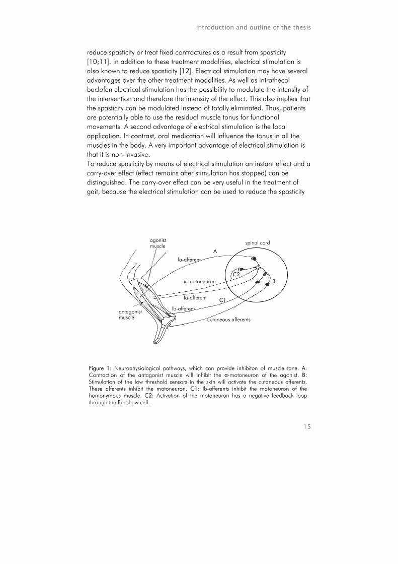
Figure 1: Neurophysiological pathways, which can provide inhibiton of muscle tone. A: Contraction of the antagonist muscle will inhibit the α-motoneuron of the agonist. B: Stimulation of the low threshold sensors in the skin will activate the cutaneous afferents. These afferents inhibit the motoneuron. C1: Ib-afferents inhibit the motoneuron of the homonymous muscle. C2: Activation of the motoneuron has a negative feedback loop through the Renshaw cell.
A
B
C1
C2 α-motoneuron
Ia-afferent
Ia-afferent Ib-afferent
cutaneous afferents antagonist muscle
agonist muscle spinal cord
Chapter 1
16
before the actual gait (training) is performed. Thus, the gait impairment caused by spasticity will be reduced or eliminated, which can facilitate gait. Several methods of electrical stimulation for spasticity reduction using the carry-over effect have been reported. These stimulation methods can be grouped by i) stimulation of the antagonist [13-15], ii) stimulation of the dermatome [16] and iii) stimulation of both the agonist and antagonist [12;17]. Figure 1 shows the neurophysiological pathways on which these stimulation methods are based. Stimulation of the antagonist initiates the reciprocal inhibition. When the dermatome is stimulated (cutaneous stimulation) the Ib-inhibitory interneuron inhibits the motoneuron and stimulation of the agonist activates both the Ib-inhibitory pathway and Renshaw cell inhibition. All these studies state that the electrical stimulation can be beneficial. On the other hand it is unclear what method provides the best result and whether this result is better than a placebo approach. The instant effect of electrical stimulation to inhibit muscle activity has been investigated in stroke patients using antagonistic nerve stimulation [18] and in SCI patients during gait using cutaneomuscular stimulation [7]. These studies reported a reduction of the reflex excitability in the triceps surae due to the interventions. The stimulation parameters applied in FES studies are different from the stimulation parameters used in the inhibitory studies [18-21]. In FES relatively low frequencies and short pulse widths are used compared to the inhibitory studies. The stimulation parameters in FES selectively activate non-nociceptive afferents, and are therefore relatively more comfortable for the subjects, and thus can be applied for a relatively long time. The required inhibition must last for 300 ms or more to be functional. This means that stimulation should be adapted to a functional and comfortable level with an inhibitory effect. This can only be done when comfortable stimulation parameters are used. It is unknown if electrical stimulation based on these parameters used in FES can cause neurophysiological changes. The goal of this thesis was to investigate the influence of electrical stimulation on spasticity of leg muscles in spinal cord injury patients and its impact on gait. Both, the carry-over effect and the instant effect of electrical stimulation during gait were investigated.
Introduction and outline of the thesis
17
OUTLINE OF THE THESIS
Chapter 2 and 3 describe a new measure for spasticity. The goal of these studies is to develop a valid and reliable measure, which, unlike other measures, objectively assesses spasticity in the functional range. In chapter 2 the development of the assessment method is described. The newly developed assessment uses of movements over the whole range of motion and the assessment provides outcomes which are comparable to outcomes of clinical measures like the (Modified) Ashworth scale [22;23] and the Tardieu scale [24]. In chapter 3 the new assessment for spasticity is evaluated on its correlation with other measures (i.e. criterion validity), and the reliability and responsiveness is presented. Because no golden standard is available for the measurement of spasticity, the Modified Ashworth scale, clonus-score and H/M-ratio [25] are used as measures to assess the criterion validity. The newly developed assessment for spasticity is used in chapter 4 to study the effect of electrical stimulation. Chapter 4 describes the effect of three methods of electrical stimulation used to reduce spasticity. These methods are based on inhibitory neurophysiological pathways; antagonistic, agonistic and low threshold sensory inhibition [26]. The carry-over effect of the stimulation is studied in ten complete spinal cord injury patients. A blinded placebo controlled study is performed, in which patients received all three stimulation interventions and a placebo approach on four separate days. To assess the carry-over effect, measurements are performed until two hours after the intervention. For the assessment of spasticity the newly developed assessment, described in chapter 2 and 3, is used in combination with the Modified Ashworth scale, clonus-score and H/M-ratio. In chapter 5 the spinal reflex excitability of the vastus lateralis during gait, measured by the H/M-ratio, is described for both healthy subjects and spastic SCI patients. The H/M-ratio for mid-stance and mid-swing and the modulation of these outcomes within the gait cycles are studied. The differences between the H/M-ratios of mid-stance and mid-swing in the healthy subjects and differences between healthy subjects and patients are presented.
Chapter 1
18
Chapter 6 is the description of the instant effect of electrical stimulation during gait provoked by stimulation of the hamstrings and L3/4 dermatome. Both, neurophysiological mechanisms and gait performance are studied. The assessments are carried out in five spastic incomplete SCI patients. The electrical stimulation is performed to inhibit the vastus lateralis during the swing phase. As a result, the knee flexion is expected to be facilitated. In chapter 7 a general discussion of the thesis is described.

Introduction and outline of the thesis
19
REFERENCE LIST
1. Dietz V. Spastic movement disorder. Spinal Cord 2000 Jul;38(7):389-93. 2. Taylor S, Ashby P, Verrier M. Neurophysiological changes following traumatic spinal
lesions in man. J Neurol Neurosurg Psychiatry 1984 Oct;47(10):1102-8. 3. Toft E. Mechanical and electromyographic stretch responses in spastic and healthy
subjects. Acta Neurol Scand Suppl 1995;163:1-24. 4. Hiersemenzel LP, Curt A, Dietz V. From spinal shock to spasticity: neuronal
adaptations to a spinal cord injury. Neurology 2000 Apr 25;54(8):1574-82. 5. Lance JW. Spasticity: disordered motor control. Feldman RG; Young RR; Koella WP.
Symposium Synopsis, Miami: Symposia Specialists; 1980. pp. 485-500. 6. Maxwell DJ, Granat M, Baardman G. CREST system: Available functions. Series title:
Clinical rehabilitation using electrical stimulation via telematics. 1997. 7. Fung J, Barbeau H. Effects of conditioning cutaneomuscular stimulation on the
soleus H- reflex in normal and spastic paretic subjects during walking and standing. J Neurophysiol 1994 Nov;72(5):2090-104.
8. Van der Salm A, Nene A, Maxwell DJ, Veltink PH, Hermens HJ, IJzerman MJ. Gait impairments in a group of patients with incomplete spinal cord injury and their relevance regarding therapeutic approaches using FES. Artificial Organs 2005 Jan;29(1):8-14.
9. Riley PO, Kerrigan DC. Torque action of two-joint muscles in the swing period of stiff-legged gait: a forward dynamic model analysis. J Biomech 1998 Sep;31(9):835-40.
10. Bhakta BB. Management of spasticity in stroke. Br Med Bull 2000;56(2):476-85. 11. Burchiel KJ, Hsu FP. Pain and spasticity after spinal cord injury: mechanisms and
treatment. Spine 2001 Dec 15;26(24 Suppl):S146-60. 12. Vodovnik L, Bowman BR, Hufford P. Effects of electrical stimulation on spinal
spasticity. Scand J Rehabil Med 1984;16(1):29-34. 13. Alfieri V. Electrical treatment of spasticity. Reflex tonic activity in hemiplegic patients
and selected specific electrostimulation. Scand J Rehabil Med 1982;14(4):177-82. 14. Robinson CJ, Kett NA, Bolam JM. Spasticity in spinal cord injured patients: 1. Short-
term effects of surface electrical stimulation. Arch Phys Med Rehabil 1988 Aug;69(8):598-604.
15. Robinson CJ, Kett NA, Bolam JM. Spasticity in spinal cord injured patients: 2. Initial measures and long-term effects of surface electrical stimulation. Arch Phys Med Rehabil 1988 Oct;69(10):862-8.
16. Bajd T, Gregoric M, Vodovnik L, Benko H. Electrical stimulation in treating spasticity resulting from spinal cord injury. Arch Phys Med Rehabil 1985 Aug;66(8):515-7.
17. Franek A, Turczynski B, Opara J. Treatment of spinal spasticity by electrical stimulation. J Biomed Eng 1988 May;10(3):266-70.
18. Veltink PH, Ladouceur M, Sinkjær T. Inhibition of the triceps surae stretch reflex by stimulation of the deep peroneal nerve in persons with spastic stroke. Arch Phys Med Rehabil 2000 Aug;81(8):1016-24.
19. Bajd T, Kralj A, Turk R, Benko H, Sega J. Use of functional electrical stimulation in the rehabilitation of patients with incomplete spinal cord injuries. J Biomed Eng 1989 Mar;11(2):96-102.
20. Braun Z, Mizrahi J, Najenson T, Graupe D. Activation of paraplegic patients by

Chapter 1
20
functional electrical stimulation: training and biomechanical evaluation. Scand J Rehabil Med Suppl 1985;12:93-101.
21. Granat MH, Ferguson AC, Andrews BJ, Delargy M. The role of functional electrical stimulation in the rehabilitation of patients with incomplete spinal cord injury--observed benefits during gait studies. Paraplegia 1993 Apr;31(4):207-15.
22. Ashworth B. Preliminary trail of carisoprodal in multiple sclerosis. 1964;192:540-2. 23. Bohannon RW, Smith MB. Interrater reliability of a modified Ashworth scale of
muscle spasticity. Physical Therapy 1987 Feb;67(2):206-7. 24. Held JP, Pierrot-Deseilligny E. Le Bilan Moteur Central. In: Reeducation motrice des
affections neurologiques. ed: Bailiere JB et fils. Paris:1969:31-42. 25. Visser, S. L. Reflexen. In: Klinische Elekromyografy. ed: Notermans Bussel:
1981:353-68. 26. Kandell ER, Schwartz JH, Jessell TM. Essentials of neural science and behavior.
McGraw-Hill; 1995.
CHAPTER 2 Development of a new method for objective assessment of spasticity using full range passive movements Arjan van der Salm, Peter H. Veltink, Hermie J. Hermens, Maarten J. IJzerman, Anand V. Nene Objective: Development of a method for assessment of spasticity, in which the whole range of motion at a wide variation of speeds is applied. The reflexive and non-reflexive components of the muscle response are measured. Design: Cross-sectional design to study construct validity. Setting: Research department affiliated with a rehabilitation hospital in the Netherlands. Patients: 9 complete spinal cord injured patients recruited from the rehabilitation hospital. Main outcome measures: 30 to 45 stretches over the whole range of motion were applied to the triceps surae muscle at varying velocities measuring from 30 to 150 º/s. EMG responses were measured in order to assess reflex excitability. The torque over the ankle joint was measured during the whole stretch. The angle and velocity at which the reflex was initiated was also determined. Results: The EMG responses increased significantly at increasing stretch velocities (p<0.001). The applied maximum angles are reproducible (ICC = 0.81) and provide representative torque responses. Conclusion: The assessment method of spasticity using full range passive movements provides objective outcomes. The angular-velocity is responsible for an exponential increase in amplitude of the EMG response. Archives of Physical Medicine & Rehabilitation. Article in Press.

Chapter 2
22
1. INTRODUCTION
Spasticity may be very impairing in patients with upper motor neuron lesions. Several authors state that spasticity can cause gait impairments [1-3]. An important aspect of this is increased plantar flexion during swing. Affected patients have to compensate for this by circumduction of the leg or hiking of the pelvis. Thus, a relatively small impairment such as the increased plantar flexion can have a large impact on the general movement pattern. Increased plantar flexion during stance, ankle vaulting, is also frequently observed [4]. These gait impairments may be due to co-contractions [5] or hyper-reflexive movements in response to muscle stretch [6-10]. Spasticity is defined as a ‘velocity dependent increase in the tonic stretch reflex (muscle tone) with exaggerated tendon jerks, resulting from the hyper excitability of the stretch reflex, as one component of the upper motor neurone syndrome’ [11]. It should be noted that spasticity is only one part of muscle stiffness which also includes passive muscle stiffness [12]. Passive muscle stiffness depends on soft tissue changes. These changes provide a biomechanical, non-reflexive, component of muscle stiffness [13]. It is important to distinguish between these components of muscle stiffness, because it may have consequences for treatment. Therefore, an assessment for spasticity should objectively measure both the reflexive and non-reflexive components of muscle stiffness. Frequently used clinical tests of spasticity are; the Ashworth scale (AS) [14], the Modified Ashworth scale (MAS) [15] and the Tardieu scale [16]. The AS is a 5 point scale grading from 0; ‘normal muscle tone’ to 4; ‘limb rigid in flexion or extension’. The modified MAS is extended with an extra grade between the 1 and 2, i.e. 1+. The scores are determined by moving the joint over its entire range of motion. Commonly used velocities are approximately 50 º/s [17;18]. A disadvantage of the AS and MAS is the poor inter-tester reliability [19;20]. The outcome of the Tardieu test is the angle at which a catch can be felt. The catch is defined as a sudden increase in muscle stiffness in response to a brisk muscle stretch. The inter- and intra-tester reliability correlation coefficient of the Tardieu test was found to vary from 0.38 to 0.90 [21]. Thus, this reliability is poor in several cases. Other measurements of spasticity have been reported, which do not require subjective assessment and therefore may be more reliable, for example the H-reflex and tendon tap [22]. However, these measurement methods do not
Development of a method for objective assessment of spasticity
23
assess spasticity in the functional range. It can be concluded that no objective assessment for spasticity in the functional range is clinically available. The goal of this study is to propose such an assessment method. It uses the same movement range as the MAS, but assesses both the reflexive and non-reflexive components of muscle stiffness using physical measures.
2. METHODS
2.1. SUBJECTS
Spinal cord injury (SCI) subjects were recruited from a database of a rehabilitation centre in the Netherlands (Het Roessingh, Enschede). Inclusion criteria were: presence of spasticity (Ashworth grade 1 or higher), absence of voluntary movements in the triceps surae, time since injury at least 6 months, triceps surae muscles and tibialis anterior muscle must be able to contract using electrical stimulation and age above 18. Patients were allowed to take anti-spasticity medication, but they approved not to change the doses 2 weeks before and during the experiment. Patients with hypersensitive skin of the legs, absence of the dorsal flexion beyond anatomical position or diseases which could temporally increase tonus (specifically bladder infection) were excluded. All subjects gave informed consent to participate and the experiment was approved by the local ethics committee. Patients were measured 3 to 4 times with 3 to 14 days in between. The time of the day at which the measurements started was kept equal for all measurement sessions.
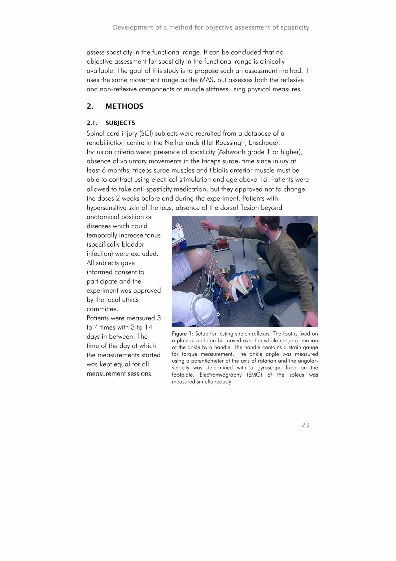
Figure 1: Setup for testing stretch reflexes. The foot is fixed on a plateau and can be moved over the whole range of motion of the ankle by a handle. The handle contains a strain gauge for torque measurement. The ankle angle was measured using a potentiometer at the axis of rotation and the angular-velocity was determined with a gyroscope fixed on the footplate. Electromyography (EMG) of the soleus was measured simultaneously.

Chapter 2
24
2.2. STRETCH REFLEX OVER THE WHOLE RANGE OF MOTION
The patients were seated upright and the knee was flexed 75 degrees, zero degrees being defined as knee extension (Figure 1). Two different devices were used. The foot was fixed to a footplate, which could be rotated around one axis, thus providing dorsal and plantar flexion at the ankle joint. The foot was strapped to the plate using a soft, flexible brace and Velcro in such a manner that the heel could not lift from the plate, yet the ankle joint could freely be moved. The rotation axis of the ankle joint, defined as the line through the malleolli, was aligned with the rotation axis of the device. Before the measurement started the range of motion of the ankle joint was determined manually. Stops were inserted at the maximal plantar flexion and dorsal flexion, to prevent movement in excess of the range of movement. Dorsal flexion and subsequently plantar flexion movements were applied manually using a handle. The movement of interest was the dorsal flexion movement to assess soleus muscle spasticity, which is clinically most relevant. Thus, a movement from maximal plantar flexion to maximal dorsal flexion was applied. Between two successive movements, at least 5 seconds rest in plantar flexion was prescribed, whereas the duration of the stretch was less than 2.3 seconds. For safety reasons, the first four stretches were slow. As time progressed, both slow and fast movements were carried out. The latter stretch velocities were applied in random order. The angles and angular-velocities were measured simultaneously. The applied stretch velocity was presented after each movement, therefore it was possible to equally spread the applied velocities over the whole range. In one session about 30 to 45 stretches were applied, ranging from 30 to 150 º/s. The duration of one session was approximately 5 minutes.
2.3. DATA RECORDING
The EMG of the soleus muscle was measured with surface electrodes (Neuroline® type 720 00-s Ag-AgCl gel electrodes; diameter 12 mm, inter-electrode space 20 mm) using a bipolar arrangement. A ground electrode was applied on the lateral malleollus. The electrodes were applicated on one third of the line through the medial malleous and the medial epicondyl of the femur [23] (Figure 1). Before application the skin was shaved, abraded and cleaned with alcohol. For the EMG recording TMSI® hard- and software was used. The sample frequency of the EMG was 2048 Hz. The EMG data was band-pass filtered applying cut-off frequencies of 20 to 200 Hz. A 22 bit-A/D-converter was used which had an effective resolution of

Development of a method for objective assessment of spasticity
25
71.9 nV. The angles, angular-velocities and torques were measured with a sample frequency of 1000 Hz. Angles were measured with a calibrated potentiometer fixed over the axis of ankle rotation. For the movement of the foot the anatomical position was defined as 0º and plantar flexion was defined as being negative. The angular-velocity was measured with a calibrated angular-velocity-sensor (gyroscope) on the foot-plate. Torque was measured with a calibrated strain gauge in the handle. The angle, angular-velocity and torque data were recorded on a laptop computer using an A/D-converter and Labview® software. The analysis of the data was performed in Matlab (Mathworks®).
2.4. DATA ANALYSIS
The stretch movements were applied manually, comparable to the stretches of (M)AS movements and stretches in daily life. The average velocity during the dorsal flexion over the whole range was defined as the stretch-velocity. The start of the EMG-burst in the filtered EMG data was detected with a threshold. This threshold was 3 times the standard deviation of the noise level, determined before the stretch started. The detection of the start of the EMG-burst was performed during the stretch. This detection time was corrected for the delay of the reflex-loop, which was estimated as 45 ms [24;25]. When a burst was detected the Root-Mean-Square (RMS) value of the EMG-signals over a 100 ms window was calculated. This window length matched the largest burst times. The window started at the beginning of the burst. The RMS-values of the EMG response to the stretch were plotted against average stretch velocities. The increase of EMG responses at increasing speed was described with an exponential fit over the 30 to 45 responses in one session. The EMG responses at 50 º/s (EMG50), 75 º/s (EMG75) and 100 º/s (EMG100) were calculated from the fitted curve. The state (angle and angular-velocity) in which the stretch reflex was initiated was determined. In detail, 45 ms before the beginning of the EMG-burst the angular-velocity and the angle of the stretch movement were determined. The results were plotted in an angular-velocity/ angle graph. The average and the standard deviation of the slope values of the linear-regression lines in the angular-velocity/ angle graphs were calculated. The angle, which triggered the burst-start (reflex-initiating angle) was defined at the velocity of 100 º/s. This velocity is commonly used for the Tardieu scale [16;21].

Chapter 2
26
Two devices were used in this study. The device, which was used for the first five measurements, had a relatively large inertia to ensure smooth movements for low velocities, but the ankle torque could not be measured due to this large moment of inertia. In order to allow measurement of ankle torque at varying velocities, a second device, with low moment of inertia (0.065 kg m2), was used in the last four experiments. The torques measured at velocities smaller than 70 º/s were averaged. At velocities higher than 70 º/s the influence of inertia to the torque may interfere with the torque from the muscle. The average values of the first, second and third 1/3 of the movement were determined and analysed, i.e. early-, mid- and late-movement-torque.
2.5. STATISTICAL ANALYSIS
EMG responses were statistically analysed. These RMS-values at the 3 speeds, 50, 75 and 100 º/s did not have a normal distribution. Therefore a non-parametric test was used to determine the velocity effect. The used test was the Friedman test for comparison of more than two related groups. For post-hoc tests the Wilcoxon signed rank test was used. To evaluate reproducibility of movement ranges Intra-Class-Correlation coefficient (ICC) was determined, using a 2-way random model with absolute agreement. The 95%-CI (Confidence Interval) was determined as well. Slopes of fitted lines in reflex-initiating angle – angular-velocities plots were determined. The average slope value was calculated with the 95%-CI. Alpha was 0.05 in all cases.

Development of a method for objective assessment of spasticity
27
3. RESULTS
3.1. SUBJECTS
33 patients were found to be suitable, according to the files. These patients were contacted. 16 patients were not willing to participate, because of other commitments or lack of interest. 17 patients were seen for intake. 7 of them were excluded because of lack of spasticity in the triceps surae muscles. One of the patients could not sit in the experimental setup due to trunk instability and it was not possible to measure this patient in his wheelchair. Finally, nine spinal cord injury patients were selected and participated in the study. The demographic data of these patients are presented in table 1. For the results of the EMG response the power was calculated. This power was found to be 0.79 (average SD is 3.35, average difference is 3.2, alfa is 0.05 and N is 8) [26].
Table 1: Demographic data of patients.
3.2. DATA
Figure 2 shows EMG responses in one subject, angles and angular-velocities for stretches at 3 speeds, 67, 84 and 102 º/s. In this session the range of motion is from 29 º plantar flexion to 14 º dorsal flexion. At increasing speed the EMG response increases. This relation between mean speed of the movement and RMS of the EMG burst is presented in figure 3 for 45 stretches applied during one measurement for the same subject. The response was found to be exponential which agrees with the sigmoid shape found in literature [27;28]. This study only evaluated the threshold of the sigmoid shape. The RMS-values of the EMG response at 50, 75 and 100 º/s are determined.
Patients Sex Age Injury level
Time since injury
(months)
Modified Ashworth
scale
Clonus
1 M 36 T4 71 1+ Y 2 M 30 T5 33 1 Y 3 M 42 C6 211 1+ Y 4 M 30 C6/7 28 3 Y 5 F 41 T8 208 1+ N 6 M 34 C6 105 3 N 7 M 37 T11 217 1 Y 8 F 21 C5 97 1 Y 9 M 41 T4/5 275 1 N
Chapter 2
28
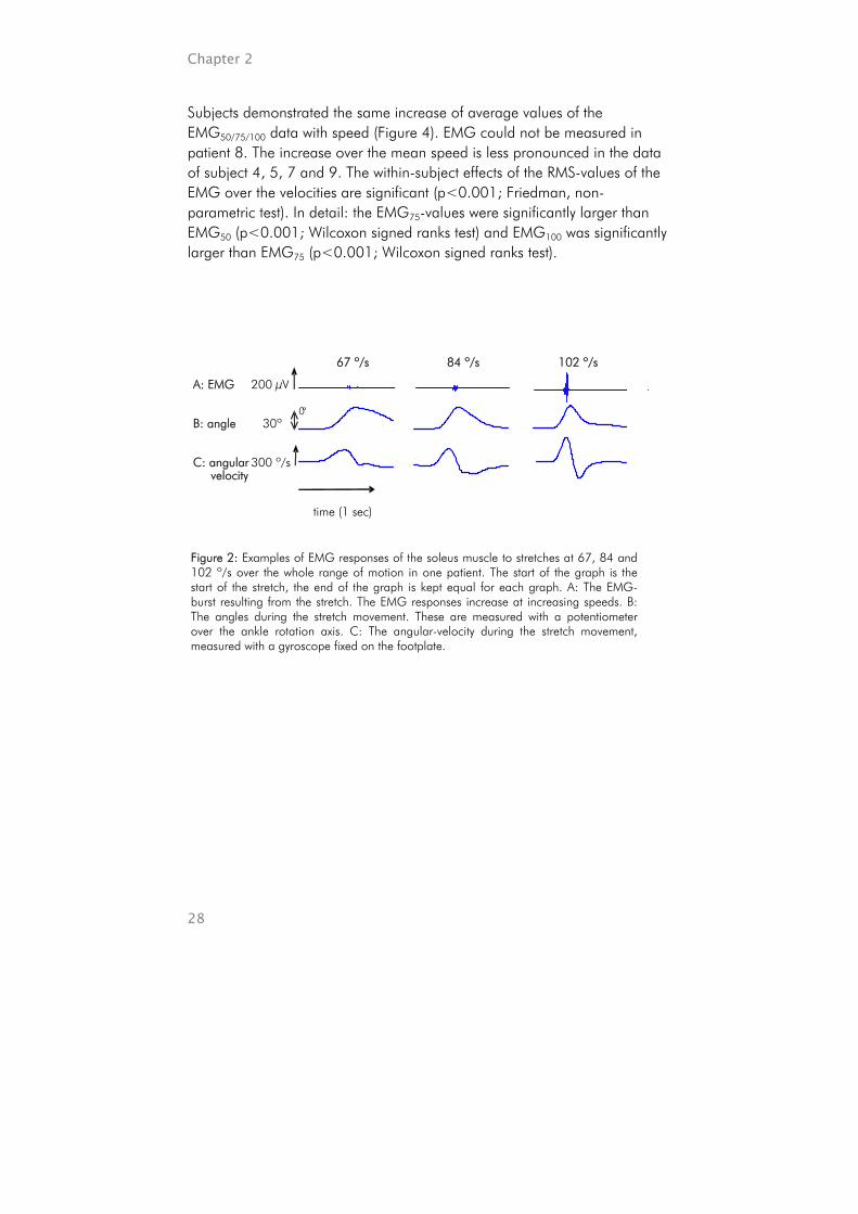
Subjects demonstrated the same increase of average values of the EMG50/75/100 data with speed (Figure 4). EMG could not be measured in patient 8. The increase over the mean speed is less pronounced in the data of subject 4, 5, 7 and 9. The within-subject effects of the RMS-values of the EMG over the velocities are significant (p<0.001; Friedman, non-parametric test). In detail: the EMG75-values were significantly larger than EMG50 (p<0.001; Wilcoxon signed ranks test) and EMG100 was significantly larger than EMG75 (p<0.001; Wilcoxon signed ranks test).
Figure 2: Examples of EMG responses of the soleus muscle to stretches at 67, 84 and 102 º/s over the whole range of motion in one patient. The start of the graph is the start of the stretch, the end of the graph is kept equal for each graph. A: The EMG-burst resulting from the stretch. The EMG responses increase at increasing speeds. B: The angles during the stretch movement. These are measured with a potentiometer over the ankle rotation axis. C: The angular-velocity during the stretch movement, measured with a gyroscope fixed on the footplate.
102 º/s 84 º/s 67 º/s 200 µV
300 º/s C: angular velocity
30º
A: EMG
time (1 sec)
B: angle 0º

Development of a method for objective assessment of spasticity
29
EMG
resp
onse
(µ
V)
0
20
40
60
80
1 2 3 4 5 6 7 9
RMS 50 º/s RMS 75 º/s RMS 100 º/s
Subject
Figure 4: Average values (+ 1 SD) for the RMS-values of the EMG responses, except subject 8. The presented RMS values represent the responses at stretches at 50, 75 and 100 º/s, respectively EMG50, EMG75 and EMG100.
Figure 3: Plot of one session in one subject (same subject and measurement as figure 2). The RMS-values of the EMG-signal over a ~100 ms window at the time of burst, are shown. These are responses to stretches ranging from 38 to 102 º/s. ● represent the RMS-values of the EMG-signal as a result from the reflexes; ○ represent the EMG responses below the threshold value, defined as 3 times the noise level. The fitted (solid) curve (+/- 1 SD, dotted curves) shows clearly the increase in the EMG response amplitudes at increasing velocities. The fitted curve is used to determine the RMS-values at 50, 75 and 100 º/s, i.e. EMG50, EMG75 and EMG100.
0 20 40 60 80 100 120 -
0
10
20
30
40
Mean Speed (º/sec)
EMG100
EMG75
EMG50
RMS
EMG
in w
indo
w (µ
V)
Chapter 2
30
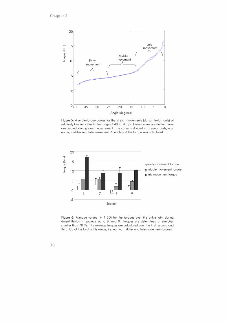
Figure 5: 4 angle-torque curves for the stretch movements (dorsal flexion only) at relatively low velocities in the range of 40 to 70 o/s. These curves are derived from one subject during one measurement. The curve is divided in 3 equal parts, e.g. early-, middle- and late-movement. At each part the torque was calculated.
40 35 30 25 20 15 10 5 0 -5
0
5
10
15
20
Torq
ue (N
m)
Angle (degrees)
Late movement
Middle movement Early
movement
Torq
ue (N
m)
Subject -5
0
5
10
15
20
6 7 8 9
early movement torque middle movement torque late movement torque
Figure 6: Average values (+ 1 SD) for the torques over the ankle joint during dorsal flexion in subjects 6, 7, 8, and 9. Torques are determined at stretches smaller than 70 o/s. The average torques are calculated over the first, second and third 1/3 of the total ankle range, i.e. early-, middle- and late-movement-torques.

Development of a method for objective assessment of spasticity
31
Figure 7: Graph of angular-velocities and angles 45 ms before the start of the EMG response. The graph is derived from one subject during one measurement involving 34 stretches. The linear regression line is fitted trough the scatter plot. In the graph the reflex-initiating angle is indicted at 100 º/s.
Figure 5 shows the torques at velocities up to 70 º/s. 4 stretches during one measurement in one subject are presented. The curves show the characteristic shape of tissue stretch. 3 ranges were distinguished: early-, middle- and late-movement-torque. Variability of the maximum angle may influence the outcome of the torque, because at the final range of dorsal flexion the torque increases rapidly. Therefore we checked the reproducibility of the maximum angles. The reproducibility of these angles was good (ICC = 0.81; 95%-CI = 0.53 - 0.96). In addition, no relevant change between the shapes of the curves is present. The average torque responses (+ 1 SD) for subject 6, 7, 8 and 9 are presented in figure 6. These results were not statistically evaluated because the torque was only measured in 4 subjects. As expected, the torque increases when the muscle is progressively stretched. Especially in subject 6 the increase in the late movement torque is remarkable. This subject shows also large EMG responses (Figure 4).
0 50 100 150 200 250 300 -35
-30
-25
-20
-15
-10
-5
Angl
e 45
ms
befo
re b
urst
sta
rt (º
)
Slope = - 0.036
Angular velocity 45 ms before burst start (º/sec)
reflex initiating angle at 100 º/s
Chapter 2
32
3.3. BURST START AND AMPLITUDE
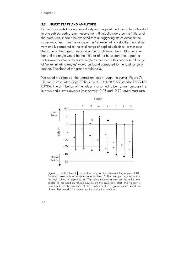
Figure 7 presents the angular velocity and angle at the time of the reflex start in one subject during one measurement. If velocity would be the initiator of the burst-start, it could be expected that all triggering states occur at the same velocities. Then the range of the ‘reflex-initiating velocities’ would be very small, compared to the total range of applied velocities. In that case, the slope of the angular-velocity/ angle graph would be ∞. On the other hand, if the angle would be the initiator of the burst-start, the triggering states would occur at the same angle every time. In this case a small range of ‘reflex-initiating angles’ would be found compared to the total range of motion. The slope of the graph would be 0. We tested the slopes of the regression lines through the curves (Figure 7). The mean calculated slope of the subjects is 0.018 º/º/s (standard deviation 0.035). The distribution of the values is assumed to be normal, because the kurtosis and curve skewness (respectively -0.08 and -0.70) are almost zero.
dorsal flexion
plantar flexion
Figure 8: The first lines (▐ ) show the range of the reflex-initiating angles at 100 º/s stretch velocity in all subjects, except subject 8. The average range of motion for each subject is presented (♦). The reflex-initiating angles are the ankle joint angles 45 ms (used as reflex delay) before the EMG-burst-start. This velocity is comparable to the stretches of the Tardieu scale. Negative values stand for plantar flexion and 0 o is defined as the anatomical position.
Subject
1 2 3 4 5 6 7 9
Angl
e (º
)
-50
-40
-30
-20
-10
0
10
20
Development of a method for objective assessment of spasticity
33
The 95%-CI of the slope is -0.011 to 0.048. Thus, the slope does not differ significantly from 0. This indicates strongly that the angle is the initiator for the start of the EMG-burst. Figure 8 shows the range of the reflex-initiating angles at 100 º/s for 3 or 4 measurements of each subject. The range of motion of the ankle joint is also presented. All responses are initiated in the mid range of the movement and are relatively small compared to the total range of motion. Only in subjects 3 and 5 the range of the reflex-initiating angle is larger than 30% of the total range of motion. Note that plantar flexion is negative and the anatomical position of the ankle joint is zero.
4. DISCUSSION
Reflex excitability is commonly assessed by grading the reflex response to an impulse delivered to the tendon of a muscle. This is a much simpler response than the complex patterns of activity which may be seen following muscle stretch caused by active or passive movement [29]. The use of the stretch reflex over the whole range of motion with velocities from 30 to 150 º/s, is a better approach to movements of daily life. In normal gait the average angular velocities of the ankle dorsal flexion ranges from about 20 to 75 º/s in stance [30]. This is slightly higher in swing, 25 to 90 º/s (estimated from normal data described by Perry [30]). Other stretch-velocities during daily life in spastic patients may be even higher, for example the shocks while wheelchair riding and sudden foot-ground-contact during transfers. The described stretch reflex over the whole range of motion also is comparable to the, clinically used, (M)AS. In literature stretch velocities of 30 to 70 º/s are reported for the MAS [17;18]. But, very brisk movements are also applied in clinical settings to determine spasticity. For the comparability to the MAS we looked at the EMG response of the muscle at a mean velocity of 50 º/s (EMG50). For comparability to stretches in daily life, also mean stretch velocities of 75 º/s (EMG75) and 100 º/s (EMG100) were determined (Figure 3). With our setup the torque is measured, which includes the active and passive responses to muscle stretch. This makes it possible to determine whether muscle stiffness is due to reflexive or non-reflexive components. Thus, in subjects with high EMG responses it can be assumed that they suffer from reflex hyper excitability. Subjects with high grades of torque without high EMG responses will suffer from muscle stiffness due to passive
Chapter 2
34
components. The treatment of increased reflex excitability is very different from more passive increased muscle stiffness. In addition, the measurement provides objective results which might increase the inter-tester-reliability. This inter-tester-reliability is poor in the (M)AS. The device, which was used for the last four measurements, allowed measurement of ankle torque, but did not impede the application of smooth movements. It is, therefore, preferred to use the second device, which can be used in almost all spastic patients. Manually performing the testing has several advantages over a motorized device. First, the setup is less complex and less expensive. Thus, this method of testing could be more easily applied in a clinical setting. Secondly, clinicians were able to feel the movement. Smooth movements could be applied by the operator, even with a low inertia of 0.065 kg m2, using a relatively long handle of approximately 0.5 meters (Figure 1). This allowed a clinical assessment of tissue stiffness. The outcome of this study may be influenced by the relatively small number of patient, but the power was almost 0.80, which is acceptable. In addition, significant results were found. Only a few patients had high grades of spasticity. This reduces the contrast in the results. It is very likely that this makes it more difficult to detect statistical significant correlations or differences. Other studies indicate that creep can be present when stretch is repeatedly applied to tissue [31]. The results in figure 5 indicate that no relevant creep was present during this representative session. Between each stretch movement there were 5 seconds rest. This may be the cause for the absence of creep. In some cases only single action potentials in the EMG-signal could be distinguished. Single action potentials will not be detected by the MAS, because the muscle will only generate a very small force. In our setup these responses will not provide large outcomes in the EMG50/75/100 because we used the RMS-value over a 100 ms window. In addition, those small responses will (generally) not impair the patient. The angle is most likely the initiator of the reflex response, because no significant difference is found from the horizontal slope in angular-velocity/
Development of a method for objective assessment of spasticity
35
angle graph. The relation between angle and angular velocity at which the reflex was estimated to be generated (Figure 7) depends on the actual reflex delay. This delay was assumed to be 45 ms, but it actually depends on leg length, since it is caused by the limited conduction velocity of the action potentials along the nerve fibres. This delay was not measured for each individual subject. Errors in the delay may influence the relation between reflex-initiating angle and velocity. In most subjects the reflex-initiating velocity was near the maximum, where the sensitivity for delay errors is minimal. However, it is advised to assess reflex delays for each individual, for example using a tendon tap. In some patients the muscle contraction due to the reflex caused a plantar flexion movement of the foot lifting the heel of the footplate. This did not influence the outcome of the reflex-initiating angle, because the value was determined as soon as the reflex started. Thus, before the heel was lifted. The reflex-initiating angle outcome is comparable to the outcome of the Tardieu-scale. Reflexes are initiated by one or more of the sensing systems in the muscles, tendons, ligaments or other stretched tissues [32]. In this study it is assumed that the reflex is initiated mainly by the muscle spindles. Other sensing systems may also provide a reflex, which subsequently appear in time, because at varying velocities other sensing systems may be active. Nevertheless, the first and most important response will be from the muscle spindles [24]. The finding that the angle is the trigger for initiation of the reflex-response means that muscle spindles become active at the same point during the stretch. This may be caused by the slack of the stretched tissue. Passive structures like the muscle tendon have a certain slack. The muscle itself may also have a slack when it is shortened excessively. Then the muscle filaments can not actively provide tension in the muscle [33]. Also, muscle spindles have a slack region [34]. In healthy muscles this slack in the muscle spindle is actively compensated with the intrinsic muscle fibres innervated by the gamma-motoneurons [35]. In patients with upper-motor-neuron lesions, this active slack compensation is dysfunctional. Of course, changes in inhibition in the reflex loop, such as pre- or postsynaptic inhibition, may also result in a fixed angle at which the response is triggered. Further research to study the initiator of the reflex will be very informative to understand reflex responses. Movement velocity influences the magnitude of the response as shown in figure 3 and 4. The exponential relation found in this study agrees with the sigmoidal input-output relation in monosynaptic reflex pathways [27;28].
Chapter 2
36
This is a common input-output relation for several neurological processes [36]. This assumption can also be explained from a neurophysiological background. When the stretch velocity is increasing, more Ia-afferents from the muscle spindles will be recruited. When a certain threshold is reached the alfa-motoneurons in the spine start generating action potentials activating the muscles. When the velocity is continuously increased, the amount of participating monosynaptic reflexes increases simultaneously. In addition, not only monosynaptic reflexes but also bisynaptic and polysynaptic reflexes will be activated, initiated by multiple sensory systems. Sensors which will be involved are; muscle-spindles, tendon organs, skin-, joint- and ligament-receptors [37;38]. This recruitment takes place gradually [38]. At a certain stretch velocity, all reflexes will be recruited. Then the saturation level is reached. In our experiment the saturation level was never reached. In subject 6 the EMG response is relatively high (Figure 4). This subject shows also a relatively low reflex-initiating angle (Figure 8). On the other hand, subject 5 shows a rather low reflex-initiating angle, which indicates a high reflex sensitivity, whereas the EMG response is low. All outcomes are reproducible. Thus, both outcomes represent other components of the reflex sensitivity. The high torque values in subject 6 (Figure 6) are likely due to the high reflexive responses to the stretches.
5. CONCLUSION
The method and device described can objectively assess muscle spasticity and distinguish between the reflexive and non-reflexive components of muscle stiffness. The stretches used in this measurement system are comparable to stretches occurring in daily life. The reflex activity is initiated at specific ankle angles, independent of the stretch velocity. The angular-velocity is responsible for the amplitude of the EMG response, with an exponential increase noted at increasing velocity of stretch.
Development of a method for objective assessment of spasticity
37
REFERENCES
1. Schindler-Ivens S, Shields RK. Low frequency depression of H-reflexes in humans with acute and chronic spinal-cord injury. Exp Brain Res 2000 Jul;133(2):233-41.
2. Turk R, Obreza P. Functional electrical stimulation as an orthotic means for the rehabilitation of paraplegic patients. Paraplegia 1985 Dec;23(6):344-8.
3. Vodovnik L, Bowman BR, Hufford P. Effects of electrical stimulation on spinal spasticity. Scand J Rehabil Med 1984;16(1):29-34.
4. Van der Salm A, Nene A, Maxwell DJ, et al. Gait impairments in a group of patients with incomplete spinal cord injury and their relevance regarding therapeutic approaches using FES. Art Org 2005 Jan; 29(1):8-14.
5. Knutsson E, Richards C. Different types of disturbed motor control in gait of hemiparetic patients. Brain 1979 Jun;102(2):405-30.
6. Dimitrijevic MR, Nathan PW. Studies of spasticity in man. 2. Analysis of stretch reflexes in spasticity. Brain 1967 Jun;90(2):333-58.
7. Benecke R, Berthold A, Conrad B. Denervation activity in the EMG of patients with upper motor neuron lesions: time course, local distribution and pathogenetic aspects. J Neurol 1983;230(3):143-51.
8. Corcos DM, Gottlieb GL, Penn RD, Myklebust B, Agarwal GC. Movement deficits caused by hyperexcitable stretch reflexes in spastic humans. Brain 1986 Oct;109 ( Pt 5):1043-58.
9. Mizrahi EM, Angel RW. Impairment of voluntary movement by spasticity. Ann Neurol 1979 Jun;5(6):594-5.
10. Knutsson E, Martensson A, Gransberg L. Influences of muscle stretch reflexes on voluntary, velocity-controlled movements in spastic paraparesis. Brain 1997 Sep; 120 ( Pt 9):1621-33.
11. Lance JW. Spasticity: disordered motor control. In: Feldman RG, Young RR, Koella WP editors. Symposium Synopsis. Miami: Symposia Specialists; 1980. pp. 485-500.
12. O'Dwyer NJ, Ada L, Neilson PD. Spasticity and muscle contracture following stroke. Brain 1996;119(5):1737-49.
13. Sheean G. Spasticity rehabilitation. 1st ed. Churchill communications Europe; 1998. 14. Ashworth B. Preliminary trail of carisoprodal in multiple sclerosis. 1964; 192: 540-
2. 15. Bohannon RW, Smith MB. Interrater reliability of a modified Ashworth scale of
muscle spasticity. Physical Therapy 1987 Feb;67(2):206-7. 16. Gracies J-M, Marosszeky JE, Renton R, Sandanam J, Gandevia SC, Burke D. Short-
term effects of dynamic lycra splints on upper limb in hemiplegic patients. Arch Phys Med Rehabil 2000;81:1547-55.
17. Sloan RL, Sinclair E, Thompson J, Taylor S, Pentland B. Inter-rater reliability of the modified Ashworth Scale for spasticity in hemiplegic patients. Int J Rehabil Res. 1992;15(2):158-61.
18. Pandyan AD, Price CI, Rodgers H, Barnes MP, Johnson GR. Biomechanical examination of a commonly used measure of spasticity. Clin Biomech (Bristol, Avon). 2001 Dec;16(10):859-65.
19. Damiano DL, Quinlivan JM, Owen BF, Payne P, Nelson KC, Abel MF. What does the Ashworth scale really measure and are instrumented measures more valid and precise? Dev Med Child Neurol 2002 Feb;44(2):112-8.
Chapter 2
38
20. Pandyan AD, Johnson GR, Price CI, et al. A review of the properties and limitations of the Ashworth and modified Ashworth scales as measures of spasticity. 1999;13: 373-83.
21. Fosang AL, Galea MP, McCoy AT, Reddihough DS, Story I. Measures of muscle and joint performance in the lower limb of children with cerebral palsy. Dev Med Child Neurol 2003;45:664-70.
22. Braddom RL, Johnson EW. H-reflex: Review and classification with suggested clinical uses. Ach Phys Med Rehabil 1974;55:412-7.
23. Hermens HJ, Freriks B, Merletti R, et al. SENIAM: European Recommendations for Surface ElectroMyoGraphy. Enschede: Roessingh Research and Development; 1999.
24. Grey MJ, Ladouceur M, Andersen JB, Nielsen JB, Sinkjær T. Group II muscle afferents probably contribute to the medium latency soleus stretch reflex during walking in humans. J Physiol 2001 Aug;534(Pt 3):925-33.
25. Sinkjær T, Nielsen J, Toft E. Mechanical and electromyographic analysis of reciprocal inhibition at the human ankle joint. J Neurophysiol 1995 Aug;74(2):849-55.
26. Altman DG. Practical statistics for medical research. Chapman & Hall; 1991. 27. Rall W. Experimental monosynaptic input - output relations in mammalian spinal
cord. Journal of Cellular Comparative Physiology 1955;46:413-37. 28. Hunt CT. Monosynaptic reflex response of spinal motoneurones to graded afferent
stimulation. Journal of General Physiology 1955;38:813-52. 29. Fellows SJ, Ross HF, Thilmann AF. The limitations of the tendon jerk as a marker of
pathological stretch reflex activity in human spasticity. J Neurol Neurosurg Psychiatry 1993 May;56(5):531-7.
30. Perry J. Gait analysis. Thorofar, USA: SLACK Incorporated; 1992. 31. Solomonow M. Ligaments: a source of work-related musculoskeletal disorders.
Journal of Electromyography and Kinesiology 2004;14:49-60. 32. Eversull BS, Solomonow M, He Zhou EE, Baratta RV, Ping Zhu M. Neuromuscular
neutral zones sensitivity to lumbar displacement rate. Clinical Biomechanics 2001;16:102-13.
33. Guyton AC. Textebook of medical physiology. 5 ed. Phildelphia, London, Toronto: WQ.B. Saunders Company; 1976. pp. 130-47.
34. Proske U, Morgan DL, Gregory JE. Thixotropy in skeletal muscle and in muscle spindles: a review. Prog Neurobiol 1993 Dec;41(6):705-21.
35. Kandell ER, Schwartz JH, Jessell TM. Essentials of neural science and behavior. McGraw-Hill; 1995.
36. Capaday C. Neurophysiological methods for studies of the motor system in freely moving human subjects. J Neurosci Methods 1997 Jun 27;74(2):201-18.
37. Cordo PJ, Flores-Vieira C, Verschueren SM, Inglis JT, Gurfinkel V. Position sensitivity of human muscle spindles: single afferent and population representations. J Neurophysiol 2002 Mar;87(3):1186-95.
38. Rothwell, J. Control of human volutary movement. 2 ed. London: Chapman & Hall; 1994.
CHAPTER 3
Criterion validity and reliability of a method for objective assessment of spastic hypertonia using full range passive movements
Arjan van der Salm, Peter H. Veltink, Hermie J. Hermens, Maarten J. IJzerman, Anand V. Nene
Objective: Evaluation of an objective method to assess spastic hypertonia, using full range passive movements at varying velocities on its reliability and comparability to other measures of spastic hypertonia (criterion validity). Design: Cross-sectional test-retest design over 3 to 4 separate days. Setting: Research department affiliated with a rehabilitation hospital in the Netherlands. Patients: 8 patients with a spinal cord injury were recruited from the rehabilitation hospital. Average age was 36.4 years (range 30 to 42) and average time since injury was 144 months. Except for one patient with an ASIA C, all had ASIA A impairment scores. Patients had no voluntary contractibility of the triceps surae. Main outcome measures: 30 to 45 stretches over the whole range of motion were applied to the triceps surae muscle at varying velocities, ranging from 30 to 150 º/s. EMG responses and the angle at which the reflex was initiated were measured. Outcome measures for concurrent validity are the Modified Ashworth Scale, clonus score and H/M-ratio. Results: The EMG responses at stretch velocities ≥ 75 º/s correlated with the H/M-ratio; Spearman’s rho was > 0.68. In addition, patients with clonus had an EMG response to the soleus muscle stretch that was approximately 3 times higher than that of patients without clonus, but this difference is not significant. The Intra-Class Correlation (ICC) coefficient for reproducibility was > 0.78 for the EMG responses at stretch velocities ≥ 75 º/s, and 0.71 for the angle at which the reflex was initiated. The responsiveness was 0.30 to 0.35 for the EMG responses and 0.54 for the reflex-initiating angle. Conclusion: The assessment of EMG response during stretches over the total range of motion with the proposed method provides a valid and reproducible value for the reflex excitability, but the responsiveness is marginal. Resubmitted for Publication in Archives of Physical Medicine & Rehabilitation
Chapter 3
40
1. Introduction
Spastic hypertonia is the resistance to passive stretch while the patient attempts to relax. This passive resistance may be caused by 1) active muscle fibres, 2) stretch reflex action and 3) passive tissue stiffness [1]. To investigate the effect of a treatment, it might be helpful to measure these components. Especially, the distinction between reflexive and non-reflexive muscle stiffness is important in spastic hypertonia, because this may have consequences for treatment [2]. Spasticity is one condition, which is embedded in spastic hypertonia [3], but according to Lance’s definition [4], spasticity defines the hyperexcitability of the reflex, whereas spastic hypertonia incorporates also non-reflexive disorders. Measurements of spastic hypertonia, which are commonly used in clinical settings are the Ashworth Scale (AS) [5;6], the Modified Ashworth Scale (MAS) [6], the Tardieu Scale [7] and the tendon tap [8]. The (M)AS is used most frequently, although its validity is debatable because no distinction can be made between the reflexive and the non-reflexive components causing the muscle stiffness [9;10]. The reliability of the Ashworth scale is good if evaluated over four joints [11], but the reliability decreases when the scale is used to evaluate only one joint [10]. It was found that the Ashworth scale has an acceptable inter-rater reliability (Kendall’s tau ≥ 0.7) for the triceps surae, but its reliability is less acceptable for the muscles around the hip (Kendall’s tau 0.55) [12]. The correlation coefficients for the reliability of the Tardieu scale in the lower limb were found to be 0.38 to 0.93 [13] but as far as we know, the validity of the Tardieu scale has not yet been studied. The tendon tap provides an objective outcome measure, but has a poor relationship with the clinical parameters of spastic hypertonia [14;15]. Thus, the currently used methods for the clinical assessment of spastic hypertonia could be improved with regard to their reliability and validity. We developed a method to objectively measure spastic hypertonia around the ankle joint, using an adequate range of movements and velocities. The advantage of this assessment is its comparability to the stretches in daily life and to the execution of the (M)AS and Tardieu assessments [2]. For this, the soleus muscle was stretched manually 30 to 45 times, while the electromyogram (EMG) was measured. Manual testing has several advantages over a motor driven device. At first the device is less complex and cheaper. Therefore, it can more easily be used in clinical settings.

Criterion validity and reliability of a measurement for spasticity
41
Secondly, the clinician can feel the movement. On the other hand, the use of a motor will provide more reproducible movements. The goal of this study was to evaluate a new measurement for spastic hypertonia on its comparability to clinical scales (MAS and clonus score) and to the H/M-ratio, as a measure for spinal reflex excitability. This comparison with external criteria is called criterion validity. In addition, the reliability and the responsiveness of the newly developed measurement are investigated to determine its usefulness in comparative trials. Responsiveness provides a value for the changes which can be detected.
2. METHODS
2.1. SUBJECTS
The patients who were recruited from the database of a rehabilitation centre (Het Roessingh, Enschede, The Netherlands), all suffered from spinal cord injury (SCI). Patients with the ability to contract the triceps surae voluntarily were excluded. Only patients with SCI in the chronic stage (> 6 months) and with lesions of T12 or above were included. Spasticity had to be present in at least one triceps surae muscle (MAS 1 or higher). Patients with excessive reduction in their range of motion (ROM < 30 degrees), or general impairments which could exacerbate hypertonus (especially bladder infection), were also excluded. The patients were allowed to take anti-spastic medication, but they were asked not to change the dose two weeks before or during the study period. All patients gave informed consent, and the study protocol was approved by the local Ethics Committee.
2.2. MEASUREMENTS
The patients were measured 3 to 4 times with, on average, a one-week interval between two subsequent measurements, and the assessments were always performed in the same order. The time of day at which the measurements started was the same for each patient.

Chapter 3
42
2.3. STRETCH REFLEX MEASUREMENT
2.3.1. Set-up
The setup of the stretch reflex measurement is described in a previous article [2]. For the stretch reflex measurement, the patients were seated upright, with the spastic leg on a footplate (Figure 1). The hip was flexed 90º and the knee was flexed 75º (full extension was defined as zero degrees), except in two cases, when the knee was kept in extension during the measurement (these two patients had insufficient trunk stability to sit in the device, so they were measured in their wheelchair). The foot was strapped to the footplate, which could be moved freely around the ankle joint by turning a handle. The axis of movement was aligned with the line through the malleolli. The movement of interest was the stretch of the soleus muscle, corresponding to dorsal flexion of the foot, which was performed over the total ROM. In one session, 35 to 45 trials (stretches) were performed manually at varying velocities in a pseudo-random order. After each stretch movement the ankle was plantar flexed slowly, followed by a five-second rest. The applied average velocities were determined immediately after each stretch movement, and ranged from 30 to 150 º/s. The angular velocities were determined with a gyroscope, which was attached to the footplate.
Figure 1: Set-up for testing stretch reflexes. The foot is fixed on a footplate, and the ankle can be moved over the whole range of motion by turning a handle. The ankle angle is measured with a potentiometer at the axis of rotation and the angular velocity is determined with a gyroscope fixed on the footplate. The EMG of the soleus is measured simultaneously.
75º
90º
EMG Angle Ang. velocity
Criterion validity and reliability of a measurement for spasticity
43
2.3.2. Data-recording
The ankle angle was measured with a calibrated potentiometer, and the angular velocity was measured with a calibrated angular velocity sensor (gyroscope). The sample frequency of the potentiometer and the gyroscope was 1 kHz. For the first sessions the gyroscope was not available, so the angular velocity was obtained by differentiating the angle signal. Electromyography (EMG) of the soleus muscle was recorded by means of a bipolar arrangement of electrodes (TMSi® hardware and software; Neuroline® Ag-AgCl gel-electrodes type 720 00-S; diameter 12 mm, inter-electrode space 20 mm). The skin was shaved, abraded and cleaned with alcohol and the electrodes were applied according to a strict protocol [16]. A ground electrode was applied to the ipsilateral malleolus. The sample frequency of the EMG was 2048 Hz, and the data were band-filtered 20 to 200 Hz.
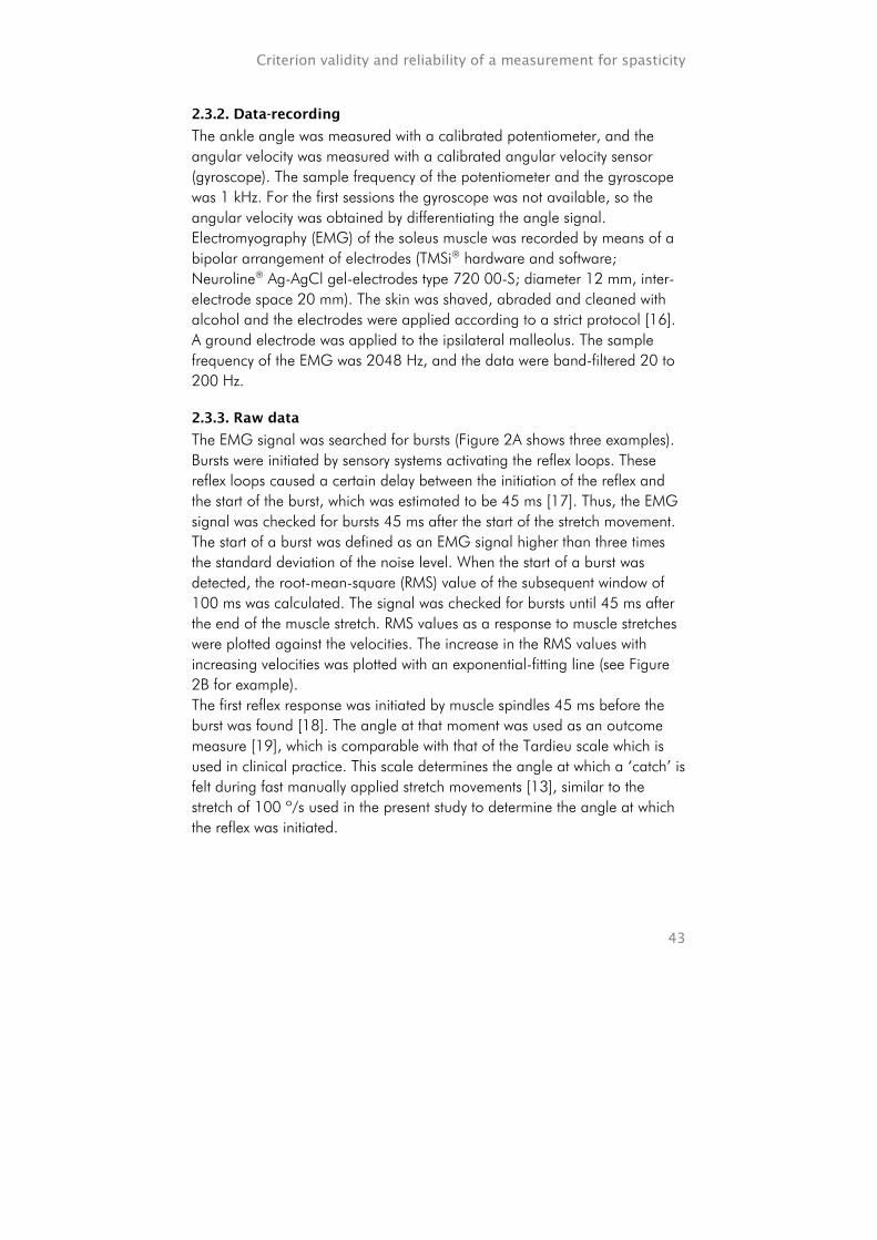
2.3.3. Raw data
The EMG signal was searched for bursts (Figure 2A shows three examples). Bursts were initiated by sensory systems activating the reflex loops. These reflex loops caused a certain delay between the initiation of the reflex and the start of the burst, which was estimated to be 45 ms [17]. Thus, the EMG signal was checked for bursts 45 ms after the start of the stretch movement. The start of a burst was defined as an EMG signal higher than three times the standard deviation of the noise level. When the start of a burst was detected, the root-mean-square (RMS) value of the subsequent window of 100 ms was calculated. The signal was checked for bursts until 45 ms after the end of the muscle stretch. RMS values as a response to muscle stretches were plotted against the velocities. The increase in the RMS values with increasing velocities was plotted with an exponential-fitting line (see Figure 2B for example). The first reflex response was initiated by muscle spindles 45 ms before the burst was found [18]. The angle at that moment was used as an outcome measure [19], which is comparable with that of the Tardieu scale which is used in clinical practice. This scale determines the angle at which a ‘catch’ is felt during fast manually applied stretch movements [13], similar to the stretch of 100 º/s used in the present study to determine the angle at which the reflex was initiated.
Chapter 3
44
2.3.4. Data-analysis
The RMS-values of the 100 ms EMG windows, determined from the burst start, of the soleus muscle, was plotted at the varying velocities. The outcome measures EMG50, EMG75 and EMG100 were calculated with the exponential-fitting line of the RMS-values at the velocities of 50, 75 and 100º/s respectively (Figure 2B). The reflex-initiating angle was defined as the angle at which the reflex was initiated, thus 45 ms before the burst start.
2.4. CRITERION STANDARD MEASUREMENTS
The measures used as criterion standards were selected for their neurophysiological and clinical relevance, i.e. the MAS, clonus score and H/M-ratio.
2.4.1. Clinical scales
The MAS was assessed by one experienced physical therapist [6] and the presence of clonus was scored. The assessor was blinded for the outcomes of the other assessments.
2.4.2. H/M-ratio
The H/M-ratio of the soleus muscle was also determined while the patient was seated. This provided a value for the reflex excitability [19]. Other studies state that the H-reflex amplitude may also depend on muscle fatigue [20]. To determine these values a rectangular current pulse with a pulse width of 1000 µs was applied to the tibial nerve in the popliteal fossa. The nerve was stimulated with a self-adhesive electrode (Neuroline®; type 720-00-s), and a self-adhesive counter electrode (5 to 9 cm) was applied just above the ipsilateral patella. Stimuli were given at a rate of less than 0.1 pulses per second. It was found that frequencies smaller than 0.2 Hz do not influence the H-reflex amplitude [20]. The amplitudes of the H-reflex and the M(Motor)-wave were determined by calculating the peak-to-peak value from the EMG signal at certain delays after the stimulus. The stimulation intensity started at 5 mA, and was increased in steps of 5 to 10 mA. First the M-wave was recorded, until saturation level was reached. The maximum M-wave was used as a reference value to exclude variance due to changes in stimulation and recording.

Criterion validity and reliability of a measurement for spasticity
45
Subsequently, the maximal H-reflex was determined [21]. The H/M-ratio was calculated by dividing the maximal H-reflex (Hmax) by the maximal M-wave (Mmax) [22]:
max
max
MH
ratioH/M =−
2.5. STATISTICAL ANALYSIS
2.5.1. Criterion validity
The correlation between the proposed set-up outcome measures and the concurrent assessment methods were determined with the Spearman’s non-parametric test [23], using the average values of all days [24]. Correlation coefficients were considered to be acceptable at values higher than 0.70, and values higher than 0.85 were considered to be good. The 95% confidence intervals (95% CI) were calculated. The differences between the patients with and without clonus were determined with the non-parametric Mann-Whitney test for the outcome measures; the EMG50/75/100 and the reflex-initiating angle.
2.5.2. Reliability and responsiveness
Reliability of the assessment (EMG50/75/100 and reflex-initiating angle) was determined with the Intra-Class Correlation (ICC) coefficients for the ranked values, because normal distribution could not be assumed. In addition, to study the effect of changes in impedance due to differences in skin and electrode placement, the ICC of the Mmax was calculated. A 2-way mixed effects model with absolute agreement was used. ICC coefficients higher than 0.60 were considered to be acceptable, and ICC coefficients higher or equal to 0.80 were considered to be good. The 95% CIs were calculated. To detect longitudinal changes in a patient, the within-subject variation is important. Therefore, the Smallest Detectable Difference (SDD) was calculated, using the following equations [25]:
MSerrorSEm =
SEm*2*2.145SDD = with MSerror being the mean square error within the subjects, calculated by means of a repeated measures test. The SEm is the standard error of the measurement, and 2.145 is the t-value, which depends on the number of subjects (n=8) and tests (T=3) (at a 2-tailed probability of 5 percent).

Chapter 3
46
Responsiveness was determined to evaluate the usefulness of the method in comparative trials. The responsiveness is a ratio of the difference which can be detected and the clinical expected change. The responsiveness was determined on the basis of the difference due to treatment of electrical stimulation. This electrical stimulation consisted of a 45 minutes cyclic stimulation of the ipsilaterla tibialis anterior, triceps surae or lateral side of the foot (S1 dermatome), and was applied immediately after the measurement [26]. For each patient the result of the most effective stimulation method was determined immediately after the intervention, and these results were averaged. The average changes caused by the treatment were divided by the SDD, providing a value for responsiveness.
3. RESULTS
3.1. SUBJECTS
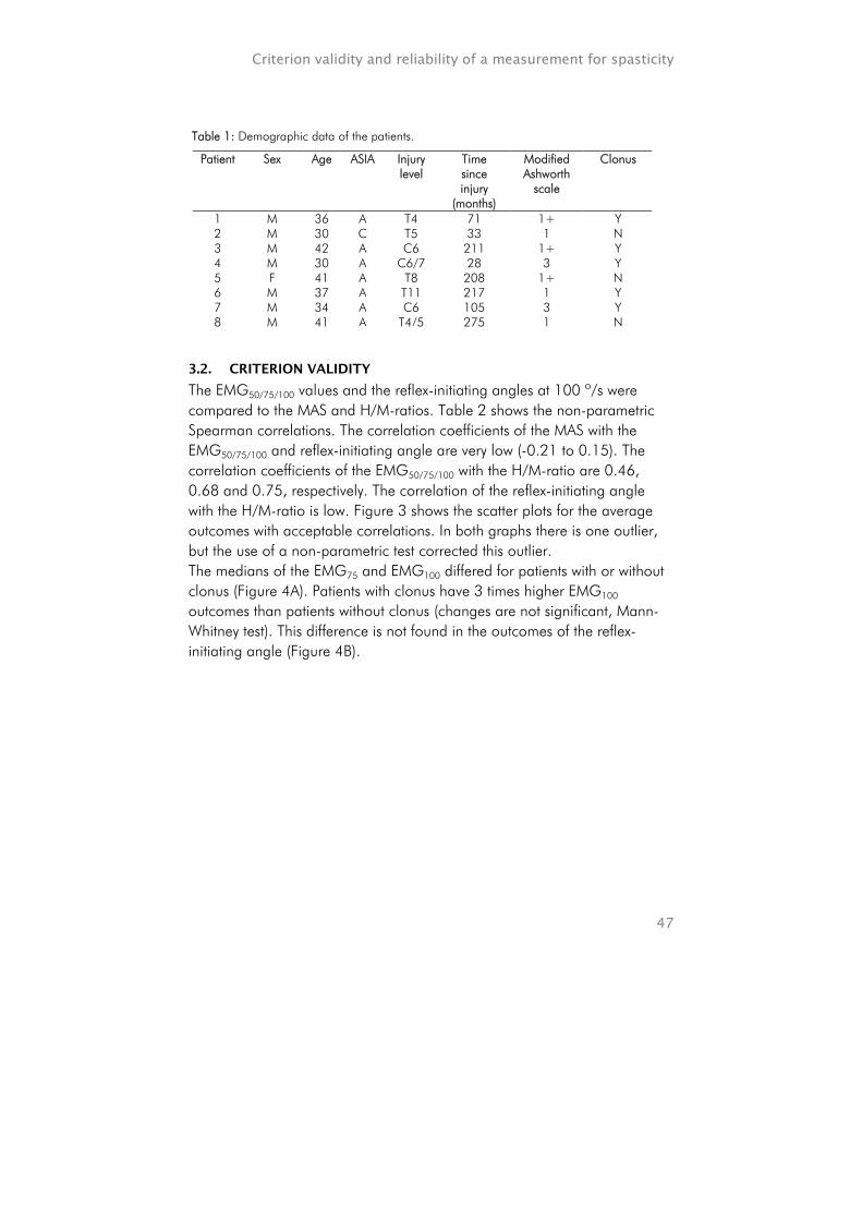
Thirty-three patients registered in the database were found to be suitable for the study. Sixteen of these patients were unwilling to participate, mainly because of other commitments. The remaining seventeen patients were screened during the intake. Seven patients were excluded because of the absence of spasticity in the relevant muscles, one patient was excluded because the set-up could not be placed near enough to his electric wheelchair, and one subject could not be measured due to technical problems with the EMG. Eight subjects were finally included in the study. The demographic data of the participating patients is presented in Table 1. One female and seven male patients participated and their age ranged from 30 to 42 years. Except for one patient with an ASIA C, all had ASIA A impairment scores, but none of the patients could voluntary contract the triceps surae. Their levels of injury were between C6 and T11. The shortest time since injury was 28 months. The MAS in the measured triceps surae ranged from 1 to 3, and 5 of the patients suffered from clonus. In most cases the average interval between two subsequent measurement sessions was one week, and the time between two measurements sessions was at least 3 days, and not longer than 2 weeks.
Criterion validity and reliability of a measurement for spasticity
47
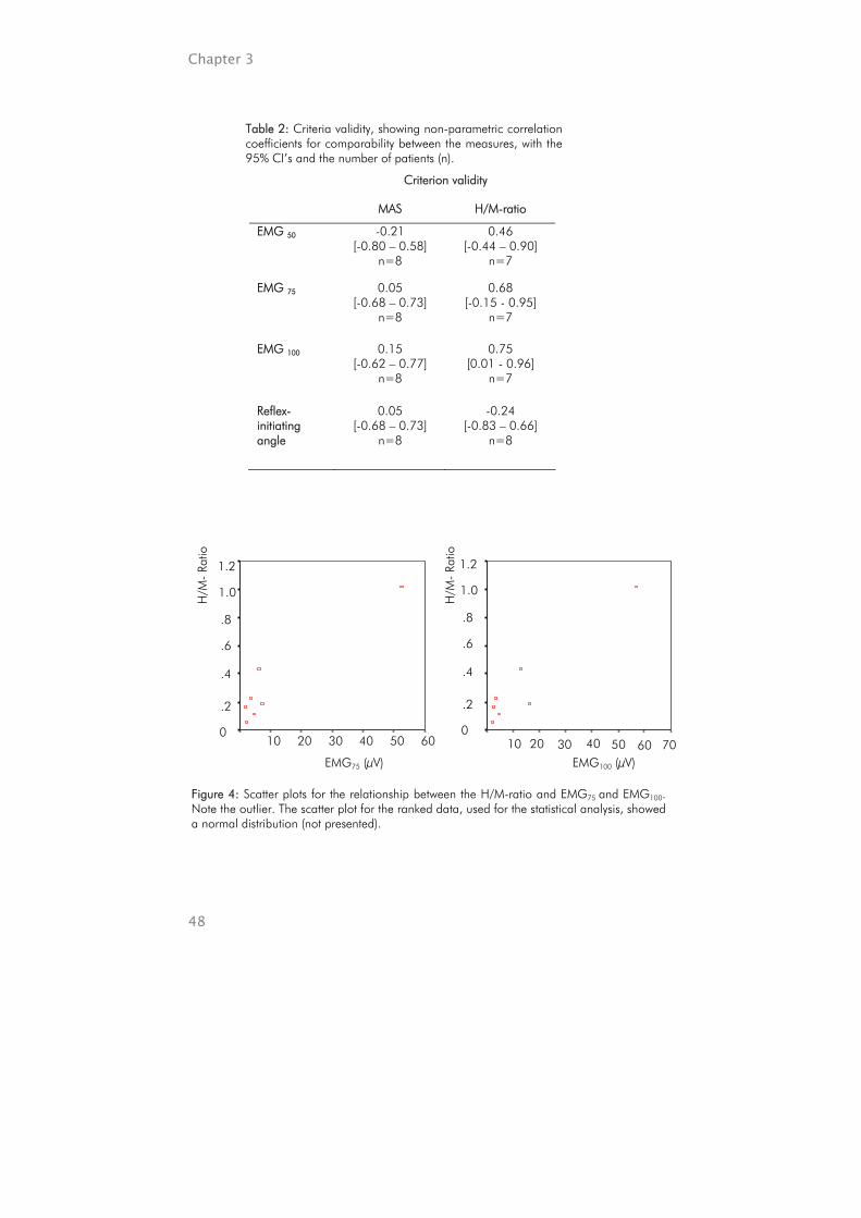
3.2. CRITERION VALIDITY
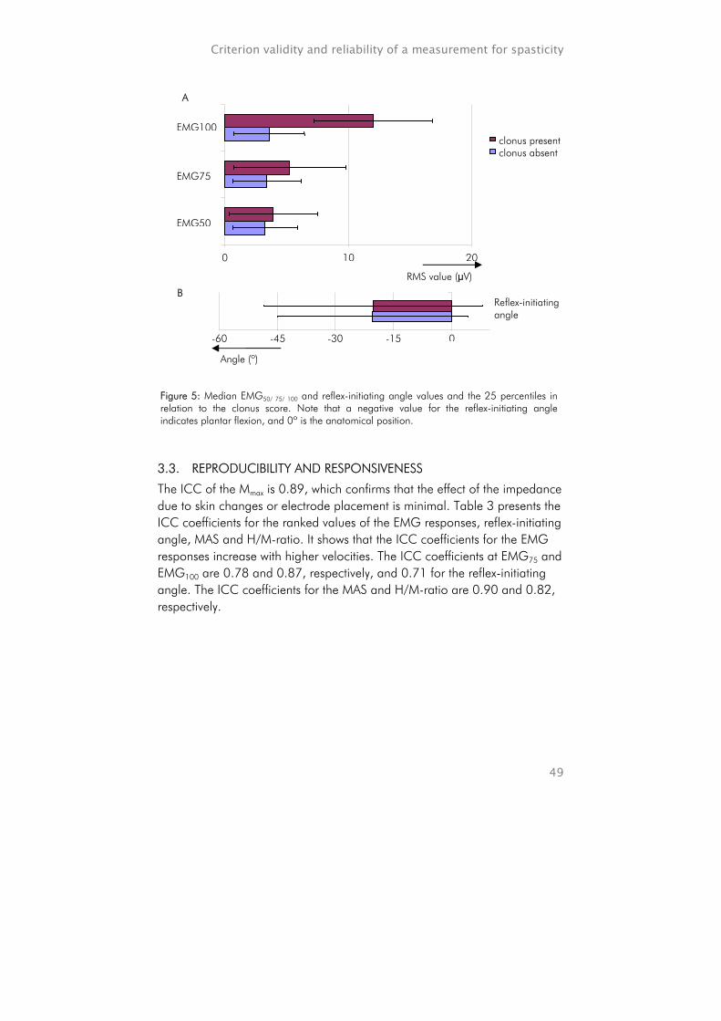
The EMG50/75/100 values and the reflex-initiating angles at 100 º/s were compared to the MAS and H/M-ratios. Table 2 shows the non-parametric Spearman correlations. The correlation coefficients of the MAS with the EMG50/75/100 and reflex-initiating angle are very low (-0.21 to 0.15). The correlation coefficients of the EMG50/75/100 with the H/M-ratio are 0.46, 0.68 and 0.75, respectively. The correlation of the reflex-initiating angle with the H/M-ratio is low. Figure 3 shows the scatter plots for the average outcomes with acceptable correlations. In both graphs there is one outlier, but the use of a non-parametric test corrected this outlier. The medians of the EMG75 and EMG100 differed for patients with or without clonus (Figure 4A). Patients with clonus have 3 times higher EMG100 outcomes than patients without clonus (changes are not significant, Mann-Whitney test). This difference is not found in the outcomes of the reflex-initiating angle (Figure 4B).
Table 1: Demographic data of the patients.
Patient Sex Age ASIA Injury level
Time since injury
(months)
Modified Ashworth
scale
Clonus
1 M 36 A T4 71 1+ Y 2 M 30 C T5 33 1 N 3 M 42 A C6 211 1+ Y 4 M 30 A C6/7 28 3 Y 5 F 41 A T8 208 1+ N 6 M 37 A T11 217 1 Y 7 M 34 A C6 105 3 Y 8 M 41 A T4/5 275 1 N
Chapter 3
48
Table 2: Criteria validity, showing non-parametric correlation coefficients for comparability between the measures, with the 95% CI’s and the number of patients (n).
Criterion validity
MAS H/M-ratio
EMG 50 -0.21 [-0.80 – 0.58]
n=8
0.46 [-0.44 – 0.90]
n=7
EMG 75 0.05 [-0.68 – 0.73]
n=8
0.68 [-0.15 - 0.95]
n=7
EMG 100 0.15 [-0.62 – 0.77]
n=8
0.75 [0.01 - 0.96]
n=7
Reflex-initiating angle
0.05 [-0.68 – 0.73]
n=8
-0.24 [-0.83 – 0.66]
n=8
Figure 4: Scatter plots for the relationship between the H/M-ratio and EMG75 and EMG100. Note the outlier. The scatter plot for the ranked data, used for the statistical analysis, showed a normal distribution (not presented).
EMG75 (µV)
60 50 40 30 20 10
1.2 1.0 .8 .6 .4
.2 0
H/M
- Ra
tio
H/M
- Ra
tio
1.2
1.0
.8
.6
.4
.2
0 605040302010
EMG100 (µV)70
Criterion validity and reliability of a measurement for spasticity
49
3.3. REPRODUCIBILITY AND RESPONSIVENESS
The ICC of the Mmax is 0.89, which confirms that the effect of the impedance due to skin changes or electrode placement is minimal. Table 3 presents the ICC coefficients for the ranked values of the EMG responses, reflex-initiating angle, MAS and H/M-ratio. It shows that the ICC coefficients for the EMG responses increase with higher velocities. The ICC coefficients at EMG75 and EMG100 are 0.78 and 0.87, respectively, and 0.71 for the reflex-initiating angle. The ICC coefficients for the MAS and H/M-ratio are 0.90 and 0.82, respectively.
A
0 10 20
EMG50
EMG75
EMG100 clonus present clonus absent
RMS value (µV)
B Reflex-initiating angle
Angle (º)
-60 -45 -30 -15 0
Figure 5: Median EMG50/ 75/ 100 and reflex-initiating angle values and the 25 percentiles in relation to the clonus score. Note that a negative value for the reflex-initiating angle indicates plantar flexion, and 0º is the anatomical position.
Chapter 3
50
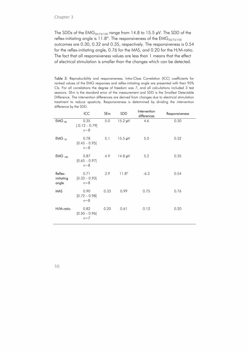
The SDDs of the EMG50/75/100 range from 14.8 to 15.5 µV. The SDD of the reflex-initiating angle is 11.8º. The responsiveness of the EMG50/75/100 outcomes are 0.30, 0.32 and 0.35, respectively. The responsiveness is 0.54 for the reflex-initiating angle, 0.76 for the MAS, and 0.20 for the H/M-ratio. The fact that all responsiveness values are less than 1 means that the effect of electrical stimulation is smaller than the changes which can be detected. Table 3: Reproducibility and responsiveness. Intra-Class Correlation (ICC) coefficients for ranked values of the EMG responses and reflex-initiating angle are presented with their 95% CIs. For all correlations the degree of freedom was 7, and all calculations included 3 test sessions. SEm is the standard error of the measurement and SDD is the Smallest Detectable Difference. The intervention differences are derived from changes due to electrical stimulation treatment to reduce spasticity. Responsiveness is determined by dividing the intervention difference by the SDD.
ICC SEm SDD Intervention differences
Responsiveness
EMG 50 0.35 [-0.12 – 0.79]
n=8
5.0 15.2 µV 4.6 0.30
EMG 75 0.78 [0.45 – 0.95]
n=8
5.1 15.5 µV 5.0 0.32
EMG 100 0.87 [0.65 – 0.97]
n=8
4.9 14.8 µV 5.2 0.35
Reflex-initiating angle
0.71 [0.33 – 0.93]
n=8
3.9 11.8º -6.3 0.54
MAS 0.90 [0.72 – 0.98]
n=8
0.33 0.99 0.75 0.76
H/M-ratio 0.82 [0.50 – 0.96]
n=7
0.20 0.61 0.12 0.20
Criterion validity and reliability of a measurement for spasticity
51
4. DISCUSSION
In this study, we presented a new approach to assess spastic hypertonia. We found that the EMG75 and EMG100 as a measure for reflex excitability correlate well with the H/M-ratio. The reliability of the outcomes EMG75, EMG100 and reflex-initiating angle was acceptable or good. The use of criterion standards like MAS, clonus score and H/M-ratio implies that only certain aspects of spastic hypertonia are investigated. Unfortunately, no golden standard is available to measure spastic hypertonia. Another limitation of the study may be the number of subjects which was relatively small, nevertheless significant correlations were found, indicating that sufficient patients were included. Other studies also performed stretches to investigate spastic hypertonia [8;22;27-31]. Except for one study [30], all studies used a motor to perform the stretch movements. Several studies used only short stretches of 3º to 5º to elicit a stretch reflex [8;29;31]. Others [22;27;30] used stretches over a relatively large range. It was found previously that EMG response amplitudes increase at increasing velocity [27;28]. Levin [22] compared the EMG response to a stretch of approximately 500º/s over 30º of ankle dorsal flexion, to the H/M-ratio. He found a correlation of 0.75, which is equal to our findings. Additionally, Levin found that the reflex-initiating angle does not correlate with the H/M-ratio, which is also consistent to our study. In the study which used manual testing, the ankle dorsal flexion was performed at one velocity [30]. The stretch was applied over the total range of motion over 5 seconds, which is relatively slow. In our study some perturbations resulted in short responses in the EMG signal. Then only a biphasic or a triphasic response was detected, providing small EMG50/75/100 values. These responses yielded a relatively low RMS value, and are not likely to impair the patients. The MAS does not correlate with the EMG50/75/100 nor reflex-initiating angle assessments, despite the fact that the setup was based on the Ashworth scale. This implies that the MAS provides an outcome about other components of muscle stiffness than to the other outcomes. The MAS was developed to detect the reflexive component of the spasticity [6], but other studies report that the MAS determines torque, including the reflexive and non-reflexive components of muscle stiffness [10]. Thus, more accurate measures are needed to determine the reflexive component only.
Chapter 3
52
The presence of clonus is related to the sensitivity of the reflex excitability [32]. There is a considerable difference in the EMG responses between the groups with and without clonus, which indicates that the EMG response provides a value for the sensitivity of the reflex excitability. The correlations of the EMG responses with the H/M-ratio point in the same direction, because the H/M-ratio is also a measure for the reflex excitability [19], whereas other studies stated that fatigue may also influence the H/M-ratio [20]. The H/M-ratio measures mainly the reflex excitability of the motoneurons to Ia-afferent monosynaptic input. The stretch reflex response over the total ROM will include more reflex loops, i.e. mono-, bi- and polysynaptic loops, initiated by multiple sensor systems. The reflex-initiating angle is not related to the H/M-ratio. Thus, the reflex-initiating angle measures a different aspect of the reflex excitability. It is very likely that the reflex-initiating angle is dependent on the sensitivity of the muscle sensors, so that the reflex-initiating angle would decrease if the muscle sensors (spindles) were more sensitive [33]. The correlations in reliability found in this study are acceptable for the EMG75 (ICC = 0.78) and reflex-initiating angle (ICC = 0.71), and good for the EMG100 (ICC = 0.87). The responsiveness is poor for all of the EMG responses (0.30 – 0.35) and for the reflex-initiating angle (0.54). The SDD, and therefore the responsiveness, depends on the variance in the assessment method (SEm) and the variance over days. Yet, the average standard deviation of one measurement series is approximately 5.6 µV, whereas the average RMS values are approximately 10 µV. The SDD may be improved when multiple stretches are performed in a smaller range of velocities, preferably at 75 º/s to 100 º/s. Although, the experimental set-up used in this study was relatively complex, in a clinical setting a one-channel EMG recording system, an electric goniometer and a personal computer would be sufficient to determine the outcomes. In fact: an instrumented version of clinically used manual tests, which is expected to have a better acceptance than more complex and more expensive motor-systems. This simplified set-up could be used to obtain useful data on spastic hypertonia of the patients related to their movements in daily life.

Criterion validity and reliability of a measurement for spasticity
53
5. CONCLUSION
The assessment of EMG response during stretches over the total range of motion with the proposed method provides reproducible outcomes for the reflex excitability, which is related to the H/M-ratio, but the responsiveness is marginal.
Chapter 3
54
REFERENCE LIST
1. Powers RK, Campbell DL, Rymer WZ. Stretch reflex dynamics in spastic elbow flexor muscles. Ann Neurol 1989;25(1):32-42.
2. Van der Salm A, Veltink PH, Hermens HJ, Nene AV, IJzerman MJ. Development of a new method for objective assessment of spasticity using full range passive movements. In Press: Arch Phys Med Rehabil.
3. Meythaler JM, Guin-Renfoe S, Brunner RC, Hadley MN. Intrathecal baclofen for spasticc hypertonia from stroke. Stroke 2001;32:2099-109.
4. Lance JW. Spasticity: disordered motor control. In Feldman RG, Young RR, Koella WP. Symposium Synopsis. Miami: Symposia Specialists; 1980:485-500.
5. Ashworth B. Preliminary trail of carisoprodal in multiple sclerosis. 1964;192:540-2. 6. Bohannon RW, Smith MB. Interrater reliability of a modified Ashworth scale of
muscle spasticity. Physical Therapy 1987 Feb;67(2):206-7. 7. Gracies J-M, Marosszeky JE, Renton R, Sandanam J, Gandevia SC, Burke D. Short-
term effects of dynamic lycra splints on upper limb in hemiplegic patients. Arch Phys Med Rehabil 2000;81:1547-55.
8. Fellows SJ, Ross HF, Thilmann AF. The limitations of the tendon jerk as a marker of pathological stretch reflex activity in human spasticity. J Neurol Neurosurg Psychiatry 1993 May;56(5):531-7.
9. Damiano DL, Quinlivan JM, Owen BF, Payne P, Nelson KC, Abel MF. What does the Ashworth scale really measure and are instrumented measures more valid and precise? Dev Med Child Neurol 2002 Feb;44(2):112-8.
10. Pandyan AD, Johnson GR, Price CI, Cureless RH, Barnes MP, Rodgers H. A review of the properties and limitations of the Ashworth and modified Ashworth scales as measures of spasticity. 1999;13:373-83.
11. Lee KC, Carson L, Kinnin E, Patterson V. The Ashworth scale: a reliable and reproducible method of measuring spasticity. J Neurol Rehabil 1989;3:205-9.
12. Nuysens G, De Weerdt W, Ketelare P. Interrater reliability of the Ashworth Scale in multiple sclerosis. Clin Rehabil 1994;8:286-92.
13. Fosang AL, Galea MP, McCoy AT, Reddihough DS, Story I. Measures of muscle and joint performance in the lower limb of children with cerebral palsy. Dev Med Child Neurol 2003;45:664-70.
14. Milanov I. Clinical and neurophysiological correlations of spasticity. Functional Neurology 1999;14:193-201.
15. Zhang L, Wang G, Nishida T, Xu D, Sliwa JA, Rymer WZ. Hyperactive tendon reflexes in spastic multiple sclerosis: measures and mechanisms of action. Arch. Physical Medicine and Rehabilitation 2000;81:901-9.
16. Hermens HJ, Freriks B, Merletti R, Stegeman D, Blok J, Rau G, Disslehorst-Klug C, Hägg G. SENIAM: European Recommendations for Surface ElectroMyoGraphy. Enschede: Roessingh Research and Development; 1999; ISBN:90-75452-15-2.
17. Sinkjær T, Nielsen J, Toft E. Mechanical and electromyographic analysis of reciprocal inhibition at the human ankle joint. J Neurophysiol 1995 Aug;74(2):849-55.
18. Grey MJ, Ladouceur M, Andersen JB, Nielsen JB, Sinkjær T. Group II muscle afferents probably contribute to the medium latency soleus stretch reflex during walking in humans. J Physiol 2001 Aug;534(Pt 3):925-33.

Criterion validity and reliability of a measurement for spasticity
55
19. Funase K, Miles TS. Observations on the variability of the H reflex in humans soleus. Muscle & Nerve 1999 Mar;22:341-6.
20. Schindler-Ivens S, Shields RK. Low frequency depression of H-reflexes in humans with acute and chronic spinal-cord injury. Exp Brain Res 2000 Jul;133(2):233-41.
21. Fisher MA. H reflexes and F waves. Fundamentals, normal and abnormal patterns. Neurol Clin Am 2002;20:339-60.
22. Levin MF, Hui-Chan C. Are H and stretch reflexes in hemiparesis reproducible and correlated with spasticity? J Neurol 1993 Feb;240(2):63-71.
23. Altman DG. Practical statistics for medical research. Chapman & Hall; 1991; ISBN:0-412-27630-5.
24. Bland MJ, Altman DG. Calculating correlation coefficients with repeated observations: Part 1 -- correlation within subjects. BMJ 1995;310(446 ).
25. IJzerman MJ, Baardman G, van 't Hof MA, Boom HBK, Hermens HJ, Veltink PH. Validity and reproducibility of crutch force and heart rate measurements to assess energy expendure of paraplegic gait. Arch Phys Med Rehabil 1999 Sep;80:1017-23.
26. Van der Salm A, Veltink PH, Nene AV, Hermens HJ, IJzerman MJ. Spasticity reduction of the triceps surae using electrical stimulation. JRRD 2004;41,2(Suppl).
27. Thilmann AF, Fellows SJ, Garms E. The mechanism of spastic muscle hypertonus. Variation in reflex gain over the time course of spasticity. Brain 1991 Feb;114 ( Pt 1A):233-44.
28. Ibrahim IK, Berger W, Trippel M, Dietz V. Stretch-induced electromyographic activity and torque in spastic elbow muscles. Differential modulation of reflex activity in passive and active tasks. Brain 1993;116(4):971-89.
29. Sinkjær T. Muscle, reflex and central components in the control of the ankle joint in healthy and spastic man. Acta Neurol Scand Suppl 1997;170:1-28.
30. Becher JG, Harlaar J, Lankhorst GJ, Vogelaar TW. Measurement of impaired muscle function of the gastrocnemius, soleus, and tibialis anterior muscles in spastic hemiplegia: A preliminary study. J of Rehab Research & Develop 1998;35(3):314-26.
31. Lambertz D, Goubel F, Perot C. A method to evaluate reflex excitability of the human ankle plantarflexors despite changes in maximal activation capacities. Exp Brain Res 2002;143:89-99.
32. Sheean G. Spasticity rehabilitation. Churchill communications; 1998. 33. Proske U, Morgan DL, Gregory JE. Thixotropy in skeletal muscle and in muscle
spindles: a review. Prog Neurobiol 1993 Dec;41(6):705-21.

Chapter 3
56
CHAPTER 4 Comparison of electrical stimulation methods for reduction of triceps surae spasticity in SCI Arjan van der Salm, Peter H. Veltink, Maarten J. IJzerman, Karin C.G. Groothuis-Oudshoorn, Anand V. Nene, Hermie J. Hermens Objective: Comparison of the effect of three methods of electrical stimulation to reduce spasticity of the triceps surae in patients with complete spinal cord injury. The carry-over effect was also investigated. Design: Placebo-controlled study with repeated measurements after the interventions. Setting: Research department affiliated with a rehabilitation hospital in the Netherlands. Patients: 10 patients with a complete spinal cord injury were recruited from the outpatient population of the rehabilitation hospital. All had ASIA A impairment scores, except for one, who had ASIA C. The patients had no voluntary triceps surae contractibility. Interventions: 45-minute cyclic electrical stimulation of the agonist, antagonist or dermatome of the triceps surae or a placebo approach. Outcome measures: Outcome measures were the Modified Ashworth scale (MAS), the clonus score, and the H/M-ratio. The EMG response to a stretch of the soleus over the whole range of motion was also determined. The magnitude and ankle angle at which the EMG response started were calculated. Results: Stimulation of the agonist provided a significant reduction in the MAS compared to the placebo approach (p<0.001). There was no significant change in the H/M-ratio or the EMG response amplitude after any of the stimulation methods, whereas stimulation of the antagonist muscle resulted in a significant reduction in the ankle angle at which the EMG response started, compared to the placebo approach (p<0.037). Conclusion: It is concluded that triceps surae stimulation reduces the MAS for that specific muscle, whereas the angle at which the reflex starts changes after antagonist stimulation. Conditionally accepted for Publication in Archives of Physical Medicine & Rehabilitation
Chapter 4
58
1. Introduction
Many treatment modalities are available to reduce spasticity, such as oral medications, chemodenervation and neurolysis, implanted pumps, physical therapy and surgery. They are applied to reduce spasticity or to treat contractures that result from spasticity [1;2]. In addition to these treatment modalities, therapeutic electrical stimulation is also known to reduce spasticity [3]. There are several advantages of electrical stimulation compared with the other treatment modalities. It can modulate the intensity of the intervention, and therefore the intensity of the effect. This also implies that the spasticity can be modulated instead of totally eliminated. Thus, patients have the potential ability to use the residual muscle tone for function. A second advantage of electrical stimulation is the local application. Oral medication, on the other hand, will influence the tonus in all the muscles in the body. The disadvantages of electrical stimulation are discomfort for the patient during stimulation and the limited duration of the effect. In the past, several studies have been performed to determine the effect of electrical stimulation on spasticity in patients with a spinal cord injury(SCI) [3-12], including studies with whole-hand stimulation (Mesh glove) and TENS. These studies found a positive effect of stimulation in patients with SCI. Pre and post-intervention assessments were made in all the studies to measure the effect of electrical stimulation. However, only one placebo-controlled study has been reported [9]. The methods used for stimulation in these studies differ greatly. Some studies describe stimulation of the antagonistic muscle [7-8;10;13]. Antagonist contraction is known to have an inhibitory effect on the agonist muscle [14], and several studies confirm that this so-called reciprocal inhibition is decreased in spastic patients [15-19]. To enhance the reciprocal inhibition in these patients, antagonistic muscle stimulation may be beneficial. In other studies the dermatome related to the spastic muscle was stimulated [5;11], using an inhibitory neurophysiologic pathway which is activated by sensory afferents from the low threshold sensors in the skin [20]. These afferents have an inhibitory effect on the muscles related to the same neurological segment [5]. Stimulation of the spastic muscle itself, which is also a method of treatment [6], is based on recurrent inhibition [3]. This is thought to be

Comparison of electrical stimulation methods
59
caused by the Renshaw cell, which has a negative feedback loop to the α-motoneuron [14], and this mechanism is found to be decreased in spastic patients [21-24]. Agonist muscle stimulation can be used to enhance the recurrent inhibition as an inhibitory pathway for the agonist muscle. The parameters used for stimulation also differed among the studies, suggesting that the optimal method of stimulation has not yet been identified. The goal of this study was to determine the effect, in general, and the carry-over effect of electrical stimulation to reduce spasticity in the triceps surae in patients with SCI. The effects of three different methods of electrical stimulation were also compared agonist, antagonist and dermatome stimulation. Spastic hypertonia was assessed using the Modified Ashworth scale (MAS), the clonus score and the H/M-ratio. In addition, the EMG response to a stretch of the soleus muscle over the whole range of motion was assessed and the response magnitude and the threshold-angle were determined. These measurements were chosen for their neurophysiological and clinical relevance, measuring either the reflex sensitivity or the mechanical components of muscle stiffness.
2. METHODS
2.1. SUBJECTS
Patients with SCI were recruited from the database of a rehabilitation centre in the Netherlands (Het Roessingh, Enschede). Inclusion criteria were: presence of spasticity (MAS grade 1 or higher), absence of voluntary contractibility in the triceps surae, time since injury at least 6 months, ability to contract triceps surae muscles and tibialis anterior muscle with electrical stimulation, and over 18 years of age. The patients were allowed to take antispastic medication, but they were asked not to change the dosage within 2 weeks before and during the study period. Patients with hypersensitive skin on the legs, with equinus deformity, or conditions which could temporally increase tonus (specifically bladder infection) were excluded. All patients gave informed consent to participate in the study, which was approved by the local ethics committee. The database was screened for information about the presence, level and chronicity of spasticity in the legs, the age of the patient and any possible reason for exclusion. This screening was performed by a physical medicine and rehabilitation physician and a researcher. The patients who met the inclusion criteria were invited to participate in the study via a letter, and one

Chapter 4
60
week later they were contacted by phone. The other in/exclusion criteria were assessed by means of a questionnaire and an intake screening. The presence of spastic hypertonia and passive muscle stiffness was measured according to the MAS, and inability to voluntarily contract the triceps surae was confirmed. Excitability of the triceps surae and the tibialis anterior with electrical stimulation was also determined.
2.2. DESIGN
Each patient received the interventions on 4 separate days, with an average interval of 7 days (range 3 days to 14 days). According to the literature the wash-out period of the effect of the electrical stimulation may last as long as 24 hours [3]. A minimum of 72 hours between two subsequent interventions was considered to be long enough to ensure that the effect of the former intervention had disappeared. For each patient the interventions started with a baseline measurement session at the same time each day. Sequentially, patients received one of the three methods of stimulation (agonist stimulation, antagonist stimulation or dermatome stimulation) or a placebo
Day 1
Day 2
Day 3
Day 4
baseline
baseline
baseline
baseline
placebo approach
Post 1 Post 2
agonist stimulation
antagonist stimulation
dermatome stimulation
Post 1
Post 1
Post 1
Post 2
Post 2
Post 2
Post 3
Post 3
Post 3
Post 3
1 hour 1 hour
Figure 1: Design of the study. The patients came on 3 or 4 days, and on each day another intervention was applied: agonist stimulation, antagonist stimulation, dermatome stimulation or a placebo approach. Each day started with a baseline measurement session, followed by an intervention. The first post-measurement session took place immediately after the intervention, and the second and third post-measurement sessions with 1-hour intervals.
45 minutes
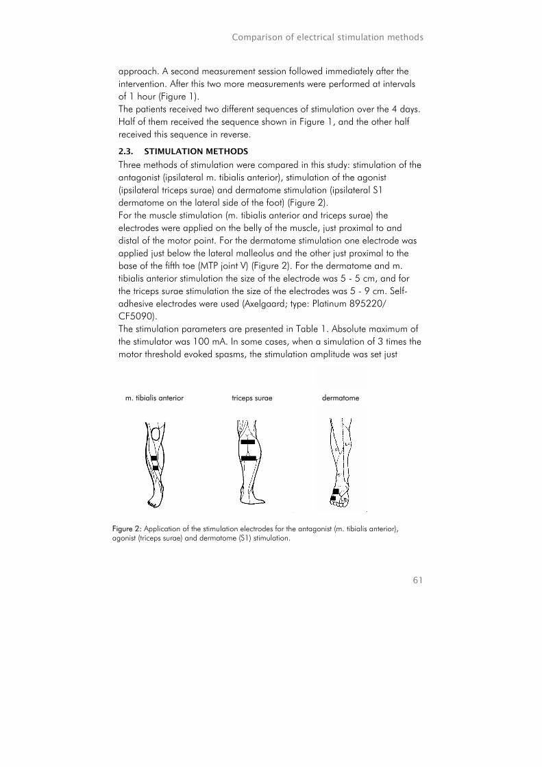
Comparison of electrical stimulation methods
61
approach. A second measurement session followed immediately after the intervention. After this two more measurements were performed at intervals of 1 hour (Figure 1). The patients received two different sequences of stimulation over the 4 days. Half of them received the sequence shown in Figure 1, and the other half received this sequence in reverse.
2.3. STIMULATION METHODS
Three methods of stimulation were compared in this study: stimulation of the antagonist (ipsilateral m. tibialis anterior), stimulation of the agonist (ipsilateral triceps surae) and dermatome stimulation (ipsilateral S1 dermatome on the lateral side of the foot) (Figure 2). For the muscle stimulation (m. tibialis anterior and triceps surae) the electrodes were applied on the belly of the muscle, just proximal to and distal of the motor point. For the dermatome stimulation one electrode was applied just below the lateral malleolus and the other just proximal to the base of the fifth toe (MTP joint V) (Figure 2). For the dermatome and m. tibialis anterior stimulation the size of the electrode was 5 - 5 cm, and for the triceps surae stimulation the size of the electrodes was 5 - 9 cm. Self-adhesive electrodes were used (Axelgaard; type: Platinum 895220/ CF5090). The stimulation parameters are presented in Table 1. Absolute maximum of the stimulator was 100 mA. In some cases, when a simulation of 3 times the motor threshold evoked spasms, the stimulation amplitude was set just
Figure 2: Application of the stimulation electrodes for the antagonist (m. tibialis anterior), agonist (triceps surae) and dermatome (S1) stimulation.
m. tibialis anterior triceps surae dermatome
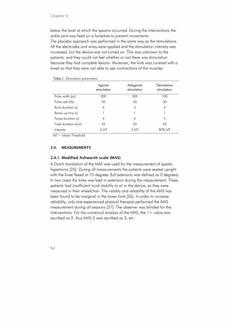
Chapter 4
62
below the level at which the spasms occurred. During the interventions the ankle joint was fixed on a footplate to prevent movements. The placebo approach was performed in the same way as the stimulations. All the electrodes and wires were applied and the stimulation intensity was increased, but the device was not turned on. This was unknown to the patients, and they could not feel whether or not there was stimulation because they had complete lesions. Moreover, the limb was covered with a towel so that they were not able to see contractions of the muscles.
Table 1: Stimulation parameters.
Agonist
stimulation Antagonist stimulation
Dermatome stimulation
Pulse width (µs) 300 300 100
Pulse rate (Hz) 30 30 30
Burst duration (s) 4 4 4
Ramp-up time (s) 1 1 1
Pause duration (s) 4 4 4
Total duration (min) 45 45 45
Intensity 3 MT 3 MT 80% MT
MT = Motor Threshold
2.4. MEASUREMENTS
2.4.1. Modified Ashworth scale (MAS)
A Dutch translation of the MAS was used for the measurement of spastic hypertonia [25]. During all measurements the patients were seated upright with the knee flexed at 75 degrees (full extension was defined as 0 degrees). In two cases the knee was kept in extension during the measurement. These patients had insufficient trunk stability to sit in the device, so they were measured in their wheelchair. The validity and reliability of the MAS has been found to be marginal in the lower limb [26]. In order to increase reliability, only one experienced physical therapist performed the MAS measurement during all sessions [27]. The observer was blinded for the interventions. For the numerical analysis of the MAS, the 1+ value was ascribed as 2, thus MAS 2 was ascribed as 3, etc.
Comparison of electrical stimulation methods
63
2.4.2. Clonus score
The same observer assessed whether or not the patients suffered from clonus of the triceps surae muscle. The clonus was elicited by a rapid perturbation with sustained pressure. The observer determined whether the clonus was maintained for more than 5 seconds or whether the clonus disappeared. Thus, the clonus score was: 0) no clonus, 1) self-limiting clonus or 2) sustaining clonus.
2.4.3. H-reflex measurement
To measure the H/M-ratio a stimulus was applied to the tibial nerve in the popliteal fossa. The optimal location was sought with a handheld probe, and when it was found an electrode (Neuroline® type 720 00-s) was placed on this location. The inactive electrode was a self-adhesive 5 by 9 cm electrode placed directly above the ipsilateral patella. For the H-reflex measurement a rectangular biphasic pulse, with a pulse width of 1000 µs, was applied. For the electromyography (EMG), Neuroline® (type 720 00-s, Ag-AgCl gel) self-adhesive electrodes with a diameter of 12 mm were applied, and a bipolar arrangement with an inter-electrode space of 20 mm was used. The ground electrode was applied to the ipsilateral medial malleolus. Before application, the leg was shaved, and the skin was abraded and cleaned with alcohol. A strict protocol was followed for placement of the electrodes [28]. The sample frequency of the EMG was 2048 Hz, and the data were band-filtered; 20 to 200 Hz. The amplitude of the H-reflex and motor-wave (M-wave) were determined by calculating the peak-to-peak value from the EMG signal at appropriate delays after the stimulus. The stimulation intensity was increased in steps of 5 - 10 mA. After saturation of the M-wave was achieved, the maximum H-reflex was determined. Approximately 15 stimuli were given, at a rate of less than 0.1 pulses per second, until an appropriate H/M-graph was obtained. The maximum H-reflex (Hmax) and maximum M-wave (Mmax) were determined, and the H/M-ratio was calculated according to the following formula [29]:
max
max
MH
ratioH/M =−
The validity of the H/M-ratio as a value for the reflex excitability may be diffused by muscle fatigue [29], but the reliability of the H/M-ratio of the soleus muscle is good [30].
Chapter 4
64
2.4.4. Stretch reflex
Set-up The muscle response was measured during a stretch of the soleus muscle over the whole range of motion at several speeds [32]. The patients were seated upright with the knee flexed at 75 degrees. In two cases the knee was kept in extension during the measurement. These patients had insufficient trunk stability to sit in the device, so they were measured in their wheelchair. The foot was strapped on a footplate which rotated around the ankle joint, and for the measurement the footplate was moved in dorsal flexion, thus stretching the triceps surae. In one session 30 - 45 stretches were applied at various different velocities, ranging from 30 to 150 º/s. Data-recording and analysis EMG was recorded in the soleus muscle, similar to the measurement of the H/M-ratio. The filtered EMG signal was searched for bursts with a threshold value, defined as 3 times the standard deviation of the noise level. When a burst was found during the stretch movement the root mean square (RMS) value was calculated over a window of 100 ms (200 samples), subsequent to the moment at which the threshold was passed. The RMS responses increased exponentially with increasing velocities. An exponential curve fit was estimated, and an average response value at a velocity of 100 º/s (EMG100) was determined with this fitted line [32]. Another outcome was the reflex-initiating angle, which was defined as the angle at which a reflex was generated, resulting in an actual EMG response at an average stretch velocity of 100 º/s. The reflex-initiating angle outcome was the angle 45 ms before the start of the EMG burst [33]. The position in which the ankle had no plantar or dorsal flexion (anatomical position) was defined as 0º. This 45 ms delay was incorporated to include the time between the initiation of the reflex and the actual EMG response. This outcome is comparable to scores on the Tardieu scale that is used in clinical practice [34]. This scale measures fast, manually applied stretch movements, similar to the stretch of 100 º/s we used to determine the reflex-initiating angle [32].
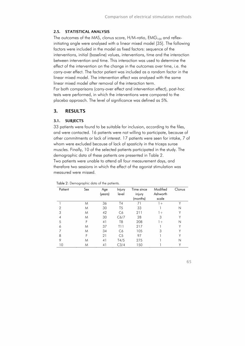
Comparison of electrical stimulation methods
65
2.5. STATISTICAL ANALYSIS
The outcomes of the MAS, clonus score, H/M-ratio, EMG100 and reflex-initiating angle were analysed with a linear mixed model [35]. The following factors were included in the model as fixed factors: sequence of the interventions, initial (baseline) values, interventions, time and the interaction between intervention and time. This interaction was used to determine the effect of the intervention on the change in the outcomes over time, i.e. the carry-over effect. The factor patient was included as a random factor in the linear mixed model. The intervention effect was analysed with the same linear mixed model after removal of the interaction term. For both comparisons (carry-over effect and intervention effect), post-hoc tests were performed, in which the interventions were compared to the placebo approach. The level of significance was defined as 5%.
3. RESULTS
3.1. SUBJECTS
33 patients were found to be suitable for inclusion, according to the files, and were contacted. 16 patients were not willing to participate, because of other commitments or lack of interest. 17 patients were seen for intake, 7 of whom were excluded because of lack of spasticity in the triceps surae muscles. Finally, 10 of the selected patients participated in the study. The demographic data of these patients are presented in Table 2. Two patients were unable to attend all four measurement days, and therefore two sessions in which the effect of the agonist stimulation was measured were missed.
Table 2: Demographic data of the patients.
Patient Sex Age (years)
Injury level
Time since injury
(months)
Modified Ashworth
scale
Clonus
1 M 36 T4 71 1+ Y 2 M 30 T5 33 1 N 3 M 42 C6 211 1+ Y 4 M 30 C6/7 28 3 Y 5 F 41 T8 208 1+ N 6 M 37 T11 217 1 Y 7 M 34 C6 105 3 Y 8 F 21 C5 97 1 Y 9 M 41 T4/5 275 1 N 10 M 41 C3/4 150 1 Y
Chapter 4
66
3.2. EFFECT OF ELECTRICAL STIMULATION
3.2.1. Clinical scales
None of the participants reported adverse effects or increased spasms after any of the interventions. On the MAS, a significant intervention effect was found; p<0.001 (Figure 3), but the clonus score showed no significant intervention effect; p<0.21. Post-hoc tests for the MAS showed that only the agonist stimulation differed significantly from the placebo approach (p<0.001). It was found that the group average on the MAS decreased from 1.6 to 0.9 (46% reduction) immediately after stimulation of the agonist. However, given the rather mild grades of spasticity, this reduction may be of limited clinical relevance. No significant carry-over effect was found on the MAS or the clonus score; p<0.113 (n=10) and p<0.586, respectively (n=10).
3.2.2. Assessments for reflex excitability
The H/M-ratio showed no significant changes in the intervention effect or carry-over effect, p<0.31 and p<0.43, respectively (n=7), because only minimal changes were present (Figure 3). The EMG100 outcomes showed no significant changes for either the intervention effect (p<0.60) or the carry-over effect (p<0.79, n=8). The reflex-initiating angle showed a significant change for the intervention effect (p<0.015, n=8), but the carry-over effect was not significant (p<0.14, n=8). Post-hoc tests for the intervention effect showed that antagonist stimulation resulted in a significant change, compared to the placebo effect, p<0.037. The reflex-initiating angle changed from 18.6 to 15.2 degrees of plantar flexion immediately after stimulation of the antagonist, which was consistent with a reduction in the reflex sensitivity. Note that 0º is the anatomical position of the foot (no plantar or dorsal flexion).
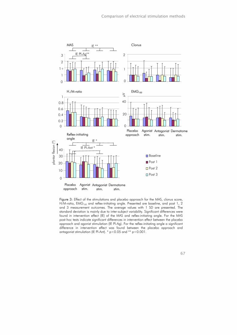
Comparison of electrical stimulation methods
67
Figure 3: Effect of the stimulations and placebo approach for the MAS, clonus score, H/M-ratio, EMG100 and reflex-initiating angle. Presented are baseline, and post 1, 2 and 3 measurement outcomes. The average values with 1 SD are presented. The standard deviation is mainly due to inter-subject variability. Significant differences were found in intervention effect (IE) of the MAS and reflex-initiating angle. For the MAS post-hoc tests indicate significant differences in intervention effect between the placebo approach and agonist stimulation (IE Pl-Ag). For the reflex-initiating angle a significant difference in intervention effect was found between the placebo approach and antagonist stimulation (IE Pl-Ant). * p<0.05 and ** p<0.001.
Baseline
Post 1
Post 2
Post 3
MAS
0
1
1+
2
3
IE **
IE Pl-Ag** -
Clonus
0
1
2
H./M-ratio
0
0.2
0.4
0.6
0.8
1
EMG100
0
20
40
µV
Reflex-initiating angle
0
10
20
30
40
Placebo approach
Agonist stim.
Antagonist stim.
Dermatome stim.
plan
tar
flexi
on (º
) IE *
IE Pl-Ant *
Placebo approach
Agonist stim.
Antagonist stim.
Dermatome stim.

Chapter 4
68
4. DISCUSSION
The MAS suggest that stimulation of the agonist muscle may be the best method, but, as the MAS measures both the spastic hypertonia and the mechanical components of muscle stiffness [26], it is not clear which component has primarily changed. The H/M-ratio showed no change, which indicates that the sensitivity of the spinal synapses [36] was not affected by any of the interventions. Moreover, the EMG100, which can be regarded as another measure for the excitability of the spinal synapses, also showed no significant change after the interventions. Therefore, our results indicate that the spinal connections in the reflex-loop are not influenced by electrical stimulation, suggesting that the changes in the MAS due to agonist stimulation are primarily caused by mechanical components of muscle stiffness or the muscle spindles. The significant change in the reflex-initiating angle indicates that antagonist stimulation is more effective to reduce spasticity. The outcome of the reflex-initiating angle depends mainly on the sensitivity of the sensors, which are controlled by the activity of the γ-motoneurons, and mechanical stiffness of the muscles and tendons. The mechanical stiffness, i.e. visco-elasticity, of a muscle may depend on the blood flow in the tissue [37]. Due to the muscle contractions the blood flow will be increased in the stimulated area, agonist and antagonist, which, in turn, can decrease the muscle stiffness. The effect of the sequence was statistically controlled. It was found that the sequence did not influence any of the outcomes significantly. We therefore concluded that the wash-out period of 72 hours was adequate. This also indicated that randomisation was not needed. In other studies it has been found that patients with spasticity may benefit from electrical stimulation [3;5;6;8-12]. However, only one of these studies included was controlled by a placebo group. This may be an important reason for the discrepancy in results between these studies and our study. If we had performed paired t-tests for the pre and post-intervention measurement outcomes only, more significant changes would have been observed. For example: both the agonist and the antagonist stimulation would result in significant changes in the MAS. In addition, two studies allowed movements of the limb during stimulation, in contrast to the isometric condition in our study [3;6]. The movement of the stimulated limbs in itself, may have caused the effect found in those studies. Limb movement, i.e. muscle stretch, is a commonly applied and effective method of treatment
Comparison of electrical stimulation methods
69
for spasticity [1]. In contrast to the inclusion criteria in our study, all the previous studies included patients with spasticity and more or less intact supraspinal control. This implies that the patients could feel the stimulation and could therefore not be blinded. This may have resulted in additional (placebo) effects. Recently, the SPASM group published a new definition of spasticity, which included a greater range of signs and symptoms than those included in the commonly used Lance definition [38;39]. The new definition includes the entire range of ‘so-called’ positive signs and symptoms, such as increased tendon reflex, clonus, spasms and increased resistance to passive movement. We measured several of these, but not all of the positive signs were included in our measurements. The reflex sensitivity depends on the sensitivity at the level of sensors (mainly the muscle spindles) and spinal synapses (inhibition and facilitation). The sensitivity of the muscle spindles is mainly controlled by the γ-motoneuron activity [14], and mechanical changes due to muscle stretch may alter the response of the muscle to stretch. In addition, mechanical changes in muscles and tendons due to muscle stretch may also, indirectly, alter the sensitivity of the muscle spindles: in muscles or tendons with higher tension the muscle spindles will be stretched at an earlier stage than in relaxed muscles. The sensitivity of the spinal synapses depends on several inhibitory and facilitating neural pathways, e.g. presynaptic inhibition [20]. According to the results of our study, the benefit of electrical stimulation to reduce spasticity is limited, but in clinical practice the effect may be enhanced when movement is allowed during stimulation. The stimulation might also have more effect if a series of stimulations are applied over a period of several weeks [9]. Additionally, the inhibitory effect of stimulation could also be effective during activies, providing an instant reduction in the reflex excitability. In patients with spasticity and intact supraspinal control, electrical stimulation may have an inhibitory effect, due to changes in the corticomotoneural excitability [12].
5. CONCLUSION
According to the MAS outcomes, triceps surae muscle stimulation results in a significant reduction in spasticity of that specific muscle. The reflex-initiating angle changed significantly after stimulation of the tibial anterior muscle.
Chapter 4
70
REFERENCE LIST
1. Bhakta BB. Management of spasticity in stroke. Br Med Bull 2000;56(2):476-85. 2. Burchiel KJ, Hsu FP. Pain and spasticity after spinal cord injury: mechanisms and
treatment. Spine 2001 Dec 15;26(24 Suppl):S146-60. 3. Vodovnik L, Bowman BR, Hufford P. Effects of electrical stimulation on spinal
spasticity. Scand J Rehabil Med 1984;16(1):29-34. 4. Daly JJ, Marsolais EB, Mendell LM, Rymer WZ, Stefanovska A, Wolpaw JR, Kantor
C. Therapeutic neural effects of electrical stimulation. IEEE Trans Rehabil Eng 1996 Dec;4(4):218-30.
5. Bajd T, Gregoric M, Vodovnik L, Benko H. Electrical stimulation in treating spasticity resulting from spinal cord injury. Arch Phys Med Rehabil 1985 Aug;66(8):515-7.
6. Franek A, Turczynski B, Opara J. Treatment of spinal spasticity by electrical stimulation. J Biomed Eng 1988 May;10(3):266-70.
7. Alfieri V. Electrical treatment of spasticity. Reflex tonic activity in hemiplegic patients and selected specific electrostimulation. Scand J Rehabil Med 1982;14(4):177-82.
8. Robinson CJ, Kett NA, Bolam JM. Spasticity in spinal cord injured patients: 1. Short-term effects of surface electrical stimulation. Arch Phys Med Rehabil 1988 Aug;69(8):598-604.
9. Chen SC, Chen YL, Chen CJ, Lai CH, Chiang WH, Chen WL. Effects of surface electrical stimulation on the muscle-tendon junction of spastic gastrocnemius in stroke patients. Disabil Rehabil 2005 Feb;27(3):105-10.
10. Mirbagheri MM, Ladouceur M, Barbeau H, Kearney RE. The effects of long-term FES-assisted walking on intrinsic and reflex dynamic stiffness in spastic spinal-cord-injured subjects. IEEE Trans Neural Syst Rehabil Eng 2002;10(4):280-9.
11. Dimitrijevic MM, Soroker N. Mesh-glove. 2. Modulation of residual upper limb motor control after stroke with whole-hand electric stimulation. Scand J Rehabil Med 1994;26(4):187-90.
12. Tinazzi M, Zarattini S, Valeriani M, Romito S, Farina S, Moretto G, Smania N, Fiaschi A, Abbruzzese G. Long-lasting modulation of human motor cortex following prolonged transcutaneous electrical nerve stimulation (TENS) of forearm muscles: evidence of reciprocal inhibition and facilitation. Exp Brain Res 2005;161(4):457-64.
13. Robinson CJ, Kett NA, Bolam JM. Spasticity in spinal cord injured patients: 2. Initial measures and long-term effects of surface electrical stimulation. Arch Phys Med Rehabil 1988 Oct;69(10):862-8.
14. Rothwell J. Control of human volutary movement. 2 ed. London: Chapman & Hall; 1994.
15. Okuma Y, Lee RG. Reciprocal inhibition in hemiplegia: correlation with clinical features and recovery. Can J Neurol Sci 1996 Feb;23(1):15-23.
16. Okuma Y, Mizuno Y, Lee RG. Reciprocal Ia inhibition in patients with asymmetric spinal spasticity. Clin Neurophysiol 2002 Feb;113(2):292-7.
17. Boorman GI, Lee RG, Becker WJ, Windhorst UR. Impaired "natural reciprocal inhibition" in patients with spasticity due to incomplete spinal cord injury. Electroencephalogr Clin Neurophysiol 1996 Apr;101(2):84-92.
18. Morita H, Crone C, Christenhuis D, Petersen NT, Nielsen JB. Modulation of presynaptic inhibition and disynaptic reciprocal Ia inhibition during voluntary

Comparison of electrical stimulation methods
71
movement in spasticity. Brain 2001 Apr;124(Pt 4):826-37. 19. Leonard CT, Diedrich PM, Matsumoto T, Moritani T, McMillan JA. H-reflex
modulations during voluntary and automatic movements following upper motor neuron damage. Electroencephalogr Clin Neurophysiol 1998 Dec;109(6):475-83.
20. Kandell ER, Schwartz JH, Jessell TM. Essentials of neural science and behavior. McGraw-Hill; 1995.
21. Raynor EM, Shefner JM. Recurrent inhibition is decreased in patients with amyotrophic lateral sclerosis. Neurology 1994 Nov;44(11):2148-53.
22. Mazzocchio R, Rossi A. Involvement of spinal recurrent inhibition in spasticity. Further insight into the regulation of Renshaw cell activity. Brain 1997 Jun;120 ( Pt 6):991-1003.
23. Mazzocchio R, Rossi A. Recurrent inhibition in human spinal spasticity. Ital J Neurol Sci 1989 Jun;10(3):337-47.
24. Katz R, Pierrot-Deseilligny E. Recurrent inhibition in humans. Prog Neurobiol 1999 Feb;57(3):325-55.
25. Bohannon RW, Smith MB. Interrater reliability of a modified Ashworth scale of muscle spasticity. Physical Therapy 1987 Feb;67(2):206-7.
26. Pandyan AD, Johnson GR, Price CI, et al. A review of the properties and limitations of the Ashworth and modified Ashworth scales as measures of spasticity. 1999;13: 373-83.
27. Pandyan AD, Price CI, Rodgers H, Barnes MP, Johnson GR. Biomechanical examination of a commonly used measure of spasticity. Clin Biomech (Bristol, Avon). 2001 Dec;16(10):859-65.
28. Hermens HJ, Freriks B, Merletti R, et al. SENIAM: European Recommendations for Surface ElectroMyoGraphy. Enschede: Roessingh Research and Development; 1999.
29. Schindler-Ivens S, Shields RK. Low frequency depression of H-reflexes in humans with acute and chronic spinal-cord injury. Exp Barin Res 2000;133(2):233-41.
30. Palmieri RM, Hoffmann MA, Ingersoll CD. Intersession reliability for the H-reflex measurements arsing from the soleus, peroneal nerve, and tibialis anterior musculature. Int J Neurosci 2002;112:841-50.
31. Visser SL. Reflexen. in: Klinische Neurofysiologie, Notermans. Bussel: 1981: 353-68.
32. Van der Salm A, Veltink PH, Hermens HJ, IJzerman MJ, Nene AV. development of a new method for objective assessment of spasticity using full range passive movements. Resubmitted to Archives of Phys Med & Rehab.
33. Sinkjær T, Nielsen J, Toft E. Mechanical and electromyographic analysis of reciprocal inhibition at the human ankle joint. J Neurophysiol 1995 Aug;74(2):849-55.
34. Fosang AL, Galea MP, McCoy AT, Reddihough DS, Story I. Measures of muscle and joint performance in the lower limb of children with cerebral palsy. Dev Med Child Neurol 2003;45:664-70.
35. Everitt B, Rabe-Hesketh S. Analyzing medical data using S-Plus. New York: Springer; 2001. Dietz K, Gail K, Krickeberg J, et al., editors. Statistics for biology and health.
36. Funase K, Miles TS. Observations on the variability of the H reflex in humans soleus. Muscle & Nerve 1999 Mar;22:341-6.
37. Evetovich TK, Nauman NJ, Conley DS, Todd JB. Effect of static stretching of the

Chapter 4
72
biceps brachii on torque, electromyography, and mechanomyography during concentric isokinetic muscle actions. Journal of Strength and Conditioning Research 2003;17(3):484-8.
38. Pandyan AD, Gregoric M, Barnes MP, Wood D, Van Wijck F, Burridge J, Hermens H, Johnson GR. Spasticity: clinical perceptions, neurological realities and meaningful measurement. Disability and Rehabilitation 2005;27(1/2):2-6.
39. Lance JW. Spasticity: disordered motor control. In: Feldman RG, Young RR, Koella WP editors. Symposium Synopsis. Miami: Symposia Specialists; 1980. pp. 485-500.
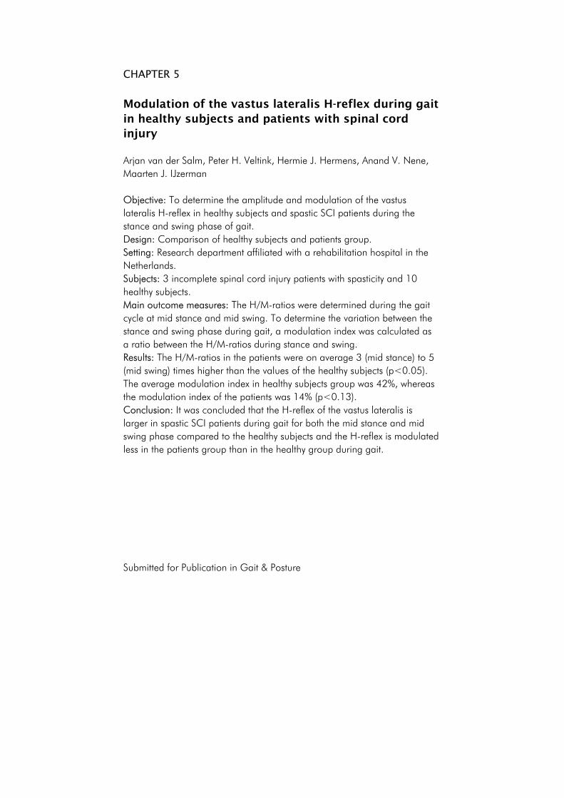
CHAPTER 5
Modulation of the vastus lateralis H-reflex during gait in healthy subjects and patients with spinal cord injury Arjan van der Salm, Peter H. Veltink, Hermie J. Hermens, Anand V. Nene, Maarten J. IJzerman Objective: To determine the amplitude and modulation of the vastus lateralis H-reflex in healthy subjects and spastic SCI patients during the stance and swing phase of gait. Design: Comparison of healthy subjects and patients group. Setting: Research department affiliated with a rehabilitation hospital in the Netherlands. Subjects: 3 incomplete spinal cord injury patients with spasticity and 10 healthy subjects. Main outcome measures: The H/M-ratios were determined during the gait cycle at mid stance and mid swing. To determine the variation between the stance and swing phase during gait, a modulation index was calculated as a ratio between the H/M-ratios during stance and swing. Results: The H/M-ratios in the patients were on average 3 (mid stance) to 5 (mid swing) times higher than the values of the healthy subjects (p<0.05). The average modulation index in healthy subjects group was 42%, whereas the modulation index of the patients was 14% (p<0.13). Conclusion: It was concluded that the H-reflex of the vastus lateralis is larger in spastic SCI patients during gait for both the mid stance and mid swing phase compared to the healthy subjects and the H-reflex is modulated less in the patients group than in the healthy group during gait. Submitted for Publication in Gait & Posture

Chapter 5
74
1. Introduction
In previous studies it was found that, the spinal inhibition of the soleus muscle is modulated during gait and other activities [1-3]. In general, this means that the spinal inhibition is much larger during the swing phase than during the stance phase in gait. This modulation is thought to be induced by an increased alfa-motoneuron activity during stance and an increased post-synaptic inhibition of these alfa-motoneurons during swing [1]. In addition, presynaptic inhibition plays an important role affecting the activity of the Ia-afferents just before their connection to the alfa-motoneurons [1;3]. This presynaptic inhibition is found to be decreased in spastic patients [4-6] which results in a altered reflex modulation for the soleus muscle in patients with spasticity [3;7]. The reflex excitability during gait can be determined with several assessments. One possibility is to apply a rapid stretch of approximately 5 degrees during several phases of gait [3]. Such measurement has been performed only for the stretch of the triceps surae. Another assessment of reflex excitability is the H (Hoffmann)-reflex, which bypasses the sensory system, but provides a value for the spinal reflex excitability [8]. The H-reflex of the triceps surae is used frequently to measure reflex modulation during gait in healthy and spastic subjects [1;3]. Dietz and colleagues described the H-reflex modulation of the vastus lateralis and the rectus femoris during the early stance phase of gait in healthy subjects [9]. It was found that the H-reflex varies during the early stance. Dietz ea. found no correlation between the H-reflex amplitude and the presence of background EMG. Schneider and colleagues also mentioned that in several movement patterns the H-reflex is not correlated with the EMG activity [1]. Nevertheless, it was found that in the soleus muscle the H-reflex amplitude is relatively high during the stance phase, when muscle activity is present, whereas during the swing phase, when no muscle activity is required, the H-reflex amplitude is low or even zero. This reflex modulation is also expected to be present in the vastus lateralis, because this muscle is active during the loading response and the mid stance, whereas no activity is required during swing [10]. Thus, the reflex excitability should be more inhibited during the swing phase than during the stance phase. In spastic SCI patients this reflex variation between stance and swing is expected to be decreased, because, in general, the

Vastus lateralis H-reflex during gait
75
spinal inhibition of these patients was found to be decreased [4]. To our knowledge no study about the H-reflex amplitudes and their variation of the vastus lateralis during gait in spastic patients is performed. In this study, the modulation of the spinal reflex excitability is defined as the variation in the H/M-ratio between the mid stance and mid swing phase. The goal of this study is to determine the change of the spinal reflex excitability in the vastus lateralis in healthy subjects in the stance and swing phase during gait. In addition, the differences in the H/M-ratios, between the healthy subjects and the spastic SCI patients, and the reflex variation in the stance and swing phase are compared. For this the H/M-ratios during mid stance and mid swing and their modulation are determined and compared.
2. METHODS
2.1. SUBJECTS
Patients were recruited from a rehabilitation centre in the Netherlands (Het Roessingh, Enschede). Only patients with spinal cord injury (SCI) for at least 6 months were included. The selection criteria were: patients had to be able to walk with or without support or orthosis and the gait had to be affected by spasticity of the quadriceps. Patients were measured 2 times on separate days. The patients’ outcomes were compared with 10 healthy subjects. All subjects gave informed consent to participate in the experiment which was approved by the local ethics committee.
2.2. DESIGN
Patients walked ten meters and during one random stride a single current pulse was applied to the femoral nerve. The timing of the pulse was at 20 or 80 percent of the gait cycle after heel strike, corresponding to mid stance or mid swing respectively [10]. The timing of the current pulse was controlled by a gyroscope (velocity sensor) attached just below the proximal head of the fibula of the measured leg [11]. An individual threshold was used, which could detect the onset of the swing phase.
2.3. MEASUREMENTS
To elicit the H-reflex, a rectangular current pulse of 500 µs was applied to the femoral nerve in the triangle of the inguinal ligament and m. sartorius [9]. The optimal location was found with a handheld probe. A self-adhesive electrode (Neuroline® Ag-AgCl gel-electrodes type 720-00-S; diameter 12
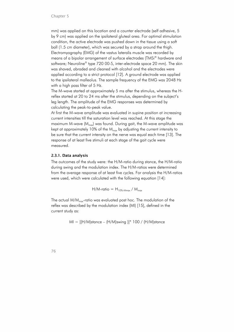
Chapter 5
76
mm) was applied on this location and a counter electrode (self adhesive, 5 by 9 cm) was applied on the ipsilateral gluteal area. For optimal stimulation condition, the active electrode was pushed down in the tissue using a soft ball (1.5 cm diameter), which was secured by a strap around the thigh. Electromyography (EMG) of the vastus lateralis muscle was recorded by means of a bipolar arrangement of surface electrodes (TMSi® hardware and software; Neuroline® type 720 00-S, inter-electrode space 20 mm). The skin was shaved, abraded and cleaned with alcohol and the electrodes were applied according to a strict protocol [12]. A ground electrode was applied to the ipsilateral malleolus. The sample frequency of the EMG was 2048 Hz with a high pass filter of 5 Hz. The M-wave started at approximately 5 ms after the stimulus, whereas the H-reflex started at 20 to 24 ms after the stimulus, depending on the subject’s leg length. The amplitude of the EMG responses was determined by calculating the peak-to-peak value. At first the M-wave amplitude was evaluated in supine position at increasing current intensities till the saturation level was reached. At this stage the maximum M-wave (Mmax) was found. During gait, the M-wave amplitude was kept at approximately 10% of the Mmax by adjusting the current intensity to be sure that the current intensity on the nerve was equal each time [13]. The response of at least five stimuli at each stage of the gait cycle were measured.
2.3.1. Data analysis
The outcomes of the study were: the H/M-ratio during stance, the H/M-ratio during swing and the modulation index. The H/M-ratios were determined from the average response of at least five cycles. For analysis the H/M-ratios were used, which were calculated with the following equation [14]:
H/M-ratio = H10%-Mmax / Mmax
The actual M/Mmax-ratio was evaluated post hoc. The modulation of the reflex was described by the modulation index (MI) [15], defined in the current study as:
MI = [(H/M)stance – (H/M)swing )]* 100 / (H/M)stance

Vastus lateralis H-reflex during gait
77
2.4. STATISTICAL ANALYSIS
The outcomes of the patients on the two separate days were averaged. The H/M-ratios during the stance phase and the swing phase were compared for the healthy subjects with a paired t-test. The differences between the healthy subjects and the patients were tested with a Mann-Whitney test (non-parametric test for independent samples). This was done for the H/M-ratio during stance, the H/M-ratio during swing and the MI.
3. RESULTS
3.1. SUBJECTS
3 SCI patients and 10 healthy subjects participated in the study. The age of the SCI patients ranged from 34 to 57 and the age of the healthy subjects ranged from 22 to 62. All patients had incomplete (ASIA D) cervical spinal cord lesions. The time since injury was from 20 months to 111 months, the Modified Ashworth scale [16] of the quadriceps ranged form 1+ to 3. All patients had decreased flexion of the knee during the swing phase. This was accompanied with an increased EMG activity of the quadriceps muscle. In table 1 demographic and raw data of the patients and healthy subjects are presented. Table 1: Demographic and raw data of the subjects. The ASIA score is an impairment score in which D means incomplete spinal cord lesions. MAS is the Modified Ashworth score of the quadriceps and MI is the modulation index of the vastus lateralis.
Patient SCI Sex Age (yrs)
ASIA Injury level
Time since injury
(months)
MAS H/M-ratio
stance
H/M-ratio swing
MI
1 Y F 57 D C5/6 111 2 0.37 0.22 32.9 2 Y M 51 D C5/6 20 3 0.60 0.47 22.0 7 Y M 34 D C4/5 75 1+ 0.40 0.48 -25.0 21 N F 25 0.25 0.074 70.9 22 N M 62 0.24 0.10 58.3 23 N M 26 0.098 0.050 49.4 24 N F 23 0.13 0.074 44.8 25 N F 22 0.22 0.062 71.3 26 N F 24 0.047 0.040 16.2 27 N M 25 0.051 0.028 44.8 28 N F 26 0.029 0.014 51.2 29 N M 39 0.039 0.040 -1.5 210 N F 26 0.41 0.33 18.0
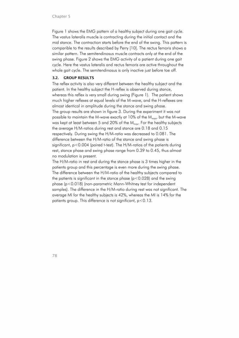
Chapter 5
78
Figure 1 shows the EMG pattern of a healthy subject during one gait cycle. The vastus lateralis muscle is contracting during the initial contact and the mid stance. The contraction starts before the end of the swing. This pattern is comparible to the results described by Perry [10]. The rectus femoris shows a similar pattern. The semitendinosus muscle contracts only at the end of the swing phase. Figure 2 shows the EMG activity of a patient during one gait cycle. Here the vastus lateralis and rectus femoris are active throughout the whole gait cycle. The semitendinosus is only inactive just before toe off.
3.2. GROUP RESULTS
The reflex activity is also very different between the healthy subject and the patient. In the healthy subject the H-reflex is observed during stance, whereas this reflex is very small during swing (Figure 1). The patient shows much higher reflexes at equal levels of the M-wave, and the H-reflexes are almost identical in amplitude during the stance and swing phase. The group results are shown in figure 3. During the experiment it was not possible to maintain the M-wave exactly at 10% of the Mmax, but the M-wave was kept at least between 5 and 20% of the Mmax. For the healthy subjects the average H/M-ratios during rest and stance are 0.18 and 0.15 respectively. During swing the H/M-ratio was decreased to 0.081. The difference between the H/M-ratio of the stance and swing phase is significant, p<0.004 (paired t-test). The H/M-ratios of the patients during rest, stance phase and swing phase range from 0.39 to 0.45, thus almost no modulation is present. The H/M-ratio in rest and during the stance phase is 3 times higher in the patients group and this percentage is even more during the swing phase. The difference between the H/M-ratio of the healthy subjects compared to the patients is significant in the stance phase (p<0.028) and the swing phase (p<0.018) (non-parametric Mann-Whitney test for independent samples). The difference in the H/M-ratio during rest was not significant. The average MI for the healthy subjects is 42%, whereas the MI is 14% for the patients group. This difference is not significant, p<0.13.
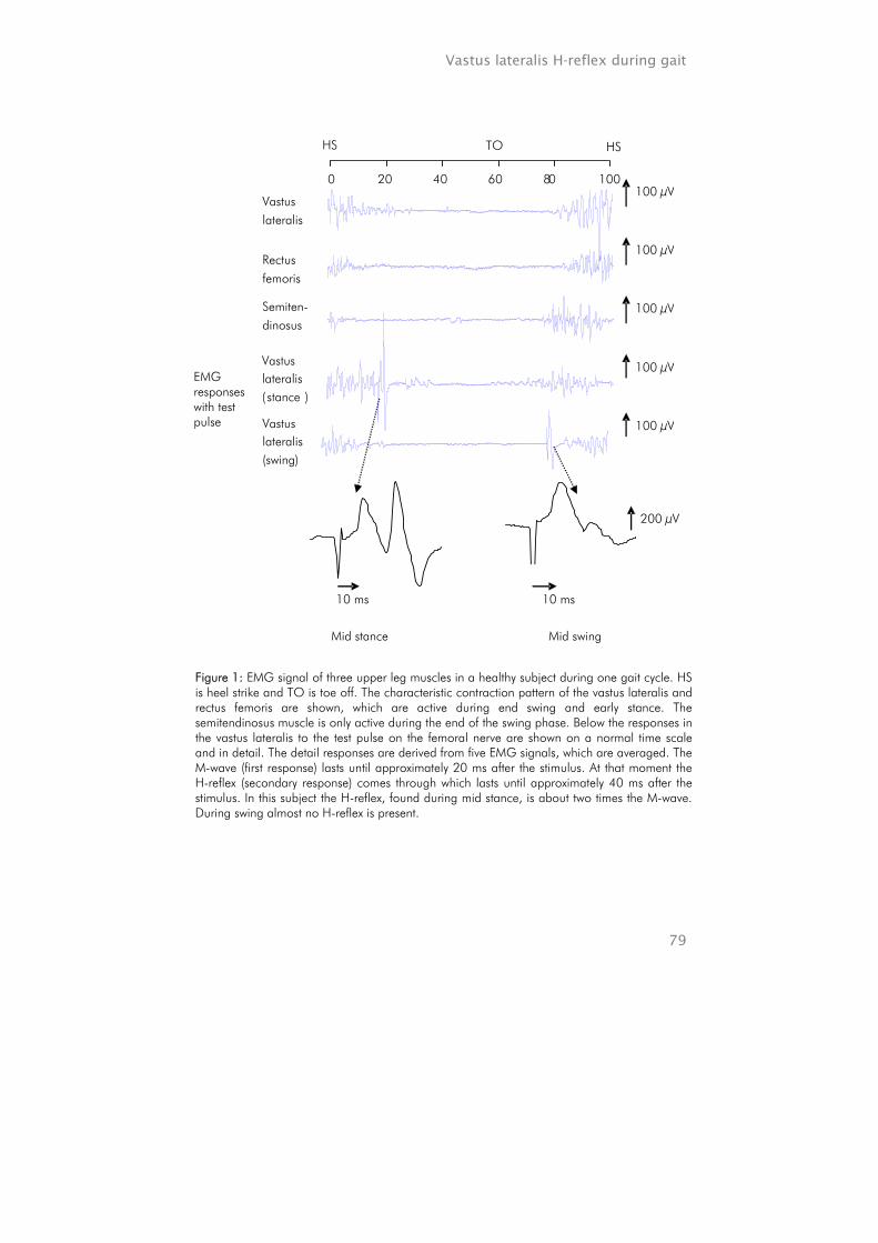
Vastus lateralis H-reflex during gait
79
Vastuslateralis
Rectusfemoris
Semiten-dinosus
Vastuslateralis ( stance )
Vastuslateralis(swing)
0 20 40 60 8 0 100
HS TO HS
100 µV
100 µV
100 µV
100 µV
100 µV
EMG responses with test pulse
10 ms
Mid stance Mid swing
10 ms
200 µV
Figure 1: EMG signal of three upper leg muscles in a healthy subject during one gait cycle. HS is heel strike and TO is toe off. The characteristic contraction pattern of the vastus lateralis and rectus femoris are shown, which are active during end swing and early stance. The semitendinosus muscle is only active during the end of the swing phase. Below the responses in the vastus lateralis to the test pulse on the femoral nerve are shown on a normal time scale and in detail. The detail responses are derived from five EMG signals, which are averaged. The M-wave (first response) lasts until approximately 20 ms after the stimulus. At that moment the H-reflex (secondary response) comes through which lasts until approximately 40 ms after the stimulus. In this subject the H-reflex, found during mid stance, is about two times the M-wave. During swing almost no H-reflex is present.
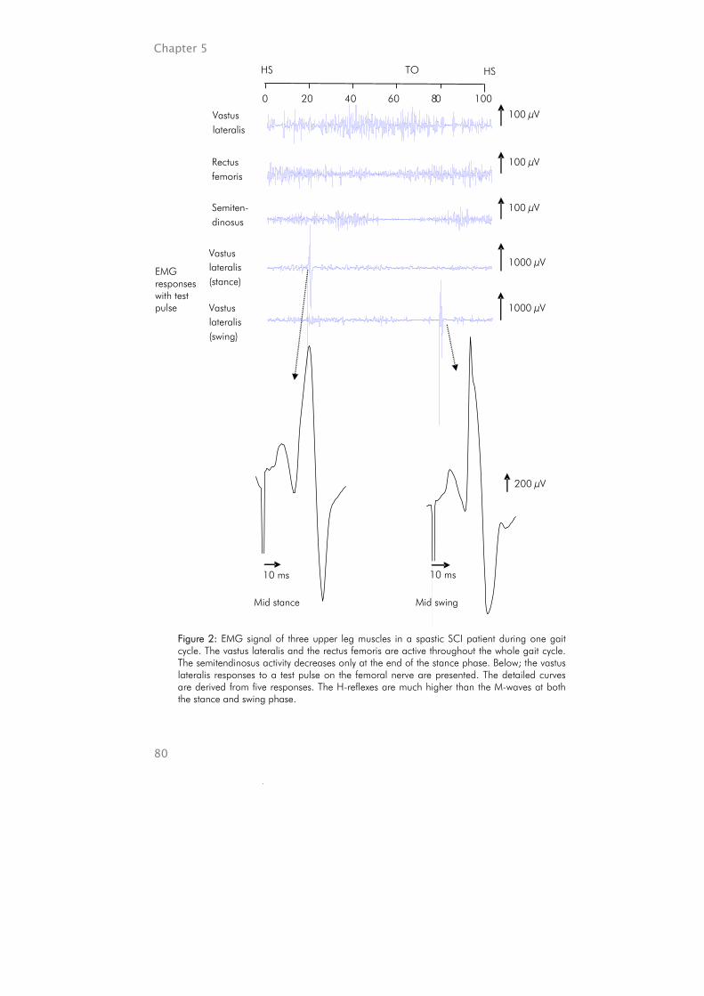
Chapter 5
80
0 20 40 60 8 0 100
HS TO HS
Vastuslateralis
Rectusfemoris
Semiten-dinosus
100 µV
100 µV
100 µV
1000 µV
1000 µV
Vastuslateralis(stance)
Vastuslateralis(swing)
EMG responses with test pulse
200 µV
10 ms
Mid stance Mid swing
10 ms
Figure 2: EMG signal of three upper leg muscles in a spastic SCI patient during one gait cycle. The vastus lateralis and the rectus femoris are active throughout the whole gait cycle. The semitendinosus activity decreases only at the end of the stance phase. Below; the vastus lateralis responses to a test pulse on the femoral nerve are presented. The detailed curves are derived from five responses. The H-reflexes are much higher than the M-waves at both the stance and swing phase.

Vastus lateralis H-reflex during gait
81
4. DISCUSSION
H/M-ratios of the healthy subjects during stance found in the current study are comparible to the data presented earlier by Dietz et al. [9]. They found the H/M-ratio in the early stance phase lies within the range of 0.08 to 0.18. We found a comparible H/M-ratio during stance, 0.15. The hypothesis that the H/M-ratio is decreased during the swing phase is confirmed. The average H/M-ratio measured during swing is almost half the value found during stance. In patients the H/M-ratios were much higher compared to the healthy subjects. In addition, patients only showed a slight modulation of the H/M-ratio. These results are comparible to the findings in the H-reflex outcomes of the soleus muscle studied by Fung & Barbeau [7]. They found almost no reflex modulation in severely impaired spastic subjects and the amplitude of the H-reflex was approximately 2 times higher than the H/M-ratio in healthy subjects. In our study the MI was not found to be significantly different between healthy subjects and SCI patients, despite a large difference between the averages of both groups (14% vs. 42%). It might be relevant to
Figure 3: H/Mmax-ratios and M/Mmax-ratios in healthy subjects and spastic SCI patients determined at rest and during mid stance and mid swing of a gait cycle. Group averages and their SD’s are shown. The M/Mmax-ratios (presented by the stock charts) are kept at approximately 0.1. The columns show the H/M-ratios. Significant differences are indicated by * (p<0.05) and ** (p<0.01).
0
0.2
0.4
0.6
0.8
rest stance swing rest stance swing
healthy patients
H/M
-rat
io, M
/Mm
ax-r
atio
*
*
**
Chapter 5
82
study this outcome in larger groups, because the power of the study would be increased, which might provide significant differences in the MI. The observed differences between healthy and spastic patients can be caused by extraneous factors. The most important factor is the age, which differs between the groups. The average age of the healthy subjects group was 29.8 (range 22 to 62) years, whereas the average age of the patients was 47.3 (range 34 to 57). The patients were relatively older than the healthy subjects. Some studies state that the H/M-ratio decreases with increasing age [17;18], whereas others found no change [19]. Thus, when age had influenced the H/M-ratio, the outcomes we found in the patient group might be decreased by the relatively high age of this group. The difference in the H/M-ratios between the patients and the healthy subjects, then, may be expected to be higher when ages would be comparable. The MI is not affected by the age, because the decrease in H/M-ratio is both for the stance and swing phase. The results of this study indicate that the inhibition in the spinal synapses is less in spastic patients than in healthy subjects. Also during the swing phase of gait, the inhibition is not increased, which may be a reason for the hyperactivity of the quadriceps muscle during this stage of the gait cycle. Other studies found decreased inhibitory neurological pathways in spastic subjects [20;21]. An important pathway, which is included in the H-reflex measurement, is the presynaptic inhibition. This pathway is controlled by supraspinal centres and is found to be decreased in patients with SCI [20]. Another pathway which has been found to have a large effect on the reflex activity is the reciprocal inhibition [22]. This is also thought to play an important role in spasticity [23]. In the treatment of spastic patients it should be considered whether the reflex excitability can be inhibited, especially during the swing phase of gait. Electrical stimulation, which can provide a momentary muscular inhibition [7], might be a good solution. This normalisation might help to improve the gait performance.
5. CONCLUSION
It can be concluded that the H-reflex modulates in the vastus lateralis muscle in healthy subjects during gait. This modulation consists of a relatively high H-reflex during mid stance and a lower H-reflex during mid swing. In spastic patients with SCI this modulation is not as pronounced and the H-reflexes during both phases of gait are 3 to 5 times higher than the H-reflexes found in healthy subjects.

Vastus lateralis H-reflex during gait
83
REFERENCES
1. Schneider A, Lavoie B, Capaday C. On the origin of the soleus H-reflex modulation pattern during human walking and its task-dependent differences. J. Neurophysiol. 2000;83:2881-90.
2. Faist M, Dietz V, Pierrot-Deseilligny E. Modulation, probably presynaptic in origin, of monosynaptic Ia excitation during human gait. Exp Brain Res 1996;109:441-9.
3. Sinkjær T. Muscle, reflex and central components in the control of the ankle joint in healthy and spastic man. Acta Neurol Scand Suppl 1997;170:1-28.
4. Delwaide PJ, Pennisi G. Tizanidine and electrophysiologic analysis of spinal control mechanisms in humans with spasticity. Neurology 1994 Nov;44(11 Suppl 9):S21-7; discussion S27-8.
5. Faist M, Mazevet D, Dietz V, Pierrot-Deseilligny E. A quantitative assessment of presynaptic inhibition of Ia afferents in spastics. Differences in hemiplegics and paraplegics. Brain 1994 Dec;117 ( Pt 6):1449-55.
6. Okuma Y, Lee RG. Reciprocal inhibition in hemiplegia: correlation with clinical features and recovery. Can J Neurol Sci 1996 Feb;23(1):15-23.
7. Fung J, Barbeau H. Effects of conditioning cutaneomuscular stimulation on the soleus H- reflex in normal and spastic paretic subjects during walking and standing. J Neurophysiol 1994 Nov;72(5):2090-104.
8. Funase K, Miles TS. Observations on the variability of the H reflex in humans soleus. Muscle & Nerve 1999 Mar;22:341-6.
9. Dietz V, Faist M, Pierrot-Deseilligny E. Amplitude modulation of the quadriceps H-reflex in the human during the early stance phase of gait. Exp Brain Res 1990;79:221-4.
10. Perry J. Gait analysis. Thorofar, USA: SLACK Incorporated; 1992. 11. Monaghan CC, Veltink PH, Bultstra G, Droog E, Kotiadis D, van Riel W. Control of
triceps surae stimulation based on shank orientation using a uniaxial gyroscope IFESS conference proceedings; Bournemouth, UK. 2004.
12. Hermens HJ, Freriks B, Merletti R, et al. SENIAM: European Recommendations for Surface ElectroMyoGraphy. Enschede: Roessingh Research and Development; 1999.
13. Capaday C. Neurophysiological methods for studies of the motor system in freely moving human subjects. J Neurosci Methods 1997 Jun 27;74(2):201-18.
14. Visser SL. Reflexen. Notermans Bussel: 1981. pp. 353-68. 15. Yang JF, Fung J, Edamura M, Blunt R, Stein RB, Barbeau H. H-reflex modulation
during walking in spastic paretic subjects. Can J Neurol Sci 1991 Nov;18(4):443-52.
16. Bohannon RW, Smith MB. Interrater reliability of a modified Ashworth scale of muscle spasticity. Physical Therapy 1987 Feb;67(2):206-7.
17. Delwaide PJ. Etude experimentale de l'hyperreflexie tendineuse en clinique neuroloque. Bruxelles: Editions Arscia; 1971.
18. Ongeboer de Visser BW, Bour LJ, Koelman JHTM, Speelman JD. Cumulative vibratory indices and the H/M ratio of the soleus H-reflex: a quantitative study in control and spastic subjects. Electroencephal Clin Neurophysiol 1989;73:162-6.
19. Jankus WR, Robinson LR, Little JW. Normal linits of side-to-side H-reflex amplitude variability. Arch Phys Med Rehabil 1994;75:3-7.

Chapter 5
84
20. Morita H, Crone C, Christenhuis D, Petersen NT, Nielsen JB. Modulation of presynaptic inhibition and disynaptic reciprocal Ia inhibition during voluntary movement in spasticity. Brain 2001 Apr;124(Pt 4):826-37.
21. Mazzocchio R, Rossi A. Involvement of spinal recurrent inhibition in spasticity. Further insight into the regulation of Renshaw cell activity. Brain 1997 Jun;120 ( Pt 6):991-1003.
22. Nielsen J, Crone C, Sinkjær T, Toft E, Hultborn H. Central control of reciprocal inhibition during fictive dorsiflexion in man. Exp Brain Res 1995;104(1):99-106.
23. Crone C, Nielsen J, Petersen N, Ballegaard M, Hultborn H. Disynaptic reciprocal inhibition of ankle extensors in spastic patients. Brain 1994 Oct;117 ( Pt 5):1161-8.
CHAPTER 6 Effect of electrical stimulation of hamstrings and l3/4 dermatome on H/M-ratio and performance of gait in spastic SCI-patients Arjan van der Salm, Peter H. Veltink, Hermie J. Hermens, Anand V. Nene, Maarten J. IJzerman Objective: To determine the effect of electrical stimulation of hamstrings and L3/4 dermatome on the swing phase of gait. Design: Intervention study in which patients are used as their own control. Setting: Research department affiliated with a rehabilitation hospital in the Netherlands. Patients: Five incomplete spinal cord injury patients with spasticity. Intervention: Two electrical stimulation methods were investigated; i.e. hamstrings and L3/4 dermatome stimulation. Both interventions were applied during the swing phase of gait. Main outcome measures: Step length, maximum hip and knee flexion during the swing phase of gait. Changes of spinal inhibition during gait were evaluated using the H/M-ratio at mid swing. Results: The hip flexion decreased 4.4º (p<0.05) when the hamstrings were stimulated during the swing phase, whereas the knee flexion was not changed. The step length did not change significantly. The H/M-ratio of 3 patients was measured. In one patient the H/M-ratio increased while L3/4 dermatome was stimulated. Another patient showed a decrease of the H/M-ratio during hamstrings stimulation. One patient showed no relevant change at any of the interventions. Conclusion: It was concluded that hamstrings stimulation during the swing phase results in a reduction of the hip flexion in all five SCI patients. The H/M-ratio of the vastus lateralis was normalised using hamstrings stimulation in one of three patients. Stimulation of the L3/4 dermatome provides no significant changes in gait performance, but in one patient the H/M-ratio increased. Submitted for Publication in Neuromodulation
Chapter 6
86
1. Introduction
An important gait impairment in spinal cord injury (SCI) patients is decreased knee flexion during the swing phase [1;2]. This impairment can cause a limitation of the swing limb advancement of the affected leg, which will decrease the step length of that leg. It is thought that spasticity may play an important role in this impairment [1;3]. Especially activity of the vastus lateralis and rectus femoris are reported to limit the knee flexion [2]. To assist SCI patients to improve their gait, electrical stimulation (ES) can be used. Several studies found positive effects using ES in patients with SCI [4-10]. One study also included stimulation of the hamstrings combined with the use of an orthosis, to enhance the knee flexion [11]. In normal gait, hamstrings are only activated at the end of the swing phase to decelerate the knee extension [12]. The knee flexion during swing is induced passively due to an active hip flexion and the push off initiated by the calf muscles. In SCI patients, the knee flexion might be actively provoked during the swing phase using stimulation of the hamstrings. This may also affect the movement of the hip, because the hamstrings are bi-articular muscles. Functional ES is mainly used to achieve a direct force production in the muscles. But, functional ES will also induce neurophysiological changes, that may influence the gait. Inhibition of spastic muscles may be one of these effects. This mechanism was investigated in a study of the effect of cutaneomuscular stimulation on soleus H-reflex [3]. It was found that the H-reflex of the soleus muscle can be inhibited by cutaneomuscular stimulation of the plantar side of the foot. Another study found that stimulation of the peroneal nerve can inhibit the triceps surae stretch reflex in stroke patients [13]. Both studies used 3 to 5 pulses of 1 ms at a frequency of 200 Hz. In contrast, in studies in which ES is used clinically, the pulses are approximately 300 µs and the frequency ranges from 20 to 25 Hz for muscle stimulation [4;5;7;14-16]. This study investigates the effect of hamstrings and L3/4 dermatome stimulation, which is used clinically, on gait parameters. Stimulation was executed during swing phase of gait. Main outcome measures were hip and knee flexion, step length and the H/M-ratio during mid swing. The effect could be very useful in the treatment of patients with SCI, especially when spastic muscles are inhibited.
Effect of electrical stimulation on H/M-ratio and gait performance
87
2. METHODS
2.1. SUBJECTS
Patients were recruited from a rehabilitation centre in the Netherlands (Het Roessingh, Enschede). Only patients with spinal cord injury (SCI) for at least 6 months were included. Patients were only included if they were able to walk with or without support or orthosis and if their gait was affected by spasticity, which was determined using EMG measurement during gait. For this, the EMG of 6 muscles (soleus, gastrocnemius, tibialis anterior, rectus femoris, vastus lateralis and hamstrings) in the leg was measured. Thereafter, these measurements were analysed by an experienced PMR-physician. All patients gave informed consent to participate in the experiment, which was approved by the local ethics committee.
2.2. DESIGN
Patients were measured two times on separate days. Both days started with a baseline measurement followed by an intervention measurement. Each baseline measurement consisted of 6 trials, during which kinematic measurements were performed and at least 5 trials, during which the H-reflex was measured. Each trial consisted of 10 meter walking. A test stimulus for the H-reflex was applied during one, randomly chosen, gait cycle in each trial. After the baseline measurement the intervention measurement was performed. All measurements, kinematic and H-reflex measurements, were executed again combined with one of the interventions. Antagonist muscle (hamstrings) was stimulated on one day, dermatome stimulation (L3/4 dermatome) was performed on the other. The sequence, however, was randomized. Each day, the time at which the measurements started was kept the same. The most affected leg of the patients was used for the measurements.
2.3. INTERVENTIONS
Self adhesive electrodes (5 to 9 cm) were used to stimulate hamstrings or dermatome. Two electrodes were placed proximally and distally over the muscle belly of the hamstrings. For dermatome stimulation one lateral electrode was placed just above the knee joint and the other electrode was placed medially just below the knee joint [17]. This placement covered part of the L3/4 dermatome. In addition, the medial electrode stimulated the sensory saphenous nerve (L3/4). The stimulation frequency was 30 Hz. The pulse width was 300 µs for muscle stimulation and 100 µs for skin stimulation. The intensity of muscle stimulation was adjusted to allow the
Chapter 6
88
knee to bend against gravity in sitting position. The intensity of the dermatome stimulation was just below the motor threshold. In both interventions the stimulation burst started at the onset of swing phase and lasted for 70% of the swing phase. The timing of the burst was controlled by an angular-velocity-sensor (gyroscope) which measured angular-velocity in the saggital plane [18]. This gyroscope was fixated on the proximal head of the fibula with a strap. An individual threshold was used to detect the onset of the swing phase.
2.4. MEASUREMENTS
2.4.1. Kinematics
A VICON® setup (version 370, 6 cameras) was used to measure the kinematics during gait. Passive markers were placed on the sacrum, anterior superior ilea, thighs, knees, lower legs, ankle and distal second metatarsal bones according to a strict protocol. These markers were placed by an experienced physical therapist. The VCM (VICON Clinical Manager, version 1.37) model was used to determine maximal knee and hip flexion in the saggital plane and step length [19].
2.4.2. EMG-recording
The EMG of the vastus lateralis muscle was measured with surface electrodes (Neuroline® type 720 00-s Ag-AgCl gel electrodes; diameter 12 mm, inter-electrode space 20 mm) using a bipolar arrangement. A ground electrode was applied on the ipsilateral lateral malleolus. The muscle electrodes were applied at 1/3 of the line from the lateral side of the patella to the anterior spina iliaca superior [20]. Before application, the skin was shaved, abraded and cleaned with alcohol. For the EMG recording TMSI® hard- and software was used. The sample frequency of the EMG was 2048 Hz with a high pass filter of 5 Hz and a digital FIR low pass filter of 553 Hz.
2.4.3. H-reflex measurement
In order to stimulate the femoral nerve, the electrode was placed in the femoral triangle and the counter electrode was applied on the ipsilateral gluteal area [21]. A 500 µs block pulse was used to stimulate the nerve. The optimal stimulation location of the nerve was found with a handheld probe. For optimal stimulation condition, the active electrode was pushed down in the tissue using a soft ball (diameter 1.5 cm), which was secured by a strap around the thigh. Before the measurements during gait were performed, the
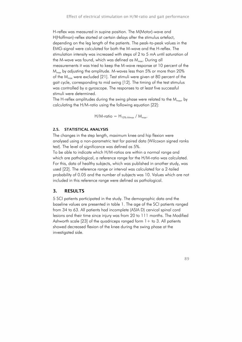
Effect of electrical stimulation on H/M-ratio and gait performance
89
H-reflex was measured in supine position. The M(Motor)-wave and H(Hoffman)-reflex started at certain delays after the stimulus artefact, depending on the leg length of the patients. The peak-to-peak values in the EMG-signal were calculated for both the M-wave and the H-reflex. The stimulation intensity was increased with steps of 2 to 5 mA until saturation of the M-wave was found, which was defined as Mmax. During all measurements it was tried to keep the M-wave response at 10 percent of the Mmax by adjusting the amplitude. M-waves less than 5% or more than 20% of the Mmax were excluded [21]. Test stimuli were given at 80 percent of the gait cycle, corresponding to mid swing [12]. The timing of the test stimulus was controlled by a gyroscope. The responses to at least five successful stimuli were determined. The H-reflex amplitudes during the swing phase were related to the Mmax, by calculating the H/M-ratio using the following equation [22]:
H/M-ratio = H10%-Mmax / Mmax.
2.5. STATISTICAL ANALYSIS
The changes in the step length, maximum knee and hip flexion were analysed using a non-parametric test for paired data (Wilcoxon signed ranks test). The level of significance was defined as 5%. To be able to indicate which H/M-ratios are within a normal range and which are pathological, a reference range for the H/M-ratio was calculated. For this, data of healthy subjects, which was published in another study, was used [22]. The reference range or interval was calculated for a 2-tailed probability of 0.05 and the number of subjects was 10. Values which are not included in this reference range were defined as pathological.
3. RESULTS
5 SCI patients participated in the study. The demographic data and the baseline values are presented in table 1. The age of the SCI patients ranged from 34 to 63. All patients had incomplete (ASIA D) cervical spinal cord lesions and their time since injury was from 20 to 111 months. The Modified Ashworth scale [23] of the quadriceps ranged form 1+ to 3. All patients showed decreased flexion of the knee during the swing phase at the investigated side.
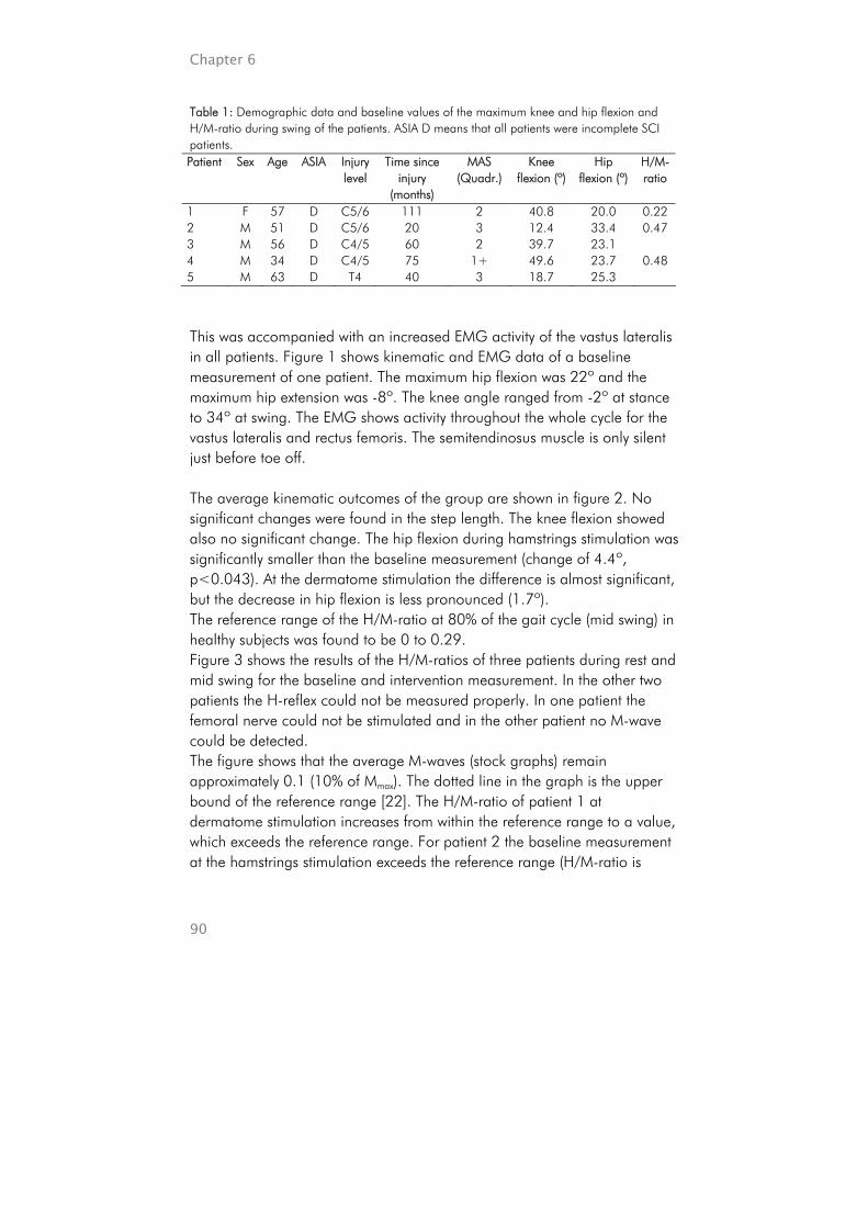
Chapter 6
90
Table 1: Demographic data and baseline values of the maximum knee and hip flexion and H/M-ratio during swing of the patients. ASIA D means that all patients were incomplete SCI patients. Patient Sex Age ASIA Injury
level Time since
injury (months)
MAS (Quadr.)
Knee flexion (º)
Hip flexion (º)
H/M-ratio
1 F 57 D C5/6 111 2 40.8 20.0 0.22 2 M 51 D C5/6 20 3 12.4 33.4 0.47 3 M 56 D C4/5 60 2 39.7 23.1 4 M 34 D C4/5 75 1+ 49.6 23.7 0.48 5 M 63 D T4 40 3 18.7 25.3
This was accompanied with an increased EMG activity of the vastus lateralis in all patients. Figure 1 shows kinematic and EMG data of a baseline measurement of one patient. The maximum hip flexion was 22º and the maximum hip extension was -8º. The knee angle ranged from -2º at stance to 34º at swing. The EMG shows activity throughout the whole cycle for the vastus lateralis and rectus femoris. The semitendinosus muscle is only silent just before toe off. The average kinematic outcomes of the group are shown in figure 2. No significant changes were found in the step length. The knee flexion showed also no significant change. The hip flexion during hamstrings stimulation was significantly smaller than the baseline measurement (change of 4.4º, p<0.043). At the dermatome stimulation the difference is almost significant, but the decrease in hip flexion is less pronounced (1.7º). The reference range of the H/M-ratio at 80% of the gait cycle (mid swing) in healthy subjects was found to be 0 to 0.29. Figure 3 shows the results of the H/M-ratios of three patients during rest and mid swing for the baseline and intervention measurement. In the other two patients the H-reflex could not be measured properly. In one patient the femoral nerve could not be stimulated and in the other patient no M-wave could be detected. The figure shows that the average M-waves (stock graphs) remain approximately 0.1 (10% of Mmax). The dotted line in the graph is the upper bound of the reference range [22]. The H/M-ratio of patient 1 at dermatome stimulation increases from within the reference range to a value, which exceeds the reference range. For patient 2 the baseline measurement at the hamstrings stimulation exceeds the reference range (H/M-ratio is

Effect of electrical stimulation on H/M-ratio and gait performance
91
0.41), whereas the H/M-ratio during stimulation lies within this reference range (H/M-ratio is 0.16). In this patient the H/M-ratio was increased during the dermatome stimulation. In patient 4 almost no change of the H/M-ratios due to one of the interventions was present.
Figure 2: Gait parameters, step length, hip flexion and knee flexion, in spastic SCI patients (averages and 1 SD). In the upper graph the step length is presented. In the graph below the maximum knee flexions and hip flexions are presented. The interventions are stimulation of the hamstrings or L3/4 dermatome. Before each intervention measurement a baseline measurement was performed. Both the outcomes of the baseline and intervention measurements at each day are presented with the P-value of the difference (Wilcoxon signed ranks test).
400
450
500
550
600
650
700
hamstrings stimulation
dermatome stimulation
Step length
Baseline Intervention
leng
th (m
m) P<0.23
0
10
20
30
40
50
60
hamstrings stimulation
dermatome stimulation
hamstrings stimulation
dermatome stimulation
Knee flexion Hip flexion
Baseline
Intervention
flexi
on (º
) P<0.043
P<0.068
P<0.14
P<0.27
P<0.69
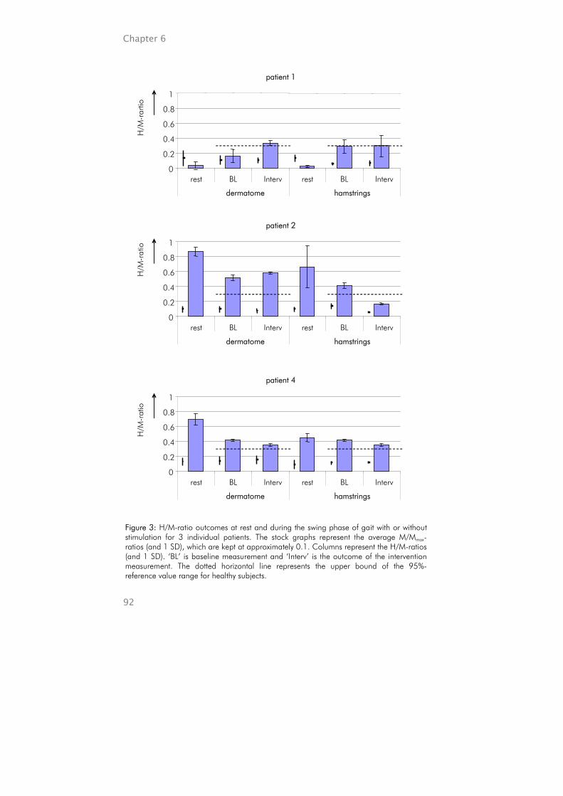
Chapter 6
92
Figure 3: H/M-ratio outcomes at rest and during the swing phase of gait with or without stimulation for 3 individual patients. The stock graphs represent the average M/Mmax-ratios (and 1 SD), which are kept at approximately 0.1. Columns represent the H/M-ratios (and 1 SD). ‘BL’ is baseline measurement and ‘Interv’ is the outcome of the intervention measurement. The dotted horizontal line represents the upper bound of the 95%-reference value range for healthy subjects.
patient 1
0
0.2
0.4
0.6
0.8
1
rest BL Interv rest BL Interv
dermatome hamstrings
patient 2
0
0.2
0.4
0.6
0.8
1
rest BL Interv rest BL Interv
dermatome hamstrings
patient 4
0
0.2
0.4
0.6
0.8
1
rest BL Interv rest BL Interv
dermatome hamstrings
H/M
-rar
tio
H/M
-rat
io
H/M
-rat
io
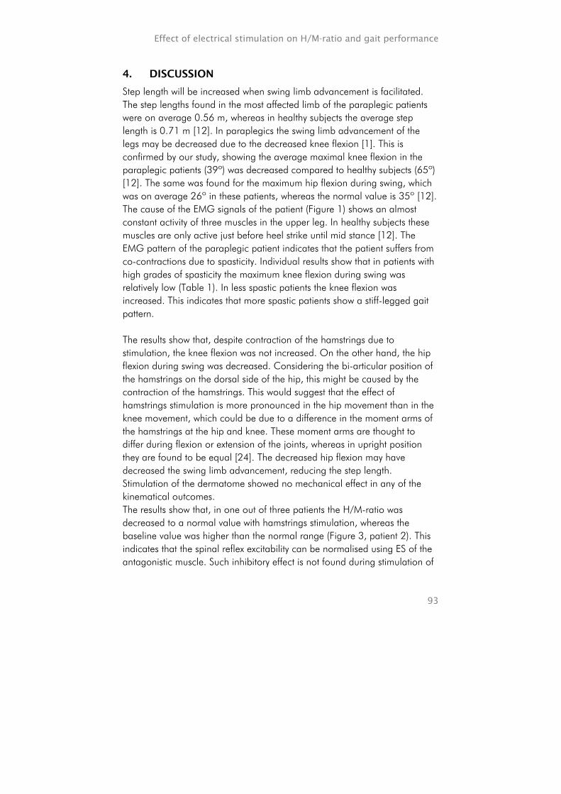
Effect of electrical stimulation on H/M-ratio and gait performance
93
4. DISCUSSION
Step length will be increased when swing limb advancement is facilitated. The step lengths found in the most affected limb of the paraplegic patients were on average 0.56 m, whereas in healthy subjects the average step length is 0.71 m [12]. In paraplegics the swing limb advancement of the legs may be decreased due to the decreased knee flexion [1]. This is confirmed by our study, showing the average maximal knee flexion in the paraplegic patients (39º) was decreased compared to healthy subjects (65º) [12]. The same was found for the maximum hip flexion during swing, which was on average 26º in these patients, whereas the normal value is 35º [12]. The cause of the EMG signals of the patient (Figure 1) shows an almost constant activity of three muscles in the upper leg. In healthy subjects these muscles are only active just before heel strike until mid stance [12]. The EMG pattern of the paraplegic patient indicates that the patient suffers from co-contractions due to spasticity. Individual results show that in patients with high grades of spasticity the maximum knee flexion during swing was relatively low (Table 1). In less spastic patients the knee flexion was increased. This indicates that more spastic patients show a stiff-legged gait pattern. The results show that, despite contraction of the hamstrings due to stimulation, the knee flexion was not increased. On the other hand, the hip flexion during swing was decreased. Considering the bi-articular position of the hamstrings on the dorsal side of the hip, this might be caused by the contraction of the hamstrings. This would suggest that the effect of hamstrings stimulation is more pronounced in the hip movement than in the knee movement, which could be due to a difference in the moment arms of the hamstrings at the hip and knee. These moment arms are thought to differ during flexion or extension of the joints, whereas in upright position they are found to be equal [24]. The decreased hip flexion may have decreased the swing limb advancement, reducing the step length. Stimulation of the dermatome showed no mechanical effect in any of the kinematical outcomes. The results show that, in one out of three patients the H/M-ratio was decreased to a normal value with hamstrings stimulation, whereas the baseline value was higher than the normal range (Figure 3, patient 2). This indicates that the spinal reflex excitability can be normalised using ES of the antagonistic muscle. Such inhibitory effect is not found during stimulation of

Chapter 6
94
the dermatome. On contrary, the H/M-ratio at dermatome stimulation increases in one patient to a pathological value, whereas the baseline value was within the normal range. In this patient dermatome stimulation has a facilitating effect in the spinal synapses. This difference in effect between the patients might be caused by a difference in the severity of spasticity. The patient, who showed a reduction, had a relatively high grade of spasticity, MAS was 3, compared to the other two patients, MAS was 1+ and 2 (Table 1). This indicates that, in patients with more severe spasticity, electrical stimulation may provide a higher level of inhibition in antagonist muscles. A second reason for the different change in the H/M-ratio may be the difference in the time since injury. In the patient in whom hamstrings stimulation reduced the H/M-ratio, the time since injury was 20 months, whereas the other patients suffered longer from SCI; 75 and 111 months (Table 1). The spinal connections, of patients who suffer relatively long from SCI and spasticity, might have adapted, which result in a change of their inhibitory pathways. This is supported by the finding that several inhibitory pathways are decreased in spastic patients [25;26], and that there is the significant change of the reflex excitability during at least the first two years after the injury in SCI patients [27]. It is not expected that changes in the cycle time of one stride due to the electrical stimulation have influenced the outcome of the H/M-ratio. In patient 2, in which the largest difference in the H/M-ratio is found, the cycle time during hamstrings stimulation had increased by 6%. This means that the test pulse was applied at approximately 75% of the gait cycle (instead of 80%). This is still during mid swing, when no vastus lateralis activity is required [12]. Other studies found that the delay between the conditioning pulses and the test pulse (or test stretch) was important for the inhibitory effect. Fung & Barbeau [3] found that the effective delay, which provided an inhibitory effect in the soleus by stimulation of the medial plantar arch, was between 0 and 50 ms (calculated from the latest pulse). Veltink et al. [13] found the inhibitory delay, between the last conditioning pulse on the peroneal nerve and a stretch of the gastrocnemius, in the range of 59 to 184 ms. Because the stimulation location we used, is different from the other studies, these delays can not be compared directly. The effective inhibitory period of the latter study lasted for at least 125 ms. To be effective the required inhibitory period for the swing phase of gait is at least 300 ms, because during this period the vastus lateralis is stretched.
Effect of electrical stimulation on H/M-ratio and gait performance
95
The used parameters can inhibit the spinal connections, but the effect might be even more pronounced when stimulation parameters are changed. Especially, at increased frequencies more effect could be expected, because, more afferent action potentials will be sent to the spinal cord, which can provide an inhibitory state [3;13]. The nerve fibre recruitment might also be increased using relatively high pulse widths and intensities. But, all parameters should be functional and comfortable for the patient, which is not the case at large frequencies, pulse widths and intensities. One option may be that arrays of short bursts, as used by Veltink et al. [13], can be applied when inhibition is required. The mechanical effect with hamstrings stimulation should be kept as low as possible.
5. CONCLUSION
It can be concluded that hamstrings stimulation during the swing phase provides a reduction in the hip flexion in SCI patients, whereas the knee flexion is not altered. The H/M-ratio at mid swing of the vastus lateralis can be normalised using ES of the hamstrings. Stimulation of the L3/4 dermatome provides no changes in gait performance, but it can facilitate spinal connections.
Chapter 6
96
REFERENCE LIST
1. Van der Salm A, Nene A, Maxwell DJ, Veltink PH, Hermens HJ, IJzerman MJ. Gait impairments in a group of patients with incomplete spinal cord injury and their relevance regarding therapeutic approaches using FES. Artificial Organs 2005 Jan;29(1):8-14.
2. Perry J, Barto P, Gronley J, Yoshida H. Limb flexion deficits: implications for FES gait assist design. Proceedings of the Eight International Symposium on ECHE, Dubrovnik 1984.
3. Fung J, Barbeau H. Effects of conditioning cutaneomuscular stimulation on the soleus H- reflex in normal and spastic paretic subjects during walking and standing. J Neurophysiol 1994 Nov;72(5):2090-104.
4. Bajd T, Andrews BJ, Kralj A, Katakis J. Restoration of walking in patients with incomplete spinal cord injuries by use of surface electrical stimulation--preliminary results. Prosthet Orthot Int 1985 Aug;9(2):109-11.
5. Bajd T, Kralj A, Turk R, Benko H, Sega J. Use of functional electrical stimulation in the rehabilitation of patients with incomplete spinal cord injuries. J Biomed Eng 1989 Mar;11(2):96-102.
6. Braun Z, Mizrahi J, Najenson T, Graupe D. Activation of paraplegic patients by functional electrical stimulation: training and biomechanical evaluation. Scand J Rehabil Med Suppl 1985;12:93-101.
7. Granat MH, Ferguson AC, Andrews BJ, Delargy M. The role of functional electrical stimulation in the rehabilitation of patients with incomplete spinal cord injury--observed benefits during gait studies. Paraplegia 1993 Apr;31(4):207-15.
8. Isakov E, Mizrahi J, Graupe D, Becker E, Najenson T. Energy cost and physiological reactions to effort during activation of paraplegics by functional electrical stimulation. Scand J Rehabil Med Suppl 1985;12:102-7.
9. Kralj A, Bajd T, Turk R, Krajnik J, Benko H. Gait restoration in paraplegic patients: a feasibility demonstration using multichannel surface electrode FES. J Rehabil R D 1983 Jul;20(1):3-20.
10. Stein RB, Belanger M, Wheeler G, Wieler M, Popovic DB, Prochazka A, Davis LA. Electrical systems for improving locomotion after incomplete spinal cord injury: an assessment. Arch Phys Med Rehabil 1993 Sep;74(9):954-9.
11. Solomonow M, Baratta R, Hirokawa S, Rightor N, Walker W, Beaudette P, Shoji H, D'Ambrosia R. The RGO Generation II: muscle stimulation powered orthosis as a practical walking system for thoracic paraplegics. Orthopedics 1989 Oct;12(10):1309-15.
12. Perry J. Gait analysis. Thorofar, USA: SLACK Incorporated; 1992. 13. Veltink PH, Ladouceur M, Sinkjær T. Inhibition of the triceps surae stretch reflex by
stimulation of the deep peroneal nerve in persons with spastic stroke. Arch Phys Med Rehabil 2000 Aug;81(8):1016-24.
14. Granat M, Keating JF, Smith AC, Delargy M, Andrews BJ. The use of functional electrical stimulation to assist gait in patients with incomplete spinal cord injury. Disabil Rehabil 1992 Apr-1992 Jun 30;14(2):93-7.
15. Brissot R, Gallien P, Le Bot MP, Beaubras A, Laisne D, Beillot J, Dassonville J. Clinical experience with functional electrical stimulation-assisted gait with Parastep in spinal cord-injured patients. Spine 2000 Feb 15;25(4):501-8.
Effect of electrical stimulation on H/M-ratio and gait performance
97
16. Sykes L, Ross ER, Powell ES, Edwards J. Objective measurement of use of the reciprocating gait orthosis (RGO) and the electrically augmented RGO in adult patients with spinal cord lesions. Prosthet Orthot Int 1996 Dec;20(3):182-90.
17. Bajd T, Gregoric M, Vodovnik L, Benko H. Electrical stimulation in treating spasticity resulting from spinal cord injury. Arch Phys Med Rehabil 1985 Aug;66(8):515-7.
18. Monaghan CC, Veltink PH, Bultstra G, Droog E, Kotiadis D, van Riel W. Control of triceps surae stimulation based on shank orientation using a uniaxial gyroscope. IFESS conference proceedings; Bournemouth, UK. 2004.
19. VICON Clinical Manager; User's Guide, Version 1.01992. 20. Hermens HJ, Freriks B, Merletti R, Stegeman D, Blok J, Rau G, Disslehorst-Klug C,
Hägg G. SENIAM: European Recommendations for Surface ElectroMyoGraphy. Enschede: Roessingh Research and Development; 1999.
21. Dietz V, Faist M, Pierrot-Deseilligny E. Amplitude modulation of the quadriceps H-reflex in the human during the early stance phase of gait. Exp Brain Res 1990;79:221-4.
22. Van der Salm A, Veltink PH, Hermens HJ, Nene AV, IJzerman MJ. Modulation of the vastus lateralis H-reflex during gait in healthy subjects and patients with spinal cord injury. submitted to Gait & Posture
23. Bohannon RW, Smith MB. Interrater reliability of a modified Ashworth scale of muscle spasticity. Physical Therapy 1987 Feb;67(2):206-7.
24. Hof AL. The force resulting from the action of mono- and biarticular muscles in a limb. Journal of Biomechanics 2001;34:1085-9.
25. Delwaide PJ, Pennisi G. Tizanidine and electrophysiologic analysis of spinal control mechanisms in humans with spasticity. Neurology 1994 Nov;44(11 Suppl 9):S21-7; discussion S27-8.
26. Okuma Y, Mizuno Y, Lee RG. Reciprocal Ia inhibition in patients with asymmetric spinal spasticity. Clin Neurophysiol 2002 Feb;113(2):292-7.
27. Hiersemenzel LP, Curt A, Dietz V. From spinal shock to spasticity: neuronal adaptations to a spinal cord injury. Neurology 2000 Apr 25;54(8):1574-82.

Chapter 6
98
CHAPTER 7
General discussion
Chapter 7
100
GENERAL DISCUSSION
The goal of this thesis was to investigate the influence of electrical stimulation on spasticity of leg muscles in spinal cord injury patients and its impact on gait. Both, the carry-over effect and the instant effect of electrical stimulation during gait were investigated. To be able to investigate these effects spasticity had to be measured adequately during both rest and gait. Therefore a new method for the measurement of spasticity in rest was developed. For the measurement of spasticity during gait we used the H-reflex measurement, which is used frequently for the triceps surae. We investigated whether this measurement could be used in the vastus lateralis and whether the outcomes were different for healthy and spastic subjects.
MEASUREMENT OF SPASTICITY
Clinicians need an objective, functionally orientated (imitating rotation in gait), assessment of spasticity. The assessment protocol described in chapter 2 may be appropriate, since it can objectively assess spasticity in a functional range of motion providing clinically useful outcomes. The outcomes of the assessment protocol provide valid and reproducible information about the reflex excitability. In addition, passive muscle stiffness can be measured. Many methods to measure spasticity in rest are available, but none of these measurements assesses spasticity objectively in a functional range of motion nor can they distinguish between reflexive and non-reflexive components. The currently used measures for spasticity can be divided into three groups; biomechanical, neurophysiological and clinical measures. The latter are most commonly used, but their validity is a point of discussion [1]. The clinical assessments of spasticity, especially the (Modified) Ashworth scale [2] and Tardieu scale [3;4], use movements in a functional range of movement, and provide a clinically relevant outcome. Neurophysiological and biomechanical assessments, on the other hand, are not directly related to clinical assessments [5] and the translation to a clinical relevant outcome is difficult [6;7]. Reflexive components can also be measured during postural control using disturbance experiments [8]. For this random force inputs are used in addition to position control by a subject, which is applicable in many central nervous disorders, but difficult in complete SCI patients. With the new developed measurement it is possible to assess the reflex activity and the passive muscle stiffness separately. The responsiveness of the reflex excitability measurement is found to be marginal. It depends on the
General discussion
101
variability of the outcomes between the days and the difference caused by an intervention. It is known that spasticity differs between days. This inter-day variation might be relatively high compared to the intervention effect, which was used to determine the responsiveness. Therefore, when the assessment is used in order to determine treatment effects over days, only relatively large effects, exceeding the inter-day variation, can be determined. In this study major differences in the reflex excitability of the vastus lateralis between healthy subjects and spastic SCI patients during gait were found, which is consistent with reflex excitability of the soleus muscle [9;10]. This means that the inhibitory state of the synaptic connection of the Ia-afferent with the alfa-motoneuron was decreased in spastic patients compared to the healthy subjects during both the stance and swing phase. The variation of the reflex excitability during the gait cycle was much more pronounced in the healthy subjects than in the spastic patients. In addition, during the stance phase the reflex excitability in patients was three times higher than in the healthy subjects and during swing this difference was even more. This difference between healthy and spastic subjects could be expected when neurophysiological differences are considered. Important factors, which are thought to modulate the spinal reflex excitability during gait, are pre- and post-synaptic inhibition [10]. Pre-synaptic inhibition has been found to be decreased in paraplegics [11;12]. Pre-synaptic inhibition causes a reduced effectiveness in several, selected afferent nerve synapses. Post-synaptic inhibition has only been investigated in hemiplegic patients, but was found to be decreased as well [13;14]. Post-synaptic inhibition reduces the sensitivity of the motoneuron for all inputs. The continuing hyper-excitability of the spinal synapses may be an important factor in the increased muscle activity during gait in spastic patients, which is thought to impair gait. It may, therefore, be useful to inhibit the reflex excitability at certain moments in time according to functional requirements, especially during swing, when reflex activity of the vastus lateralis should be decreased.
CARRY-OVER EFFECT OF ELECTRICAL STIMULATION
In literature it was found that spasticity can be reduced using the carry-over effect of electrical stimulation [15-19]. In these studies it was stated that changes in spinal connections may have caused this reduction of spasticity, but no study was performed in which the spinal reflex excitability was actually measured. We found no spinal changes in any of the electrical stimulation methods. However, several other changes after electrical stimulation were

Chapter 7
102
detected. It was concluded that agonist stimulation can decrease the MAS outcome and that stimulation of the antagonist may reduce the reflex sensitivity, which is consistent with the outcomes of other studies [15;17]. The intervention effect in the MAS immediately after stimulation was a 46% reduction, which can be stated as clinically relevant. The reflex sensitivity was expressed as the timing of the angle at which the reflex was initiated, which is comparable to the Tardieu scale [20]. The change of the timing at which the angle was initiated was 3.4º, which is 7% of the averaged total range of motion. Both effects may be explained by changes in the muscle stiffness due to the electrical stimulation, while the spinal reflex excitability remains unchanged as indicated by H-reflex tests. An increase in the blood flow, which may change the visco-elasticity of the tissue, can be the reason for the difference found in the MAS in this study. But, also the results found in the other studies, might be due to visco-elastic changes, because, the assessment methods used incorporate both the reflexive and non-reflexive components of spasticity. The other studies reported larger effects of electrical stimulation. This difference may be caused by the difference in study designs. In our study, the ankle joint was fixated to prevent movements during the interventions, whereas other studies allowed movements [17;19]. In addition, none of the studies included a placebo intervention to control the outcomes, whereas we blinded the patients and compared the intervention effects with the effect of a placebo intervention. Our patients had complete lesions, thus supraspinal influences were excluded and other studies included a more heterogeneous spastic population. The spastic patients in our study suffered from marginal spasticity, whereas in other studies patients suffered from severe spasticity. In conclusion, therapeutic electrical stimulation can be useful to reduce spasticity in complete SCI patients, but spinal changes are not likely to occur when the limb is fixated during stimulation, because the H/M-ratio and the EMG response to stretch were not influenced by the electrical stimulation. The reduction of spasticity, expressed in the MAS, immediately after 45 minutes of stimulation was 46% and decreased to 38% and 23%, respectively, 1 and 2 hours after the stimulation. The effect is most evident in the stimulation of the spastic muscle itself. This effect might be enhanced when movements are allowed during stimulation and in patients with relatively severe spasticity.

General discussion
103
INSTANT EFFECT OF ELECTRICAL STIMULATION
Earlier studies showed that electrical stimulation can provide an instant spinal inhibition [21;22]. Veltink and colleagues found that antagonist nerve stimulation can inhibit spastic muscles. In this particular study the peroneal nerve was stimulated, which resulted in a reciprocal inhibition of the triceps surae in stroke patients. Voormolen et al. described a similar effect in stroke patients during gait [23]. In our study we found in one out of three SCI-patient that the spinal reflex activity in the quadriceps can be normalized using electrical stimulation of the hamstrings during the swing phase of gait, which indicates reciprocal inhibition as found by Veltink et al. and Voormolen et al. [21;23]. This effect might depend on the severity of spasticity. We found the inhibitory effect in a severely spastic patient, whereas the other two patients, with moderate spasticity, showed no (or opposite) change. This is consistent with the finding of Fung & Barbeau that severely spastic patients profit most from electrical stimulation to inhibit reflex excitability [22]. Fung & Barbeau found a decreased triceps surae reflex excitability of the triceps surae due to cutaneomuscular stimulation on the plantar side of the foot [22]. We were not able to elicit such changes using L3/4 dermatome stimulation to inhibit the vastus lateralis, probably due to differences in the localisation of the stimulation and the used stimulation parameters. Especially, the pulse width and stimulation frequency differed between the studies. In order to selectively stimulate low threshold sensors in the skin, we used a pulse width of 100 µs for the dermatome stimulation, whereas Fung & Barbeau, Veltink ea. and Voormolen ea. used a pulse width of 1000 µs. For hamstrings muscle stimulation we used a pulse width of 300 µs. In addition, we used a stimulation frequency of 30 Hz, compared to 200 Hz of Fung & Barbeau and Veltink et al. A disadvantage of the relatively large pulse width is that both low threshold sensors and nociceptors are stimulated. Thus, other neurophysiological reflex pathways are included. Additionally, it means that pain is felt at relatively low intensities and, therefore, the duration of the stimulation can not be very large. The burst duration used by Fung & Barbeau was 11 ms and the effect lasted for 50 ms, whereas the burst duration in the study of Veltink et al. [21] was 21 ms and the effect lasted for at least 124 ms. The effect should last for approximately 300 ms to be functional. The use of ‘mild’ stimulation parameters allowed us to stimulate over a relatively long time. Another option to optimize the stimulation parameters would be to repeatedly apply

Chapter 7
104
short bursts of high frequency stimulation, whenever required to continue the inhibition effect. In addition to the neurophysiological changes, electrical stimulation of the hamstrings during gait, may also provoke kinetic and kinematic changes. The found kinematic changes did not match the hypothesis that during hamstrings stimulation the knee flexion would be increased. The bi-articular nature of the hamstrings muscle caused a significant reduction in the hip extension during the swing phase, whereas the knee flexion did not change. Thus, other applications are preferable to evoke knee flexion with electrical stimulation. For example, an improved push off could facilitate knee flexion [24;25]. In other muscles or at different stimulation parameters the negative kinetic and kinematic effects may be less pronounced and the inhibitory effect may be more distinct. The inhibitory effect of electrical stimulation might, for example, be improved using uni-directional afferent stimulation, avoiding efferent activity of muscles e.g. using an ‘anodal block’ or ‘high frequency stimulation’ [26-28]. The anodal block method blocks at least part of the nerve fibres excited by the cathode electrode by hyperpolarisation at the anode, in most cases using a tripolar cuff electrode configuration surrounding the nerve. It has been investigated in animals only. High frequency stimulation can block the action potentials completely, which is probably caused by a continuous depolarisation of the nerve membrane [29]. For these methods cuff electrodes have to be implanted and wrapped around a nerve.
GENERALISATION
The developed method for the assessment of spasticity can be used in other patients with spasticity. The measurement of spasticity is not different for SCI patients compared to other patients with spasticity. Spasticity depends on the visco-elasticity of the tissue and neurological factors. The contribution of the components may be different for every syndrome, but the components remain the same. The effect of electrical stimulation may also be effective for other spastic patients’ populations, but it should be investigated further. As in SCI-patients, the neurophysiological pathways are changed in syndromes like in stroke or ALS. For example, pre-synaptic inhibition was found to be decreased in hemiplegic, SCI and MS patients [11;14;30;31]. It should be noted that the effect of electrical stimulation may be different for each syndrome, as we found that the effect even differs within the SCI-population.
General discussion
105
FUTURE RESEARCH
The measurement of spasticity, as described in this study, might be investigated further: the assessment of the passive component of spasticity should be validated and the reproducibility of this outcome should be investigated. In addition, the setup might be simplified to make it more useful in clinical settings. Therefore, the equipment should be applicable while the patient stays within the (wheel)chair. The instant effect of the electrical stimulation used for reflex inhibition should be investigated in more muscles of the leg; i.e. soleus, flexors digitorum, hamstrings etc. to determine the clinical relevance of this treatment modulation. The instant inhibiting effect of electrical stimulation might also be useful for upper limb muscles. Finally, the optimal stimulation parameters for spasticity reduction should be determined, especially the effect of increased stimulation frequency, pulse width and uni-directional stimulation using ‘anodal block’ and ‘high frequency stimulation’.
Chapter 7
106
REFERENCE LIST
1. Pandyan AD, Johnson GR, Price CI, et al. A review of the properties and limitations of the Ashworth and modified Ashworth scales as measures of spasticity. Clinical Rehab 1999;13:373-83.
2. Ashworth B. Preliminary trail of carisoprodal in multiple sclerosis. 1964;192:540-2. 3. Fosang AL, Galea MP, McCoy AT, Reddihough DS, Story I. Measures of muscle and
joint performance in the lower limb of children with cerebral palsy. Dev Med Child Neurol 2003;45:664-70.
4. Bohannon RW, Smith MB. Interrater reliability of a modified Ashworth scale of muscle spasticity. Physical Therapy 1987 Feb;67(2):206-7.
5. Milanov I. Clinical and neurophysiological correlations of spasticity. Functional Neurology 1999;14:193-201.
6. Katz RT, Rymer WZ. Spastic hypertonia: mechanisms and measurement. Arch Phys Med Rehabil 1989 Feb;70(2):144-55.
7. Levin MF, Hui-Chan C. Are H and stretch reflexes in hemiparesis reproducible and correlated with spasticity? J Neurol 1993 Feb;240(2):63-71.
8. van der Helm FCT, Schouten AC, de Vlugt E, Brouwn GG. Identification of intrinsic and reflexive components of human arm dynamics during postural control. J Neurosci Methods 2002 Sep;119(1):1-14.
9. Sinkjær T. Muscle, reflex and central components in the control of the ankle joint in healthy and spastic man. Acta Neurol Scand Suppl 1997;170:1-28.
10. Schneider A, Lavoie B, Capaday C. On the origin of the soleus H-reflex modulation pattern during human walking and its task-dependent differences. J. Neurophysiol. 2000;83:2881-90.
11. Faist M, Mazevet D, Dietz V, Pierrot-Deseilligny E. A quantitative assessment of presynaptic inhibition of Ia afferents in spastics. Differences in hemiplegics and paraplegics. Brain 1994 Dec;117 ( Pt 6):1449-55.
12. Katz R. Presynaptic inhibition in humans: a comparison between normal and spastic patients. J Physiol Paris 1999 Sep-1999 Oct 31;93(4):379-85.
13. Aymard C, Katz R, Lafitte C, Lo E, Penicaud A, Pradat-Diehl P, Raoul S. Presynaptic inhibition and homosynaptic depression: a comparison between lower and upper limbs in normal human subjects and patients with hemiplegia. Brain 2000 Aug;123 ( Pt 8):1688-702.
14. Milanov I. Examination of the segmental pathophysiological mechanisms of spasticity. Electromyogr Clin Neurophysiol 1994 Mar;34(2):73-9.
15. Alfieri V. Electrical treatment of spasticity. Reflex tonic activity in hemiplegic patients and selected specific electrostimulation. Scand J Rehabil Med 1982;14(4):177-82.
16. Bajd T, Gregoric M, Vodovnik L, Benko H. Electrical stimulation in treating spasticity resulting from spinal cord injury. Arch Phys Med Rehabil 1985 Aug;66(8):515-7.
17. Franek A, Turczynski B, Opara J. Treatment of spinal spasticity by electrical stimulation. J Biomed Eng 1988 May;10(3):266-70.
18. Robinson CJ, Kett NA, Bolam JM. Spasticity in spinal cord injured patients: 1. Short-term effects of surface electrical stimulation. Arch Phys Med Rehabil 1988 Aug;69(8):598-604.
19. Vodovnik L, Bowman BR, Hufford P. Effects of electrical stimulation on spinal spasticity. Scand J Rehabil Med 1984;16(1):29-34.
General discussion
107
20. Gracies J-M, Marosszeky JE, Renton R, Sandanam J, Gandevia SC, Burke D. Short-term effects of dynamic lycra splints on upper limb in hemiplegic patients. Arch Phys Med Rehabil 2000;81:1547-55.
21. Veltink PH, Ladouceur M, Sinkjaer T. Inhibition of the triceps surae stretch reflex by stimulation of the deep peroneal nerve in persons with spastic stroke. Arch Phys Med Rehabil 2000 Aug;81(8):1016-24.
22. Fung J, Barbeau H. Effects of conditioning cutaneomuscular stimulation on the soleus H- reflex in normal and spastic paretic subjects during walking and standing. J Neurophysiol 1994 Nov;72(5):2090-104.
23. Voormolen MM, Ladouceur M, Veltink PH, Sinkjaer T. Soleus stretch reflex inhibition in the early swing phase of gait using deep peroneal nerve stimulation in spastic stroke participants. Neuromodulation 2000;3(2):107-17.
24. Bajd T, Stefancic M, Matjacic Z, Kralj A, Savrin R, Benko H, Karcnik T, Obreza P. Improvement in step clearance via calf muscle stimulation. Med Biol Eng Comput 1997 Mar;35(2):113-6.
25. Van der Salm A, Nene A, Maxwell DJ, Veltink PH, Hermens HJ, IJzerman MJ. Gait impairments in a group of patients with incomplete spinal cord injury and their relevance regarding therapeutic approaches using FES. Artificial Organs 2005 Jan;29(1):8-14.
26. Solomonow M, Eldred E, Lyman J, Foster J. Control of muscle contractile force through indirect high-frequency stimulation. American Journal of Physical Medicine 1983;62(2):71-82.
27. Fang ZP, Mortimer JT. Selective activation of small motor axons by quasi-trapezoidal current pulses. IEEE Trans Biomed Eng 1991 Feb;38(2):168-74.
28. Bhadra N, Kilgore KL. Direct current electrical conduction block of peripheral nerve. IEEE Trans Neural Syst Rehabil Eng 2004 Sep;12(3):313-24.
29. Kilgore KL, Bhadra N. Nerve conduction block utilising high-frequency alternating current. Med Biol Eng Comput 2004 May;42(3):394-406.
30. Okuma Y, Lee RG. Reciprocal inhibition in hemiplegia: correlation with clinical features and recovery. Can J Neurol Sci 1996 Feb;23(1):15-23.
31. Morita H, Crone C, Christenhuis D, Petersen NT, Nielsen JB. Modulation of presynaptic inhibition and disynaptic reciprocal Ia inhibition during voluntary movement in spasticity. Brain 2001 Apr;124(Pt 4):826-37.

Chapter 7
108
Summary
109
SUMMARY
In The Netherlands approximately 9.000 people suffer from a Spinal Cord Injury (SCI) and 19 percent of them have the ability to walk. Especially, patients with walking abilities are anxious to improve their gait performance. The gait of SCI patients is often impaired due to a decreased muscle activity, paralysis, or an increased muscle activity, spasticity. It has been found that important impairments in these patients are an exceeded plantar flexion and a decreased knee flexion, both occurring during swing. These impairments may decrease the swing limb advancement, which may affect the step length. One of the treatment modalities, which is thought to reduce spasticity is electrical stimulation. Several studies describe that electrical stimulation has a carry-over effect, which can be used to reduce spasticity up to approximately 24 hours. In addition, it has also been found that electrical stimulation has an instant (or direct) effect. Both these effects may be useful to improve the gait performance in SCI patients. The goal of the thesis was to study the effect of electrical stimulation in SCI patients to reduce spasticity in muscles of the leg and to improve gait performance, using both the carry-over and the instant effect. To achieve this goal it was necessary to have appropriate spasticity measures for the carry-over effect and the instant effect, thus during rest and gait. Using these measures, we studied the carry-over and the instant effect of electrical stimulation on spasticity. In chapter two the development of a new method for the measurement of spasticity is described. The method is based on the (modified) Ashworth scale and Tardieu scale, which are used frequently by clinicians. For the assessment, 30 to 45 stretches of the triceps surea over the whole range of motion were applied at varying velocities, measuring from 30 to 150 º/s. In normal gait the maximal angular velocities of ankle dorsal flexion are approximately 90 º/s. Other stretch velocities during daily life in spastic patients may be even higher, for example sudden foot-ground-contact during transfers. In addition, during activities of daily life the whole range of joint motion is used, therefore we used this range during the measurement. The electromyography (EMG) responses were measured to assess the reflex excitability, and the torque over the ankle joint was determined. The angle at which the reflex was initiated was also measured. The assessment method was used in 9 complete SCI patients. The results showed a significant

Summary
110
increase in the EMG response at increasing velocities. It was concluded that the assessment method of spasticity using full range passive movements provides objective outcomes and distinguishes between reflexive and non-reflexive components of muscle stiffness. In chapter three the assessment method for spasticity was studied for its criterion validity and reliability. A cross-sectional test-retest design over 3 to 4 separate days was performed in 8 complete SCI patients. To study the criterion validity the assessment outcomes were compared to the modified Ashworth scale, clonus score and H/M-ratio. The Ashworth scale and the clonus score are clinically used scales to determine spasticity. The H/M-ratio is an objective, neurophysiological measure, which provides an outcome for the spinal reflex excitability. The reliability was determined with the Intra-Class correlation (ICC) coefficient and the responsiveness was calculated. It was found that the EMG responses at stretches with a velocity of 75 or 100 º/s correlated significantly with the H/M-ratio (Spearman’s rho ≥ 0.68). In addition, the results indicated that the clonus score was related to the EMG responses. No correlations were found with the modified Ashworth scale, but this might be expected because only measures for reflex excitability were studied, whereas, the torque outcomes were not included in this study, because the data of only 4 patients were available. The Modified Ashworth scale might be correlated to the torque outcomes. The reliability was good for the EMG responses at stretch velocities of 75 º/s and 100 º/s (ICC ≥ 0.78), and for the angle at which the reflex was initiated (ICC 0.71). On the other hand, the calculated responsiveness was relatively low, being approximately 0.30 for the EMG responses and 0.54 for the reflex-initiating angle. This is mainly due to the relatively high variance caused by the variability in spasticity between the days, compared to the relatively low intervention effect of electrical stimulation we used. Chapter four describes the carry-over effect of electrical stimulation, to reduce spasticity in the triceps surae, for three stimulation methods. The used design was a placebo controlled study with repeated measurements after the interventions. Ten complete SCI patients were included in the study. These patients were measured on 4 separate days. The intervention consisted of a 45 minutes cyclic electrical stimulation on the antagonist, agonist or dermatome. Alternatively a placebo approach was applied. Each day another intervention was investigated. The outcomes measures were the
Summary
111
modified Ashworth scale, clonus score, H/M-ratio, stretch response over the whole range of motion and the reflex-initiating angle. It was found that stimulation of the agonist provided a significant reduction in the modified Ashworth scale of 46%. The antagonist stimulation reduced the reflex-initiating angle significantly (7%). The outcomes of the reflex excitability showed no significant changes in any of the stimulation methods. The effects found, in both the modified Ashworth scale and the reflex-initiating angle, were associated with changes in the visco-elasticity of the surrounding tissue. It was concluded that the carry-over effect of electrical stimulation provides no changes in the spinal reflex excitability. The reflex excitability and its variability during gait are described in chapter five. To determine the spinal reflex excitability of the vastus lateralis, we measured the H-reflex during the mid stance and mid swing phases. This measurement was done in 10 healthy and 3 spastic incomplete SCI subjects. The H/M-ratios were determined during both phases of gait and their variability was determined using a modulation index. Results pointed out that the H/M-ratios of the spastic patients were approximately 3 times higher than the outcomes of the healthy subjects (P<0.05). The average modulation index in the healthy subjects group was 42%, whereas the modulation index in the patients group was 14%. Due to the large variation in the outcomes, this difference was not significant. In chapter six the effect of hamstrings and L3/4 dermatome stimulation during the swing phase of gait was investigated. Both interventions were studied on separate days, and the intervention effect was compared with a baseline measurement. Gait performance outcomes, i.e. step length, maximum hip flexion and maximum knee flexion during swing, were determined in 5 spastic SCI patients. It was found that the hip flexion decreased during swing due to the electrical stimulation of the hamstrings, rather than an increase of knee flexion. Concerning the bi-articular position of the hamstrings on the dorsal side, this was explicable. No other significant changes in gait performance were found. Additionally, we investigated the effect of the electrical stimulation on the spinal reflex excitability during the swing phase of gait. For this the H/M-ratios of the vastus lateralis muscles of 3 patients were measured. In one patient the H/M-ratio was increased while the L3/4 dermatome was stimulated. Another patient showed a decrease of the H/M-ratio during hamstrings stimulation. One patient showed no

Summary
112
relevant change at any of the interventions. This indicates that spastic muscles can be inhibited using the instant effect of electrical stimulation.

Samenvatting
113
SAMENVATTING
In Nederland zijn er ongeveer 9000 mensen met een dwarslaesie. Van deze populatie kan 19 procent lopen. Het is gebleken dat deze groep die loopfunctie verbeterd zou willen hebben. Er zijn twee factoren die de loopfunctie bij dwarslaesie patiënten kunnen verstoren. De eerste is verlamming van de spieren en de tweede is de spasticiteit die vaak optreedt bij mensen met een dwarslaesie. De meest voorkomende bewegingen die verstoord zijn tijdens het lopen zijn de toegenomen plantairflexie (voet blijft naar beneden hangen) en de verminderde knieflexie (buiging) tijdens de zwaaifase. Het is zeer waarschijnlijk dat deze factoren de zwaaibeweging van het been beperken en dat daardoor de staplengte van het been verminderd wordt. Elektrische stimulatie is één van de behandelmogelijkheden voor spasticiteit. Een effect van elektrische stimulatie is het zogenoemde ‘carry-over’ effect, dat wil zeggen dat het effect enige tijd blijft bestaan nadat de stimulatie is gestopt. Er zijn studies die aangeven dat dit effect tot 24 uur kan blijven nawerken. Naast het carry-over effect heeft elektrische stimulatie ook een direkt effect, dus, op het moment van stimulatie. Beide effecten kunnen een positieve invloed hebben op het lopen bij patiënten met een dwarsleasie, doordat de belemmerende invloed van spasticiteit verminderd wordt. Het doel van het onderzoek was om door middel van elektrische stimulatie spasticiteit in beenspieren te verminderen en het lopen te verbeteren bij patiënten met een dwarslaesie. Hiervoor is gebruik gemaakt van het ‘carry-over’ effect en het ‘direkte’ effect van elektrische stimulatie. Om dit doel te bereiken was het van belang om op een juiste manier spasticiteit te kunnen meten in rust en tijdens het lopen. Ten eerste is er een nieuwe meetmethode ontwikkeld om spasticiteit te meten. Dit is beschreven in hoofdstuk twee. Deze meetmethode is gebaseerd op de Gemodificeerde Ashworth-schaal en de Tardieu-schaal. Deze schalen worden veelvuldig gebruikt door artsen en paramedici om de mate van spasticiteit te bepalen. De hoeken en hoeksnelheden van de enkel die gebruikt worden voor de meting zijn onder andere gebaseerd op het lopen. Tijdens het lopen kunnen snelheden tot 90 º/s optreden, daarentegen kunnen bij andere bewegingen, zoals bij het maken van transfers, nog hogere snelheden voorkomen. Tijdens deze bewegingen wordt de hele bewegingsuitslag van de enkel gebruikt. Voor de nieuwe meetmethode
Samenvatting
114
werden de kuitspieren 30 tot 45 keer gerekt bij verschillende snelheden, te weten 30 tot 150 º/s. Tijdens deze bewegingen werd het elektromyografie(EMG)-signaal gemeten om een maat te krijgen voor de reflexactiviteit. Tevens werd het moment rond de enkel bepaald. Ook werd de enkelhoek, op het moment dat de reflex uitgelokt werd, geschat. De meting is verricht bij negen compleet geleadeerde dwarsleasie patiënten. Gezien de resultaten is er geconcludeerd dat de meetmethode een objectief inzicht geeft in de mate van reflexactiviteit en daarnaast de mate van passieve spierstijfheid kan bepalen. In hoofdstuk drie is de hierboven beschreven meetmethode onderzocht op de criterium-validiteit en betrouwbaarheid. Hiervoor zijn acht compleet gelaedeerde dwaslaesie patiënten gemeten in een cross-sectionele test-hertest studie. De patiënten werden op 3 of 4 verschillende dagen gemeten. De uitkomsten van de nieuwe meetmethode werden vergeleken met de uitkomsten van de gemodificeerde Ashworth-schaal, clonus-score en H/M-ratio. De Ashworth-schaal en clonus-score zijn klinische maten die de spasticiteit en reflexactiviteit meten. De H/M-ratio is een objectieve, neurofysiologische meting voor de mate van spinale reflexactiviteit die onder andere bepaald wordt met EMG. De betrouwbaarheid is bepaald door middel van de Intra-Class correlatie coëfficiënt (ICC). Eveneens is de responsiviteit van de meetmethode bepaald. Uit de resultaten bleek dat de EMG signalen bij hogere bewegingssnelheden (75 en 100 º/s) significant correleerden met de H/M-ratio (Spearman rho >0.68). Daarnaast leek er een duidelijke relatie te bestaan tussen de mate van clonus en de EMG-signalen bij rek. De reflexactiviteit was dus goed te meten met de meetopstelling. De gemodificeerde Ashworth-schaal liet geen significante correlatie zien. Mogelijk zijn er wel correlaties aan te tonen tussen de gemodificeerde Ashworth-schaal en de gemeten momenten tijdens de rekbeweging, maar deze momentmetingen zijn niet meegenomen aangezien er maar van vier patiënten meetdata waren. De betrouwbaarheid van de EMG-signalen tijdens rek was goed bij hogere bewegingssnelheden van 75 º/s en 100 º/s (ICC > 0.78). De betrouwbaarheid van de enkelhoek op het moment, dat de reflex uitgelokt werd, was eveneens goed (ICC = 0.71). De responsiviteit daarentegen, was matig voor alle uitkomstmaten. Voor het berekenen van de responsiviteit werden de meetvariabiliteit en een interventie-effect gebruikt. Door de relatief hoge variabiliteit in de spasticiteit tussen de meetdagen en het
Samenvatting
115
relatief kleine effect door de elektrische stimulatie viel de responsiviteit laag uit. In hoofdstuk vier is het ‘carry-over’ effect beschreven van drie verschillende methoden voor elektrische stimulatie ter reductie van spasticiteit in de kuitspier. Tien compleet gelaedeerde dwarslaesie patiënten zijn gemeten in een placebo gecontroleerde studie. Om het ‘carry-over’ effect te bepalen is er herhaald gemeten na de interventies. De interventies bestonden uit cyclische elektrische stimulatie van de antagonist (spier met tegenovergestelde functie), agonist (spastische spier) en S1 dermatoom (huiddeel, neurologisch gerelateerd aan de spastische spier) gedurende 45 minuten. Daarnaast is er een placebo-interventie gegeven. De patiënten kwamen 4 dagen en elke dag werd een andere interventie onderzocht. De uitkomstmaten waren: de gemodificeerde Ashworth schaal, de clonus score, de H/M-ratio, het EMG signaal tijdens rek en de enkelhoek waarbij de rekreflex geïnitieerd werd. De gemodificeerde Ashworth schaal was significant lager na stimulatie van de agonist. De reductie direkt na de stimulatie was 46%. Stimulatie van de antagonist bleek een significante reductie van 7% in de reflex-enkelhoek te geven. Geen van de metingen voor de spinale reflexactiviteit bleek te veranderen door de interventies. De gevonden effecten in de gemodificeerde Ashworth-schaal en de reflex-enkelhoek kunnen worden verklaard door veranderingen in de visco-elasticiteit van het omliggende weefsel. Daarbij is geconcludeerd dat er geen ‘carry-over’ effect is in de reflexactiviteit na elektrische stimulatie. Hoofdstuk vijf beschrijft de reflexactiviteit van de vastus lateralis (bovenbeen spier) tijdens de midden stand en midden zwaaifase van het lopen. Daarbij is ook gekeken naar de fluctuatie van de reflexactiviteit tussen de midden stand- en midden zwaaifase. De reflexactiviteit is gemeten door middel van de H/M-ratio. De meting is uitgevoerd bij tien gezonde proefpersonen en drie spastische, incompleet gelaedeerde dwarslaesie patiënten. De fluctuatie van de H/M-ratio tussen de stand- en zwaaifase werd bepaald m.b.v. een modulatie-index. Uit de resultaten bleek dat de H/M-ratios van de patiënten ongeveer 3 keer hoger waren dan de waarden van de gezonde proefpersonen (p<0.05). De gemiddelde modulatie-index voor de gezonden was 42%, daarentegen was de gemiddelde modulatie-index van
Samenvatting
116
de patiënten 14%. Dit verschil was niet significant vanwege de relatief grote variaties in de uitkomsten. Het effect van hamstrings en L3/4 dermatoomstimulatie tijdens de zwaaifase van het lopen is beschreven in hoofdstuk zes. Deze interventies zijn op twee verschillende dagen onderzocht en de uitkomsten tijdens de interventies zijn steeds gerelateerd aan een voormeting. Bij vijf spastische, incompleet gelaedeerde dwarslaesie patiënten zijn de staplengte, de maximale heuphoek en de maximale kniehoek tijdens de zwaaifase bepaald. Het bleek dat de maximale heuphoek tijdens de zwaaifase significant kleiner werd bij stimulatie van de hamstrings. Daarentegen was er geen verandering in de maximale kniehoek. Voorafgaand aan de studie was er wel verwacht dat de kniehoek zou toenemen, maar de biarticulaire ligging van de hamstrings aan de achterzijde van het been maakt het zeker aannemelijk dat alleen de heuphoek is afgenomen. Verder zijn er geen relevante verschillen in deze uitkomstmaten van het lopen gevonden. De spinale reflexactiviteit, met en zonder elektrische stimulatie, is eveneens onderzocht tijdens de zwaaifase van het lopen. Hiervoor is de H/M-ratio bij patiënten gemeten. Bij één patiënt nam de H/M-ratio toe tijdens stimulatie van het dermatoom. De H/M-ratio van een andere patiënt werd lager tijdens stimulatie van de hamstrings en één patiënt vertoonde geen verandering bij beide interventies. Hieruit blijkt dat het mogelijk is om de reflexactiviteit direkt te beïnvloeden door elektrische stimulatie.

Nawoord
117
NAWOORD
Klaar. Het staat allemaal op papier. En nu alles af is, is het goed om de mensen die een bijdrage hebben geleverd te bedanken, want een promotie onderzoek kun je niet alleen doen, dat doe je met een heel team. Op de eerste plaats wil ik mijn promotoren bedanken. 4 jaar geleden kwamen Maarten IJzerman en Peter Veltink met de vraag of ik dit promotie onderzoek wilde doen. Ik hoefde er niet lang over na te denken en enkele maanden later was ik al druk bezig. Maarten heeft altijd weer benadrukt om de te volgen lijn in het oog te houden. Dit is misschien wel het belangrijkste aspect van een promotie. Ik was ook blij dat er iemand bij betrokken was die dezelfde achtergrond had en daardoor goed begreep wat ik wilde. Peter was inhoudelijk altijd erg goed en heeft mij ondersteund om meerdere richtingen van het onderzoeksgebied uit te spitten. Beiden mijn dank voor de begeleiding. Daarnaast ben ik Anand Nene dankbaar voor zijn kritische, klinische blik op mijn onderzoek. Hij heeft er voor gewaakt dat dit onderzoek niet verzandde in een fundamenteel geheel. Hermie Hermens wil ik bedanken voor zijn inhoudelijke adviezen. Het lukte hem steeds weer om een belangrijk en interessant discussiepunt aan te boren. Maar ook wil ik Hermie bedanken voor de gezellige en leerzame momenten tijdens congressen. Hier heb ik geleerd hoe belangrijk het is om goede partners in onderzoek te hebben. Dit onderzoek had nooit plaats kunnen vinden zonder de ondersteuning van het koepelproject: ’Functional strain, work capacity and mechanisms of restoration of mobility in the rehabilitation of persons with spinal cord injury’, van de ZONMW. Ik wil Luc van der Woude, als projectleider van het koepelproject, dankzeggen voor het vertrouwen om mij het deelproject uit te laten voeren. De klinische metingen tijdens mijn experimenten heeft Victorien Erren uitgevoerd. Ik moest haar hiervoor steeds wegplukken vanachter haar bureau, maar desondanks zorgde zij even voor een welkome afwisseling voor de proefpersonen en voerde zij deskundig de metingen uit. Colleen Monaghan is even na mij gestart met haar promotie onderzoek. Vooral in het begin hebben we veel opgetrokken en gediscussieerd over de opzet van het onderzoek. Later heb ik erg veel gehad aan de ervaring die zij
Nawoord
118
opdeed met haar experimenten. Daarnaast heeft Dimitrios Kotiadis mij veelvuldig geholpen bij de experimenten. Ik waardeerde de gezelligheid tijdens de metingen. Zelfs als er ’s avonds experimenten werden uitgevoerd, kon ik op Dimitrios rekenen. Anke Kottink, Joke de Kroon en Judith Fleuren heb ik regelmatig als klankbord kunnen gebruiken. De projecten waar zij mee bezig waren hadden een sterke link met mijn project en daardoor konden we ervaringen uitwisselen. Voor de statistische vraagstukken heb ik veelvuldig gebruik gemaakt van de kennis van Karin Groothuis. Meestal kwam ik binnen met: ‘Karin, ik heb een korte vraag’. Toch was het dan niet zo eenvoudig als ik dacht en moesten we soms enkele uren overleggen voordat we een antwoord hadden dat naar beider tevredenheid was. Gelukkig lukte dit wel altijd. Tijdens mijn promotie heeft Jarno Ursum, als afstudeerder van biomedische gezondheidswetenschappen, een afstudeerstage bij mij gelopen. Jarno heeft ervoor gezorgd dat ik duidelijke gegevens kreeg uit de brij van data. Erg belangrijk voor het uitvoeren van metingen is de technische ondersteuning. Voor het bouwen van de stimulatoren die ik heb gebruikt ben ik Gerrit Bultstra erkentelijk. De programmatuur die ik gebruikt heb voor de metingen zijn gemaakt door Willem van Riel, Ed Droog en Leendert Schaake. Leendert was natuurlijk ook onmisbaar voor de meetopstellingen in het lab. Ook de verdere ondersteuning door Jos Spoelstra, Wil de Groot, Diane Muller en Inger Makaske is belangrijk geweest om goed te werken aan het project. Niet in de laatste plaats ben ik erg dankbaar voor de medewerking van de proefpersonen. Voor hen waren de metingen niet altijd even spectaculair, maar toch stelden de meeste proefpersonen interesse in de experimenten. De laatste tijd heb ik regelmatig met verschillende mensen gediscussieerd over mijn project. In het bijzonder wil ik Wiebe de Vries bedanken voor het luisterend oor, zodat ik in de discussie de grote hoeveelheid informatie weer op orde kon krijgen. Natuurlijk heeft de gezellige werksfeer binnen RRD waarvoor alle collega’s verantwoordelijk zijn, ook een positieve bijdrage gehad.

Nawoord
119
Als laatste wil ik mijn ouders bedanken voor de interesse die zij steeds weer toonden in mijn bezigheden, zodat ik gestimuleerd werd om een goed resultaat neer te zetten. Ook Tamare heeft hieraan een grote bijdrage geleverd. Zonder haar begrip was dit niet gelukt. Iedereen bedankt.

Nawoord
120
Curriculum Vitae
121
CURRICULUM VITAE
Arjan van der Salm werd geboren op 20 juli 1971 te Delft. Nadat hij zijn middelbare school had afgerond is hij in 1990 fysiotherapie gaan studeren aan de Hogeschool Enschede, de huidige Saxion Hogescholen. Na de opleiding fysiotherapie is hij aan de Katholieke Universiteit Nijmegen de tweejarige kopstudie biomedische gezondheidswetenschappen gaan volgen, met de afstudeerrichting bewegingswetenschappen. Als afstudeerstage heeft hij een onderzoek verricht bij de afdeling fysiotherapie van het Radboud ziekenhuis, tegenwoordig het Universitair Medisch Centrum St Radboud. Dit onderzoek was gericht op het looppatroon van patiënten met een voorste kruisband reconstructie. Deze opleiding heeft hij in 1996 afgerond en hij is daarna enkele jaren blijven werken bij het Universitair Medisch Centrum St Radboud als onderzoeker en fysiotherapeut. In 2000 is Arjan gaan werken bij het Roessingh Research & Development. Het eerste project duurde een jaar en betrof een onderzoek naar de zorgketen bij kinderen met een cerebrale parese. In maart 2001 is Arjan begonnen aan zijn promotie onderzoek met de titel ‘Verbetering van het looppatroon bij personen met een incomplete paraplegie door middel van elektrostimulatie’. Dit onderzoek vormde een onderdeel van een landelijk project, dat bekend is onder de naam ‘Functionele belasting, belastbaarheid en mechanismen van herstel van mobiliteit in de revalidatie van personen met een dwarslaesie’ van het ZONmw revalidatie programma. Tijdens het promotie onderzoek is Arjan blijven werken als fysiotherapeut, eerst in een praktijk in Enter en later in Hengelo. In april 2003 heeft hij eveneens de opleiding manuele therapie aan de SOMT te Amersfoort afgerond. Op dit moment is Arjan werkzaam aan de Saxion Hogescholen Enschede als docent binnen de opleiding fysiotherapie en daarnaast is hij werkzaam als fysio-/manueeltherapeut in de fysiotherapie pratijk ‘de Elsbeek’ te Hengelo.
Curriculum vitae
122
PUBLICATIONS
Journal papers 1. A. van der Salm, P.H. Veltink, H.J. Hermens, M.J. IJzerman, A.V. Nene.
Development of a new method for objective assessment of spasticity using full range passive movements. In press: Archives of Physical Medicine & Rehabilitation.
2. A. van der Salm, A.V. Nene, D.J. Maxwell, P.H. Veltink, H.J. Hermens, M.J. IJzerman. Gait impairments in a group of patients with incomplete spinal cord injury and their relevance regarding therapeutic approaches using FES. Artificial Organs, Jan 2005. 29(1) 8-14.
3. A. van der Salm, P.H. Veltink, H.J. Hermens, M.J. IJzerman, A.V. Nene. Criterion validity and reliability of a method for objective assessment of spastic hypertonia using full range passive movements. Resubmitted to Archives of PM&R.
4. A. van der Salm, P.H. Veltink, M.J. IJzerman, K.C.G. Groothuis-Oudshoorn, A.V. Nene, H.J. Hermens. Comparison of electrical stimulation methods for reduction of triceps surae spasticity in SCI-patients. Conditionally accepted: Archives of Physical Medicine & Rehabilitation.
5. A. van der Salm, P.H. Veltink, H.J. Hermens, A.V. Nene, M.J. IJzerman. Modulation of the vastus lateralis H-reflex during gait in healthy subjects and patients with spinal cord injury. Submitted: Gait & Posture.
6. A. van der Salm, P.H. Veltink, H.J. Hermens, A.V. Nene, M.J. IJzerman. Effect of electrical stimulation of hamstrings and L3/4 dermatome on H/M-ratio and performance of gait in spastic SCI-patients. Submitted: Neuromodulation.
Books 1. A. van der Salm, W.H. van Harten, C.G.B.Maathuis. Progress 8, 2001. Quality of
the cerebral palsy care chain (in Dutch)
Conference contributions
1. A. van der Salm, P.H. Veltink, H.J. Hermens, A.V. Nene, M.J. IJzerman. Comparison of Electrical Stimulation Methods to reduce Tricpes Surae Spasticity in SCI-patients. Proceedings of Congress ‘Spasticity evidence based measurement and treatment’. Newcastle upon Tyne, December 2004. Oral presentation.
2. A. van der Salm, P.H. Veltink, A.V. Nene, H.J. Hermens, M.J. IJzerman. Spasticity reduction of the triceps surae using electrical stimulation. JRRD Vol 41(2) Suppl. 2 March/ April 2004 (oral presentation Third International Congress on Restoration of (wheeled) Mobility in SCI Rehabilitation: State of the Art III)
3. A. van der Salm, P.H. Veltink, H.J. Hermens, A.V. Nene, M.J. IJzerman. Comparison of three electrical stimulation methods for reduction of triceps surae spasticity in SCI-patients. Proceeding of the Dutch annual conference on biomedical engineering. Papendal 2004. Poster presentation
4. A. van der Salm. Spasticiteit: het meten en beïnvloeden d.m.v. elektrische stimulatie. Minisymposium: ’Functional strain, work capacity and mechanisms of restoration of mobility in the rehabilitation of persons with spinal cord injury’. Nijmegen 2003, 19 december
5. A. van der Salm, P.H. Veltink, H.J. Hermens, A.V. Nene, M.J. IJzerman. Stretch reflex measurement validity and reliability in patients with spasticity in the triceps surae.

Curriculum Vitae
123
Proceeding of the Dutch annual conference on biomedical engineering, Papendal 2003. Poster presentation
6. A. van der Salm, M.J. IJzerman, H.J. Hermens, A.V. Nene, P.H. Veltink. Impairments and walking disabilities in incomplete Spinal Cord Injured persons - a survey. IFESS congress 2002 Ljubljana. Oral presentation
7. A. van der Salm, J. Ursum, P.H. Veltink, H.J. Hermens. Stretch reflex measurement in patients with spasticity. Proceedings of the Dutch Annual Conference on Biomedical Engineering, Papendal 2002. Oral presentation
8. Verbetering van het looppatroon bij personen met een incomplete paraplegie door middel van functionele elektrostimulatie; Stap voor stap naar verbetering van het gaan. Minisymposium: ’Functional strain, work capacity and mechanisms of restoration of mobility in the rehabilitation of persons with spinal cord injury’. Haren 2002, 7 juni
9. A. van der Salm, P.H. Veltink, H.J. Hermens, M.J. IJzerman and A.V. Nene. Reduction of spasticity in patients with incomplete spinal cord injury using electrical stimulation to improve gait. Proceedings of the Dutch Annual Conference on Biomedical Engineering, Papendal 2001. poster presentation
10. A. van der Salm. Verbetering van het looppatroon bij personen met een incomplete paraplegie door middle van functionele elektrostimulatie. Minisymposium: ’Functional strain, work capacity and mechanisms of restoration of mobility in the rehabilitation of persons with spinal cord injury’. Utrecht 2001, 22 juni
11. A. van der Salm, W.H. Van Harten, C.G.B. Maathuis. Quality in the Cerebral Parese Health Chain. Sven Jerring symposium, European academy of childhood disability. 13th annual meeting Götenborg 2001. Poster presentation