Embed Size (px)
Citation preview
Developmental Biology 331 (2009) 38–49
Contents lists available at ScienceDirect
Developmental Biology
j ourna l homepage: www.e lsev ie r.com/deve lopmenta lb io logy
Sonic hedgehog signalling inhibits palatogenesis and arrests tooth development in amouse model of the nevoid basal cell carcinoma syndrome
Martyn T. Cobourne a,⁎, Guilherme M. Xavier b, Michael Depew b, Louise Hagan a, Jane Sealby c,Zoe Webster c, Paul T. Sharpe b,d
a Department of Craniofacial Development and Orthodontics, Dental Institute, King's College London, Floor 27, Guy's Hospital, London SE1 9RT, UKb Department of Craniofacial Development, Dental Institute, King's College London, Floor 27, Guy's Hospital, London SE19RT, UKc Embryonic Stem Cell Facility, MRC Clinical Sciences Centre, Imperial College London, Hammersmith Hospital, London W12 0NN, UKd Biomedical Research Centre, Guy's and St Thomas' NHS Foundation Trust, Guy's Hospital, UK
⁎ Corresponding author. Fax: +44 2071881674.E-mail address: [email protected] (M.T. Co
0012-1606 © 2009 Elsevier Inc.doi:10.1016/j.ydbio.2009.04.021
Open access under CC BY
a b s t r a c t
a r t i c l e i n f oArticle history:Received for publication 19 November 2008Revised 16 April 2009Accepted 20 April 2009Available online 24 April 2009
Keywords:Sonic hedgehogNevoid basal cell carcinoma syndromeCleft palateHypertelorismHypodontiaCraniofacial developmentPtch1Gli1TransgenicKeratin-14
Nevoid basal cell carcinoma syndrome (NBCCS) is an autosomal dominant or spontaneous disordercharacterized by multiple cutaneous basal cell carcinomas, odontogenic keratocysts, skeletal anomalies andfacial dysmorphology, including cleft lip and palate. Causative mutations for NBCCS occur in the PTCH1 geneon chromosome 9q22.3–q31, which encodes the principle receptor for the Hedgehog signalling pathway. Wehave investigated the molecular basis of craniofacial defects seen in NBCCS using a transgenic mouse modelexpressing Shh in basal epithelium under a Keratin-14 promoter. These mice have an absence of flat boneswithin the skull vault, hypertelorism, open-bite malocclusion, cleft palate and arrested tooth development.Significantly, increased Hedgehog signal transduction in these mice can influence cell fate within thecraniofacial region. In medial edge epithelium of the palate, Shh activity prevents apoptosis and subsequentpalatal shelf fusion. In contrast, high levels of Shh in odontogenic epithelium arrests tooth development atthe bud stage, secondary to a lack of cell proliferation in this region. These findings illustrate the importanceof appropriately regulated Hedgehog signalling during early craniofacial development and demonstrate thatoro-facial clefting and hypodontia seen in NBCCS can occur as a direct consequence of increased Shh signalactivity within embryonic epithelial tissues.
© 2009 Elsevier Inc.Open access under CC BY license.
Introduction
The nevoid basal cell carcinoma (Gorlin–Goltz) syndrome (NBCCS[MIM #109400]) (Gorlin and Goltz, 1960) is an autosomal dominantor spontaneous disorder characterized by multiple basal cell carcino-mas (BCC) and epidermal cysts affecting the skin, medulloblastoma,multiple and recurrent odontogenic keratocysts of the jaws, palmarand plantar pits, dural calcification, spine and rib anomalies, andcraniofacial defects (Evans et al., 1993; Gorlin, 1995; Kimonis et al.,1997; Shanley et al., 1994). Basal cell carcinomas in NBCCS arenumerous and slow-growing, generally appearing from pubertyonwards and rarely metastasize. In contrast, medulloblastomanormally arises during early childhood and requires immediatesurgery in combination with radiation or chemotherapy. However,the five year survival rate is only around 50% and the consequences of
bourne).
license.
these necessary therapeutic interventions can be devastating in thedeveloping child (Rossi et al., 2008).
The gene mutated in NBCCS maps to chromosome 9q22.3–q31 andhas been identified as PATCHED-1 (PTCH1 [MIM #601309]) (Hahn etal., 1996; Johnson et al., 1996). PTCH1 is the human homologue of theDrosophila segment polarity gene patched, which encodes a twelve-pass multispan transmembrane-domain tumour-suppressor proteinthat contains a sterol-sensing domain (Martin et al., 2001; Strutt et al.,2001), demonstrates extensive homology to the Neimann–Pickdisease (NPC1 [MIM #257220]) protein (Carstea et al., 1997; Loftuset al., 1997) and is partially related to the bacterial RND family of smallmolecule membrane pumps (Davies et al., 2000). In vertebrates, thepredominant role of Ptch1 is thought to be as a receptor andtranscriptional target within the Hedgehog signalling pathway(Goodrich et al., 1996) and in particular, for the ligand Sonic hedgehog(Shh).
Shh represents an important secreted signalling molecule that canact at both short and long-range in a variety of vertebrate organisms.Shh is essential for normal development of many regions in theembryo, in addition to subsequent homeostasis of multiple tissue

39M.T. Cobourne et al. / Developmental Biology 331 (2009) 38–49
lineages in the adult (Ingham and McMahon, 2001; McMahon et al.,2003). Signalling is mediated in target cells by binding of ligand toPtch1 (Goodrich et al., 1996), an interaction facilitated by severalother negatively regulated membrane-associated proteins, includingCdo, Boc and Gas1 (Allen et al., 2007; Martinelli and Fan, 2007;Seppala et al., 2007; Tenzen et al., 2006); whilst a furthertransmembrane protein, Hip1 is able to sequester ligand andattenuate signalling (Chuang and McMahon, 1999). Paradoxically, inthe absence of ligand Ptch1 inhibits the activity of Smoothened (Smo)(Stone et al., 1996; Taipale et al., 2002), a seven-pass multispantransmembrane-domain protein absolutely required for intracellulartransduction (Zhang et al., 2001). Binding of Shh derepresses Smofunction and allows pathway activation, although as a directtranscriptional target of signalling, Ptch1-mediated sequestrationand degradation of Shh rapidly inhibits pathway activity in respond-ing cells (Casali and Struhl, 2004; Chen and Struhl, 1996). Thisrelative buffering by Ptch1 influences both the concentration andduration of signal activity in determining the cellular response (Casaliand Struhl, 2004; Dessaud et al., 2007). Within the cell, vertebrateHedgehog signalling is mediated through the modification of Gliprotein transcriptional activity (Bai et al., 2002, 2004; Mo et al., 1997;Park et al., 2000; Stamataki et al., 2005). Primarily, this occurs bypreventing degradation of Gli2 (and Gli1) transcriptional activators(Pan et al., 2006) and promoting suitable processing of the Gli3transcriptional repressor (Tempe et al., 2006; Wang and Li, 2006).More recently, it has become clear that normal Shh function alsorequires the primary cilium (Huangfu and Anderson, 2005; Huangfuet al., 2003) and that fine control of Gli protein transcriptional activitytakes place within this organelle; a process dependent upon normalintra-flagellar transport (Corbit et al., 2005; Haycraft et al., 2005; Liuet al., 2005; May et al., 2005; Rohatgi et al., 2007). The majority ofgermline PTCH1 mutations in NBCCS are nonsense or frameshift andlead to the synthesis of a truncated protein (Lindstrom et al., 2006),with haploinsufficiency thought to form the basis of the develop-mental abnormalities (Wicking et al., 1997). In the absence of normalPtch1 function, regulation of Hedgehog signalling is compromised(Goodrich et al., 1997) with a failure to repress Smo leading to cellautonomous activation in a ligand-independent manner, increasingboth the range and duration of the effective signal (Chen and Struhl,1996). Together, these consequences of reduced PTCH1 functionlead to a marked increase in Hedgehog signal activity withinaffected target cells and this is thought to be the developmentalbasis of NBCCS.
The craniofacial anomalies described in association with NBCCSinclude macrocephaly, frontal and parietal bossing, broad nasal root,ocular hypertelorism and mandibular prognathia; which togetherproduce a characteristic facies. In addition there is a significantlyincreased incidence of cleft lip and palate (Evans et al., 1993; Shanleyet al., 1994). Currently, little is known regarding the molecular basis ofthese malformations affecting the head and face, although Shh isknown to play an important role during early craniofacial develop-ment. In particular, a loss of signalling has been associated withholoprosencephaly, cleft palate and disrupted tooth morphogenesis(Chiang et al., 1996; Dassule et al., 2000; Gritli-Linde et al., 2002; Riceet al., 2004; Seppala et al., 2007); whilst increased signalling in theearly facial processes can lead to hypertelorism (Hu and Helms, 1999).Here we have investigated the craniofacial phenotype of NBCCS usinga previously described transgenic mouse model that expresses Shh inbasal epithelium from the early stages of embryogenesis (Adolphe etal., 2004; Oro et al., 1997). In particular, increased signalling in theearly maxillary processes causes cleft palate, preventing fusion of thesecondary palatal shelves; whilst in both incisor and molar toothgerms, odontogenesis arrests at the bud stage. These mice provideevidence that in addition to the epidermis, inappropriate regulation ofShh signal activity in epithelial tissues of the craniofacial region is animportant determinant of the NBCCS phenotypic spectrum.
Materials and methods
Generation of K14-Shh transgenic mice
A DNA construct incorporating the complete open readingframe of mouse Shh cloned downstream of a human Keratin-14(K14) promoter was obtained as a kind gift from BrandonWainwright (University of Queensland). The human K14 promotercan drive gene expression in stratified squamous epithelia oftransgenic mice (Vassar et al., 1989) and this includes oral anddental epithelium of the maxilla and mandible from E11.75(Dassule et al., 2000). The K14-Shh transgene was isolatedfollowing an EcoRI/HindIII double digest and pronuclear injectionof DNA performed on embryonic (E) 0.5 CBA/C56 BL6 embryos.Injected embryos were transferred into the uteri of pseudopreg-nant female CBA/C56 BL6 recipients and harvested at embryonicstages ranging from E13.5–E17.5, following maternal sacrifice withcervical dislocation. The embryonic phenotype of these transgenicmice is associated with perinatal lethality, which precludesestablishment of a line (Adolphe et al., 2004). Transgenic progenywere identified using PCR analysis of tail snip DNA isolated usinga GenElute™ mammalian genomic DNA miniprep kit (SigmaAldrich) and amplified with human K14-specific primers (F: 5′-TCT CGC CTC TCT CTG GTC AT-3′ and R: 5′-CCT GAT ACA CAA AAACAT CAG GA-3′). These primers generate a 328 bp fragment underthe following conditions: 94 °C 2 min, 94 °C 45 s, 50 °C 45 s,72 °C 45 s for 30 cycles (Adolphe et al., 2004). A total of 42transgenic embryos were identified from a total of 238 produced,which gave an 18% success rate following pronuclear injection andmaternal transfer.
Histological and skeletal analysis
For histological analysis, embryos were fixed in 4% paraformalde-hyde (PFA) at 4 °C, dehydrated through a graded ethanol series,embedded in paraffin wax, sectioned at 7 μm and stained withhaematoxylin and eosin. Differential staining of bone and cartilagewas carried out on E17.5 mice fixed overnight in 95% ethanol thenskinned and eviscerated. Cartilage staining was achieved by soaking ina solution of 76% ethanol, 20% glacial acetic acid and 0.015% alcian blue8GX (Sigma Aldrich) for 24 h, differentiating for 7 days in 95% ethanol,macerating in 1% KOH for 24 h and then washing overnight underrunning tap water. Bone staining was carried out by subsequentimmersion in 0.1% aqueous alizarin red S (Sigma Aldrich), with theaddition of several drops 1% KOH to enhance darkness of the red.Samples were washed for 30 min under running tap water,decolorized in 20% glycerol in 1% KOH for 1–2 weeks and preparedin increasing concentrations of glycerol in 70% ethanol to a finalconcentration of 100% glycerol. Skeletal preparations were photo-graphed in light-field, submerged in 100% glycerol using a Leicastereomicroscope.
Explant culture
For explant culture, palatal shelves (E13.5) and developingmandibles (E12.5 and E13.5) were dissected under a stereomicro-scope and placed on 0.1 μm Millipore filters on 0.25 mm diameterstainless steel wire mesh in a Falcon organ culture dish containingDMEM (Sigma Aldrich), 10% fetal calf serum and 20 U/ml penicillin/streptomycin. For palatal shelves, the medial edges were orientatedin contact with each other on the filter (Brunet et al., 1993). After a72 h period of culture in an air incubator at 37 °C, explants wereeither prepared for proliferation assay (see below) or fixed directlyin 4% PFA for 24 h at 4 °C overnight and then dehydrated through agraded series of ethanols, embedded in paraffin wax and sectionedat 7 μm.

Fig.1.Developmental defects in K14-Shhmice. (A–D) Comparison of E16.5WTand K14-Shh embryos using scanning electron microscopy. (A, B) WT and (C, D) K14-Shhembryos in frontal (upper panels) and profile (lower panels) view. K14-Shh embryoswere larger than their WT littermates and a number of anomalies affecting thecraniofacial region were present, including prominence of the frontal region (compareblue dots), hypertelorism (compare white double arrows), cleft palate (orange arrow)skeletal open bite and glossoptosis (blue arrowheads). In addition, the limbs weretruncated and there was both hard and soft tissue polysyndactyly (compare yellowarrowheads). Histological analysis of WT and K14-Shh skin at E16.5. (E) WT and (F)K14-Shh mutant. The mutant skin was characterized by fragility and an epidermislacking any hair follicles or sebaceous glands. Scale bar in A=1 mm for A–D and inF=50 μm for E, F.
40 M.T. Cobourne et al. / Developmental Biology 331 (2009) 38–49
Proliferation assay
Assays for cell proliferation were carried out using a Zymed BrdULabelling and Detection Kit (Invitrogen). Both palatal and mandibularcultures were labelled with BrdU-labelling reagent (Invitrogen)diluted 1:100 in culture medium 2 h prior to fixation. Embryos werefixed in Carnoy's fixative at 4 °C overnight, dehydrated in methanol,embedded in paraffin wax and sectioned at 7 μm. For the palatal shelfcultures, the percentage of BrdU-positive cells were calculated afterblind counting by one individual on two separate occasions one weekapart and the mean value taken. Left and right explants from a total of4 WT and 4 K14-Shhmice were analyzed, which included a total of 32WT and 28 mutant sections. BrdU-positive cells were counted withinthe epithelium and mesenchyme of the anterior and posterior palateusing an ocular scale grid. Specifically, this covered an area ofmesenchyme 0.013 mm2 bounded by a 0.2 mm length of epithelium.Student's t-test was used to analyze the significance of the differencein the rates of BrdU incorporation and a P value less than 0.05 wasconsidered statistically significant.
In situ hybridisation
Section radioactive in situ hybridisation was carried out aspreviously described (Cobourne et al., 2004). Light and dark-fieldimages of sections were photographed using a Zeiss Axioscopmicroscope and merged in Adobe photoshop CS.
Apoptosis
Immunohistochemical detection of apoptotic cell death wascarried out using Terminal deoxynucleotidyl transferase-mediateddUTP Nick End Labeling (TUNEL). Briefly, tissues were fixed overnightin 4% PFA at 4 °C, embedded in paraffin wax and sectioned at 7 μm.TUNELwas carried out using an In Situ Cell Death Detection Kit (RocheDiagnostics) according to the manufacturer's instructions.
Scanning electron microscopy
For scanning electron microscopy, tissues were fixed and stored in2.5% glutaraldehyde in 0.1 M sodium cacodylate buffer, rinsed in 0.1Mcacodylate buffer and postfixed in 1% osmium tetroxide in water for90 min. This was followed by dehydration through a graded series ofacetone inwater, critical point drying in liquid CO2 and sputter coatingwith gold. Tissues were examined and recorded in a Phillips SEM501Bscanning electron microscope fitted with a Deben Pixie digital scangenerator and recorder.
Results
Multiple defects in K14-Shh mice phenocopy NBCCS
The spectrum of developmental anomalies seen in NBCCS isreflected in the phenotype of K14-Shh mice and consistent withprevious observations that overproduction of Shhmimics loss of Ptch1function (Adolphe et al., 2004; Oro et al., 1997). In particular, thesemice have defects that affect the skeletal tissues and skinwith varyingseverity and include proximal–distal truncation and polysyndactylyaffecting both fore and hindlimbs; ectopic cartilagenous and bonyossifications between the digits and soft tissue syndactyly. In addition,there is spina bifida, absence of the vertebral spinal processes, bifidsternum and tail kinking. The skin of K14-Shh transgenic mice ischaracterized by multiple BCC-like proliferations, associated with twodistinct cellular phenotypes within the epidermis, which range frommarked progenitor-cell hyperplasia and wrinkled, blistered skinthrough to a complete loss of tissue renewal within the epidermisand taut, shiny, translucent skin. This latter phenotype has been
associated with the most severe developmental defects, includingthose affecting the craniofacial region (Adolphe et al., 2004). Wefound that a number of transgenic K14-Shh embryos (n=34/42)demonstrated a severe craniofacial phenotype, characterized primar-ily by a prominence of the frontal region, hypertelorism, open-bitemalocclusion and cleft palate (Figs. 1A–D and Table 1) and this wasseen in association with a translucent skin phenotype (Figs. 1E, F).
NBCCS can also present with congenital anomalies of the skull andface (Kimonis et al., 1997) but to date, the aetiological basis of thesedefects has received little attention. We therefore analyzed the severecraniofacial phenotype of K14-Shhmutantmice in detail using skeletalpreparation at E17.5 (n=3). The most striking feature of the mutantskulls in comparison to WT was the absence of calvarial bonyelements. In particular, the endochondral supraoccipital and theintramembranous interparietal and parietal bones were missing intheir entirety, as was much of the caudal frontal bone (Figs. 2A, B).
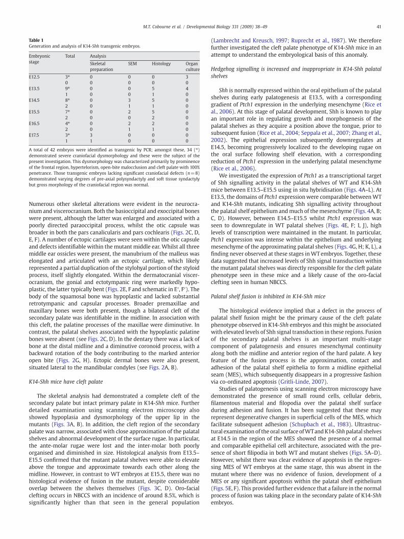
Table 1Generation and analysis of K14-Shh transgenic embryos.
Embryonicstage
Total Analysis
Skeletalpreparation
SEM Histology Organculture
E12.5 3⁎ 0 0 0 30 0 0 0 0
E13.5 9⁎ 0 0 5 41 0 0 1 0
E14.5 8⁎ 0 3 5 02 0 1 1 0
E15.5 7⁎ 0 2 5 02 0 0 2 0
E16.5 4⁎ 0 2 2 02 0 1 1 0
E17.5 3⁎ 3 0 0 01 1 0 0 0
A total of 42 embryos were identified as transgenic by PCR; amongst these, 34 (⁎)demonstrated severe craniofacial dysmorphology and these were the subject of thepresent investigation. This dysmorphology was characterized primarily by prominenceof the frontal region, hypertelorism, open-bite malocclusion and cleft palate with 100%penetrance. Those transgenic embryos lacking significant craniofacial defects (n=8)demonstrated varying degrees of pre-axial polysyndactyly and soft tissue syndactylybut gross morphology of the craniofacial region was normal.
41M.T. Cobourne et al. / Developmental Biology 331 (2009) 38–49
Numerous other skeletal alterations were evident in the neurocra-nium and viscerocranium. Both the basioccipital and exoccipital boneswere present, although the latter was enlarged and associated with apoorly directed paraoccipital process, whilst the otic capsule wasbroader in both the pars canalicularis and pars cochlearis (Figs. 2C, D,E, F). A number of ectopic cartilages were seenwithin the otic capsuleand defects identifiable within the mutant middle ear. Whilst all threemiddle ear ossicles were present, the manubrium of the malleus waselongated and articulated with an ectopic cartilage, which likelyrepresented a partial duplication of the stylohyal portion of the styloidprocess, itself slightly elongated. Within the dermatocranial viscer-ocranium, the gonial and ectotympanic ring were markedly hypo-plastic, the latter typically bent (Figs. 2E, F and schematic in E′, F′). Thebody of the squamosal bone was hypoplastic and lacked substantialretrotympanic and capsular processes. Broader premaxillae andmaxillary bones were both present, though a bilateral cleft of thesecondary palate was identifiable in the midline. In association withthis cleft, the palatine processes of the maxillae were diminutive. Incontrast, the palatal shelves associated with the hypoplastic palatinebones were absent (see Figs. 2C, D). In the dentary there was a lack ofbone at the distal midline and a diminutive coronoid process, with abackward rotation of the body contributing to the marked anterioropen bite (Figs. 2G, H). Ectopic dermal bones were also present,situated lateral to the mandibular condyles (see Figs. 2A, B).
K14-Shh mice have cleft palate
The skeletal analysis had demonstrated a complete cleft of thesecondary palate but intact primary palate in K14-Shh mice. Furtherdetailed examination using scanning electron microscopy alsoshowed hypoplasia and dysmorphology of the upper lip in themutants (Figs. 3A, B). In addition, the cleft region of the secondarypalate was narrow, associated with close approximation of the palatalshelves and abnormal development of the surface rugae. In particular,the ante-molar rugae were lost and the inter-molar both poorlyorganised and diminished in size. Histological analysis from E13.5–E15.5 confirmed that the mutant palatal shelves were able to elevateabove the tongue and approximate towards each other along themidline. However, in contrast to WT embryos at E15.5, there was nohistological evidence of fusion in the mutant, despite considerableoverlap between the shelves themselves (Figs. 3C, D). Oro-facialclefting occurs in NBCCS with an incidence of around 8.5%, which issignificantly higher than that seen in the general population
(Lambrecht and Kreusch, 1997; Ruprecht et al., 1987). We thereforefurther investigated the cleft palate phenotype of K14-Shh mice in anattempt to understand the embryological basis of this anomaly.
Hedgehog signalling is increased and inappropriate in K14-Shh palatalshelves
Shh is normally expressed within the oral epithelium of the palatalshelves during early palatogenesis at E13.5, with a correspondinggradient of Ptch1 expression in the underlying mesenchyme (Rice etal., 2006). At this stage of palatal development, Shh is known to playan important role in regulating growth and morphogenesis of thepalatal shelves as they acquire a position above the tongue, prior tosubsequent fusion (Rice et al., 2004; Seppala et al., 2007; Zhang et al.,2002). The epithelial expression subsequently downregulates atE14.5, becoming progressively localized to the developing rugae onthe oral surface following shelf elevation, with a correspondingreduction of Ptch1 expression in the underlying palatal mesenchyme(Rice et al., 2006).
We investigated the expression of Ptch1 as a transcriptional targetof Shh signalling activity in the palatal shelves of WT and K14-Shhmice between E13.5–E15.5 using in situ hybridisation (Figs. 4A–L). AtE13.5, the domains of Ptch1 expressionwere comparable betweenWTand K14-Shh mutants, indicating Shh signalling activity throughoutthe palatal shelf epithelium andmuch of themesenchyme (Figs. 4A, B;C, D). However, between E14.5–E15.5 whilst Ptch1 expression wasseen to downregulate in WT palatal shelves (Figs. 4E, F; I, J), highlevels of transcription were maintained in the mutant. In particular,Ptch1 expression was intense within the epithelium and underlyingmesenchyme of the approximating palatal shelves (Figs. 4G, H; K, L), afinding never observed at these stages inWTembryos. Together, thesedata suggested that increased levels of Shh signal transduction withinthe mutant palatal shelves was directly responsible for the cleft palatephenotype seen in these mice and a likely cause of the oro-facialclefting seen in human NBCCS.
Palatal shelf fusion is inhibited in K14-Shh mice
The histological evidence implied that a defect in the process ofpalatal shelf fusion might be the primary cause of the cleft palatephenotype observed in K14-Shh embryos and this might be associatedwith elevated levels of Shh signal transduction in these regions. Fusionof the secondary palatal shelves is an important multi-stagecomponent of palatogenesis and ensures mesenchymal continuityalong both the midline and anterior region of the hard palate. A keyfeature of the fusion process is the approximation, contact andadhesion of the palatal shelf epithelia to form a midline epithelialseam (MES), which subsequently disappears in a progressive fashionvia co-ordinated apoptosis (Gritli-Linde, 2007).
Studies of palatogenesis using scanning electron microscopy havedemonstrated the presence of small round cells, cellular debris,filamentous material and filopodia over the palatal shelf surfaceduring adhesion and fusion. It has been suggested that these mayrepresent degenerative changes in superficial cells of the MES, whichfacilitate subsequent adhesion (Schupbach et al., 1983). Ultrastruc-tural examination of the oral surface ofWTandK14-Shh palatal shelvesat E14.5 in the region of the MES showed the presence of a normaland comparable epithelial cell architecture, associated with the pre-sence of short filipodia in both WT and mutant shelves (Figs. 5A–D).However, whilst there was clear evidence of apoptosis in the regres-sing MES of WT embryos at the same stage, this was absent in themutant where there was no evidence of fusion, development of aMES or any significant apoptosis within the palatal shelf epithelium(Figs. 5E, F). This provided further evidence that a failure in the normalprocess of fusion was taking place in the secondary palate of K14-Shhembryos.
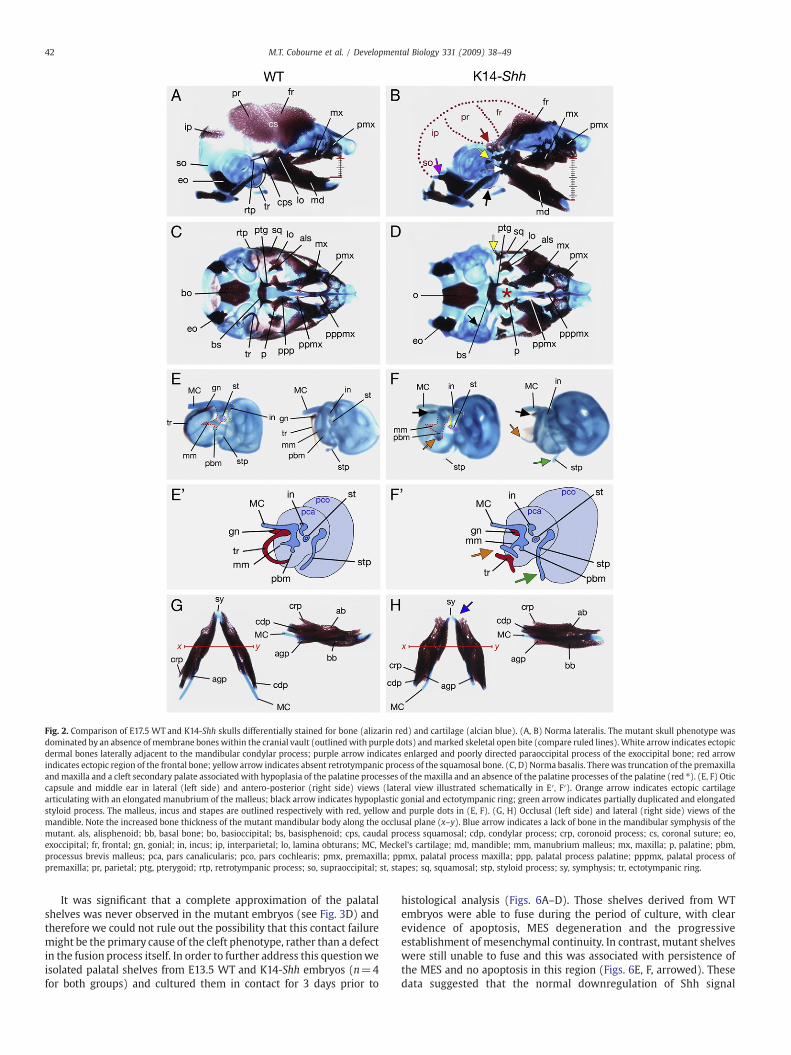
Fig. 2. Comparison of E17.5 WT and K14-Shh skulls differentially stained for bone (alizarin red) and cartilage (alcian blue). (A, B) Norma lateralis. The mutant skull phenotype wasdominated by an absence ofmembrane bones within the cranial vault (outlinedwith purple dots) andmarked skeletal open bite (compare ruled lines).White arrow indicates ectopicdermal bones laterally adjacent to the mandibular condylar process; purple arrow indicates enlarged and poorly directed paraoccipital process of the exoccipital bone; red arrowindicates ectopic region of the frontal bone; yellow arrow indicates absent retrotympanic process of the squamosal bone. (C, D) Norma basalis. Therewas truncation of the premaxillaandmaxilla and a cleft secondary palate associated with hypoplasia of the palatine processes of the maxilla and an absence of the palatine processes of the palatine (red ⁎). (E, F) Oticcapsule and middle ear in lateral (left side) and antero-posterior (right side) views (lateral view illustrated schematically in E′, F′). Orange arrow indicates ectopic cartilagearticulating with an elongated manubrium of the malleus; black arrow indicates hypoplastic gonial and ectotympanic ring; green arrow indicates partially duplicated and elongatedstyloid process. The malleus, incus and stapes are outlined respectively with red, yellow and purple dots in (E, F). (G, H) Occlusal (left side) and lateral (right side) views of themandible. Note the increased bone thickness of the mutant mandibular body along the occlusal plane (x–y). Blue arrow indicates a lack of bone in the mandibular symphysis of themutant. als, alisphenoid; bb, basal bone; bo, basioccipital; bs, basisphenoid; cps, caudal process squamosal; cdp, condylar process; crp, coronoid process; cs, coronal suture; eo,exoccipital; fr, frontal; gn, gonial; in, incus; ip, interparietal; lo, lamina obturans; MC, Meckel's cartilage; md, mandible; mm, manubrium malleus; mx, maxilla; p, palatine; pbm,processus brevis malleus; pca, pars canalicularis; pco, pars cochlearis; pmx, premaxilla; ppmx, palatal process maxilla; ppp, palatal process palatine; pppmx, palatal process ofpremaxilla; pr, parietal; ptg, pterygoid; rtp, retrotympanic process; so, supraoccipital; st, stapes; sq, squamosal; stp, styloid process; sy, symphysis; tr, ectotympanic ring.
42 M.T. Cobourne et al. / Developmental Biology 331 (2009) 38–49
It was significant that a complete approximation of the palatalshelves was never observed in the mutant embryos (see Fig. 3D) andtherefore we could not rule out the possibility that this contact failuremight be the primary cause of the cleft phenotype, rather than a defectin the fusion process itself. In order to further address this questionweisolated palatal shelves from E13.5 WT and K14-Shh embryos (n=4for both groups) and cultured them in contact for 3 days prior to
histological analysis (Figs. 6A–D). Those shelves derived from WTembryos were able to fuse during the period of culture, with clearevidence of apoptosis, MES degeneration and the progressiveestablishment of mesenchymal continuity. In contrast, mutant shelveswere still unable to fuse and this was associated with persistence ofthe MES and no apoptosis in this region (Figs. 6E, F, arrowed). Thesedata suggested that the normal downregulation of Shh signal
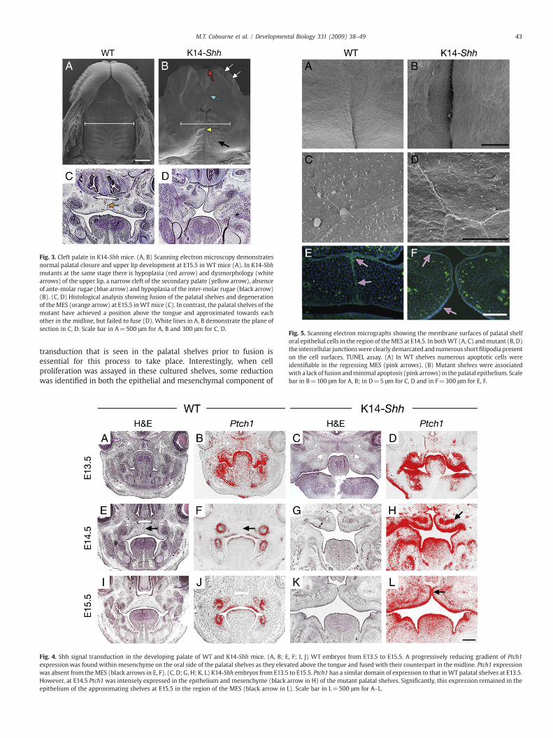
Fig. 3. Cleft palate in K14-Shh mice. (A, B) Scanning electron microscopy demonstratesnormal palatal closure and upper lip development at E15.5 in WT mice (A). In K14-Shhmutants at the same stage there is hypoplasia (red arrow) and dysmorphology (whitearrows) of the upper lip, a narrow cleft of the secondary palate (yellow arrow), absenceof ante-molar rugae (blue arrow) and hypoplasia of the inter-molar rugae (black arrow)(B). (C, D) Histological analysis showing fusion of the palatal shelves and degenerationof theMES (orange arrow) at E15.5 inWTmice (C). In contrast, the palatal shelves of themutant have achieved a position above the tongue and approximated towards eachother in the midline, but failed to fuse (D). White lines in A, B demonstrate the plane ofsection in C, D. Scale bar in A=500 μm for A, B and 300 μm for C, D.
Fig. 5. Scanning electron micrographs showing the membrane surfaces of palatal shelforal epithelial cells in the region of theMES at E14.5. In bothWT (A, C) andmutant (B, D)the intercellular junctionswere clearly demarcated andnumerous shortfilipodia presenton the cell surfaces. TUNEL assay. (A) In WT shelves numerous apoptotic cells wereidentifiable in the regressing MES (pink arrows). (B) Mutant shelves were associatedwith a lack of fusion andminimal apoptosis (pink arrows) in the palatal epithelium. Scalebar in B=100 μm for A, B; in D=5 μm for C, D and in F=300 μm for E, F.
43M.T. Cobourne et al. / Developmental Biology 331 (2009) 38–49
transduction that is seen in the palatal shelves prior to fusion isessential for this process to take place. Interestingly, when cellproliferation was assayed in these cultured shelves, some reductionwas identified in both the epithelial and mesenchymal component of
Fig. 4. Shh signal transduction in the developing palate of WT and K14-Shh mice. (A, B; E, F; I, J) WT embryos from E13.5 to E15.5. A progressively reducing gradient of Ptch1expressionwas found within mesenchyme on the oral side of the palatal shelves as they elevated above the tongue and fused with their counterpart in the midline. Ptch1 expressionwas absent from theMES (black arrows in E, F). (C, D; G, H; K, L) K14-Shh embryos from E13.5 to E15.5. Ptch1 has a similar domain of expression to that inWT palatal shelves at E13.5.However, at E14.5 Ptch1 was intensely expressed in the epithelium and mesenchyme (black arrow in H) of the mutant palatal shelves. Significantly, this expression remained in theepithelium of the approximating shelves at E15.5 in the region of the MES (black arrow in L). Scale bar in L=500 μm for A–L.

Fig. 6. Organ culture and BrdU-labelling of palatal shelves derived from E13.5 WT and K14-Shh embryos. (A, C) WT palatal shelves are able to complete the process of fusion over athree day period of culture. Note regression of the MES (yellow arrow). (B, D) K14-Shh palatal shelves fail to fuse over three days, with persistence of the MES (red arrow). In A and B,the boxes indicate regions of mesenchymal cells counted within each palatal shelf; purple dots indicate the region of epithelial cells counted. TUNEL assay for apoptosis in culturedpalatal shelves. (E) Apoptosis in the regressing MES (white arrows) of fusing WT palatal cultures. (F) In mutant palatal shelves there is no evidence of fusion or apoptosis in themedial edge epithelium (white arrow). (G) Percentage of BrdU incorporation in cultured palatal shelves. Proliferation was reduced in the epithelium and mesenchyme of bothanterior and posterior K14-Shh palatal shelves in comparison to WT; however, this was only significant in the mesenchyme. epi, epithelium; mes, mesenchyme. Data are mean±SD.n=4 mice per group (a total of 32 WT and 28 mutant palatal shelf sections). ⁎Pb0.05 versus WT. Scale bar in D=50 μm for A–D and in F=200 μm for E, F.
44 M.T. Cobourne et al. / Developmental Biology 331 (2009) 38–49
the mutant, although this was only significant in the mesenchyme(Figs. 6G). Thus, the level of Shh signalling from the palatal shelfepitheliumwould also seem to be important in determining the levelsof proliferation within the mesenchymal compartment and may havecontributed to the failure of approximation that occurred in themutant.
Tooth development arrests at the bud stage in K14-Shh embryos
Odontogenic keratocysts represent the predominant anomalyfound in the jaws of NBCCS subjects; however, tooth impactions andhypodontia are also seen with a significantly increased incidencecompared to the general population (Manfredi et al., 2004). Shh isknown to demonstrate a localized and specific expression pattern inthe epithelial compartment of the tooth germ during early odonto-genesis (Bitgood and McMahon, 1995; Cobourne et al., 2004;Hardcastle et al., 1998; Sarkar et al., 2000). In particular, beingexpressed in the early localized thickenings of odontogenic epithe-lium that demarcate the sites of tooth development and later, in theprimary enamel knot; a transitory signalling centre situated withinthe cap stage tooth germ. These domains of expression are responsiblefor signalling both within the epithelial compartment of the toothgerm and from epithelium to mesenchyme; directing epithelialgrowth during the initiation of tooth development, establishment ofthe tooth bud (Cobourne et al., 2001; Hardcastle et al., 1998; Sarkar etal., 2000) and subsequent growth and morphogenesis of the toothgerm during the cap and bell stages of development (Dassule et al.,2000; Gritli-Linde et al., 2002; Jeong et al., 2004).
We examined the odontogenic phenotype of WT and K14-Shhembryos between E13.5 and E16.5. Significantly, in mutant embryosmolar tooth development failed to progress beyond a rudimentarybud stage (Figs. 7A, F; K, P). At E13.5, this was associated with either acomplete absence of odontogenesis or a marked reduction in depth ofthe epithelial buds that were present. In many cases, multiplesuperficial invaginations represented the only evidence of odontogen-esis that were seen, with none demonstrating any of the organisationof the bud stage invaginations observed in WT embryos (Figs. 7A, F).At E14.5, in those mutant tooth germs that had developed, somelimited progression in development was seen; however, the organisa-tion remained poor and none of these mutant molar teeth reached thewell-defined cap stage seen in WT embryos (Figs. 7K, P).
These phenotypic changes were accompanied by a markedalteration in the tempero-spatial distribution of Shh signalling withinthe molar tooth germs. In WT embryos, Shh exhibits restricted andlocalized expression in the epithelial compartment of the tooth germ,whilst the downstreamHedgehog targets Ptch1 and Gli1 are expressedin both the epithelium andmesenchyme (Figs. 7B–D; L–N). In K14-Shhmutants, Shh was intensely expressed throughout the odontogenicepithelium and this was accompanied by upregulation of Ptch1 andGli1 expression in both compartments, but particularly the epithelium(Figs. 7G–I; Q–S). The mutant molar tooth germs consistently failed toform a recognizable tooth bud and progress to the cap stage. Thisphenotype was accompanied by high-level Shh signal transduction inthese teeth and suggested that regulating the level of signalling in theepithelial compartment is important during early growth anddevelopment of the tooth germ. We further investigated this arrestedtooth phenotype by assaying for the presence of cell death in bothWTand mutant molar tooth germs. Shh has previously been associatedwith the prevention of early apoptosis in the developing tooth(Cobourne et al., 2001); however, no apoptotic cells were detectedin the mutant molars at either E13.5 or E14.5. This was in contrastto E14.5 WT cap stage molar tooth germs, where apoptotic cellswere clearly visible in the enamel knot (Figs. 7E, J; O, T) (Vaahtokariet al., 1996).
Analysis of the mutant incisor phenotype demonstrated similarfindings to those found in the molar. In contrast to WT, at E14.5 theearly mutant incisor tooth germs failed to develop into clearlyrecognizable tooth buds. Instead, multiple localized invaginationswere identifiable in the maxillary incisor region, associated with highlevels of Shh throughout the epithelium and both Ptch1 and Gli1expression in epithelium and mesenchyme. No progression of incisortooth development was seen to take place from E14.5 through to E16.5and this was associated with continued high levels of Shh transcrip-tion in these regions, in addition to Ptch1 and Gli1 (Figs. 8A–X).
Arrested odontogenesis is associated with a lack of proliferation in K14-Shh mutant tooth germs
A potential explanation for the failure of normal development inthe mutant tooth germs was a lack of cellular proliferation. Weinvestigated this by culturing molar tooth germs harvested from WTand K14-Shh mandibular processes at E12.5 (n=3) and E13.5 (n=4)

Fig. 7. Molar tooth development in WT and K14-Shh mice. (A–J) At E13.5, the bud stage of development was reached in WT embryos (A) and whilst Shh expression wasdownregulated at the tip of the mature bud at this stage (B), Ptch1 (C) and Gli1 (D) were strongly expressed in both the epithelium and mesenchyme of the tooth germ. In mutantembryos, the teeth failed to reach the bud stage (F), with multiple shallow epithelial invaginations the only evidence of odontogenesis (black arrows). The expression of Shh (G) wasintense in the epithelium of these teeth at this stage; whilst Ptc1 (H) and Gli1 (I) were strongly expressed in both the epithelium and mesenchyme. No apoptotic cells were detectedin theWT bud stage tooth germ (E), although somewere evident in the mutant (J, red arrow). (K–T) At E14.5, the cap stage of development had been reached inWTembryos (K) andwhilst Shh expression was restricted to the enamel knot (L); Ptch1 (M) and Gli1 (N) were strongly expressed in both tissue compartments. Tooth germs failed to reach the cap stagein mutant embryos (P), the odontogenic epithelium remaining poorly organised with Shh (Q), Ptch1 (R) and Gli1 (S) very strongly expressed, particularly in the epithelial tissues. Atthis stage, apoptotic cells were detected in the primary enamel knot of WT tooth germs (O, red arrow) but none in the mutant (T). Scale bar in T=50 μm for A–T.
45M.T. Cobourne et al. / Developmental Biology 331 (2009) 38–49
for 3 days. Over the period of culture, WT tooth germs progressed tothe bud and cap stages of development and this was associated withabundant cellular proliferation, particularly in the epithelial compart-ment (Figs. 9A, B). In contrast, mutant cultures failed to progressbeyond the bud stage, those tooth germs that did form beingabnormally shaped, with the majority only developing into verysuperficial invaginations or rudimentary buds. In all cases, this wasassociated with low levels of proliferating cells in the epithelial andmesenchymal tissues of the mutant teeth (Figs. 9C, D).
Together, these data show a dramatic and significant effect ofincreased Hedgehog signal transduction within basal epithelium ondevelopment of the craniofacial region. In particular, appropriatelyregulated levels of Shh are important for regulating cell fate inepithelium of the developing palate and tooth. The consequences ofupregulated Shh activity form the molecular basis of the oro-facialclefting and hypodontia seen NBCCS subjects.
Discussion
NBCCS occurs with an incidence of around 1:60,000 in Caucasians,with males and females equally affected (Manfredi et al., 2004). Thevast majority of causative mutations reside in the PTCH1 gene andinheritance is autosomal dominant, although as many as half of allcases are thought to represent new mutations (Gorlin, 1995). Mostgermline NBCCS mutations are truncating and concentrated in thelarge extracellular loops, large intracellular loop and the N-terminal
region of PTCH1 (Boutet et al., 2003; Lindstrom et al., 2006; Wickinget al., 1997). However, whilst little correlation exists between themutational type and clinical features of the NBCCS phenotype,haploinsufficiency and increased Hedgehog signal activity are thoughtto provide the basis of this condition (Wicking et al., 1997). Theprinciple clinical features of NBCCS comprise BCC, odontogenickeratocysts of the jaws and skeletal anomalies; all of which canmanifest in the first few decades of life (Manfredi et al., 2004).However, the diagnostic criteria are wide and up to 70% of patientswill have some form of additional craniofacial anomaly. Thecraniofacial features associated with NBCCS can be a useful aid toearly diagnosis, which is important because of the increased risk ofdeveloping juvenile carcinoma, particularly medulloblastoma. How-ever, the aetiological basis of these developmental defects hasreceived little attention. We therefore sought to further investigatethe molecular mechanisms underlying these phenotypes using amouse model expressing Shh in basal epithelium of the early face andjaws.
Hedgehog signalling is important for early craniofacial develop-ment. Targeted disruption of Shh in mice and mutations in humansubjects can lead to holoprosencephaly [MIM #236100], which ischaracterized by a variable spectrum of abnormalities in the fore-brain and face (Belloni et al., 1996; Chiang et al., 1996; Roessler et al.,1996). Alterations in signalling from epithelium of the early facialprocesses can also affect normal development. A transient loss of Shhfunction in the frontonasal process compromises cellular survival and
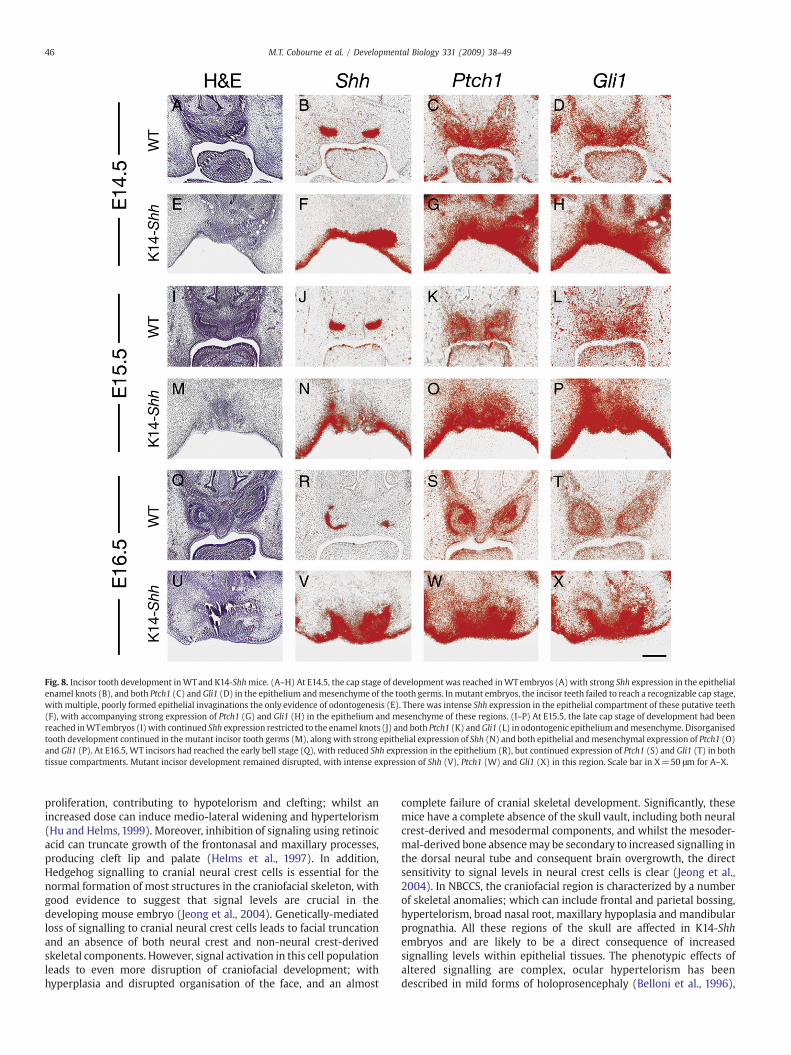
Fig. 8. Incisor tooth development inWTand K14-Shhmice. (A–H) At E14.5, the cap stage of development was reached inWTembryos (A) with strong Shh expression in the epithelialenamel knots (B), and both Ptch1 (C) and Gli1 (D) in the epithelium andmesenchyme of the tooth germs. Inmutant embryos, the incisor teeth failed to reach a recognizable cap stage,with multiple, poorly formed epithelial invaginations the only evidence of odontogenesis (E). There was intense Shh expression in the epithelial compartment of these putative teeth(F), with accompanying strong expression of Ptch1 (G) and Gli1 (H) in the epithelium and mesenchyme of these regions. (I–P) At E15.5, the late cap stage of development had beenreached inWTembryos (I) with continued Shh expression restricted to the enamel knots (J) and both Ptch1 (K) andGli1 (L) in odontogenic epithelium andmesenchyme. Disorganisedtooth development continued in themutant incisor tooth germs (M), along with strong epithelial expression of Shh (N) and both epithelial andmesenchymal expression of Ptch1 (O)and Gli1 (P). At E16.5, WT incisors had reached the early bell stage (Q), with reduced Shh expression in the epithelium (R), but continued expression of Ptch1 (S) and Gli1 (T) in bothtissue compartments. Mutant incisor development remained disrupted, with intense expression of Shh (V), Ptch1 (W) and Gli1 (X) in this region. Scale bar in X=50 μm for A–X.
46 M.T. Cobourne et al. / Developmental Biology 331 (2009) 38–49
proliferation, contributing to hypotelorism and clefting; whilst anincreased dose can induce medio-lateral widening and hypertelorism(Hu and Helms, 1999). Moreover, inhibition of signaling using retinoicacid can truncate growth of the frontonasal and maxillary processes,producing cleft lip and palate (Helms et al., 1997). In addition,Hedgehog signalling to cranial neural crest cells is essential for thenormal formation of most structures in the craniofacial skeleton, withgood evidence to suggest that signal levels are crucial in thedeveloping mouse embryo (Jeong et al., 2004). Genetically-mediatedloss of signalling to cranial neural crest cells leads to facial truncationand an absence of both neural crest and non-neural crest-derivedskeletal components. However, signal activation in this cell populationleads to even more disruption of craniofacial development; withhyperplasia and disrupted organisation of the face, and an almost
complete failure of cranial skeletal development. Significantly, thesemice have a complete absence of the skull vault, including both neuralcrest-derived and mesodermal components, and whilst the mesoder-mal-derived bone absencemay be secondary to increased signalling inthe dorsal neural tube and consequent brain overgrowth, the directsensitivity to signal levels in neural crest cells is clear (Jeong et al.,2004). In NBCCS, the craniofacial region is characterized by a numberof skeletal anomalies; which can include frontal and parietal bossing,hypertelorism, broad nasal root, maxillary hypoplasia and mandibularprognathia. All these regions of the skull are affected in K14-Shhembryos and are likely to be a direct consequence of increasedsignalling levels within epithelial tissues. The phenotypic effects ofaltered signalling are complex, ocular hypertelorism has beendescribed in mild forms of holoprosencephaly (Belloni et al., 1996),
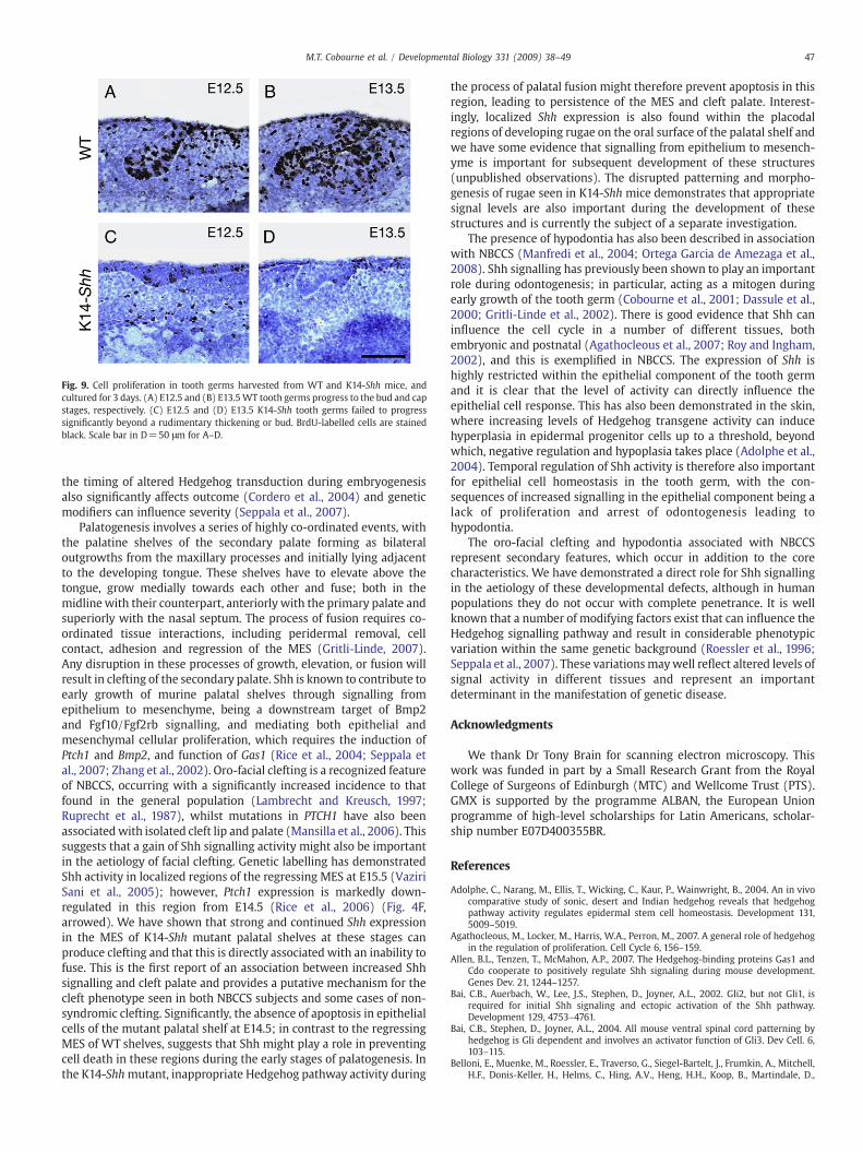
Fig. 9. Cell proliferation in tooth germs harvested from WT and K14-Shh mice, andcultured for 3 days. (A) E12.5 and (B) E13.5WT tooth germs progress to the bud and capstages, respectively. (C) E12.5 and (D) E13.5 K14-Shh tooth germs failed to progresssignificantly beyond a rudimentary thickening or bud. BrdU-labelled cells are stainedblack. Scale bar in D=50 μm for A–D.
47M.T. Cobourne et al. / Developmental Biology 331 (2009) 38–49
the timing of altered Hedgehog transduction during embryogenesisalso significantly affects outcome (Cordero et al., 2004) and geneticmodifiers can influence severity (Seppala et al., 2007).
Palatogenesis involves a series of highly co-ordinated events, withthe palatine shelves of the secondary palate forming as bilateraloutgrowths from the maxillary processes and initially lying adjacentto the developing tongue. These shelves have to elevate above thetongue, grow medially towards each other and fuse; both in themidlinewith their counterpart, anteriorly with the primary palate andsuperiorly with the nasal septum. The process of fusion requires co-ordinated tissue interactions, including peridermal removal, cellcontact, adhesion and regression of the MES (Gritli-Linde, 2007).Any disruption in these processes of growth, elevation, or fusion willresult in clefting of the secondary palate. Shh is known to contribute toearly growth of murine palatal shelves through signalling fromepithelium to mesenchyme, being a downstream target of Bmp2and Fgf10/Fgf2rb signalling, and mediating both epithelial andmesenchymal cellular proliferation, which requires the induction ofPtch1 and Bmp2, and function of Gas1 (Rice et al., 2004; Seppala etal., 2007; Zhang et al., 2002). Oro-facial clefting is a recognized featureof NBCCS, occurring with a significantly increased incidence to thatfound in the general population (Lambrecht and Kreusch, 1997;Ruprecht et al., 1987), whilst mutations in PTCH1 have also beenassociatedwith isolated cleft lip and palate (Mansilla et al., 2006). Thissuggests that a gain of Shh signalling activity might also be importantin the aetiology of facial clefting. Genetic labelling has demonstratedShh activity in localized regions of the regressing MES at E15.5 (VaziriSani et al., 2005); however, Ptch1 expression is markedly down-regulated in this region from E14.5 (Rice et al., 2006) (Fig. 4F,arrowed). We have shown that strong and continued Shh expressionin the MES of K14-Shh mutant palatal shelves at these stages canproduce clefting and that this is directly associated with an inability tofuse. This is the first report of an association between increased Shhsignalling and cleft palate and provides a putative mechanism for thecleft phenotype seen in both NBCCS subjects and some cases of non-syndromic clefting. Significantly, the absence of apoptosis in epithelialcells of the mutant palatal shelf at E14.5; in contrast to the regressingMES of WT shelves, suggests that Shh might play a role in preventingcell death in these regions during the early stages of palatogenesis. Inthe K14-Shhmutant, inappropriate Hedgehog pathway activity during
the process of palatal fusion might therefore prevent apoptosis in thisregion, leading to persistence of the MES and cleft palate. Interest-ingly, localized Shh expression is also found within the placodalregions of developing rugae on the oral surface of the palatal shelf andwe have some evidence that signalling from epithelium to mesench-yme is important for subsequent development of these structures(unpublished observations). The disrupted patterning and morpho-genesis of rugae seen in K14-Shh mice demonstrates that appropriatesignal levels are also important during the development of thesestructures and is currently the subject of a separate investigation.
The presence of hypodontia has also been described in associationwith NBCCS (Manfredi et al., 2004; Ortega Garcia de Amezaga et al.,2008). Shh signalling has previously been shown to play an importantrole during odontogenesis; in particular, acting as a mitogen duringearly growth of the tooth germ (Cobourne et al., 2001; Dassule et al.,2000; Gritli-Linde et al., 2002). There is good evidence that Shh caninfluence the cell cycle in a number of different tissues, bothembryonic and postnatal (Agathocleous et al., 2007; Roy and Ingham,2002), and this is exemplified in NBCCS. The expression of Shh ishighly restricted within the epithelial component of the tooth germand it is clear that the level of activity can directly influence theepithelial cell response. This has also been demonstrated in the skin,where increasing levels of Hedgehog transgene activity can inducehyperplasia in epidermal progenitor cells up to a threshold, beyondwhich, negative regulation and hypoplasia takes place (Adolphe et al.,2004). Temporal regulation of Shh activity is therefore also importantfor epithelial cell homeostasis in the tooth germ, with the con-sequences of increased signalling in the epithelial component being alack of proliferation and arrest of odontogenesis leading tohypodontia.
The oro-facial clefting and hypodontia associated with NBCCSrepresent secondary features, which occur in addition to the corecharacteristics. We have demonstrated a direct role for Shh signallingin the aetiology of these developmental defects, although in humanpopulations they do not occur with complete penetrance. It is wellknown that a number of modifying factors exist that can influence theHedgehog signalling pathway and result in considerable phenotypicvariation within the same genetic background (Roessler et al., 1996;Seppala et al., 2007). These variationsmaywell reflect altered levels ofsignal activity in different tissues and represent an importantdeterminant in the manifestation of genetic disease.
Acknowledgments
We thank Dr Tony Brain for scanning electron microscopy. Thiswork was funded in part by a Small Research Grant from the RoyalCollege of Surgeons of Edinburgh (MTC) and Wellcome Trust (PTS).GMX is supported by the programme ALBAN, the European Unionprogramme of high-level scholarships for Latin Americans, scholar-ship number E07D400355BR.
References
Adolphe, C., Narang, M., Ellis, T., Wicking, C., Kaur, P., Wainwright, B., 2004. An in vivocomparative study of sonic, desert and Indian hedgehog reveals that hedgehogpathway activity regulates epidermal stem cell homeostasis. Development 131,5009–5019.
Agathocleous, M., Locker, M., Harris, W.A., Perron, M., 2007. A general role of hedgehogin the regulation of proliferation. Cell Cycle 6, 156–159.
Allen, B.L., Tenzen, T., McMahon, A.P., 2007. The Hedgehog-binding proteins Gas1 andCdo cooperate to positively regulate Shh signaling during mouse development.Genes Dev. 21, 1244–1257.
Bai, C.B., Auerbach, W., Lee, J.S., Stephen, D., Joyner, A.L., 2002. Gli2, but not Gli1, isrequired for initial Shh signaling and ectopic activation of the Shh pathway.Development 129, 4753–4761.
Bai, C.B., Stephen, D., Joyner, A.L., 2004. All mouse ventral spinal cord patterning byhedgehog is Gli dependent and involves an activator function of Gli3. Dev Cell. 6,103–115.
Belloni, E., Muenke, M., Roessler, E., Traverso, G., Siegel-Bartelt, J., Frumkin, A., Mitchell,H.F., Donis-Keller, H., Helms, C., Hing, A.V., Heng, H.H., Koop, B., Martindale, D.,

48 M.T. Cobourne et al. / Developmental Biology 331 (2009) 38–49
Rommens, J.M., Tsui, L.C., Scherer, S.W., 1996. Identification of Sonic hedgehog as acandidate gene responsible for holoprosencephaly. Nat. Genet. 14, 353–356.
Bitgood, M.J., McMahon, A.P., 1995. Hedgehog and Bmp genes are coexpressed atmany diverse sites of cell–cell interaction in the mouse embryo. Dev. Biol. 172,126–138.
Boutet, N., Bignon, Y.J., Drouin-Garraud, V., Sarda, P., Longy, M., Lacombe, D., Gorry, P.,2003. Spectrum of PTCH1 mutations in French patients with Gorlin syndrome. J.Invest. Dermatol. 121, 478–481.
Brunet, C.L., Sharpe, P.M., Ferguson, M.W., 1993. The distribution of epidermalgrowth factor binding sites in the developing mouse palate. Int. J. Dev. Biol. 37,451–458.
Carstea, E.D., Morris, J.A., Coleman, K.G., Loftus, S.K., Zhang, D., Cummings, C., Gu, J.,Rosenfeld, M.A., Pavan, W.J., Krizman, D.B., Nagle, J., Polymeropoulos, M.H., Sturley,S.L., Ioannou, Y.A., Higgins, M.E., Comly, M., Cooney, A., Brown, A., Kaneski, C.R.,Blanchette-Mackie, E.J., Dwyer, N.K., Neufeld, E.B., Chang, T.Y., Liscum, L., Strauss III,J.F., Ohno, K., Zeigler, M., Carmi, R., Sokol, J., Markie, D., O'Neill, R.R., van Diggelen,O.P., Elleder, M., Patterson, M.C., Brady, R.O., Vanier, M.T., Pentchev, P.G., Tagle, D.A.,1997. Niemann–Pick C1 disease gene: homology to mediators of cholesterolhomeostasis. Science 277, 228–231.
Casali, A., Struhl, G., 2004. Reading the Hedgehog morphogen gradient by measuringthe ratio of bound to unbound Patched protein. Nature 431, 76–80.
Chen, Y., Struhl, G., 1996. Dual roles for patched in sequestering and transducingHedgehog. Cell 87, 553–563.
Chiang, C., Litingtung, Y., Lee, E., Young, K.E., Corden, J.L., Westphal, H., Beachy, P.A., 1996.Cyclopia and defective axial patterning in mice lacking Sonic hedgehog genefunction. Nature 383, 407–413.
Chuang, P.T., McMahon, A.P., 1999. Vertebrate Hedgehog signalling modulated byinduction of a Hedgehog-binding protein. Nature 397, 617–621.
Cobourne, M.T., Hardcastle, Z., Sharpe, P.T., 2001. Sonic hedgehog regulates epithelialproliferation and cell survival in the developing tooth germ. J. Dent. Res. 80,1974–1979.
Cobourne, M.T., Miletich, I., Sharpe, P.T., 2004. Restriction of sonic hedgehog signallingduring early tooth development. Development 131, 2875–2885.
Corbit, K.C., Aanstad, P., Singla, V., Norman, A.R., Stainier, D.Y., Reiter, J.F., 2005.Vertebrate Smoothened functions at the primary cilium. Nature 437, 1018–1021.
Cordero, D., Marcucio, R., Hu, D., Gaffield, W., Tapadia, M., Helms, J.A., 2004. Temporalperturbations in sonic hedgehog signaling elicit the spectrum of holoprosencephalyphenotypes. J. Clin. Invest. 114, 485–494.
Dassule, H.R., Lewis, P., Bei, M., Maas, R., McMahon, A.P., 2000. Sonic hedgehog regulatesgrowth and morphogenesis of the tooth. Development 127, 4775–4785.
Davies, J.P., Chen, F.W., Ioannou, Y.A., 2000. Transmembrane molecular pump activity ofNiemann–Pick C1 protein. Science 290, 2295–2298.
Dessaud, E., Yang, L.L., Hill, K., Cox, B., Ulloa, F., Ribeiro, A., Mynett, A., Novitch, B.G.,Briscoe, J., 2007. Interpretation of the sonic hedgehog morphogen gradient by atemporal adaptation mechanism. Nature 450, 717–720.
Evans, D.G., Ladusans, E.J., Rimmer, S., Burnell, L.D., Thakker, N., Farndon, P.A., 1993.Complications of the naevoid basal cell carcinoma syndrome: results of apopulation based study. J. Med. Genet. 30, 460–464.
Goodrich, L.V., Johnson, R.L., Milenkovic, L., McMahon, J.A., Scott, M.P., 1996.Conservation of the hedgehog/patched signaling pathway from flies to mice:induction of a mouse patched gene by Hedgehog. Genes Dev. 10, 301–312.
Goodrich, L.V., Milenkovic, L., Higgins, K.M., Scott, M.P., 1997. Altered neural cell fatesand medulloblastoma in mouse patched mutants. Science 277, 1109–1113.
Gorlin, R.J., 1995. Nevoid basal cell carcinoma syndrome. Dermatol. Clin. 13, 113–125.Gorlin, R.J., Goltz, R.W., 1960. Multiple nevoid basal-cell epithelioma, jaw cysts and bifid
rib. A syndrome. N. Engl. J. Med. 262, 908–912.Gritli-Linde, A., 2007. Molecular control of secondary palate development. Dev. Biol.
301, 309–326.Gritli-Linde, A., Bei, M., Maas, R., Zhang, X.M., Linde, A., McMahon, A.P., 2002. Shh
signalling within the dental epithelium is necessary for cell proliferation, growthand polarization. Development 129, 5323–5337.
Hahn, H., Wicking, C., Zaphiropoulous, P.G., Gailani, M.R., Shanley, S., Chidambaram, A.,Vorechovsky, I., Holmberg, E., Unden, A.B., Gillies, S., Negus, K., Smyth, I., Pressman,C., Leffell, D.J., Gerrard, B., Goldstein, A.M., Dean, M., Toftgard, R., Chenevix-Trench,G., Wainwright, B., Bale, A.E., 1996. Mutations of the human homolog of Drosophilapatched in the nevoid basal cell carcinoma syndrome. Cell 85, 841–851.
Hardcastle, Z., Mo, R., Hui, C.C., Sharpe, P.T., 1998. The Shh signalling pathway in toothdevelopment: defects in Gli2 and Gli3 mutants. Development 125, 2803–2811.
Haycraft, C.J., Banizs, B., Aydin-Son, Y., Zhang, Q., Michaud, E.J., Yoder, B.K., 2005. Gli2and Gli3 localize to cilia and require the intraflagellar transport protein polaris forprocessing and function. PLoS Genet. 1, e53.
Helms, J.A., Kim, C.H., Hu, D., Minkoff, R., Thaller, C., Eichele, G., 1997. Sonic hedgehogparticipates in craniofacial morphogenesis and is down-regulated by teratogenicdoses of retinoic acid. Dev. Biol. 187, 25–35.
Hu, D., Helms, J.A., 1999. The role of sonic hedgehog in normal and abnormalcraniofacial morphogenesis. Development 126, 4873–4884.
Huangfu, D., Anderson, K.V., 2005. Cilia and Hedgehog responsiveness in the mouse.Proc. Natl. Acad. Sci. U. S. A. 102, 11325–11330.
Huangfu, D., Liu, A., Rakeman, A.S., Murcia, N.S., Niswander, L., Anderson, K.V., 2003.Hedgehog signalling in the mouse requires intraflagellar transport proteins. Nature426, 83–87.
Ingham, P.W., McMahon, A.P., 2001. Hedgehog signaling in animal development:paradigms and principles. Genes Dev. 15, 3059–3087.
Jeong, J., Mao, J., Tenzen, T., Kottmann, A.H., McMahon, A.P., 2004. Hedgehog signaling inthe neural crest cells regulates the patterning and growth of facial primordia. GenesDev. 18, 937–951.
Johnson, R.L., Rothman, A.L., Xie, J., Goodrich, L.V., Bare, J.W., Bonifas, J.M., Quinn, A.G.,Myers, R.M., Cox, D.R., Epstein Jr., E.H., Scott, M.P., 1996. Human homolog ofpatched, a candidate gene for the basal cell nevus syndrome. Science 272,1668–1671.
Kimonis, V.E., Goldstein, A.M., Pastakia, B., Yang, M.L., Kase, R., DiGiovanna, J.J., Bale, A.E.,Bale, S.J., 1997. Clinical manifestations in 105 persons with nevoid basal cellcarcinoma syndrome. Am. J. Med. Genet. 69, 299–308.
Lambrecht, J.T., Kreusch, T., 1997. Examine your orofacial cleft patients for Gorlin–Goltzsyndrome. Cleft Palate-Craniofac. J. 34, 342–350.
Lindstrom, E., Shimokawa, T., Toftgard, R., Zaphiropoulos, P.G., 2006. PTCH mutations:distribution and analyses. Hum. Mutat. 27, 215–219.
Liu, A., Wang, B., Niswander, L.A., 2005. Mouse intraflagellar transport proteins regulateboth the activator and repressor functions of Gli transcription factors. Development132, 3103–3111.
Loftus, S.K., Morris, J.A., Carstea, E.D., Gu, J.Z., Cummings, C., Brown, A., Ellison, J., Ohno,K., Rosenfeld, M.A., Tagle, D.A., Pentchev, P.G., Pavan, W.J., 1997. Murine model ofNiemann–Pick C disease: mutation in a cholesterol homeostasis gene. Science 277,232–235.
Manfredi, M., Vescovi, P., Bonanini, M., Porter, S., 2004. Nevoid basal cell carcinomasyndrome: a review of the literature. Int. J. Oral Maxillofac. Surg. 33, 117–124.
Mansilla, M.A., Cooper, M.E., Goldstein, T., Castilla, E.E., Lopez Camelo, J.S., Marazita, M.L.,Murray, J.C., 2006. Contributions of PTCH gene variants to isolated cleft lip andpalate. Cleft Palate-Craniofac. J. 43, 21–29.
Martin, V., Carrillo, G., Torroja, C., Guerrero, I., 2001. The sterol-sensing domain ofPatched protein seems to control Smoothened activity through Patched vesiculartrafficking. Curr. Biol. 11, 601–607.
Martinelli, D.C., Fan, C.M., 2007. Gas1 extends the range of Hedgehog action byfacilitating its signaling. Genes Dev. 21, 1231–1243.
May, S.R., Ashique, A.M., Karlen, M., Wang, B., Shen, Y., Zarbalis, K., Reiter, J., Ericson, J.,Peterson, A.S., 2005. Loss of the retrograde motor for IFT disrupts localization ofSmo to cilia and prevents the expression of both activator and repressor functionsof Gli. Dev. Biol. 287, 378–389.
McMahon, A.P., Ingham, P., Tabin, C., 2003. Developmental roles and clinical significanceof hedgehog signalling. Curr. Top. Dev. Biol. 53, 1–114.
Mo, R., Freer, A.M., Zinyk, D.L., Crackower, M.A., Michaud, J., Heng, H.H., Chik, K.W., Shi,X.M., Tsui, L.C., Cheng, S.H., Joyner, A.L., Hui, C., 1997. Specific and redundantfunctions of Gli2 and Gli3 zinc finger genes in skeletal patterning and development.Development 124, 113–123.
Oro, A.E., Higgins, K.M., Hu, Z., Bonifas, J.M., Epstein Jr., E.H., Scott, M.P., 1997.Basal cell carcinomas in mice overexpressing sonic hedgehog. Science 276,817–821.
Ortega Garcia de Amezaga, A., Garcia Arregui, O., Zepeda Nuno, S., Acha Sagredo, A.,Aguirre Urizar, J.M., 2008. Gorlin–Goltz syndrome: clinicopathologic aspects. Med.Oral Patol. Oral Cir. Bucal. 13, E338–E343.
Pan, Y., Bai, C.B., Joyner, A.L., Wang, B., 2006. Sonic hedgehog signaling regulates Gli2transcriptional activity by suppressing its processing and degradation. Mol. CellBiol. 26, 3365–3377.
Park, H.L., Bai, C., Platt, K.A., Matise, M.P., Beeghly, A., Hui, C.C., Nakashima, M., Joyner,A.L., 2000. Mouse Gli1 mutants are viable but have defects in SHH signaling incombination with a Gli2 mutation. Development 127, 1593–1605.
Rice, R., Spencer-Dene, B., Connor, E.C., Gritli-Linde, A., McMahon, A.P., Dickson, C.,Thesleff, I., Rice, D.P., 2004. Disruption of Fgf10/Fgfr2b-coordinated epithelial–mesenchymal interactions causes cleft palate. J. Clin. Invest. 113, 1692–1700.
Rice, R., Connor, E., Rice, D.P., 2006. Expression patterns of Hedgehog signallingpathway members during mouse palate development. Gene Expr. Patterns 6,206–212.
Roessler, E., Belloni, E., Gaudenz, K., Jay, P., Berta, P., Scherer, S.W., Tsui, L.C., Muenke, M.,1996. Mutations in the human Sonic Hedgehog gene cause holoprosencephaly. Nat.Genet. 14, 357–360.
Rohatgi, R., Milenkovic, L., Scott, M.P., 2007. Patched1 regulates hedgehog signaling atthe primary cilium. Science 317, 372–376.
Rossi, A., Caracciolo, V., Russo, G., Reiss, K., Giordano, A., 2008. Medulloblastoma: frommolecular pathology to therapy. Clin. Cancer Res. 14, 971–976.
Roy, S., Ingham, P.W., 2002. Hedgehogs tryst with the cell cycle. J. Cell Sci. 115,4393–4397.
Ruprecht, A., Austermann, K.H., Umstadt, H., 1987. Cleft lip and palate, seldom seenfeatures of the Gorlin–Goltz syndrome. Dentomaxillofac. Radiol. 16, 99–103.
Sarkar, L., Cobourne, M., Naylor, S., Smalley, M., Dale, T., Sharpe, P.T., 2000. Wnt/Shhinteractions regulate ectodermal boundary formation during mammalian toothdevelopment. Proc. Natl. Acad. Sci. U. S. A. 97, 4520–4524.
Schupbach, P.M., Chamberlain, J.G., Schroeder, H.E., 1983. Development of the secondarypalate in the rat: a scanning electron microscopic study. J. Craniofac. Genet. Dev.Biol. 3, 159–177.
Seppala, M., Depew, M.J., Martinelli, D.C., Fan, C.M., Sharpe, P.T., Cobourne, M.T., 2007.Gas1 is a modifier for holoprosencephaly and genetically interacts with sonichedgehog. J. Clin. Invest. 117, 1575–1584.
Shanley, S., Ratcliffe, J., Hockey, A., Haan, E., Oley, C., Ravine, D., Martin, N., Wicking, C.,Chenevix-Trench, G., 1994. Nevoid basal cell carcinoma syndrome: review of 118affected individuals. Am. J. Med. Genet. 50, 282–290.
Stamataki, D., Ulloa, F., Tsoni, S.V., Mynett, A., Briscoe, J., 2005. A gradient of Gli activitymediates graded Sonic Hedgehog signaling in the neural tube. Genes Dev. 19,626–641.
Stone, D.M., Hynes, M., Armanini, M., Swanson, T.A., Gu, Q., Johnson, R.L., Scott, M.P.,Pennica, D., Goddard, A., Phillips, H., Noll, M., Hooper, J.E., de Sauvage, F., Rosenthal,A., 1996. The tumour-suppressor gene patched encodes a candidate receptor forSonic hedgehog. Nature 384, 129–134.

49M.T. Cobourne et al. / Developmental Biology 331 (2009) 38–49
Strutt, H., Thomas, C., Nakano, Y., Stark, D., Neave, B., Taylor, A.M., Ingham, P.W., 2001.Mutations in the sterol-sensing domain of Patched suggest a role for vesiculartrafficking in Smoothened regulation. Curr. Biol. 11, 608–613.
Taipale, J., Cooper, M.K., Maiti, T., Beachy, P.A., 2002. Patched acts catalytically tosuppress the activity of Smoothened. Nature 418, 892–897.
Tempe, D., Casas, M., Karaz, S., Blanchet-Tournier, M.F., Concordet, J.P., 2006. Multisiteprotein kinase A and glycogen synthase kinase 3beta phosphorylation leads to Gli3ubiquitination by SCFbetaTrCP. Mol. Cell. Biol. 26, 4316–4326.
Tenzen, T., Allen, B.L., Cole, F., Kang, J.S., Krauss, R.S., McMahon, A.P., 2006. The cellsurface membrane proteins Cdo and Boc are components and targets of theHedgehog signaling pathway and feedback network in mice. Dev. Cell 10, 647–656.
Vaahtokari, A., Aberg, T., Thesleff, I., 1996. Apoptosis in the developing tooth: associationwith an embryonic signaling center and suppression by EGF and FGF-4.Development 122, 121–129.
Vassar, R., Rosenberg, M., Ross, S., Tyner, A., Fuchs, E., 1989. Tissue-specific anddifferentiation-specific expression of a human K14 keratin gene in transgenic mice.Proc. Natl. Acad. Sci. U. S. A. 86, 1563–1567.
Vaziri Sani, F., Hallberg, K., Harfe, B.D., McMahon, A.P., Linde, A., Gritli-Linde, A., 2005.Fate-mapping of the epithelial seam during palatal fusion rules out epithelial–mesenchymal transformation. Dev. Biol. 285, 490–495.
Wang, B., Li, Y., 2006. Evidence for the direct involvement of {beta}TrCP in Gli3 proteinprocessing. Proc. Natl. Acad. Sci. U. S. A. 103, 33–38.
Wicking, C., Shanley, S., Smyth, I., Gillies, S., Negus, K., Graham, S., Suthers, G., Haites, N.,Edwards, M., Wainwright, B., Chenevix-Trench, G., 1997. Most germ-line mutationsin the nevoid basal cell carcinoma syndrome lead to a premature termination of thePATCHED protein, and no genotype–phenotype correlations are evident. Am. J.Hum. Genet. 60, 21–26.
Zhang, X.M., Ramalho-Santos, M., McMahon, A.P., 2001. Smoothened mutants revealredundant roles for Shh and Ihh signaling including regulation of L/R asymmetry bythe mouse node. Cell 105, 781–792.
Zhang, Z., Song, Y., Zhao, X., Zhang, X., Fermin, C., Chen, Y., 2002. Rescue of cleft palate inMsx1-deficient mice by transgenic Bmp4 reveals a network of BMP and Shhsignaling in the regulation of mammalian palatogenesis. Development 129,4135–4146.


![Regulation of Hedgehog Signalling Inside and Outside the Celleprints.whiterose.ac.uk/119986/1/jdb_04_00023.pdfJ. Dev. Biol. 2016, 4, 23 2 of 20 elsewhere [11,12]. This review will](https://img.pdfslide.us/doc/110x75/6128f9d9bafd3e72b224dcda/regulation-of-hedgehog-signalling-inside-and-outside-the-j-dev-biol-2016-4.jpg)