Embed Size (px)
Citation preview
2221RESEARCH REPORT
INTRODUCTIONThe nasal capsule is dorsoventrally divided into two parts: the upper
part, the ectethmoid, serves olfaction and is composed of the lamina
cribosa, the crista galli apophysis and the conchae. The lower part,
the mesethmoid, is a thick cartilage bar extending from the corpus
sphenoidalis to the rostral extremity of the nose (Fig. 1A-B�). In the
avian embryo, the mesethmoid constitutes the cartilage primordium
of the upper beak.
Lineage experiments have shown that Hox-negative cephalic
neural crest cells (CNCCs) emigrating from the prosencephalic and
the anterior mesencephalic neural folds give rise to the nasal capsule
and to first pharyngeal arch (PA) structures (Couly et al., 1993;
Creuzet et al., 2002; Kontges and Lumsden, 1996; Noden, 1992;
Trainor and Tam, 1995). By contrast, Hox-positive NCCs, which are
only found posteriorly to rhombomere 2, do not contribute to the
facial skeleton, but generate more-posterior structures of the embryo
(Couly et al., 1996; Kontges and Lumsden, 1996; Santagati and
Rijli, 2003). Premigratory, Hox-negative CNCCs behave as an
equivalence group and lack the topographic information needed to
give rise to the different structures of the craniofacial skeleton
(Couly et al., 2002). We have shown by surgical deletion and
grafting of different parts of the foregut endoderm that this
epithelium harbours the instructive signals (Couly et al., 2002;
Noden, 1992; Ruhin et al., 2003), although their molecular nature
remains to be determined. Among candidate cues, sonic hedgehog
(Shh) is expressed in the most anterior part of the endoderm,
endoderm zone I (EZ-I), of the early chick embryo (Brito et al.,
2006). Later, incoming CNCCs express the Shh receptor patched 1,
indicating a potentially active Shh signalling (Jeong et al., 2004).
The importance of Shh signalling in the control of different aspects
of craniofacial development has been demonstrated in several
models including zebrafish (Wada et al., 2005), mouse (Chiang et
al., 1996; Jeong et al., 2004) and chicken (Brito et al., 2006; Cordero
et al., 2004; Helms et al., 1997; Hu et al., 2003). Furthermore, human
syndromes with nasal malformations, such as foetal alcohol
syndrome, Smith-Lemli-Opitz syndrome and holoprosencephaly,
have been associated with defective SHH signalling (Herman, 2003;
Traiffort et al., 2004; Yamada et al., 2005).
MATERIALS AND METHODSAvian embryosFertilised eggs were obtained from Morizeau Farms, France (chicken,
Gallus gallus) or Cailles de Chanteloup Farms, France (quail, Coturnixcoturnix japonica) and incubated at 38°C in a humidified atmosphere for
approximately 32 hours to reach the 5-somite stage HH8+ (Hamburger and
Hamilton, 1992; Teillet et al., 1998). Embryos were collected and dissected
at room temperature in phosphate-buffered saline (PBS).
Embryo processingImmunoperoxidase detection was performed as previously described (Couly
et al., 2002). Quail nuclei were detected with the quail-specific monoclonal
antibody QCPN at 1/500 (obtained from the Developmental Studies
Hybridoma Bank developed under the auspices of the NICHD and
maintained by The University of Iowa, Department of Biological Sciences,
Iowa City, IA 52242). Chick and quail Gli1 proteins were detected using
rabbit polyclonal antibody #2553S (Cell Signaling Technologies) at 1/200.
Shh transcripts were detected by in situ hybridisation as previously described
(Couly et al., 2002). Whole-mount skeletons were visualised according to
standard staining protocols using Alcian Blue for cartilage and Alizarin Red
for bone (Couly et al., 2002).
Ablation of EZ-IExperiments were carried out in ovo on windowed chick embryos at the 5-
somite stage HH8+ (Couly et al., 2002). Two bilateral excisions were first
performed on the superficial ectoderm on each side of the neural tube at the
level of the prosencephalon and down to the mesencephalon (see Fig. S1 in
the supplementary material). A very fine curved tungsten microknife was
then passed under the neural tube and the notochord, resulting in their
separation from the dorsal foregut endoderm. A transversal incision was then
performed on the neural plate at the level of the posterior mesencephalon.
The neural epithelium was reclined rostrally in order to gain access to the
endoderm. The ventral endoderm was separated from the ventral ectoderm
by passing a very fine curved tungsten microknife between the two tissues.
The rostral-most attachment between the ventral endoderm and the anterior
neural fold was then cut. At this point, the ventrolateral endoderm was free
of any attachment and we could then easily remove the EZ-I by performing
Sonic hedgehog signalling from foregut endoderm patternsthe avian nasal capsuleLaurence Benouaiche1,2,*, Yorick Gitton1,*, Christine Vincent3, Gérard Couly1,2,† and Giovanni Levi1,†
Morphogenesis of the facial skeleton depends on inductive interactions between cephalic neural crest cells and cephalic epithelia,including the foregut endoderm. We show that Shh expression in the most rostral zone of the endoderm, endoderm zone I (EZ-I), isnecessary to induce the formation of the ventral component of the avian nasal capsule: the mesethmoid cartilage. Surgical removalof EZ-I specifically prevented mesethmoid formation, whereas grafting a supernumerary EZ-I resulted in an ectopic mesethmoid.EZ-I ablation was rescued by Shh-loaded beads, whereas inhibition of Shh signalling suppressed mesethmoid formation. Thisinteraction between the endoderm and cephalic neural crest cells was reproduced in vitro, as evidenced by Gli1 induction. Our workbolsters the hypothesis that early endodermal regionalisation provides the blueprint for facial morphogenesis and that itsdisruption might cause foetal craniofacial defects, including those of the nasal region.
KEY WORDS: Sonic hedgehog, Cephalic neural crest cells, Endoderm, Foregut, Mesethmoid, Nasal capsule, Chick
Development 135, 2221-2225 (2008) doi:10.1242/dev.020123
1Evolution des Régulations Endocriniennes, CNRS UMR 5166, Muséum Nationald’Histoire Naturelle, Paris, France. 2Service de Chirurgie Plastique, Maxillofaciale etStomatologie, Hôpital Necker-Enfants Malades, 149, rue de Sèvres, 75015 Paris,France. 3Biologie du Développement, CNRS UMR 7622, Université Pierre et MarieCurie, Paris, France.
*These authors contributed equally to this work.†Authors for correspondence (e-mails: [email protected]; [email protected])
Accepted 29 April 2008 DEVELO
PMENT
2222
a transversal incision at a distance of ~150 μm from its rostral end, and ~100
μm in front of the anterior intestinal portal vein. At this stage, the caudal
domain is EZ-II (Couly et al., 2002). The neural tube and the ectoderm were
then delicately placed back in their initial position, where they rapidly
reconnected with the rest of the embryonic tissues, resuming their
development with no obvious defects. In a series of experiments, before
closing the embryo, we placed a heparin acrylic bead (Sigma) in the vacant
region where the EZ-I had been ablated. Embryos were reincubated until
stage HH35 for morphological and chondrocranial analysis.
Grafts of quail EZ-I in chick embryosGrafts of EZ-I were obtained from 5-somite quail embryos following the
same dissection procedure described for EZ-I ablation in the chick. EZ-I
grafts were transplanted into chick embryos at the 5-somite stage in ovo after
performing a unilateral ectodermal incision at the appropriate axial level.
The graft was placed either in the anteroventral mesenchyme (below the
level of the prosencephalon) or into the presumptive mesenchyme of the first
branchial arch. Some embryos were fixed and sectioned 24 hours after
operation (HH14) to analyse Shh expression, to detect quail nuclei using the
QCPN antibody, and to examine Gli1 expression. In some cases, a
simultaneous graft of both EZ-I and anterior neural crest was used to reveal,
by QCPN immunostaining, the relative position of migrating CNCCs and of
the grafted endoderm.
Effects of Shh- or cyclopamine-loaded beadsExperiments were carried out in ovo on windowed chick embryos at the 5-
somite stage (Couly et al., 2002). For rescue experiments using Shh, heparin
beads (120 μm diameter; Sigma, St Louis, MO) were soaked in 100 μg/ml
mouse recombinant Shh (R&D systems, Minneapolis, MN) in PBS for 1
hour at 37°C, then rinsed three times in PBS immediately prior to use. Beads
soaked in 0.1% BSA were used as control. EZ-I was ablated as previously
described and then one bead was placed in the vacated territory. For
experiments with cyclopamine, heparin beads were soaked in crystalline 11-
deoxyjervine (Toronto Research Chemicals, Toronto, Canada) at 4 mg/ml in
95% ethanol, and then rinsed three times in PBS prior to use (Watkins et al.,
2003). Control beads were soaked in PBS.
Culture and analysis of endoderm and CNCC explantsMicrodissected tissue fragments from transverse domains of the
ventrolateral endoderm or bilateral neural plate apical ridges were collected
as illustrated in Fig. 4A (Couly et al., 2002; Ruhin et al., 2003). Endodermal
stripes and neural crest fragments were washed in PBS, then either deposited
onto 0.4 μm porosity Millipore nylon inserts (Gitton et al., 1999) or onto
glass Lab-Tek multichambered slides (Nunc) for up to 48 hours and cultured
in Dulbecco’s Modified Eagle Medium (Gibco, France). This defined
medium was supplemented with a 1:1 mix of serum replacement medium
(Sigma) and B27 solution (Gibco), a 1:1 mix of penicillin and streptomycin
antibiotic mix (25 μg/ml and 25 U/ml) and L-glutamine (2 mM, Gibco) as
previously described (Dahmane et al., 2001). Pharmacological treatments
included 20 μM cyclopamine (R&D Systems) or 10 μM Shh [recombinant
mouse N-terminal fragment (Shh-N), R&D Systems] diluted in the culture
medium. Significant cell emigration was observed around neural crest
explants as early as 12 hours after incubation.
At the end of the culture period, the explants and surrounding cells
were gently washed in culture medium, scraped and detached from the
support and collected for total RNA extraction using the RNeasy Microkit
(Qiagen). To obtain equivalent amounts of extracted material, equivalent
numbers of tissue fragments were used, i.e. two EZ-1 or two bilateral
CNCC fragments were equated with each co-culture of EZ-1 and CNCCs.
For random hexamer-primed reverse transcription into total cDNA, we
used the RT-PCR First-Strand Synthesis System (Invitrogen) including
DNaseI treatment. Negative controls that omitted the Superscript II
reverse transcriptase demonstrated that all samples were free of
contaminant nucleic material (data not shown). Total cDNAs were
analysed by PCR in the linear amplification range (30 cycles). Expression
of chicken Gapdh, Shh and Gli1 was monitored by PCR; primers and
conditions are available upon request.
RESULTS AND DISCUSSIONTo determine the function of EZ-I in the control of craniofacial
development, we first ablated this endodermal territory in ovo from
5-somite chick embryos, well before CNCC migration (Fig. 1C; see
Fig. S1 in the supplementary material) and analysed their skeletal
morphology at Hamburger Hamilton stage 35 (HH35). In all
surviving embryos (8/19), the mesethmoid cartilage was absent or
severely reduced, whereas the ectethmoid and the infraorbital
septum were always present and of relatively normal size and shape
(Fig. 1D,D�). The adjacent ectoderm, neural tube and first arch
CNCC derivatives developed normally.
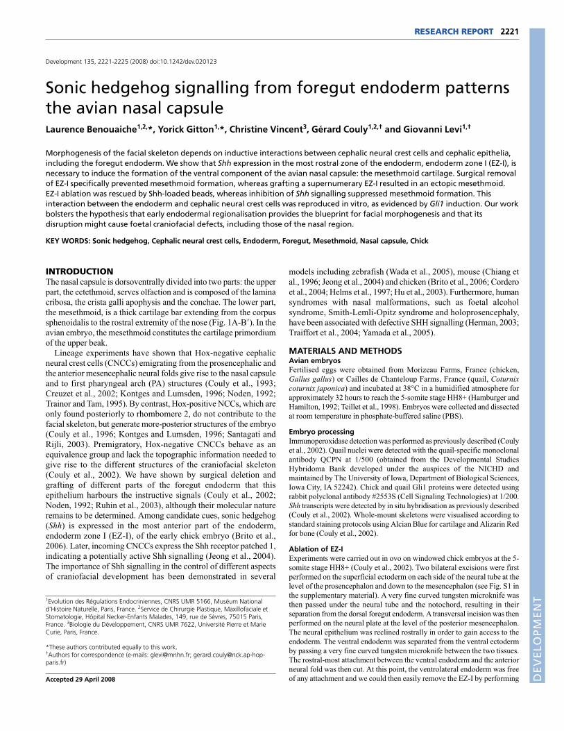
Next we transplanted a supplementary EZ-I from 5-somite
quail embryos into the presumptive nasal capsule territory of
stage-matched chick embryos and analysed skeletal morphology
at HH35. All survivors (12/21) displayed one ectopic,
supernumerary mesthemoid-like cartilage (Fig. 2A,B,B�) next to
the normal and complete nasal capsule. Heterotopic quail EZ-I
grafts, implanted within the 5-somite chick presumptive first PA
territory, induced a supernumerary mesthemoid-like element in
10/12 survivors (Fig. 2C,D). Dermatocranial elements [putatively
premaxillary bones (Couly et al., 1993)] formed in close
proximity to the supernumerary cartilage. All other first and
RESEARCH REPORT Development 135 (13)
Fig. 1. Endoderm zone I ablation prevents mesethmoidformation. (A-B�) Frontal (A) and lateral (B) views of the cephalicskeleton of a chick embryo at 9 days (HH35) with correspondingdrawings (A�,B�), highlighting the elements of the nasal capsule: blue,mesethmoid (mes); red, ectethmoid (ect); green, infraorbital septum (is).Mc, Meckel’s cartilage. (C) Schematic of the surgical procedure ofendoderm zone I (EZ-I) ablation. I and II, endoderm zones I and II; r1-r8,rhombomeres 1 to 8. (D) Lateral view of the chondrochranium of arepresentative HH35 embryo in which EZ-I has been ablated at the 5-somite stage. (Upper inset) The same embryo before skeletalpreparation. (Lower inset) Frontal view of the same embryo showingthe normal size and shape of the ectethmoid. (D�) Drawingcorresponding to D, using the same colour code as above. Note theabsence of the mesethmoid, whereas the ectethmoid and theinfraorbital septum are still present.
DEVELO
PMENT
second PA derivatives developed normally. Both homotopic and
heterotopic grafts suggest that EZ-I is necessary and sufficient to
induce the differentiation of any Hox-negative CNCC contingent
into a mesethmoid cartilage. Ectopic cartilage elements
consistently failed to develop when we grafted EZ-I into the Hox-
positive neural crest domain (data not shown).
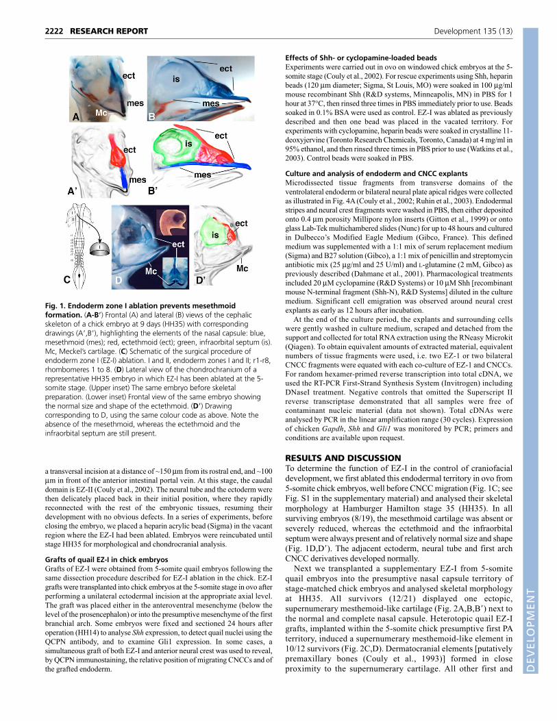
We then examined whether Shh expression by EZ-I (Fig. 3A,B)
(Brito et al., 2006) could be responsible for its capacity to induce
mesethmoid formation. First, we doubly transplanted EZ-I and
CNCCs from 5-somite quail into stage-matched chicken embryos.
In all cases, the grafted EZ-I was strongly Shh-positive and was in
close contact with grafted post-migratory CNCCs (Fig. 3A-C). We
2223RESEARCH REPORTShh patterning of the nasal capsule
Fig. 2. Supernumerary EZ-I graft in thecephalic region induces an ectopicmesethmoid. (A) Dorsal schematic view of5-somite quail and chick embryos showingthe dissection of EZ-I from a quail embryoand its transplantation into the lateralanterior mesenchyme of a chick embryo. (B) Ventral view at HH35 of thechondrochranium of an operated embryo.A supernumerary mesethmoid developswithin the nasofrontal regionperpendicularly to the host nasal capsule,which develops normally. (B�) Drawingcorresponding to B, with the same colourcode as in Fig. 1. (C) Transplantation of aquail EZ-I into the mesenchyme of thepresumptive first PA of a recipient chickembryo. (D) Lateral view of a skeletalpreparation from a 12-day (HH38) operatedembryo. A supernumerary mesethmoid canbe seen developing within the maxillaryarch under the native Meckel’s cartilage.ect, ectethmoid; I and II, endoderm zones Iand II; Mc, Meckel’s cartilage; mes,mesethmoid; mes*, supernumerarymesethmoid.
Fig. 3. Shh is expressed in EZ-I and can rescue the mesethmoidloss induced by EZ-I ablation. (A-C) This HH14 chick embryosimultaneously received two quail grafts at the 5-somite stage. Onewas a homotopic substitution of the anterior neural crest and the othera supernumerary EZ-1 graft in the mesenchyme of the presumptive firstPA. (A,B) Frontal section analysed by in situ hybridisation with a Shhprobe, showing strong expression in the floor plate, in the notochordand in the grafted EZ-I (boxed). (C) Immunodetection of quail nucleiconfirms the quail origin of the grafted endoderm and of theneighbouring CNCCs. (D) Dorsal schematic view of a 5-somite chickembryo showing the strategy of Shh rescue. After EZ-I ablation (Step 1),a Shh-loaded bead was implanted in the vacant region (Step 2).(E) Representative rescued embryo (HH35) showing almost normaldevelopment of the mesethmoid. (E�) The same embryo before skeletalpreparation showing the development of an upper beak. 1stpa, firstpharyngeal arch; I and II, endoderm zones I and II; ect, ectethmoid;en*, supernumerary graft of EZ-I; fp, floor plate; Mc, Meckel’s cartilage;mes, mesethmoid; n, neural tube; nt, notochord; QNCC, quail neuralcrest cells; r1-r8, rhombomeres 1-8.
DEVELO
PMENT
2224
then transplanted EZ-I into the presumptive first PA close to the
ocular region and observed a strong ectopic induction of Gli1, a Shh
target (Dahmane et al., 2001) not normally expressed in this area
(see Fig. S2 in the supplementary material). To determine whether
Shh expression from EZ-I directly contributed to mesethmoid
induction, we ablated EZ-I from 5-somite embryos and implanted a
Shh-loaded bead in the vacant territory (Fig. 3D). In all surviving
embryos (18/36), the nasal capsule presented a mesethmoid-like
cartilage correctly located under a normal ectethmoid (Fig. 3E,E�).Control BSA-loaded beads did not rescue the lack of mesethmoid
resulting from EZ-I ablation (n=6).
To assess whether Shh-Gli1 signalling was necessary to
generate a mesthemoid cartilage, we then implanted a
cyclopamine-loaded bead in contact with EZ-I at the 5-somite
stage. In all survivors (14/36), the mesethmoid was absent,
whereas the ectethmoid could be recognised (see Fig. S3 in the
supplementary material).
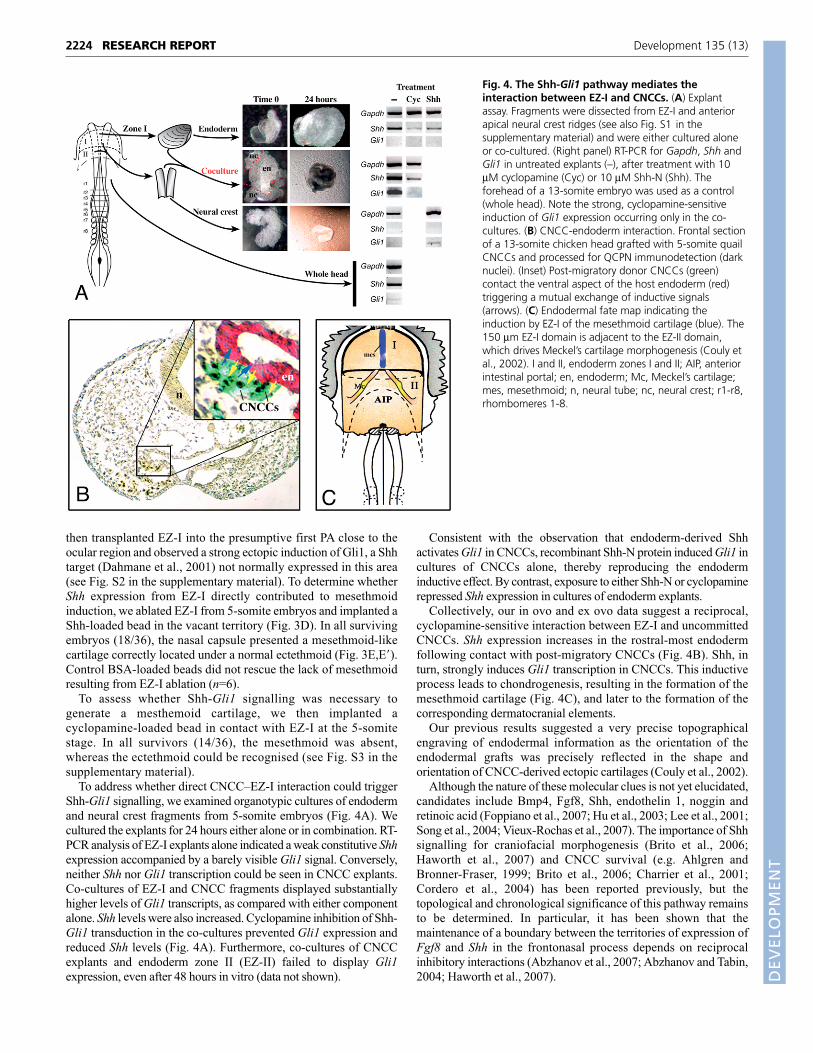
To address whether direct CNCC–EZ-I interaction could trigger
Shh-Gli1 signalling, we examined organotypic cultures of endoderm
and neural crest fragments from 5-somite embryos (Fig. 4A). We
cultured the explants for 24 hours either alone or in combination. RT-
PCR analysis of EZ-I explants alone indicated a weak constitutive Shhexpression accompanied by a barely visible Gli1 signal. Conversely,
neither Shh nor Gli1 transcription could be seen in CNCC explants.
Co-cultures of EZ-I and CNCC fragments displayed substantially
higher levels of Gli1 transcripts, as compared with either component
alone. Shh levels were also increased. Cyclopamine inhibition of Shh-
Gli1 transduction in the co-cultures prevented Gli1 expression and
reduced Shh levels (Fig. 4A). Furthermore, co-cultures of CNCC
explants and endoderm zone II (EZ-II) failed to display Gli1expression, even after 48 hours in vitro (data not shown).
Consistent with the observation that endoderm-derived Shh
activates Gli1 in CNCCs, recombinant Shh-N protein induced Gli1 in
cultures of CNCCs alone, thereby reproducing the endoderm
inductive effect. By contrast, exposure to either Shh-N or cyclopamine
repressed Shh expression in cultures of endoderm explants.
Collectively, our in ovo and ex ovo data suggest a reciprocal,
cyclopamine-sensitive interaction between EZ-I and uncommitted
CNCCs. Shh expression increases in the rostral-most endoderm
following contact with post-migratory CNCCs (Fig. 4B). Shh, in
turn, strongly induces Gli1 transcription in CNCCs. This inductive
process leads to chondrogenesis, resulting in the formation of the
mesethmoid cartilage (Fig. 4C), and later to the formation of the
corresponding dermatocranial elements.
Our previous results suggested a very precise topographical
engraving of endodermal information as the orientation of the
endodermal grafts was precisely reflected in the shape and
orientation of CNCC-derived ectopic cartilages (Couly et al., 2002).
Although the nature of these molecular clues is not yet elucidated,
candidates include Bmp4, Fgf8, Shh, endothelin 1, noggin and
retinoic acid (Foppiano et al., 2007; Hu et al., 2003; Lee et al., 2001;
Song et al., 2004; Vieux-Rochas et al., 2007). The importance of Shh
signalling for craniofacial morphogenesis (Brito et al., 2006;
Haworth et al., 2007) and CNCC survival (e.g. Ahlgren and
Bronner-Fraser, 1999; Brito et al., 2006; Charrier et al., 2001;
Cordero et al., 2004) has been reported previously, but the
topological and chronological significance of this pathway remains
to be determined. In particular, it has been shown that the
maintenance of a boundary between the territories of expression of
Fgf8 and Shh in the frontonasal process depends on reciprocal
inhibitory interactions (Abzhanov et al., 2007; Abzhanov and Tabin,
2004; Haworth et al., 2007).
RESEARCH REPORT Development 135 (13)
Fig. 4. The Shh-Gli1 pathway mediates theinteraction between EZ-I and CNCCs. (A) Explantassay. Fragments were dissected from EZ-I and anteriorapical neural crest ridges (see also Fig. S1 in thesupplementary material) and were either cultured aloneor co-cultured. (Right panel) RT-PCR for Gapdh, Shh andGli1 in untreated explants (–), after treatment with 10μM cyclopamine (Cyc) or 10 μM Shh-N (Shh). Theforehead of a 13-somite embryo was used as a control(whole head). Note the strong, cyclopamine-sensitiveinduction of Gli1 expression occurring only in the co-cultures. (B) CNCC-endoderm interaction. Frontal sectionof a 13-somite chicken head grafted with 5-somite quailCNCCs and processed for QCPN immunodetection (darknuclei). (Inset) Post-migratory donor CNCCs (green)contact the ventral aspect of the host endoderm (red)triggering a mutual exchange of inductive signals(arrows). (C) Endodermal fate map indicating theinduction by EZ-I of the mesethmoid cartilage (blue). The150 μm EZ-I domain is adjacent to the EZ-II domain,which drives Meckel’s cartilage morphogenesis (Couly etal., 2002). I and II, endoderm zones I and II; AIP, anteriorintestinal portal; en, endoderm; Mc, Meckel’s cartilage;mes, mesethmoid; n, neural tube; nc, neural crest; r1-r8,rhombomeres 1-8.
DEVELO
PMENT
Our observations imply that endoderm-derived Shh signalling,
which takes place already at the 5-somite stage, predates the
ectodermal Shh contribution to craniofacial patterning (Hu and
Helms, 2001; Hu et al., 2003). Globally, these results indicate that
whereas early mesethmoid differentiation requires EZ-I Shhexpression, ectodermal expression of Fgf8, Shh and Bmp4 controls
the size and shape of the beak at later stages of development (Cordero
et al., 2004; Hu et al., 2003; Wu et al., 2004). Shh is both a long-range
diffusible morphogen and a short-range contact-dependent factor
(Chuang and McMahon, 1999; Johnson and Tabin, 1995). Here,
ablation of EZ-I, a topographically restricted source of Shh in the
embryo, leads to a morphological lesion limited to the mesethmoid.
This implies that, at least in this context, the action of Shh takes place
in a localised paracrine, possibly contact-mediated, fashion. The
human mesethmoid cartilage gives rise to the nasal septum and the
vomer. It thus contributes to determining the proximodistal size of the
nose and the position of the premaxillary bone. Our results could help
to clarify the origin of a number of malformations of the nasofrontal
bud (Couly, 1981) that are characterised by abnormal mesethmoid
development, with normal ectethmoid. These pathologies include, for
example, cases of hypo- and hyper-septoethmoidism such as a flat
nose, short nose or very pronounced nose and a median nasal cleft
without cerebral anomalies. Thus, although as yet we cannot answer
Cyrano de Bergerac’s question “De quoi sert cette oblongue capsule?”
(What use this oblong capsule?), we may begin to understand where
this oblong capsule comes from.
We gratefully acknowledge the helpful comments of Prof. Nicole Le Douarin,Prof. Barbara Demeneix and Dr Anne Grappin-Botton, and the logistical helpof Dr Marie-Aimée Teillet. We thank Sophie Gourmet and Michel Fromaget forillustrations. This research was partially supported by the EU ConsortiumCRESCENDO (LSHM-CT-2005-018652) and the ANR project ‘GENDACTYL’.
Supplementary materialSupplementary material for this article is available athttp://dev.biologists.org/cgi/content/full/135/13/2221/DC1
ReferencesAbzhanov, A. and Tabin, C. J. (2004). Shh and Fgf8 act synergistically to drive
cartilage outgrowth during cranial development. Dev. Biol. 273, 134-148.Abzhanov, A., Cordero, D. R., Sen, J., Tabin, C. J. and Helms, J. A. (2007).
Cross-regulatory interactions between Fgf8 and Shh in the avian frontonasalprominence. Congenit Anom (Kyoto) 47, 136-148.
Ahlgren, S. C. and Bronner-Fraser, M. (1999). Inhibition of sonic hedgehogsignaling in vivo results in craniofacial neural crest cell death. Curr. Biol. 9, 1304-1314.
Brito, J. M., Teillet, M. A. and Le Douarin, N. M. (2006). An early role for sonichedgehog from foregut endoderm in jaw development: ensuring neural crestcell survival. Proc. Natl. Acad. Sci. USA 103, 11607-11612.
Charrier, J. B., Lapointe, F., Le Douarin, N. M. and Teillet, M. A. (2001). Anti-apoptotic role of Sonic hedgehog protein at the early stages of nervous systemorganogenesis. Development 128, 4011-4020.
Chiang, C., Litingtung, Y., Lee, E., Young, K. E., Corden, J. L., Westphal, H.and Beachy, P. A. (1996). Cyclopia and defective axial patterning in micelacking Sonic hedgehog gene function. Nature 383, 407-413.
Chuang, P. T. and McMahon, A. P. (1999). Vertebrate Hedgehog signallingmodulated by induction of a Hedgehog-binding protein. Nature 397, 617-621.
Cordero, D., Marcucio, R., Hu, D., Gaffield, W., Tapadia, M. and Helms, J. A.(2004). Temporal perturbations in sonic hedgehog signaling elicit the spectrumof holoprosencephaly phenotypes. J. Clin. Invest. 114, 485-494.
Couly, G. (1981). Neurocristopathies of the human nasofrontal bud. Ethmoidalsyndromes (hypo- and hyper-septoethmoidism). Rev. Stomatol. Chir. Maxillofac.82, 213-225.
Couly, G. F., Coltey, P. M. and Le Douarin, N. M. (1993). The triple origin of skullin higher vertebrates: a study in quail-chick chimeras. Development 117, 409-429.
Couly, G., Grapin-Botton, A., Coltey, P. and Le Douarin, N. M. (1996). Theregeneration of the cephalic neural crest, a problem revisited: the regeneratingcells originate from the contralateral or from the anterior and posterior neuralfold. Development 122, 3393-3407.
Couly, G., Creuzet, S., Bennaceur, S., Vincent, C. and Le Douarin, N. M.(2002). Interactions between Hox-negative cephalic neural crest cells and the
foregut endoderm in patterning the facial skeleton in the vertebrate head.Development 129, 1061-1073.
Creuzet, S., Couly, G., Vincent, C. and Le Douarin, N. M. (2002). Negativeeffect of Hox gene expression on the development of the neural crest-derivedfacial skeleton. Development 129, 4301-4313.
Dahmane, N., Sanchez, P., Gitton, Y., Palma, V., Sun, T., Beyna, M., Weiner,H. and Ruiz i Altaba, A. (2001). The Sonic Hedgehog-Gli pathway regulatesdorsal brain growth and tumorigenesis. Development 128, 5201-5212.
Foppiano, S., Hu, D. and Marcucio, R. S. (2007). Signaling by bonemorphogenetic proteins directs formation of an ectodermal signaling center thatregulates craniofacial development. Dev. Biol. 312, 103-114.
Gitton, Y., Cohen-Tannoudji, M. and Wassef, M. (1999). Specification ofsomatosensory area identity in cortical explants. J. Neurosci. 19, 4889-4898.
Hamburger, V. and Hamilton, H. L. (1992). A series of normal stages in thedevelopment of the chick embryo. 1951. Dev. Dyn. 195, 231-272.
Haworth, K. E., Wilson, J. M., Grevellec, A., Cobourne, M. T., Healy, C.,Helms, J. A., Sharpe, P. T. and Tucker, A. S. (2007). Sonic hedgehog in thepharyngeal endoderm controls arch pattern via regulation of Fgf8 in headectoderm. Dev. Biol. 303, 244-258.
Helms, J. A., Kim, C. H., Hu, D., Minkoff, R., Thaller, C. and Eichele, G. (1997).Sonic hedgehog participates in craniofacial morphogenesis and is down-regulated by teratogenic doses of retinoic acid. Dev. Biol. 187, 25-35.
Herman, G. E. (2003). Disorders of cholesterol biosynthesis: prototypic metabolicmalformation syndromes. Hum. Mol. Genet. 12 Spec No 1, R75-R88.
Hu, D. and Helms, J. (2001). Organ culture of craniofacial primordia. Methods24, 49-54.
Hu, D., Marcucio, R. S. and Helms, J. A. (2003). A zone of frontonasal ectodermregulates patterning and growth in the face. Development 130, 1749-1758.
Jeong, J., Mao, J., Tenzen, T., Kottmann, A. H. and McMahon, A. P. (2004).Hedgehog signaling in the neural crest cells regulates the patterning and growthof facial primordia. Genes Dev. 18, 937-951.
Johnson, R. L. and Tabin, C. (1995). The long and short of hedgehog signaling.Cell 81, 313-316.
Kontges, G. and Lumsden, A. (1996). Rhombencephalic neural crestsegmentation is preserved throughout craniofacial ontogeny. Development 122,3229-3242.
Lee, S. H., Fu, K. K., Hui, J. N. and Richman, J. M. (2001). Noggin and retinoicacid transform the identity of avian facial prominences. Nature 414, 909-912.
Noden, D. M. (1992). Vertebrate craniofacial development: novel approaches andnew dilemmas. Curr. Opin. Genet. Dev. 2, 576-581.
Ruhin, B., Creuzet, S., Vincent, C., Benouaiche, L., Le Douarin, N. M. andCouly, G. (2003). Patterning of the hyoid cartilage depends upon signals arisingfrom the ventral foregut endoderm. Dev. Dyn. 228, 239-246.
Santagati, F. and Rijli, F. M. (2003). Cranial neural crest and the building of thevertebrate head. Nat. Rev. Neurosci. 4, 806-818.
Song, Y., Hui, J. N., Fu, K. K. and Richman, J. M. (2004). Control of retinoic acidsynthesis and FGF expression in the nasal pit is required to pattern thecraniofacial skeleton. Dev. Biol. 276, 313-329.
Teillet, M., Watanabe, Y., Jeffs, P., Duprez, D., Lapointe, F. and Le Douarin,N. M. (1998). Sonic hedgehog is required for survival of both myogenic andchondrogenic somitic lineages. Development 125, 2019-2030.
Traiffort, E., Dubourg, C., Faure, H., Rognan, D., Odent, S., Durou, M. R.,David, V. and Ruat, M. (2004). Functional characterization of sonic hedgehogmutations associated with holoprosencephaly. J. Biol. Chem. 279, 42889-42897.
Trainor, P. A. and Tam, P. P. (1995). Cranial paraxial mesoderm and neural crestcells of the mouse embryo: co-distribution in the craniofacial mesenchyme butdistinct segregation in branchial arches. Development 121, 2569-2582.
Vieux-Rochas, M., Coen, L., Sato, T., Kurihara, Y., Gitton, Y., Barbieri, O., LeBlay, K., Merlo, G., Ekker, M., Kurihara, H. et al. (2007). Molecular dynamicsof retinoic acid-induced craniofacial malformations: implications for the origin ofgnathostome jaws. PLoS ONE 2, e510.
Wada, N., Javidan, Y., Nelson, S., Carney, T. J., Kelsh, R. N. and Schilling, T. F.(2005). Hedgehog signaling is required for cranial neural crest morphogenesisand chondrogenesis at the midline in the zebrafish skull. Development 132,3977-3988.
Watkins, D. N., Berman, D. M., Burkholder, S. G., Wang, B., Beachy, P. A. andBaylin, S. B. (2003). Hedgehog signalling within airway epithelial progenitorsand in small-cell lung cancer. Nature 422, 313-317.
Wu, P., Jiang, T. X., Suksaweang, S., Widelitz, R. B. and Chuong, C. M.(2004). Molecular shaping of the beak. Science 305, 1465-1466.
Yamada, Y., Nagase, T., Nagase, M. and Koshima, I. (2005). Gene expressionchanges of sonic hedgehog signaling cascade in a mouse embryonic model offetal alcohol syndrome. J. Craniofac. Surg. 16, 1055-1061; discussion 1062-1063.
2225RESEARCH REPORTShh patterning of the nasal capsule
DEVELO
PMENT