Embed Size (px)
Citation preview

Guidelines: Class 1 (C1) – Definitely perform (good evidence) Class 2 (C2) – Consider performing (some evidence) Class 3 (C3) – Do not perform (unsound evidence and/or deleterious)
AES – Protocols\Excellence Program\Snake Envenomation Page 1 of 18
Snake Envenomation
PATHOPHYSIOLOGY
Snake envenomation is a toxicity that requires immediate action regardless of whether the owner has identified the snake or not. Snake detection can be difficult and owner recognition is reported as being poor. Given the major neurological consequences (ie: death) and coagulopathies that can arise, all owners are advised to have their pet assessed and a snake detection test performed where applicable. Owners are advised NOT to capture the snake, though if it has been obviously killed in the process (ie: in more than one piece), then identification of the snake can be made in house (ie: counting scales – ventral, dorsal mid-body, anal division or single, and sub-caudal tail division, single or both).
In Queensland, there are approx. 120 known species of snakes, of which 65% are venomous (80). The 2x main varieties include the front-fanged snakes (Elapids) and the rear-fanged snakes (Colubrids). Elapids contain some of the most venomous snakes in the world (see Table 1). In comparison, come Colubrids produce weak venom and as their fangs are located at the back of their mouth, they are also considered poor envenomators and non-life threatening. A list of common Colubrids is located in the diagnostic section to allow direct identification when required. This also includes the Boids which are considered non-venomous. The majority of the toxic enzymes in venomous snakes are phospholipases, more specifically a phospholipase A2. As a result they act on cell membranes, leading to neurotoxic, myotoxic, haemotoxic, cardiotoxic and nephrotoxic effects. Coagulopathies are also present due to either activation of the common coagulation pathway (pro-coagulant) or from anti-coagulant effects, or a combination of both. Pre-paralytic signs have been shown to be a result of acute prothrombin activation and obstruction to the outflow of the right ventricle, leading to cor pulmonale, impedance of left ventricular filling and sudden acute hypotension. It is now also suspected to be related to an acute hypersensitivity response and release of vasoactive substances causing an acute hypotensive response.
Neurotoxicity – The severity of neurological impairment depends on whether the particular toxin attaches to the pre-synaptic terminal or post-synaptic terminal at the neuromuscular junction (NMJ). Neurotoxin’s attached to the pre-synaptic terminal tend to have a greater affinity for their receptors, are less responsive to antivenene administration, and cause more severe neurological disease.
CLINICAL SIGNS
• Pre-paralytic signs - Collapse with recovery vs lethal - Tachypnea - Vomiting/Defecation - Ptyalism - Muscle tremors
• Paralytic signs
• loss of corneal and/or pupillary light reflex
• reduced gag reflex
• ataxic vs paralytic
• mydriasis
• Bradypnoea vs Apnoea
• Haematuria +/- Oliguria
DIAGNOSTICS
• PCV/TS
• Electrolytes + Blood gas
• PT/APTT +/- ACT
• Biochemistry
• Urinalysis
• Indirect identification – SVDK (Snake Venom Detection Kit, on urine or blood)
• Direct Identification – Scale and colour recognition

Guidelines: Class 1 (C1) – Definitely perform (good evidence) Class 2 (C2) – Consider performing (some evidence) Class 3 (C3) – Do not perform (unsound evidence and/or deleterious)
AES – Protocols\Excellence Program\Snake Envenomation Page 2 of 18
Coagulopathy – There are varying degrees of coagulopathies amongst snake species, and their severity depends on whether they are caused pro-coagulant or anti-coagulant toxins. The most severe coagulopathies are caused by pro-thrombotic toxins and mimic factor-Xa, combining with endogenous factor Va to cleave prothrombin into thrombin and proceed into Venom Induced Consumptive Coagulopathy (VICC) - increased fibrin degradation products (FDP), and prolonged PT, APTT and ACT. Copperhead snakes are the only snake that show anti-PLT activity, so most snakes will have a normal PLT number initially, though can be reduced in time due to blood loss. Nephrotoxicity – Although this is not well understood, it is thought to arise from indirect actions of tubular damage associated with myoglobinuria and bilirubinuria, hypovolaemia, procoagulation and hypoxaemia-ischemia injury at the glomerulus. Myotoxicity – Rhabdomyolysis from a myotoxin is common and can lead to elevated creatine kinase and subsequent renal tubular damage if not treated. Other intra-cellular components will also lead to hyperphosphatemia, hyperkalemia, hypermagnesemia and a metabolic acidosis. Haemotoxicity – Haemolysis is also variable amongst snake venom, however, their potency’s tend to be more exaggerated in the eastern states and can require blood transfusions if global hypoxia is evident (ie: elevated lactate, low ScvO2, low base excess, tachycardia). Disruption to the cell membrane by phospholipase causes water to enter the cell, allowing it to swell and causing cell destruction. Cardiac toxicity – This is typically specific for Taicotoxin from the Taipan, and it has been shown to inhibit calcium
channels in the myocardium, leading to prolonged repolarisation and arrhythmias. Caution is used in relation to
possible fluid overload due to its effects as a negative inotrope and chronotrope, and positive lusitrope. Other
cardiac toxins have not been ruled out from other elapids, though are weak in nature if present.
DIAGNOSTICS
PCV/TS
• Evidence of anaemia maybe present due to the presence of haemolysis. The serum will likely be icteric as well and should be noted. Occasionally a blood transfusion is required if global hypoxia is present from rapid cell destruction.
Electrolytes
• Metabolic acidosis can occur due to release of intracellular contents for rhabomyolysis and haemolysis. This also includes elevated potassium, phosphate and magnesium.
• Monitor PvCO2 and PvO2 to determine effects of pro and anti-thrombotic effects on the pulmonary vessels and parenchyma. NB: DO NOT obtain an arterial or jugular sample if coagulopathic
Coagulation parameters
• Perform PT/APTT, or ACT if cost prohibitive. Due to the high fibrinogen consumption from pro-coagulant envenomation, clotting tests may not return to normal until 18-24hrs post venom neutralisation, when fibrinogen is re-synthesised. PT/APTT or ACT is measured every 6hrs till normalised. *Cats commonly don’t show a coagulopathy
Biochemistry
• CK and AST are measured to determine the significance of rhabomyolysis and if ongoing muscle damage is occurring. CK can take 2hrs to rise after envenomation, and half-life of CK is approx 3-6hrs and AST 12hrs, so any reduction in AST is significant.

Guidelines: Class 1 (C1) – Definitely perform (good evidence) Class 2 (C2) – Consider performing (some evidence) Class 3 (C3) – Do not perform (unsound evidence and/or deleterious)
AES – Protocols\Excellence Program\Snake Envenomation Page 3 of 18
• UREA and CREA are used to determine if there are any delayed effects from envenomation, and if there is impaired renal function. This can be due to effects from pigmenturia, hypovolaemia or direct nephrotoxins, though the latter are not described as yet.
• TBIL can be also be measured to monitor effects of haemolysis and possible renal tubular damage
Urinalysis
• Monitor for the presence of pigmenturia (myoglobin) +/- haematuria (red cells) as indications for possible renal tubular damage
• Measure the pH of the urine as alkaline pH has been shown to solubilise myoglobin, leading to reduced incidence of renal tubular necrosis and improved prognosis NB: DO NOT perform cystocentesis if coagulopathic
Indirect Identification (SVDK)
*Please follow instructions as outlined in the CSL SVDK. They have been included below for convenience as well
• Can be performed in serum/plasma or urine in the dog and cat due to ease
• Blood – spin the sample down and use the serum or plasma content. Collection by peripheral venipuncture is
preferred due to a likely coagulopathy.
*can give false positives (urine is preferred sample method)
• Urine – gentle palpation to gain adequate sample for SVDK, or place a temporary urinary catheter to obtain a
sample. If the patient is obtunded-stuporous and is to be admitted, place a permanent urinary catheter.
*Urine is the preferred sample as venom is up to 4x more concentrated than in blood
*Cystocentesis is contraindicated in the coagulopathic animal.
o DOG – detected in the urine between 1-24hr post envenomation, anectodally may be sooner
o CAT – detected in the urine after 8hrs, anectodally may be sooner
o Apparent period from time of envenomation and presentation if under 1 hour should not
prevent the test being done (historical account may be inaccurate etc)

Guidelines: Class 1 (C1) – Definitely perform (good evidence) Class 2 (C2) – Consider performing (some evidence) Class 3 (C3) – Do not perform (unsound evidence and/or deleterious)
AES – Protocols\Excellence Program\Snake Envenomation Page 4 of 18

Guidelines: Class 1 (C1) – Definitely perform (good evidence) Class 2 (C2) – Consider performing (some evidence) Class 3 (C3) – Do not perform (unsound evidence and/or deleterious)
AES – Protocols\Excellence Program\Snake Envenomation Page 5 of 18

Guidelines: Class 1 (C1) – Definitely perform (good evidence) Class 2 (C2) – Consider performing (some evidence) Class 3 (C3) – Do not perform (unsound evidence and/or deleterious)
AES – Protocols\Excellence Program\Snake Envenomation Page 6 of 18

Guidelines: Class 1 (C1) – Definitely perform (good evidence) Class 2 (C2) – Consider performing (some evidence) Class 3 (C3) – Do not perform (unsound evidence and/or deleterious)
AES – Protocols\Excellence Program\Snake Envenomation Page 7 of 18
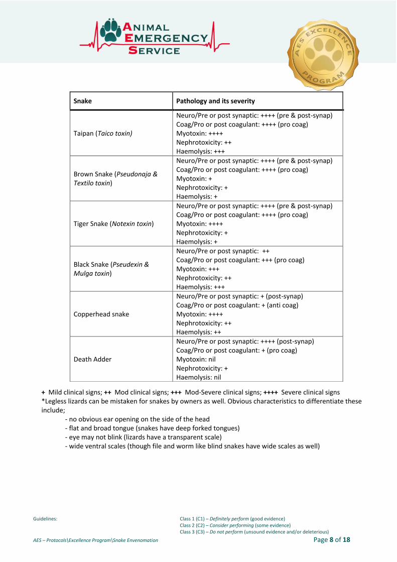
Direct Identification
Table 1: Snake Grouping and Characteristics – Snakes mentioned are diagnosed only with CSL SVDK
Guidelines: Class 1 (C1) – Definitely perform (good evidence) Class 2 (C2) – Consider performing (some evidence) Class 3 (C3) – Do not perform (unsound evidence and/or deleterious)
AES – Protocols\Excellence Program\Snake Envenomation Page 8 of 18
+ Mild clinical signs; ++ Mod clinical signs; +++ Mod-Severe clinical signs; ++++ Severe clinical signs *Legless lizards can be mistaken for snakes by owners as well. Obvious characteristics to differentiate these include;
- no obvious ear opening on the side of the head - flat and broad tongue (snakes have deep forked tongues) - eye may not blink (lizards have a transparent scale) - wide ventral scales (though file and worm like blind snakes have wide scales as well)
Snake Pathology and its severity
Taipan (Taico toxin)
Neuro/Pre or post synaptic: ++++ (pre & post-synap) Coag/Pro or post coagulant: ++++ (pro coag) Myotoxin: ++++ Nephrotoxicity: ++ Haemolysis: +++
Brown Snake (Pseudonaja & Textilo toxin)
Neuro/Pre or post synaptic: ++++ (pre & post-synap) Coag/Pro or post coagulant: ++++ (pro coag) Myotoxin: + Nephrotoxicity: + Haemolysis: +
Tiger Snake (Notexin toxin)
Neuro/Pre or post synaptic: ++++ (pre & post-synap) Coag/Pro or post coagulant: ++++ (pro coag) Myotoxin: ++++ Nephrotoxicity: + Haemolysis: +
Black Snake (Pseudexin & Mulga toxin)
Neuro/Pre or post synaptic: ++ Coag/Pro or post coagulant: +++ (pro coag) Myotoxin: +++ Nephrotoxicity: ++ Haemolysis: +++
Copperhead snake
Neuro/Pre or post synaptic: + (post-synap) Coag/Pro or post coagulant: + (anti coag) Myotoxin: ++++ Nephrotoxicity: ++ Haemolysis: ++
Death Adder
Neuro/Pre or post synaptic: ++++ (post-synap) Coag/Pro or post coagulant: + (pro coag) Myotoxin: nil Nephrotoxicity: + Haemolysis: nil

Guidelines: Class 1 (C1) – Definitely perform (good evidence) Class 2 (C2) – Consider performing (some evidence) Class 3 (C3) – Do not perform (unsound evidence and/or deleterious)
AES – Protocols\Excellence Program\Snake Envenomation Page 9 of 18
Below is a list of common snakes seen within Queensland. ELAPIDS
• Taipan (Coastal or Inland)
• Brown snake (Eastern, Western, Common, Speckled or Ringed) (approximately 85% of bites treated at AES)
• Tiger snake (Easter or Western) (approximately 5% of bites at AES)
• Black snake (King Brown/Mulga, Red-Bellied, Blue-bellied/Spotted) (approximately 10% of bites at AES)
• Death Adder (Common or Northern)
• Stephen’s Banded Snake
• Coiled Copperhead snake
• Whip snake (yellow-faced, black or collared) (occasional presumed cause of weakness in cats)
• Crowned snake (Dwarf, Northern or White)
• Rough scaled snake
• Naped snake (Red, Yellow or Orange)
• Black bellied swamp/Marsh snake
• Eastern small eyed snake (occasional bites seen at AES)
• Coral snake
• Myall snake
• Pale headed snake
• Bandy-Bandy
• Colletts snake COLUBRIDS
• Tree snakes (Northern, Common, Green or Brown)
• Keelback or Freshwater snake
• Slaty-grey snake
• Macleay’s water snake BOIDS
• Pythons (Spotted, Water, Amethystine, Childrens, Olive, Stimsons, Black-headed or Carpet)

Guidelines: Class 1 (C1) – Definitely perform (good evidence) Class 2 (C2) – Consider performing (some evidence) Class 3 (C3) – Do not perform (unsound evidence and/or deleterious)
AES – Protocols\Excellence Program\Snake Envenomation Page 10 of 18
VENOMOUS (All ELAPIDS)

Guidelines: Class 1 (C1) – Definitely perform (good evidence) Class 2 (C2) – Consider performing (some evidence) Class 3 (C3) – Do not perform (unsound evidence and/or deleterious)
AES – Protocols\Excellence Program\Snake Envenomation Page 11 of 18
MILD-MOD VENOMOUS (ELAPIDS & COLUBRIDS)

Guidelines: Class 1 (C1) – Definitely perform (good evidence) Class 2 (C2) – Consider performing (some evidence) Class 3 (C3) – Do not perform (unsound evidence and/or deleterious)
AES – Protocols\Excellence Program\Snake Envenomation Page 12 of 18
NON-VENOMOUS (BOIDS, Other)
For specific locations and markings, refer to ;
http://www.ehp.qld.gov.au/wildlife/livingwith/snakes/near-you/

Guidelines: Class 1 (C1) – Definitely perform (good evidence) Class 2 (C2) – Consider performing (some evidence) Class 3 (C3) – Do not perform (unsound evidence and/or deleterious)
AES – Protocols\Excellence Program\Snake Envenomation Page 13 of 18
TREATMENT
EMERGENCY PATIENT MANAGEMENT
• Place an IVC immediately on presentation (C1)
• Have intubation equipment at the ready – deterioration can occur within 2-5 mins from clinically normal to absolute respiratory paralysis (C1)
• Manage haemodynamic abnormalities by administration of shock fluid boluses of crystalloids if necessary and maintain fluids at 1x maintenance at minimum. Caution should be exercised in patients with pre-existing cardiac conditions, or where large volumes of crystalloids are given with antivenin administration (C1)
• Pre-medication prior to anti-venin administration is not recommended, though adrenaline should be on hand if an anaphylactic reaction is seen. NB: Premedicant administration has been shown to not prevent anaphylaxis - Adrenaline 0.01mg/kg IV or IM (i.e. 1ml/10kg IV for a reaction followed by a CRI at 0.05mg/kg/min if
required to control hypotension) (C1) *Adrenaline and fluid boluses are the primary treatments for anaphylaxis *Prednisolone sodium succinate is no longer available in Australia. Do not use Dexamethasone due to its long conversion time into an active state
• Snake Anti-venin administration and neutralization DOG & CAT: Dilute single vial of anti-venin 1:1 with saline and give over 20 mins (C1) NB: all known Brown snake envenomations are recommended to receive 2x vials of multi Tiger/Brown anti-venom due to their coagulopathic tendencies and to neutralize the venom
*Irreversible, pre-synaptic binding of venom is usually present by 48 hours after envenomation. Antivenin is not indicated from this point.

Guidelines: Class 1 (C1) – Definitely perform (good evidence) Class 2 (C2) – Consider performing (some evidence) Class 3 (C3) – Do not perform (unsound evidence and/or deleterious)
AES – Protocols\Excellence Program\Snake Envenomation Page 14 of 18
NB: Varying levels of units required to neutralize each snake venom varies
VENOM-INDUCED CONSUMPTION COAGULOPATHY (VICC)
There is evidence that suggests (in tiger snake envenomation) resolution of a coagulopathy may take more than
24 hours (possibly up to 36 hours). If adequate antivenin has been administered, recent evidence shows the anti-
venom should be neutralised, and production of fibrinogen and other clotting factors by the liver is required to
overcome the coagulopathy. Re-testing venom levels in the urine is no longer recommended after research
published by Leister and Padula showed that all patients had serum venom levels neutralised by 8000 units of
brown snake anti-venom alone (2x vials of multi Tiger-Brown anti-venom). In the same study, residual venom was
found in urine despite serum venom neutralisation.
Once the venom is neutralised, consider FFP if there is clinical bleeding seen. Failure to not provide enough anti-
venom (1x vial) when giving FFP can exacerbate the procoagulant syndrome. FFP may be considered if a
coagulopathy and clinical bleeding (epistaxis, pulmonary haemorrhage, haematemesis, haematuria on
sedimentation of urine etc) persists after suitable administration of antivenin (neutralized). Studies in humans
illustrate improved survival and reduced hospitalization with the use of fresh frozen plasma for coagulopathic
patient’s only once sufficient antivenin has been administered.
Detection of Venom Neutralisation
• Neutralization of all venom occurs with 4000- 8000 units of Tiger/Brown anti-venom in high serum levels, so
the current recommendation is to administer 2 x 4000 units of antivenene and not to repeat the SVDK. The

Guidelines: Class 1 (C1) – Definitely perform (good evidence) Class 2 (C2) – Consider performing (some evidence) Class 3 (C3) – Do not perform (unsound evidence and/or deleterious)
AES – Protocols\Excellence Program\Snake Envenomation Page 15 of 18
patients coagulopathic, myotoxic and haemotoxic effects should be monitored, but these should not be used
as triggers for further antivenene administration.
If the patient remains coagulopathic WITH clinical bleeding (determined by clinical and de novo bleeding, or by prolonged PT/aPTT or ACT), fresh frozen plasma is indicated. (C1)
• FFP dose 10-20mL/kg given over 2-4 hours. (care with volume overload) (C1)
CRITICAL MONITORING AND SUPPORTIVE CARE
Intravenous Fluid Therapy
Isotonic crystalloid solution with electrolyte supplements as needed (eg: KCl, KPO4, MgCl)
• 1-2x maintenance to ensure diuresis (C1)
• If cardiovascular compromise is present at shock rates of 10-20mL/kg titrate to effect (C1)
• Maintenance rates once adequate mean arterial pressure and urine production can be established (C1)
• Fresh frozen plasma at 10-20mL/kg over 2-4 hours as needed for treatment of coagulopathies which persist
after venom has been neutralised (reduced crystalloid rate commensurately during administration) (C1)
Respiratory support
Envenomation with procoagulant venom and neurotoxins may result in hypoxaemia by a number of mechanisms:
• Pulmonary haemorrhage: this is a severe and immediately life-threatening development of brown and tiger
snake envenomation. Early signs may include haemoptysis only. Treatment goals should be directed to
maintaining adequate oxygenation whilst neutralizing the venom, and then reversing the coagulopathy as
quickly as possible. Early and adequate neutralization of venom and identification of a consumption
coagulopathy is essential. The prognosis for this clinical development, in the author’s opinion, is grave.
• Aspiration pneumonia: Megaoesophagus is also recognized in canine patients envenomated by Tiger snakes.
This clinical entity reportedly may persist for up to 6 weeks even after resolution of generalized peripheral
neuropathy. Aspiration pneumonia is a well-known risk of this problem. Measures must be taken to prevent
aspiration such as suctioning the oropharynx if saliva pooling occurs or intubation under general anesthesia if
severe loss of gag reflex is present. If an animal can be intubated without anaesthetic agents, then mechanical
ventilation is usually necessary.
• Hypoventilation: this results from profound respiratory muscle paralysis. Monitoring blood gas analyses is
helpful in tracking a trend towards respiratory failure (PCO2 > 60mmHg) due to hypoventilation. Venous blood
gas is as useful in this setting as collecting an arterial sample is contraindicated in coagulopathic animals.
PcvCO2 is approximately 4-5mmHg higher than arterial blood. Peripheral venous CO2 is approximately 3-
8mmHg above arterial.
Treatment modalities for hypoxemia
Oxygen supplementation (C1)
• Administer by nasal oxygen line (care in coagulopathic patients) or oxygen cage (delivers FiO2 of 40—60%)
• Administer by insufflation in patients which are intubated
Mechanical Ventilation (C1)

Guidelines: Class 1 (C1) – Definitely perform (good evidence) Class 2 (C2) – Consider performing (some evidence) Class 3 (C3) – Do not perform (unsound evidence and/or deleterious)
AES – Protocols\Excellence Program\Snake Envenomation Page 16 of 18
• Appropriate for patients demonstrating either hypoventilation (PCO2>60mmHg) or hypo-oxygenation
(determined by arterial blood gas if obtainable, or by persistently low pulse oximetry (SpO2<90%))
• Length of ventilation requirement is variable and may be dependent on factors such as reversibility of paralysis.
Paralysis due to pre-synaptic toxins (eg: tiger, brown, black, taipan), are more difficult to reverse than those
caused by post-synaptic neurotoxins (eg: copperheads).
• Hypocapnoeic patients with normal lungs, select least aggressive ventilator settings.
ADDITIONAL SUPPORTIVE MANAGEMENT
Medications
• Mannitol (1-2mg/kg/min) if pigmenturia is present as it is a proximally acting diuretic, a potent renal
vasodilator and a potential reducer of haem iron units induced oxidant stress. (C2)
• Consider sodium bicarbonate therapy to alkalinise the urine and promote excretion of haeme products. It
may also be of use in treating the accompanying metabolic acidosis. (C2)
• Frusemide should be given if anuria or oliguria exists; 1-2mg/kg to a total of 4mg/kg to promote diuresis. (C2)
• Sedatives may be of use with anxious animals developing lower motor neuropathies
- Butorphanol CRI at 0.04-0.4mg/kg/h (C1)
• Analgesia with a mu agonist if significant rhabdomyolysis is present.
- Methadone 0.1-0.3mg/kg SC q4hrs (C2)
- Fentanyl CRI at 2-4ug/kg/hr (C2)
• Prokinetic agents may be helpful where megaoesophagus exists
- Metoclopramide CRI at 0.04-0.08mg/kg/h or 1-2mg/kg/day (C1)
• Consider gastric protectants to increase gastric pH in anticipation of aspiration, or in managing oesophageal
ulceration secondary to reflux
- Esomeprazole 0.7-1.0mg/kg IV q24hrs (C2)
Nutrition
Early enteral nutrition should be instituted as soon as the gag reflex is normal and the animal is able to swallow. If the animal remains paralysed and a gag reflex remains absent, consider the use of an appropriately selected feeding tube:
• nasogastric tube placement for severely affected cases for gastric decompression or feeding (C2)
• oesophagostomy tube for cats or small dogs (C2)
• gastrostomy tube for documented tiger snake-induced megaoesophagus may be warranted (C1)
NURSING CARE
• keep warm
• appropriate padding to prevent pressure sores and contusions
• elevation of head to 30°
• ensure adequate analgesia
• physiotherapy for the mobilized patient

Guidelines: Class 1 (C1) – Definitely perform (good evidence) Class 2 (C2) – Consider performing (some evidence) Class 3 (C3) – Do not perform (unsound evidence and/or deleterious)
AES – Protocols\Excellence Program\Snake Envenomation Page 17 of 18
• eye care for the paralysed patient
• bowel and bladder care (placement of urinary catheter and closed collection system to ensure urine is
produced at 1-2mL/kg/h (and “ins=outs”) (C1)
ANTICIPATE
• Sudden death
• Need for ventilation
• VICC
• Renal failure
• Anemia
COSTS AND HOSPITALISATION
• Hospitalisation time to expect: 1-10 days
• Costs whilst hospitalized: $3000-10,000 with or without mechanical ventilation, depending on species of snake, severity of signs and resolution of peripheral neuropathy.
PROGNOSIS AND RISK FACTORS
• Survival range for dogs and cats with treatment ranges between 75-91%
• Survival range for dogs and cats without treatment for dogs is 33%, and cats is 66%
• Alkaline urine reduces the incidence of possible nephrotoxicity
• Delayed time in getting to a hospital following Tiger snake envenomation increases mortality in dogs
REFERENCES
1. Lewis, P.F. (1994) Common Tiger snake envenomation in dogs and mice – relationship between the amount of venom and the onset of clinical signs. Aus Vet J, 71: 130-132
2. Lewis, P.F. (1994) Myotoxicity and Nephrotoxicity of the common Tiger snake (Notechis scutatus) venom in the dog. Aus Vet J, 71: 136-139
3. Hopper, K. et al (2001) Megaoesophagus in adult dogs secondary to Australian Tiger snake envenomation. Aus Vet J 79(10): 672-675
4. Swindells, K. (2010) Poisoning in Small Animal Practice – Course notes. Murdoch University 5. Holloway, S.A. & Parry, P.W. (1989) Observations on blood coagulation after snakebite in dogs and cats. Aus
Vet J 66: 364-366 6. Heller, J. et al (2007) Elapid snake envenomation in dogs in New South Wales: A Review. Aus Vet J 85: 469-
479 7. www.vin.com. 2007-2013. Veterinary Information Network 8. http://www.ehp.qld.gov.au. 2011-2013. Queensland Australia, Department of Environment and Heritage
Protection 9. Ong, R.K.C., Swindells, K. and Mansfield, C.S. (2010) Prospective determination of the specificity of a
commercial snake venom detection kit in urine samples from dogs and cats. Aus Vet J, 88(6): 222-224 10. Ong, R.K.C., Lenard, Z.M., Swindells, K.L. and Raisis, A.L. (2009) Extradural haematoma secondary to brown
snake (Pseudonajaspecies) envenomation. Aus Vet J, 87(4): 152-156 11. Swindells, K.L., Russell, N.J., Angles, J.M. and Foster, S.F. (2006) Four cases of snake envenomation responsive
to death adder antivenom. Aus Vet J, 84 (1-2): 22-29

Guidelines: Class 1 (C1) – Definitely perform (good evidence) Class 2 (C2) – Consider performing (some evidence) Class 3 (C3) – Do not perform (unsound evidence and/or deleterious)
AES – Protocols\Excellence Program\Snake Envenomation Page 18 of 18
12. Shea, G.M. (1999) The Distribution and Identification of dangerously venomous Australian terrestrial snakes. Aus Vet J, 77: 791-798
13. G.K. Isbister et al (2013) A randomised controlled trial of fresh frozen plasma for treating venom-induced consumption coagulopathy in cases of Australian snakebite (ASP-18) J Thrombo Haemost 11: 1310-8
14. Swindells, K. (2005) Management of Snake Envenomation, Proceedings 358 Centre for Veterinary Education, University of Sydney. Chp 11
15. Swindells, K.L, Russel, N.J, Angles, J.M and Foster, S.F. (2006) Four case of snake envenomation responsive to death adder antivenom. Aust Vet J, 84: 22-29
16. Heller J, Mellor DJ, Hodgson JL, Reid SWJ, Hodgson DR, Bosward KL. Elapid snake envenomation in dogs in New South Wales: a review. Australian Veterinary Journal. 2007;85(11):469–479.
17. Lewis PF. Myotoxicity and nephrotoxicity of common tiger snake (Notechis scutatus) venom in the dog. Australian veterinary journal. 1994;71(5):136–139
18. Jacoby-Alner TE, Stephens N, Davern KM, Balmer L, Brown SGA, Swindells K. Histopathological analysis and in situ localisation of Australian tiger snake venom in two clinically envenomed domestic animals. Toxicon. 2011;58(4):304–314
19. Heller J, Bosward KL, Hodgson DR, Pottie R. Anuric renal failure in a dog after Red-bellied Black snake (Pseudechis porphyriacus) envenomation. Australian Veterinary Journal. 2006;84(5):158–162
20. Padula A, Leister E. Eastern Brown Snake (Pseudonaja textilis) envenomation in dogs and cats: clinical signs, coagulation changes, brown snake venom antigen levels, and treatment with a novel caprylic acid fractionated bivalent whole IgG equine antivenom. Toxicon 138 (2017);89-97.
21. Leong O, Padula A, Leister E. Severe acute pulmonary haemorrhage and haemoptysis in 10 dogs following eastern brown snake (Pseudonaja textilis) envenomation: clinical signs, treatment and outcomes. Toxicon 150 (2018) 188-194.
*Use of any of this material is not for retail or public disclosure. It is intended for personal use and knowledge only Author(s): Dr Courtney Reddrop, Dr Michele Rebelo Reviewed: Dr Courtney Reddrop, Dr Ellie Lester & Dr Gerardo Poli Date: 9th October 2017