Embed Size (px)
Citation preview

Small molecules as regulators of bacterial Quorum Sensing. New strategy
in the development of antimicrobial agents
Carlos Mario Meléndez Gómez1 and Vladimir V. Kouznetsov
2*
1 Grupo de Investigación en Química Orgánica y Biomédica, Programa de Química, Facultad de Ciencias Básicas,
Universidad del Atlántico, A.A.1890, Barranquilla, Colombia 2 Laboratorio de Química Orgánica y Biomolecular, CMN, Universidad Industrial de Santander, Parque Tecnológico
Guatiguara, Km 2 vía refugio, Piedecuesta, A.A. 681011, Colombia
The cell-to-cell communication process (quorum sensing, QS) is an important signaling phenomenon used by bacteria and
relies on small, secreted signaling molecules. Because bacterial QS circuits regulates the expression of virulence genes
through intercellular communication, any mode of their disruption is emerged as an anti-virulence strategy with enormous
therapeutic potential The objective of this chapter is to show and discuss the advances in the bacterial QS networks,
evaluating this complex process through an analysis of the structure and function of natural and synthetic signal molecules
(autoinducers), the molecular diversification tactics and strategies and structure-activity relationship (SAR), as well as the
chemical methods controlling the regulatory activities. Discussion is divided into diverse parts: 1) Bacterial QS and the
development of new antimicrobial agents; 2) Backgrounds. Signaling molecules in the regulations of bacterial QS
network; 3) Enzymatic autoinducer inactivation as a strategy in the modulation of bacterial quorum sensing; 4) Molecular
modifications of signal molecules for LasR/RhlR regulatory activity; 5) Perspectives and conclusions.
Keywords: Quorum sensing; autoinducers; quorum quenching; small molecules; structural diversification
1. Bacterial QS and the development of new antimicrobial agents
Development of antibiotic resistance in pathogenic microorganisms is an ongoing public health threat. Emerging drug
resistance problem, which conducts to global economic and healthcare crisis, is a major and serious task of chemistry,
biology and medicine [1]. Among various approaches that can be undertaken in order to better control the emergence
and spread of drug-resistant pathogenic microorganisms, quorum sensing (QS) stands out relatively new attractive tactic
of combat against this problem. QS is a widespread phenomenon of cell-to-cell communication in several
microorganisms that is associated with the coordination of the expression of beneficial phenotypes, regulation of local
population densities and multiple virulence factors [2]. This coordination consists of the producing, releasing and
detecting small diffusible signaling molecules known as “autoinducers” (AIs). At a threshold signal concentration and
population, these molecules bind to a receptor protein and initiate changes in gene expression.
This mechanism was first discovered in 1970 during a bioluminescence study using Vibrio fischeri, a marine
bacterium associated with Hawaiian squid [3]. In that time, it was thought to be restricted to only a limited species.
Later on, extensive studies in this area have been performed with pathogenic bacteria. Nowadays, similar systems are
recognized in many organisms, including animal and plant pathogens. Over 100 species of bacteria are identified to
produce AIs in a cell density dependent manner similar to V. fischeri. It is well known that while controlling
synchronously the cell-to-cell communication process, Gram-negative, opportunistic pathogens as Bacillus subtilis,
Pseudomonas aeruginosa, Staphylococcus aureus and others can regulate biofilm formation, group motility, an arsenal
of excreted virulence factors and initiate chronic and severe infections. Several lines of evidence indicate that QS
enhances virulence of bacterial pathogens in animal models as well as in human infections hence, due to their critical
role in regulating virulence, disruption of bacterial QS is considered as a new, perspective anti-infective strategy.
Noteworthy that in contrast to traditional bacteriocidal or bacteriostatic antibiotics, disrupting QS does not cause
lethality but rather inhibits pathogen virulence.
Functional QS circuits are particularly attractive therapeutic targets for the development of new antibacterial agents,
as well as a starting point for new biochemical investigations on bacterial interactions. Actually, there is renewed
interest in drugs, which attenuate virulence rather than bacterial growth. Thus, some recent progress in exploiting this
information through the design of anti-virulence deception strategies that disrupt QS through signal molecule
inactivation, inhibition of signal molecule biosynthesis or the blockade of signal transduction are very significant and
vital to resolve drug resistance problem [4,5].
2. Backgrounds. Signaling molecules in the regulations of bacterial QS network
Many of bacterial pathogens are found to control virulence factor expression by a cell-to-cell communication system, in
which a signal molecule is generated and secreted into the surrounding environment. While the bacterial population
grows, the concentration of the signal molecule (molecules, AIs) increases until it reaches the threshold concentration at
which it binds and activates the related receptor protein. In this manner, all members of the population receive a
Antimicrobial research: Novel bioknowledge and educational programs (A. Méndez-Vilas, Ed.)
610
_____________________________________________________________________________

physiological call to reprogram gene expression throughout the population. Thus, QS regulates a wide variety of
physiological processes including bioluminescence, competence, root nodulation, sporulation, antibiotic biosynthesis,
motility, plasmid conjugal transfer, biofilm maturation, and the expression of key virulence factors in plant, animal and
human pathogens. Among these characteristic and beneficial phenotypes, biofilm, a biological architecture of
aggregated microbes on a surface, is closely associated with virulence which task consists of overwhelm host defenses.
As result, biofilm infections tend to be chronic and difficult to eradicate [6,7].
In order to control these processes mentioned above, bacteria use three classes of AIs (Fig. 1). The first identified
molecule of the most common class of AIs is N-acetyl-L-homoserine lactone (AHL). This latter molecule and its
analogs are usually used by Gram-negative bacteria, whereas oligopeptides are mainly found in Gram-positive bacteria
[8]. The N-acyl-L-homoserine lactone and its analogues, acylated homoserine lactones (AHLs), are a class of small
neutral lipid molecules composed of a homoserine lactone ring with an acyl chain, which often contains 4 to 18 carbon
atoms [9].
Fig. 1 General structures of known main autoinducers.
The third class of AIs comprises by various alifatic bifunctional compounds. Studying QS in Vibrio cholerae, the
causative agent of the disease cholera, it was found that a precursor based on the 4,5-dihydroxy-2,3-pentanedione
(DPD) structure spontaneously cyclizes into a signal molecule called as autoinducer-2 (AI-2), which chelates borate to
give a new signal molecule, identified as (2S,4S)-2-methyl-2,3,4-tetrahydroxytetrahydrofuran-borate, termed (AI-2-
borate) [10,11]. The AI-2 has been also proposed to be of biological significance in Salmonella typhimurium [11] and in
Vibrio harveyi [12]. Another signal molecule of this class was identified to be (S)-3-hydroxytridecan-4-one and called
as cholera autoinducer-1 (CAI-1) [13-15] (Fig. 1). It seems to be that autoinducers of the third class are non-species
specific signals, which mediate interspecies communication among Gram-negative and Gram-positive bacteria.
2.1 Signaling molecules used by Gram-negative bacteria
The QS signaling system in Gram-negative bacteria usually corresponds to the biosynthesis of N-acyl-L-homoserine
lactones, which are synthetized by AHL synthases. As AHLs are small neutral molecules, they can diffuse in and out of
cells by different mechanisms and when their concentration eventually reaches a sufficiently high concentration, can
bind to specific receptors that belong to a large class of DNA-binding transcription factors called “R-proteins,” which
are the second component of the QS system. This binding (complex Receptor-AHLs) regulates the transcription of
target genes required for bacterial group behavior [16] (Fig. 2).
A lot of the basic information on the bacterial QS systems has been learned from the first study of QS systems in the
light-producing Gram-negative bacteria Vibrio fischeri, which are living in light organ of the Hawaiian bobtail squid
Euprymna scolopes. When the bacteria density is sufficiently high, genes involved in bioluminescence are expressed
and light is produced [3].
Fig. 2 Simplified scheme of AHLs quorum sensing system in Gram-negative bacteria.
Antimicrobial research: Novel bioknowledge and educational programs (A. Méndez-Vilas, Ed.)
611
_____________________________________________________________________________
It was found that there are three elements: a signal generator LuxI (synthase), the cognate receptor LuxR (“R-
proteins”) and acyl-L-homoserine lactone, in this intraspecial bacterial cell-to-cell communication. Thus, an inducer
synthase (LuxI–type protein in the lux operon in V. fischeri) produces a special small AHLs molecule, which is
accumulated while the bacteria population grows. This molecule can be detected by its related cytoplasmic receptor
(LuxR–type protein). The protein complex AHL–LuxR often homodimerizes and binds adjacent to QS promoters („„lux
boxes‟‟), which can initiate the transcription of target genes required for bacterial group behavior.
2.1.1 QS circuits in Vibrio fischeri
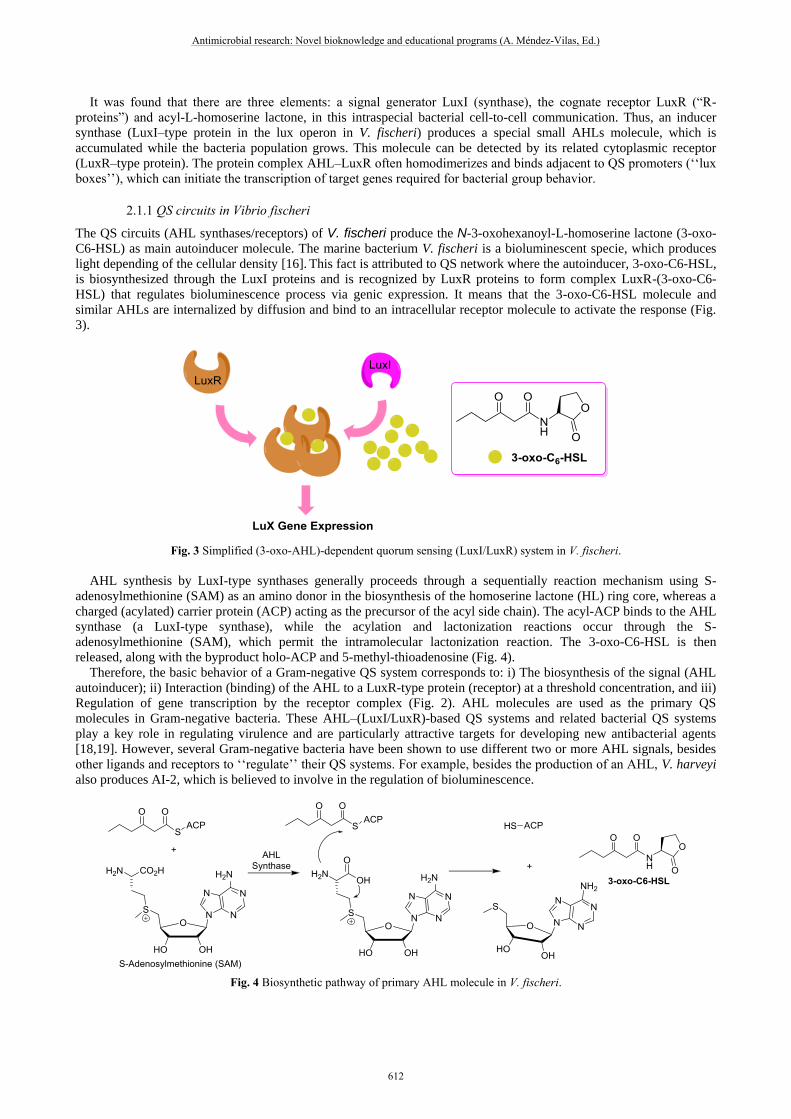
The QS circuits (AHL synthases/receptors) of V. fischeri produce the N-3-oxohexanoyl-L-homoserine lactone (3-oxo-
C6-HSL) as main autoinducer molecule. The marine bacterium V. fischeri is a bioluminescent specie, which produces
light depending of the cellular density [16]. This fact is attributed to QS network where the autoinducer, 3-oxo-C6-HSL,
is biosynthesized through the LuxI proteins and is recognized by LuxR proteins to form complex LuxR-(3-oxo-C6-
HSL) that regulates bioluminescence process via genic expression. It means that the 3-oxo-C6-HSL molecule and
similar AHLs are internalized by diffusion and bind to an intracellular receptor molecule to activate the response (Fig.
3).
Fig. 3 Simplified (3-oxo-AHL)-dependent quorum sensing (LuxI/LuxR) system in V. fischeri.
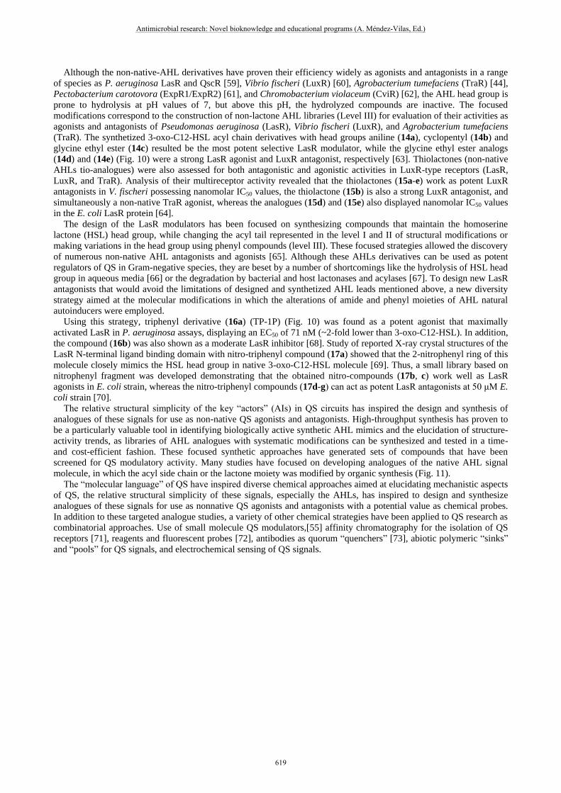
AHL synthesis by LuxI-type synthases generally proceeds through a sequentially reaction mechanism using S-
adenosylmethionine (SAM) as an amino donor in the biosynthesis of the homoserine lactone (HL) ring core, whereas a
charged (acylated) carrier protein (ACP) acting as the precursor of the acyl side chain). The acyl-ACP binds to the AHL
synthase (a LuxI-type synthase), while the acylation and lactonization reactions occur through the S-
adenosylmethionine (SAM), which permit the intramolecular lactonization reaction. The 3-oxo-C6-HSL is then
released, along with the byproduct holo-ACP and 5-methyl-thioadenosine (Fig. 4).
Therefore, the basic behavior of a Gram-negative QS system corresponds to: i) The biosynthesis of the signal (AHL
autoinducer); ii) Interaction (binding) of the AHL to a LuxR-type protein (receptor) at a threshold concentration, and iii)
Regulation of gene transcription by the receptor complex (Fig. 2). AHL molecules are used as the primary QS
molecules in Gram-negative bacteria. These AHL–(LuxI/LuxR)-based QS systems and related bacterial QS systems
play a key role in regulating virulence and are particularly attractive targets for developing new antibacterial agents
[18,19]. However, several Gram-negative bacteria have been shown to use different two or more AHL signals, besides
other ligands and receptors to „„regulate‟‟ their QS systems. For example, besides the production of an AHL, V. harveyi
also produces AI-2, which is believed to involve in the regulation of bioluminescence.
Fig. 4 Biosynthetic pathway of primary AHL molecule in V. fischeri.
Antimicrobial research: Novel bioknowledge and educational programs (A. Méndez-Vilas, Ed.)
612
_____________________________________________________________________________
Due to the emerging threat of multidrug resistance in relevant pathogens such as Pseudomonas aeruginosa,
Klebsiella pneumoniae and others, the research trends are directed to develop new molecules, which alter bacterial QS
system and, thus could mitigate virulence without having risks of resistance development, since interference with
virulence generally does not affect the growth and fitness of the bacteria [17].
Particularly, there is a wide interest in inhibiting AHL-mediated QS in some pathogenic species like P. aeruginosa,
due to the clinical importance of this opportunistic bacteria in life threatening hospital-acquired infections [20]. P.
aeruginosa is a ubiquitous environmental bacterium that is one of the top three causes of opportunistic human infections
and commonly infects immunocompromised patients [22]. P. aeruginosa infection is often aggravated by the formation
of biofilm, a mode of bacterial growth associated with antibiotic tolerance. The treatment of its infection confronts
major challenges due to the constant emergence of antibiotic-resistant variants. Antibiotic resistance to this bacterium
increases the rate of disease occurrence and mortality. With these problems, there is an urgent need to develop novel
antibiotic and anti-virulence strategies, which may be facilitated by an approach that explores more QS networks in P.
aeruginosa.
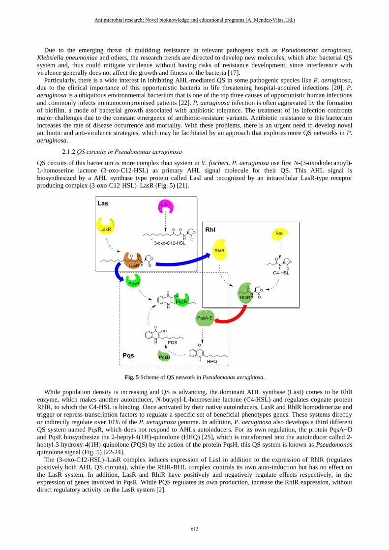
2.1.2 QS circuits in Pseudomonas aeruginosa
QS circuits of this bacterium is more complex than system in V. fischeri. P. aeruginosa use first N-(3-oxododecanoyl)-
L-homoserine lactone (3-oxo-C12-HSL) as primary AHL signal molecule for their QS. This AHL signal is
biosynthesized by a AHL synthase type protein called LasI and recognized by an intracellular LasR-type receptor
producing complex (3-oxo-C12-HSL)–LasR (Fig. 5) [21].
Fig. 5 Scheme of QS network in Pseudomonas aeruginosa.
While population density is increasing and QS is advancing, the dominant AHL synthase (LasI) comes to be RhlI
enzyme, which makes another autoinducer, N-butyryl-L-homoserine lactone (C4-HSL) and regulates cognate protein
RhlR, to which the C4-HSL is binding. Once activated by their native autoinducers, LasR and RhlR homodimerize and
trigger or repress transcription factors to regulate a specific set of beneficial phenotypes genes. These systems directly
or indirectly regulate over 10% of the P. aeruginosa genome. In addition, P. aeruginosa also develops a third different
QS system named PqsR, which does not respond to AHLs autoinducers. For its own regulation, the protein PqsA−D
and PqsE biosynthesize the 2-heptyl-4(1H)-quinolone (HHQ) [25], which is transformed into the autoinducer called 2-
heptyl-3-hydroxy-4(1H)-quinolone (PQS) by the action of the protein PqsH, this QS system is known as Pseudomonas
quinolone signal (Fig. 5) [22-24].
The (3-oxo-C12-HSL)–LasR complex induces expression of LasI in addition to the expression of RhlR (regulates
positively both AHL QS circuits), while the RhlR-BHL complex controls its own auto-induction but has no effect on
the LasR system. In addition, LasR and RhlR have positively and negatively regulate effects respectively, in the
expression of genes involved in PqsR. While PQS regulates its own production, increase the RhlR expression, without
direct regulatory activity on the LasR system [2].
Antimicrobial research: Novel bioknowledge and educational programs (A. Méndez-Vilas, Ed.)
613
_____________________________________________________________________________

Pseudomonas aeruginosa is a complex organism with multiple QS modules and signals. There are three types of
signaling molecules that function in a growth stage-dependent manner. Because of this design and development of new
selective antibacterial agents against this bacterium is difficult problem.
2.2 QS in Gram-positive bacteria
The Gram-positive bacteria QS network is different to QS circuitry of Gram-negative bacteria. First of all, the Gram-
positive bacterium does not harbor LuxI or LuxR homologues and primarily uses modified oligopeptides as
autoinducers (autoinducer peptides, AIP or QS peptides) [26,27]. These peptides are genetically encoded and are
generated ribosomally within the cell.
Noteworthy, as the oligopeptide molecules are encoded by genes, each species of bacteria is capable of producing a
peptide signal with a unique sequence. In general, the secretion of the AIP is facilitated by a membrane associated ATP-
binding cassette (ABC) transporter. Due to the physico-chemical parameters, these peptides are permeable to biological
membranes; therefore, secretion of QS peptides is mediated by specialized transporters, which are not capable to
permeate the cell membrane. As the population density increases, the AIPs accumulate in the environment. When a
certain threshold level is reached, binding of an AIP to a receptor occurs.
While the LuxR-type receptors are cytoplasmic, the sensors (receptors) for oligopeptide autoinducers in Gram-
positive bacteria are membrane-bound. Thus, there is two-component signaling proteins (membrane-bound receptors)
system, which transduce information via a series of phosphorylation events. A typical system consists of a membrane-
bound histidine kinase receptor and a cognate cytoplasmic response regulator, which functions as a transcriptional
regulator. The binding process between a signal peptide molecules and a membrane-bound sensor kinase leads to its
autophosporylation, resulting in ATP- guided phosphorylation of a conserved histidine residue (H) in the cytoplasm.
The phosphate group is transferred to the conserved aspartate residue (D) of a cognate response regulator (Fig. 6)
[28].
Fig. 6 Simplified scheme of QS network in Gram-positive bacteria.
As results, the activated response regulator effects the transcription of target genes, including the AIP, the ABC
transporter and genes for the receptor kinase and response regulator [27]. Taking into consideration that the bacterial
species are quite different, the nomenclature of the QS mechanisms can be diverse, due to the involved genes and
receptor(s). Examples of Gram-positive bacteria, which use quorum sensing peptide-based systems, include the
Streptococcus pneumonia, which use ComD/ComE to control competence development [29] or the QS system
AgrC/AgrA in the Staphylococcus aureus, which regulate the pathogenesis process [30].
3. Enzymatic autoinducer inactivation as a strategy in the modulation of bacterial
quorum sensing
As QS networks of several important bacterial pathogens, like P. aeruginosa, S. aureus, V. cholerae, and others play a
key role in the expression profile of diverse genes, including antibiotic tolerance and virulence determinants, their QS
circuits are logical, potential targets for antimicrobial chemotherapy. Disruption of bacterial quorum sensing systems
can be realized through various general approaches: 1) Inhibition of signal autoinducer production, 2) Degrading
Antimicrobial research: Novel bioknowledge and educational programs (A. Méndez-Vilas, Ed.)
614
_____________________________________________________________________________

signals, 3) Antagonizing signal binding to LuxR-family receptor, 4) Trapping signals, and 5) Suppression of synthase
and receptor activities, stabilities or production. Among them, the first three methods stand out as more important and
more developed. All these processes interfere with QS and could be coined as “quorum quenching” (QQ).
QQ enzymes mechanism aims to degrade or inactivate the AHL autoinducers. There are three known classes of
enzymes which target AHL signals: lactonases, acylases, and oxidoreductases. All these enzymes can modify the AHLs
structure. Any change in the structure of AHLs will significantly lower their affinity for their response regulators,
diminishing their ability to affect gene regulation.
3.1 AHL Lactonases
The first AHL lactonase was a 250-amino acid protein characterized as a metalloprotease, identified in treated soil
samples, encoded by the gene aiiA (autoinducer inactivation gene) from a Gram-positive Bacillus sp. 240B1 [32]. The
enzymes catalyze the hydrolysis of the ester bond in the lactone ring of a wide variety of AHLs [33]. Subsequently,
diverse AiiA-like enzymes were found and isolated from various Bacillus species such as B. thuringiensis, B. cereus, B.
anthracis, and B. mycoides, which are sharing more that 90% sequence homology in many cases [32].
The AHL lactonase enzyme family possesses hydrolytic activity toward a broad spectrum of AHLs, acting on the
acyl chain length and the oxidation state at the C3 position of the acyl chain that leads to the generation of acyl
homoserine [33]. The catalytic mechanism proposed has been based on the two independent crystal structures of an
AHL lactonase of the B. thuringiensis, and suggesting that two zinc ions are present in the binding site, coordinated by
five histidine and two aspartate residues.
Another family of AHL-lactonases was identified in diverse species as Agrobacterium tumefaciens (attM) [34],
Klebsiella pneumoniae (ahlK) and Arthrobacter sp. (ahlD) [35], which share about 30-58% homology in amino acid
sequence. These have the same conserved zinc-binding motif; therefore, it is likely that the catalytic mechanism is quite
similar. Recently, a novel group of lactonases encoded by BpiB genes (for biofilm phenotype inhibiting genes) was
discovered [36]. It was found in three BpiB genes, originating from Nitrobacter sp. strain Nb-311A, Pseudomonas
fluorescence, and Xanthomonas campestris, encode BpiB01, BpiB04, and BpiB07, respectively. Their activity was
characterized as lactonase protein due to its role in degradation of 3-oxo-C12-HSL (Fig. 7A).
Fig. 7. Enzymatic degradation of AHL autoinducers.
3.2 AHL Acylases
The acylases protein are AHL-degrading enzymes, targeted the amide bond that connects the fatty acyl chain to the
homoserine lactone (Fig. 7B). Acylases were first identified in strains of Variovorax sp. and Rhodococcus erythropolis
[37], that are able to degrade and utilize multiple AHLs as a source of both energy and nitrogen [31,38]. Degradation
required an AHL acylase, which forms HSL and the corresponding fatty acid (Fig. 7B). The fatty acid supported
growth, whereas the HSL only served as nitrogen source.
The first enzymes characterized as HSL acylases were isolated in betaproteobacterium Ralstonia strain XJ12B and
named AiiD. This protein shares amino acid sequence homology with members of the N-terminal nucleophile hydrolase
(Ntn-hydrolase, aminohydrolase) superfamily, which can catalyze the hydrolysis of amide function, so-called post-
translational amide cleavage [39]. Recombinant expression of AiiD protein in P. aeruginosa proved to prevent AHL
accumulation in the culture medium and reduce virulence. Thus, it was concluded that AHL acylases catalyze the
complete and irreversible degradation of the AHLs through the hydrolysis of their amide bond. However, six genes that
encode AHL-acylase have been characterized. These AHL-acylases degrade long-chain AHLs more efficiently than
short-chain forms.
For example, it was found that the PA2385 protein in P. aeruginosa, previously labeled as pVdQ played a role in QQ
emzyme process. Its overexpression in P. aeruginosa PAO1 inhibited accumulation of 3-oxo-C12-HSL. However, a
PvdQ knockout strain was still able to utilize HSL as a sole source of carbon, implying that another enzyme is involved
Antimicrobial research: Novel bioknowledge and educational programs (A. Méndez-Vilas, Ed.)
615
_____________________________________________________________________________

in the degradation process. That way, a second AHL acylase was found and was named QuiP (quorum signal utilization
and inactivation). QuiP enzyme shares 21% amino acid sequence homology with PvdQ and 23% homology with AiiD
from Ralstonia spp., and it is another member of the Ntn-hydrolase family, with substrate preference for long-chain
AHLs. In a related experiment, expression of PvdQ in P. aeruginosa was shown to abolish not only accumulation of 3-
oxo-C12-HSL, but also accumulation of Pseudomonas quinolone signal [40]. Noteworthy, certain enzymes, such as
PvdQ and QuiP appear to be unable to degrade AHL that has an acyl chain shorter than eight carbons.
4. Molecular modifications of signal molecules for LasR/RhlR regulatory activity
A decisive role in the regulation of bacterial QS network belongs to the small signal molecules (AHLs, AI-2, AIPs,
PQS, etc.) [41-44], which regulate cell–cell signaling process both in Gram-negative and Gram-positive bacteria. Their
binding to the cognate protein receptors does not only depend on their critic concentration, but also on their molecular
structure. Thus, in order to modulate QS circuits, many molecular signal-like small molecules have been obtained using
synthetic methods [45]. Generally, this strategy would work affecting more the LuxR-type receptor protein of QS
communication circuit. Homologues of LuxI/ LuxR have been identified in diverse bacterial genomes with a variety of
different AHLs regulating a range of physiological functions. However, now it is known that each bacterial species
responds specifically to its own unique AHL autoinducer; in general, the same molecular core is maintained, with
changes only in the size of the side chain (Fig. 8) [46].
Fig. 8 AHL core with different alkyl chain in diverse bacteria. Molecular specificity vs biological specifity.
Synthetic molecules capable of modulating diverse QS pathway (regulation of LuxR-type protein) have been
discovered through a design and high-throughput synthesis process, emulating and diversifying the molecular structure
of the natural AHL signaling molecule as a template, exhibit valuables structure-activity trends [47].
Non-native AHLs represent the most extensively studied class of synthetic QS modulators, being decisive in
identifying relevant results on SAR analysis. Critical structural aspects for the non-native synthetic molecules derived
from natural AHL corresponds to: i) The length of the lateral acyl chain; ii) The modification at the 3-carbon of the acyl
chain (is important, but not essential for activity); iii) The stereochemistry of the lactone ring (the L-stereoisomer is
needed for activity); iv) Molecular modifications to the lactone ring (Not in all cases result in active compounds); v)
The incorporation of aromatic functionality into AHLs (in the lactone ring or side lateral chain), generally yields
analogues with inhibitory activity [48].
4.1 General approaches looking for new QS modular
Structural aspects denote the basis of the focused molecular libraries that can guide the SAR studies, related to the
diverse structural modifications that must be realized. Based in the AHL native structure (for example, 3-oxo-C6-HSL,
3-oxo-C12-HSL and C4-HSL) three levels of molecular complexity are presented in which synthetic modifications are
made on the lateral acyl chain (Levels I and II), as well as changes on the lactam core (Level III) (Fig. 9).
Antimicrobial research: Novel bioknowledge and educational programs (A. Méndez-Vilas, Ed.)
616
_____________________________________________________________________________

Fig. 9 Chemical modifications of AHL signal molecules.
Several structural modifications are focused on the carbonyl-alkyl chain (4–14 carbons) (Level I), diverse C-4 phenyl
substituted N-acyl-L-homoserine lactone analogues (1) were evaluated for both their inducing activity and their ability
to competitively inhibit the action of AHL, 3-oxo-C6-HSL, responsible for the bioluminescence in V. fischeri [49].
Almost of these phenyls substituted analogues displayed significant antagonist activity, possibly due to the interaction
between the aryl group and aromatic amino acids of the LuxR receptor that prevents it from adopting the active dimeric
form. On the other hand, replacement of the β-methylene group to the ketoamide in the acyl chain of 3-oxo-C12-HSL
autoinducers (2) with functions containing heteroatoms as an NH or sulfonyl combined with alkyl chains of diverse
length, resulted in a strong QS antagonist activity of these designed molecules in P. aeruginosa bioluminescent assay
(Fig. 9) [50].
The structural level II consists in the complete modification of the side alkyl chain of AHL autoinducer. These
modifications showed that the alkyl chain length is a very significant factor in the antagonist activity, evidenced in the
QS inhibition of α-(N-alkyl-carboxamide)-γ-butyrolactones (3) evaluated in bacteria V. fischeri. These studies revealed
that the tested compounds with slightly shorter chain resulted be less active as antogonists (Fig. 9) [51]. On the other
hand, it was also found that the decrease in the LasA production in P. aeruginosa caused by the C-14 substituted
alkaloid (R)-norbgugaine ((R)-2-tetradecylpyrrolidine) (8), natural molecules [52]. The realized incorporation of
alkylthiomethyl substituent in γ-lactame derivatives of AHL showed important effects on the P. aeruginosa Las and Rhl
QS pathways. The synthetized 5-((alkylthio)methyl) pyrrolidin-2-one and its (propyl-, hexyl-) derivatives (5) were
found to strongly inhibit both QS networks [53]. Meanwhile, the alkyl sulfonamide chain in N-sulfonyl-HSL (6)
displayed antagonist activity of the QS transcriptional regulator LuxR in V. fischeri [54].
The molecular level III is related to the modification of lactone ring [55]. Introduction of simple (hetero)aromatic and
alicyclic replacements for the lactone, as nonchiral 2-aminothiazole, 2-cyclohexanol, pyridine ring system to construct
new small heterocyclic molecules (7) among others (Fig. 9) abolished LasR-induction ability or did not show any
improvement over native AHL autoinducer in P. aeruginosa. These results suggested special chemical features in the
core, which are related with the antagonist activity [50]. In order to analyze the inhibition or activation ability of an
aromatic ring for the R-protein - DNA binding, it was developed a set of substituted aniline derivatives with hidroxyl or
carboxyamide (substituted in orhto- or meta-positions) (8), which can act as H bond acceptors leading to the potent
antagonists of LasR in P. aeruginosa [56,57]. The new structural features identified in this study for both agonists and
antagonists are currently being used to design new focused libraries of analogs that should contain more potent
antagonists.
Diverse structural modifications as alkyl side chains, H donor groups or substituted phenyl groups defined the lines
may be denoted in the construction of focused libraries based on the AHL core, in the three-diverse level of focused
Antimicrobial research: Novel bioknowledge and educational programs (A. Méndez-Vilas, Ed.)
617
_____________________________________________________________________________
molecular library, based on AHL autoinducers structures: non-native AHL (level I-II), non-lactone modulators (level
III).
4.2 Further development of new modulators (agonists and antogonists) of native AHL autoinducers
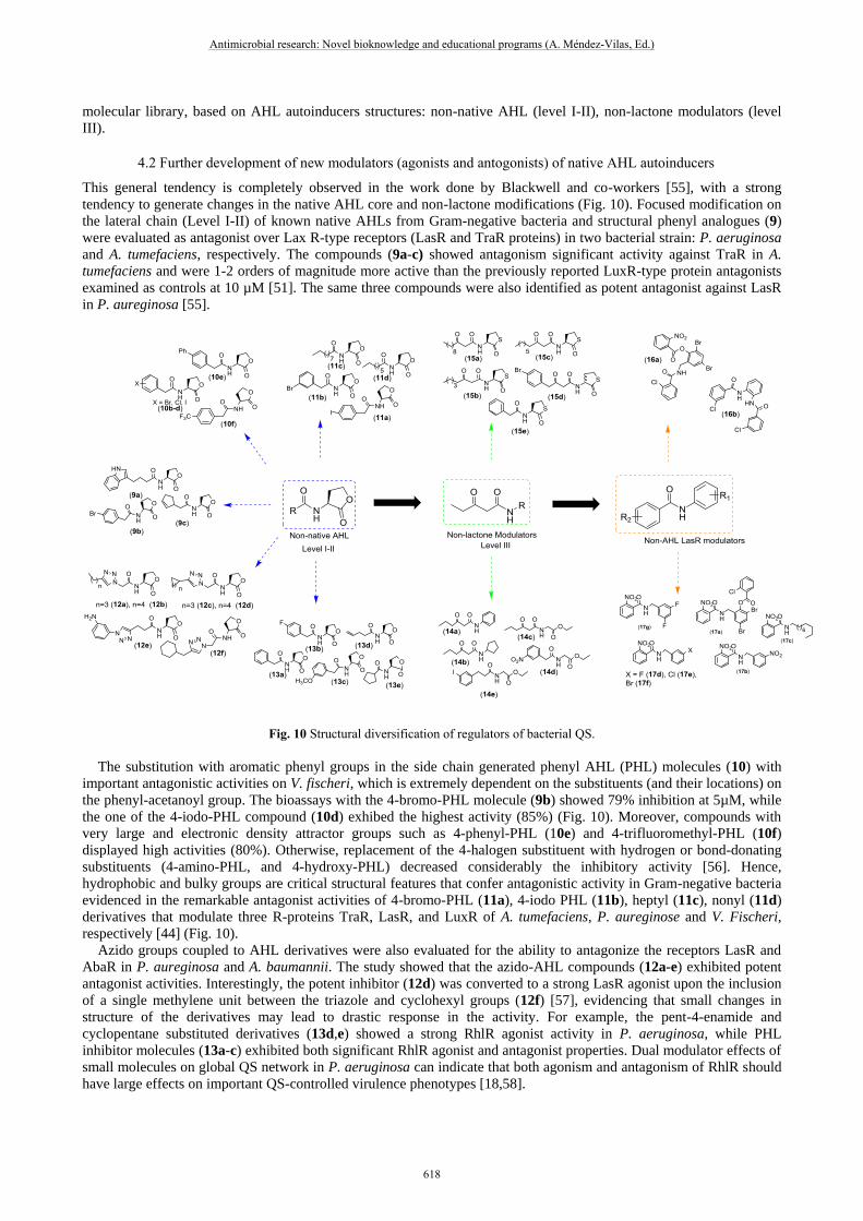
This general tendency is completely observed in the work done by Blackwell and co-workers [55], with a strong
tendency to generate changes in the native AHL core and non-lactone modifications (Fig. 10). Focused modification on
the lateral chain (Level I-II) of known native AHLs from Gram-negative bacteria and structural phenyl analogues (9)
were evaluated as antagonist over Lax R-type receptors (LasR and TraR proteins) in two bacterial strain: P. aeruginosa
and A. tumefaciens, respectively. The compounds (9a-c) showed antagonism significant activity against TraR in A.
tumefaciens and were 1-2 orders of magnitude more active than the previously reported LuxR-type protein antagonists
examined as controls at 10 µM [51]. The same three compounds were also identified as potent antagonist against LasR
in P. aureginosa [55].
Fig. 10 Structural diversification of regulators of bacterial QS.
The substitution with aromatic phenyl groups in the side chain generated phenyl AHL (PHL) molecules (10) with
important antagonistic activities on V. fischeri, which is extremely dependent on the substituents (and their locations) on
the phenyl-acetanoyl group. The bioassays with the 4-bromo-PHL molecule (9b) showed 79% inhibition at 5µM, while
the one of the 4-iodo-PHL compound (10d) exhibed the highest activity (85%) (Fig. 10). Moreover, compounds with
very large and electronic density attractor groups such as 4-phenyl-PHL (10e) and 4-trifluoromethyl-PHL (10f)
displayed high activities (80%). Otherwise, replacement of the 4-halogen substituent with hydrogen or bond-donating
substituents (4-amino-PHL, and 4-hydroxy-PHL) decreased considerably the inhibitory activity [56]. Hence,
hydrophobic and bulky groups are critical structural features that confer antagonistic activity in Gram-negative bacteria
evidenced in the remarkable antagonist activities of 4-bromo-PHL (11a), 4-iodo PHL (11b), heptyl (11c), nonyl (11d)
derivatives that modulate three R-proteins TraR, LasR, and LuxR of A. tumefaciens, P. aureginose and V. Fischeri,
respectively [44] (Fig. 10).
Azido groups coupled to AHL derivatives were also evaluated for the ability to antagonize the receptors LasR and
AbaR in P. aureginosa and A. baumannii. The study showed that the azido-AHL compounds (12a-e) exhibited potent
antagonist activities. Interestingly, the potent inhibitor (12d) was converted to a strong LasR agonist upon the inclusion
of a single methylene unit between the triazole and cyclohexyl groups (12f) [57], evidencing that small changes in
structure of the derivatives may lead to drastic response in the activity. For example, the pent-4-enamide and
cyclopentane substituted derivatives (13d,e) showed a strong RhlR agonist activity in P. aeruginosa, while PHL
inhibitor molecules (13a-c) exhibited both significant RhlR agonist and antagonist properties. Dual modulator effects of
small molecules on global QS network in P. aeruginosa can indicate that both agonism and antagonism of RhlR should
have large effects on important QS-controlled virulence phenotypes [18,58].
Antimicrobial research: Novel bioknowledge and educational programs (A. Méndez-Vilas, Ed.)
618
_____________________________________________________________________________
Although the non-native-AHL derivatives have proven their efficiency widely as agonists and antagonists in a range
of species as P. aeruginosa LasR and QscR [59], Vibrio fischeri (LuxR) [60], Agrobacterium tumefaciens (TraR) [44],
Pectobacterium carotovora (ExpR1/ExpR2) [61], and Chromobacterium violaceum (CviR) [62], the AHL head group is
prone to hydrolysis at pH values of 7, but above this pH, the hydrolyzed compounds are inactive. The focused
modifications correspond to the construction of non-lactone AHL libraries (Level III) for evaluation of their activities as
agonists and antagonists of Pseudomonas aeruginosa (LasR), Vibrio fischeri (LuxR), and Agrobacterium tumefaciens
(TraR). The synthetized 3-oxo-C12-HSL acyl chain derivatives with head groups aniline (14a), cyclopentyl (14b) and
glycine ethyl ester (14c) resulted be the most potent selective LasR modulator, while the glycine ethyl ester analogs
(14d) and (14e) (Fig. 10) were a strong LasR agonist and LuxR antagonist, respectively [63]. Thiolactones (non-native
AHLs tio-analogues) were also assessed for both antagonistic and agonistic activities in LuxR-type receptors (LasR,
LuxR, and TraR). Analysis of their multireceptor activity revealed that the thiolactones (15a-e) work as potent LuxR
antagonists in V. fischeri possessing nanomolar IC50 values, the thiolactone (15b) is also a strong LuxR antagonist, and
simultaneously a non-native TraR agonist, whereas the analogues (15d) and (15e) also displayed nanomolar IC50 values
in the E. coli LasR protein [64].
The design of the LasR modulators has been focused on synthesizing compounds that maintain the homoserine
lactone (HSL) head group, while changing the acyl tail represented in the level I and II of structural modifications or
making variations in the head group using phenyl compounds (level III). These focused strategies allowed the discovery
of numerous non-native AHL antagonists and agonists [65]. Although these AHLs derivatives can be used as potent
regulators of QS in Gram-negative species, they are beset by a number of shortcomings like the hydrolysis of HSL head
group in aqueous media [66] or the degradation by bacterial and host lactonases and acylases [67]. To design new LasR
antagonists that would avoid the limitations of designed and synthetized AHL leads mentioned above, a new diversity
strategy aimed at the molecular modifications in which the alterations of amide and phenyl moieties of AHL natural
autoinducers were employed.
Using this strategy, triphenyl derivative (16a) (TP-1P) (Fig. 10) was found as a potent agonist that maximally
activated LasR in P. aeruginosa assays, displaying an EC50 of 71 nM (~2-fold lower than 3-oxo-C12-HSL). In addition,
the compound (16b) was also shown as a moderate LasR inhibitor [68]. Study of reported X-ray crystal structures of the
LasR N-terminal ligand binding domain with nitro-triphenyl compound (17a) showed that the 2-nitrophenyl ring of this
molecule closely mimics the HSL head group in native 3-oxo-C12-HSL molecule [69]. Thus, a small library based on
nitrophenyl fragment was developed demonstrating that the obtained nitro-compounds (17b, c) work well as LasR
agonists in E. coli strain, whereas the nitro-triphenyl compounds (17d-g) can act as potent LasR antagonists at 50 μM E.
coli strain [70].
The relative structural simplicity of the key “actors” (AIs) in QS circuits has inspired the design and synthesis of
analogues of these signals for use as non-native QS agonists and antagonists. High-throughput synthesis has proven to
be a particularly valuable tool in identifying biologically active synthetic AHL mimics and the elucidation of structure-
activity trends, as libraries of AHL analogues with systematic modifications can be synthesized and tested in a time-
and cost-efficient fashion. These focused synthetic approaches have generated sets of compounds that have been
screened for QS modulatory activity. Many studies have focused on developing analogues of the native AHL signal
molecule, in which the acyl side chain or the lactone moiety was modified by organic synthesis (Fig. 11).
The “molecular language” of QS have inspired diverse chemical approaches aimed at elucidating mechanistic aspects
of QS, the relative structural simplicity of these signals, especially the AHLs, has inspired to design and synthesize
analogues of these signals for use as nonnative QS agonists and antagonists with a potential value as chemical probes.
In addition to these targeted analogue studies, a variety of other chemical strategies have been applied to QS research as
combinatorial approaches. Use of small molecule QS modulators,[55] affinity chromatography for the isolation of QS
receptors [71], reagents and fluorescent probes [72], antibodies as quorum “quenchers” [73], abiotic polymeric “sinks”
and “pools” for QS signals, and electrochemical sensing of QS signals.
Antimicrobial research: Novel bioknowledge and educational programs (A. Méndez-Vilas, Ed.)
619
_____________________________________________________________________________
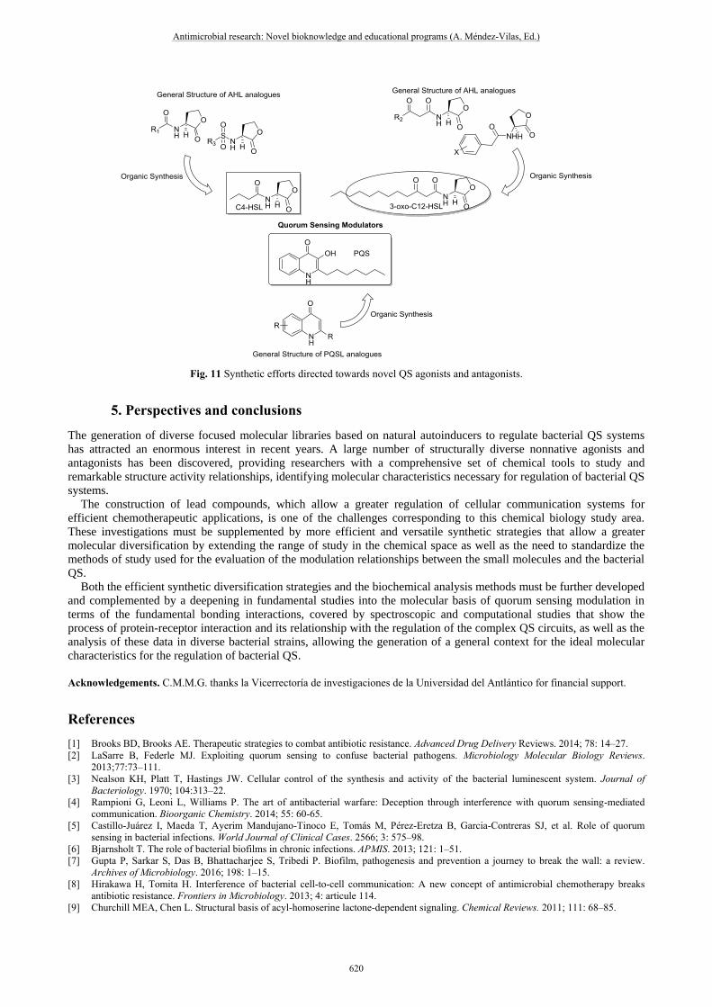
Fig. 11 Synthetic efforts directed towards novel QS agonists and antagonists.
5. Perspectives and conclusions
The generation of diverse focused molecular libraries based on natural autoinducers to regulate bacterial QS systems
has attracted an enormous interest in recent years. A large number of structurally diverse nonnative agonists and
antagonists has been discovered, providing researchers with a comprehensive set of chemical tools to study and
remarkable structure activity relationships, identifying molecular characteristics necessary for regulation of bacterial QS
systems.
The construction of lead compounds, which allow a greater regulation of cellular communication systems for
efficient chemotherapeutic applications, is one of the challenges corresponding to this chemical biology study area.
These investigations must be supplemented by more efficient and versatile synthetic strategies that allow a greater
molecular diversification by extending the range of study in the chemical space as well as the need to standardize the
methods of study used for the evaluation of the modulation relationships between the small molecules and the bacterial
QS.
Both the efficient synthetic diversification strategies and the biochemical analysis methods must be further developed
and complemented by a deepening in fundamental studies into the molecular basis of quorum sensing modulation in
terms of the fundamental bonding interactions, covered by spectroscopic and computational studies that show the
process of protein-receptor interaction and its relationship with the regulation of the complex QS circuits, as well as the
analysis of these data in diverse bacterial strains, allowing the generation of a general context for the ideal molecular
characteristics for the regulation of bacterial QS.
Acknowledgements. C.M.M.G. thanks la Vicerrectoría de investigaciones de la Universidad del Antlántico for financial support.
References
[1] Brooks BD, Brooks AE. Therapeutic strategies to combat antibiotic resistance. Advanced Drug Delivery Reviews. 2014; 78: 14–27.
[2] LaSarre B, Federle MJ. Exploiting quorum sensing to confuse bacterial pathogens. Microbiology Molecular Biology Reviews.
2013;77:73–111.
[3] Nealson KH, Platt T, Hastings JW. Cellular control of the synthesis and activity of the bacterial luminescent system. Journal of
Bacteriology. 1970; 104:313–22.
[4] Rampioni G, Leoni L, Williams P. The art of antibacterial warfare: Deception through interference with quorum sensing-mediated
communication. Bioorganic Chemistry. 2014; 55: 60-65.
[5] Castillo-Juárez I, Maeda T, Ayerim Mandujano-Tinoco E, Tomás M, Pérez-Eretza B, Garcia-Contreras SJ, et al. Role of quorum
sensing in bacterial infections. World Journal of Clinical Cases. 2566; 3: 575–98.
[6] Bjarnsholt T. The role of bacterial biofilms in chronic infections. APMIS. 2013; 121: 1–51.
[7] Gupta P, Sarkar S, Das B, Bhattacharjee S, Tribedi P. Biofilm, pathogenesis and prevention a journey to break the wall: a review.
Archives of Microbiology. 2016; 198: 1–15.
[8] Hirakawa H, Tomita H. Interference of bacterial cell-to-cell communication: A new concept of antimicrobial chemotherapy breaks
antibiotic resistance. Frontiers in Microbiology. 2013; 4: articule 114.
[9] Churchill MEA, Chen L. Structural basis of acyl-homoserine lactone-dependent signaling. Chemical Reviews. 2011; 111: 68–85.
Antimicrobial research: Novel bioknowledge and educational programs (A. Méndez-Vilas, Ed.)
620
_____________________________________________________________________________
[10] Schauder S, Shokat K, Surette MG, Bassler BL. The LuxS family of bacterial autoinducers: Biosynthesis of a novel quorum-sensing
signal molecule. Molecular Microbiology. 2001;41:463–476.
[11] Chen X, Schauder S, Potier N, Van Dorsselaer A, Pelczer I, Bassler BL, et al. Structural identification of a bacterial quorum-sensing
signal containing boron. Nature. 2002; 415: 545–549.
[12] Miller ST, Xavier KB, Campagna SR, Taga ME, Semmelhack MF, Bassler BL, et al. Salmonella typhimurium recognizes a chemically
distinct form of the bacterial quorum-sensing signal AI-2. Molecular Cell. 2004; 15: 677–687.
[13] Mok KC, Wingreen NS, Bassler BL. Vibrio harvei quorum sensing: A coincidence detector for two autoinducers controls gene
expression. EMBO Journal. 2003; 22: 870–881.
[14] Kelly RC, Bolitho ME, Higgins DA, Lu W, Ng W-L, Jeffrey PD, et al. The Vibrio cholerae quorum-sensing autoinducer CAI-1:
analysis of the biosynthetic enzyme CqsA. Nature Chemical Biology. 2009; 5: 891–895.
[15] Wei Y, Perez LJ, Ng WL, Semmelhack MF, Bassler BL. Mechanism of Vibrio cholerae autoinducer-1 biosynthesis. ACS Chemical
Biology. 2011; 6: 356–365.
[16] Li Z, Nair SK. Quorum sensing: How bacteria can coordinate activity and synchronize their response to external signals?. Protein
Science. 2012; 21: 1403– 1417.
[17] Parvez S, Venkataraman C, Mukherji S. A review on advantages of implementing luminescence inhibition test (Vibrio fischeri) for
acute toxicity prediction of chemicals. Environment Internacional. 2006; 32: 265–268.
[18] Welsh MA, Eibergen NR, Moore JD, Blackwell HE. Small molecule disruption of quorum sensing cross-regulation in Pseudomonas
aeruginosa causes major and unexpected alterations to virulence phenotypes. Journal of the American Chemical Society. 2015;
137:1510–1509.
[19] McInnis CE, Blackwell HE. Non-native N-Aroyl L -homoserine lactones are potent modulators of the quorum sensing receptor RpaR
in Rhodopseudomonas palustris. ChemBioChem. 2014; 15: 87–93.
[20] Tay SB, Yew WS. Development of quorum-based anti-virulence therapeutics targeting Gram-negative bacterial pathogens.
International Journal of Molecular Science. 2013; 14:16570–16599.
[21] Waters CM, Bassler BL. Quorum Sensing : Cell to cel communication in Bacteria. Annual review of cell and developmental biology.
2005; 21; 319-346.
[22] Stover CK, Pham XQ, Erwin L, Mizoguchi SD, Warrener P, Hickey MJ, et al. Complete genome sequence of Pseudomonas
aeruginosa PAO1, an opportunistic pathogen. Nature. 2000; 406: 959–964.
[23] Camilli A. Bacterial Small-Molecule Signaling Pathways. Science. 2006; 311: 1113–1116.
[24] Drees SL, Fetzner S. PqsE of Pseudomonas aeruginosa acts as pathway-specific thioesterase in the biosynthesis of alkylquinolone
signaling molecules. Chemistry and Biology. 2015; 22: 611–618.
[25] Cao H, Krishnan G, Goumnerov B, Tsongalis J, Tompkins R, Rahme LG. A quorum sensing-associated virulence gene of
Pseudomonas aeruginosa encodes a LysR-like transcription regulator with a unique self-regulatory mechanism. Proceedings of the
National Academy of Sciences of the United States of America. 2001; 98: 14613–14618.
[26] Pesci EC, Milbank JB, Pearson JP, McKnight S, Kende AS, Greenberg EP, et al. Quinolone signaling in the cell-to-cell communication
system of Pseudomonas aeruginosa. Proceedings of the National Academy of Sciences of the United States of America. 1999; 96:
11229–11234.
[27] Häussler S, Becker T. The pseudomonas quinolone signal (PQS) balances life and death in Pseudomonas aeruginosa populations.
PLoS Pathogens. 2008; 4: e1000166.
[28] Jimenez PN, Koch G, Thompson JA, Xavier KB, Cool RH, Quax WJ. The multiple signaling systems regulating virulence in
Pseudomonas aeruginosa. Microbiology and Molecular Biology Reviews. 2012; 76: 46–65.
[29] Thoendel M, Horswill AR. Biosynthesis of peptide signals in gram-positive bacteria. Advances in applied microbiology. 2010; 71: 91-
112.
[30] Gardan R, Besset C, Guillot A, Gitton C, Monnet V. The oligopeptide transport system is essential for the development of natural
competence in Streptococcus thermophilus strain LMD-9. Journal of Bacteriology. 2009; 191:4647– 4655.
[31] Skerker JM, Prasol MS, Perchuk BS, Biondi EG, Laub MT. Two-component signal transduction pathways regulating growth and cell
cycle progression in a bacterium: A system-level analysis. PLoS Biology. 2005; 3: e334.
[32] Cvitkovitch DG, Li YH, Ellen RP. Quorum sensing and biofilm formation in streptococcal infections. Journal of Clinical Investigation.
2003; 112: 1626–1632.
[33] Dong Y, Gusti AR, Zhang Q, Xu J, Zhang L. Identification of quorum-quenching N -acyl homoserine lactonases from Bacillus species.
Applied Environmental Microbiology. 2002; 68: 1754–1759.
[34] Dong YH, Wang LH, Xu JL, Zhang HB, Zhang XF, Zhang LH. Quenching quorum-sensing-dependent bacterial infection by an N-acyl
homoserine lactonase. Nature. 2001; 411: 813–817.
[35] Liu D, Thomas PW, Momb J, Hoang QQ, Petsko GA, Ringe D, et al. Structure and specificity of a quorum-quenching lactonase (AiiB
) from Agrobacterium tumefaciens. Biochemistry. 2007; 46: 11789–11799.
[36] Park SY, Lee SJ, Oh TK, Oh JW, Koo BT, Yum DY, et al. AhlD, an N-acylhomoserine lactonase in Arthrobacter sp., and predicted
homologues in other bacteria. Microbiology. 2003; 149:1541–1550.
[37] Schipper C, Hornung C, Bijtenhoorn P, Quitschau M, Grond S, Streit WR. Metagenome-derived clones encoding two novel lactonase
family proteins involved in biofilm inhibition in Pseudomonas aureginosa. Applied and Environmental Microbiology. 2009; 75: 224–
233.
[38] Uroz S, Chhabra SR, Cámara M, Williams P, Oger P, Dessaux Y. N-acylhomoserine lactone quorum-sensing molecules are modified
and degraded by Rhodococcus erythropolis W2 by both amidolytic and novel oxidoreductase activities. Microbiology. 2005;151:
3313–3322.
[39] Chen F, Gao Y, Chen X, Yu Z, Li X. Quorum quenching enzymes and their application in degrading signal molecules to block quorum
sensing-dependent infection. International Journal of Molecular Sciences. 2013; 14:17477–17500.
[40] Leadbetter JR, Greenberg EP. Metabolism of acyl-homoserine lactone quorum-sensing signals by Variovorax paradoxus. Journal of
Bacteriology. 2000; 182: 6921–6926.
[41] Oinonen C, Rouvinen J. Structural comparison of Ntn-hydrolases. Protein science. 2000; 9: 2329–2337.
[42] Sio CF, Otten LG, Cool RH, Diggle SP, Braun PG, Bos R, et al. Quorum quenching by an N-acyl-homoserine lactone acylase from
Pseudomonas aeruginosa PAO1. Infection and Immunity. 2006; 74: 1673–1682.
[43] Pomianek ME, Semmelhack MF. Making bacteria behave: New agonists and antagonists of quorum sensing. ACS Chemical Biology.
2007; 2: 293–295.
Antimicrobial research: Novel bioknowledge and educational programs (A. Méndez-Vilas, Ed.)
621
_____________________________________________________________________________

[44] Geske GD, Neill JCO, Miller DM, Mattmann ME, Blackwell HE. Modulation of bacterial quorum sensing with synthetic ligands:
systematic evaluation of N-acylated homoserine lactones in multiple species and new insights into their mechanisms of action. Journal
of the American Chemical Society. 2007; 8: 13613–13625.
[45] Geske GD, O’Neill JC, Blackwell HE. Expanding dialogues: from natural autoinducers to non-natural analogues that modulate quorum
sensing in Gram-negative bacteria. Chemical Society Reviews. 2008; 37: 1432–1447.
[46] Reverchon S, Chantegrel B, Deshayes C, Cotte-pattat N. New synthetic analogues of N-acyl homoserine lactones as agonists or
antagonists of transcriptional regulators involved in bacterial quorum sensing. Bioorganic and Medicinal Chemistry Letters. 2002; 12:
1153–1157.
[47] Jadhav GP, Chhabra SR, Telford G, Hooi DSW, Righetti K, Williams P, et al. Immunosuppressive but non-LasR-inducing analogues
of the Pseudomonas aeruginosa quorum-sensing molecule N-(3-Oxododecanoyl )-L -homoserine lactone. Journal of Medicinal
Chemistry. 2011; 54: 3348–3359.
[48] Boukraa M, Sabbah M, Soulère L, El ML. AHL-dependent quorum sensing inhibition : Synthesis and biological evaluation of α-(N -
alkyl-carboxamide )-γ-butyrolactones. Bioorganic and Medicinal Chemistry Letters. 2011; 21: 6876–6879.
[49] Majik MS, Naik D, Bhat C, Tilve S, Tilvi S, Souza LD. Synthesis of (R)-norbgugaine and its potential as quorum sensing inhibitor
against Pseudomonas aeruginosa. Bioorganic and Medicinal Chemistry Letters. 2013; 23: 2353–2356.
[50] Praneenararat T, Beary TMJ, Breitbach AS, Blackwell HE. Synthesis and application of an N-acylated L -homoserine lactone
derivatized affinity matrix for the isolation of quorum sensing signal receptors. Bioorganic and Medicinal Chemistry Letters. 2011; 21:
5054–5057.
[51] Castang S, Chantegrel B, Deshayes C, Gouet P, Haser R, Reverchon S, et al. N -Sulfonyl homoserine lactones as antagonists of
bacterial quorum sensing. Bioorganic and Medicinal Chemistry Letters. 2004; 14: 5145–5149.
[52] Olsen JA, Severinsen R, Rasmussen TB, Hentzer M, Givskov M, Nielsen J. Synthesis of new 3- and 4-substituted analogues of acyl
homoserine lactone quorum sensing autoinducers. Bioorganic and Medicinal Chemistry Letters. 2002; 12: 325–328.
[53] Smith KM, Bu Y, Suga H. Library screening for synthetic agonists and antagonists of a Pseudomonas aeruginosa autoinducer.
Chemistry and Biology. 2003; 10: 563–571.
[54] Smith KM, Bu Y, Suga H. Induction and inhibition of Pseudomonas aeruginosa quorum sensing by synthetic autoinducer analogs.
Chemistry and Biology. 2003; 10: 81–89.
[55] Geske GD, Wezeman RJ, Siegel AP, Blackwell HE. Small molecule inhibitors of bacterial quorum sensing and biofilm formation.
Journal of the American Chemical Society. 2005; 2: 12762–12763.
[56] Geske GD, Neill JCO, Blackwell HE. N-phenylacetanoyl-L-homoserine lactones can strongly antagonize or superagonize quorum
sensing in Vibrio fischeri. ACS Chemical Biology. 2007; 2: 315–320.
[57] Stacy DM, Welsh MA, Rather PN, Blackwell HE. Attenuation of quorum sensing in the pathogen Acinetobacter baumannii using non-
native N-acyl homoserine lactones. ACS Chemical Biology. 2012; 7, 1719-1728.
[58] Eibergen NR, Moore JD, Mattmann ME, Blackwell HE. Potent and selective modulation of the RhlR quorum sensing receptor by using
non-native ligands: An emerging target for virulence control in Pseudomonas aeruginosa. ChemBioChem. 2015; 16: 2348–2356.
[59] Mattmann ME, Shipway PM, Heth NJ, Blackwell HE. potent and selective synthetic modulators of a quorum sensing repressor in
pseudomonas aeruginosa identified from second-generation libraries of N-acylated L-homoserine lactones. ChemBioChem. 2011;12:
942–949.
[60] Geske GD, O’Neill JC, Blackwell HE. N-phenylacetanoyl-L-homoserine lactones can strongly antagonize or superagonize quorum
sensing in Vibrio fischeri. ACS Chemical Biology. 2007; 2:315–319.
[61] Palmer AG, Streng E, Jewell KA, Blackwell HE. Quorum sensing in bacterial species that use degenerate autoinducers can be tuned by
using structurally identical non-native ligands. ChemBioChem. 2011; 12: 138–47.
[62] Praneenararat T, Geske GD, Blackwell HE. Efficient synthesis and evaluation of quorum-sensing modulators using small molecule
macroarrays. Organic Letters. 2009; 11:4600–4603.
[63] Mcinnis CE, Blackwell HE. Design , synthesis , and biological evaluation of abiotic , non-lactone modulators of LuxR-type quorum
sensing. Bioorganic and Medicinal Chemistry. 2011; 19: 4812–4819.
[64] Mcinnis CE, Blackwell HE. Thiolactone modulators of quorum sensing revealed through library design and screening. Bioorganic and
Medicinal Chemistry. 2011; 19: 4820–4828.
[65] Mattmann ME, Blackwell HE. Small molecules that modulate quorum sensing and control virulence in Pseudomonas aeruginosa.
Journal of Organic Chemistry. 2010; 75: 6737–6746.
[66] Galloway WRJD, Hodgkinson JT, Bowden SD, Welch M, Spring DR. Quorum sensing in gram-negative bacteria : small-molecule
modulation of ahl and ai-2 quorum sensing pathways. Chemical Reviews. 2011; 44: 28–67.
[67] Yates EA, Philipp B, Buckley C, Atkinson S, Chhabra SR, Sockett RE, et al. N-acylhomoserine lactones undergo lactonolysis in a pH-,
temperature-, and acyl chain length-dependent manner during growth of Yersinia pseudotuberculosis and Pseudomonas aeruginosa.
Infection and Immunity. 2002; 70: 5635–5646.
[68] Moore JD, Rossi FM, Welsh MA, Nyffeler KE, Blackwell HE. A comparative analysis of synthetic quorum sensing modulators in
pseudomonas aeruginosa: new insights into mechanism, active efflux susceptibility, phenotypic response, and next-generation ligand
design. Journal of the American Chemical Society. 2015; 137: 14626–14639.
[69] Zou Y, Nair SK. Molecular basis for the recognition of structurally distinct autoinducer mimics by the pseudomonas aeruginosa lasr
quorum-sensing signaling receptor. Chemistry and Biology. 2009; 16: 961–970.
[70] Reilly MCO, Blackwell HE. Structure-based design and biological evaluation of triphenyl sca ff old-based hybrid compounds as
hydrolytically stable modulators of a luxr-type quorum sensing receptor. ACS Infectious Diseases. 2016; 2: 32-38.
[71] Spandl RJ, Nicholson RL, Marsden DM, Hodgkinson JT, Su X, Thomas GL, et al. Synthesis of a biotin-labeled quorum-sensing
molecule: Towards a general method for target identification. Synlett. 2008; 14: 2122–2126.
[72] Rayo J, Amara N, Krief P, Meijler MM. Live cell labeling of native intracellular bacterial receptors using aniline-catalyzed oxime
ligation. Journal of the American Chemical Society. 2011; 133: 7469–7475.
[73] Kaufmann GF, Park J, Mee JM, Ulevitch RJ, Janda KD. The quorum quenching antibody RS2-1G9 protects macrophages from the
cytotoxic effects of the Pseudomonas aeruginosa quorum sensing signalling molecule N-3-oxo-dodecanoyl-homoserine lactone.
Molecular Immunology. 2008; 45: 2710–2714.
Antimicrobial research: Novel bioknowledge and educational programs (A. Méndez-Vilas, Ed.)
622
_____________________________________________________________________________