Embed Size (px)
Citation preview
THE OTOSCOPEA View Through
Distinguishing Acute Otitis MediaFrom Otitis Media With Effusion
THE OTOSCOPE
Sponsored by
Slide-Lecture Book
Date of original release: January 2000
Date of re-release: June 2002
Term of approval: through June 2004
The Johns Hopkins University School of Medicineand The Institute for Johns Hopkins Nursing
This continuing medical education program is supported byan unrestricted educational grant from Pfizer Inc.
1. Title slide
2. Otitis Media With Effusion
3. Acute Otitis Media
4. Pneumatic Otoscopy
5. Tympanometry: Compliance
6. Tympanometry: Middle-Ear Pressure
7. Tympanometry: Peak
8. Spectral Gradient Acoustic Reflectometry
Cases also discussed in the video presentation on this CD-ROM
9. Case 1
10. Case 2
11. Case 3
12. Case 4
13. Case 5
14. Case 6
15. Case 7
16. Case 8
17. Case 9
18. Case 10
19. Case 11
20. Case 12
21. Case 13
22. Case 14
23. Case 15
Additional cases
24. Case 16
25. Case 17
26. Case 18
27. Case 19
28. Case 20
29. Case 21
30. Case 22
31. Case 23
32. Case 24
Index of Slides

3
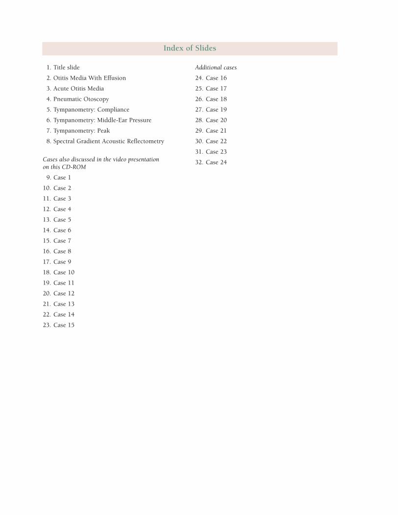
Middle-ear effusion is defined as the presence, on pneumatic otoscopy, of at least two of thefollowing three tympanic membrane (TM) abnormalities:
• abnormal color, such as white, yellow, amber, or blue discoloration;
• opacification other than due to scarring; or
• decreased or absent mobility;
or the presence of bubbles or air-fluid interfaces.1,2
1. Paradise JL. On classifying otitis media as suppurative or nonsuppurative, with a suggested clinical schema. J Pediatr.1987;111(6 Pt 1):948-951. 2. Kaleida PH. The COMPLETES exam for otitis. Contemp Pediatr. 1997;14:93-101.
4
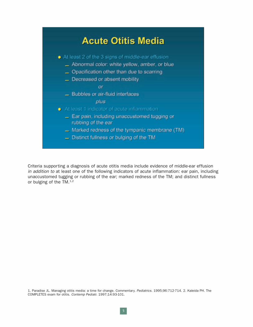
Criteria supporting a diagnosis of acute otitis media include evidence of middle-ear effusion in addition to at least one of the following indicators of acute inflammation: ear pain, includingunaccustomed tugging or rubbing of the ear; marked redness of the TM; and distinct fullnessor bulging of the TM.1,2
1. Paradise JL. Managing otitis media: a time for change. Commentary. Pediatrics. 1995;96:712-714. 2. Kaleida PH. TheCOMPLETES exam for otitis. Contemp Pediatr. 1997;14:93-101.
5
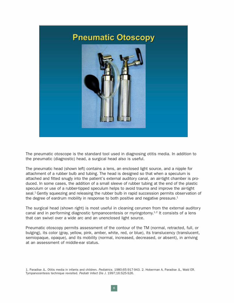
The pneumatic otoscope is the standard tool used in diagnosing otitis media. In addition tothe pneumatic (diagnostic) head, a surgical head also is useful.
The pneumatic head (shown left) contains a lens, an enclosed light source, and a nipple forattachment of a rubber bulb and tubing. The head is designed so that when a speculum isattached and fitted snugly into the patient’s external auditory canal, an air-tight chamber is pro-duced. In some cases, the addition of a small sleeve of rubber tubing at the end of the plasticspeculum or use of a rubber-tipped speculum helps to avoid trauma and improve the air-tightseal.1 Gently squeezing and releasing the rubber bulb in rapid succession permits observation ofthe degree of eardrum mobility in response to both positive and negative pressure.1
The surgical head (shown right) is most useful in cleaning cerumen from the external auditorycanal and in performing diagnostic tympanocentesis or myringotomy.1,2 It consists of a lensthat can swivel over a wide arc and an unenclosed light source.
Pneumatic otoscopy permits assessment of the contour of the TM (normal, retracted, full, orbulging), its color (gray, yellow, pink, amber, white, red, or blue), its translucency (translucent,semiopaque, opaque), and its mobility (normal, increased, decreased, or absent), in arrivingat an assessment of middle-ear status.
1. Paradise JL. Otitis media in infants and children. Pediatrics. 1980;65:917-943. 2. Hoberman A, Paradise JL, Wald ER.Tympanocentesis technique revisited. Pediatr Infect Dis J. 1997;16:S25-S26.
6
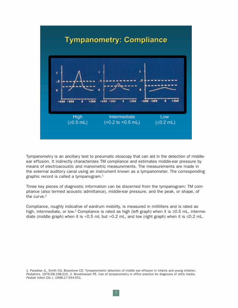
Tympanometry is an ancillary test to pneumatic otoscopy that can aid in the detection of middle-ear effusion. It indirectly characterizes TM compliance and estimates middle-ear pressure bymeans of electroacoustic and manometric measurements. The measurements are made inthe external auditory canal using an instrument known as a tympanometer. The correspondinggraphic record is called a tympanogram.1
Three key pieces of diagnostic information can be discerned from the tympanogram: TM com-pliance (also termed acoustic admittance), middle-ear pressure, and the peak, or shape, ofthe curve.2
Compliance, roughly indicative of eardrum mobility, is measured in milliliters and is rated ashigh, intermediate, or low.2 Compliance is rated as high (left graph) when it is ≥0.5 mL, interme-diate (middle graph) when it is <0.5 mL but >0.2 mL, and low (right graph) when it is ≤0.2 mL.
1. Paradise JL, Smith CG, Bluestone CD. Tympanometric detection of middle ear effusion in infants and young children.Pediatrics. 1976;58:198-210. 2. Brookhauser PE. Use of tympanometry in office practice for diagnosis of otitis media. Pediatr Infect Dis J. 1998;17:544-551.
7
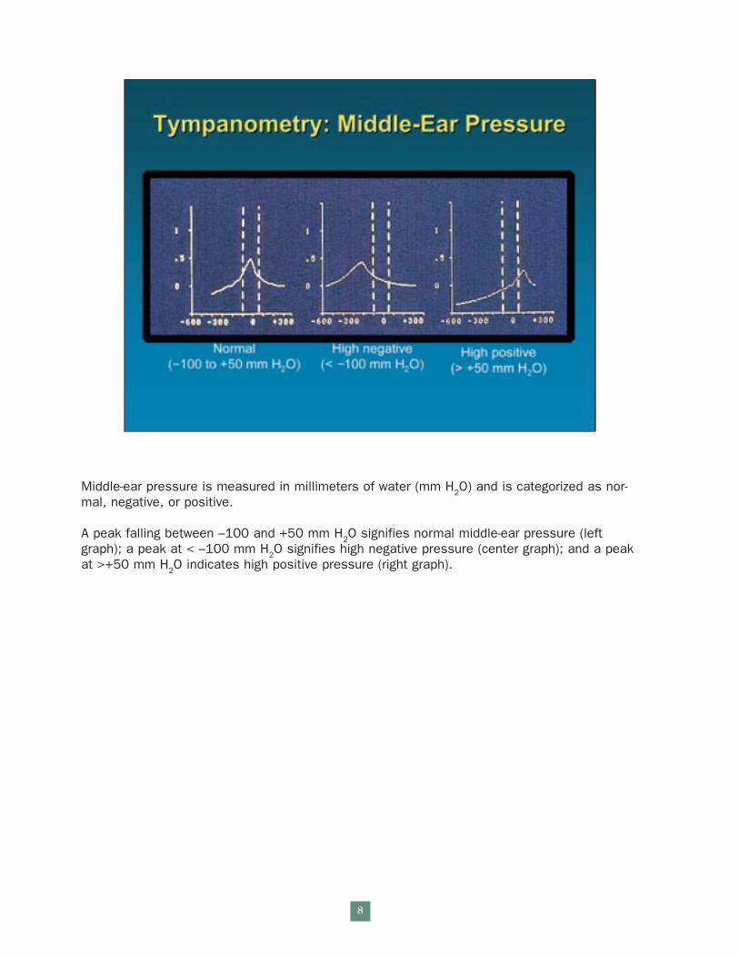
Middle-ear pressure is measured in millimeters of water (mm H2O) and is categorized as nor-mal, negative, or positive.
A peak falling between –100 and +50 mm H2O signifies normal middle-ear pressure (leftgraph); a peak at < –100 mm H2O signifies high negative pressure (center graph); and a peakat >+50 mm H2O indicates high positive pressure (right graph).
8
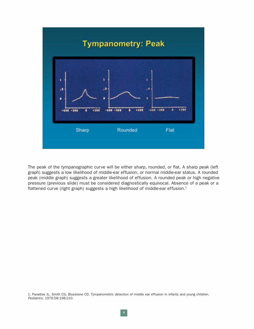
The peak of the tympanographic curve will be either sharp, rounded, or flat. A sharp peak (leftgraph) suggests a low likelihood of middle-ear effusion, or normal middle-ear status. A roundedpeak (middle graph) suggests a greater likelihood of effusion. A rounded peak or high negativepressure (previous slide) must be considered diagnostically equivocal. Absence of a peak or aflattened curve (right graph) suggests a high likelihood of middle-ear effusion.1
1. Paradise JL, Smith CG, Bluestone CD. Tympanometric detection of middle ear effusion in infants and young children.Pediatrics. 1976;58:198-210.
9
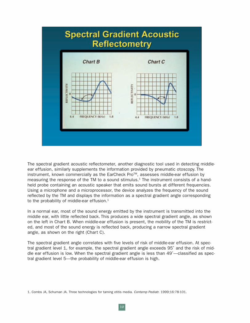
The spectral gradient acoustic reflectometer, another diagnostic tool used in detecting middle-ear effusion, similarly supplements the information provided by pneumatic otoscopy. Theinstrument, known commercially as the EarCheck ProTM, assesses middle-ear effusion bymeasuring the response of the TM to a sound stimulus.1 The instrument consists of a hand-held probe containing an acoustic speaker that emits sound bursts at different frequencies.Using a microphone and a microprocessor, the device analyzes the frequency of the soundreflected by the TM and displays the information as a spectral gradient angle corresponding to the probability of middle-ear effusion.1
In a normal ear, most of the sound energy emitted by the instrument is transmitted into themiddle ear, with little reflected back. This produces a wide spectral gradient angle, as shownon the left in Chart B. When middle-ear effusion is present, the mobility of the TM is restrict-ed, and most of the sound energy is reflected back, producing a narrow spectral gradientangle, as shown on the right (Chart C).
The spectral gradient angle correlates with five levels of risk of middle-ear effusion. At spec-tral gradient level 1, for example, the spectral gradient angle exceeds 95˚ and the risk of mid-dle ear effusion is low. When the spectral gradient angle is less than 49˚—classified as spec-tral gradient level 5—the probability of middle-ear effusion is high.
1. Combs JA, Schuman JA. Three technologies for taming otitis media. Contemp Pediatr. 1999;16:78-101.
10
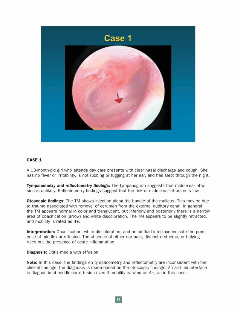
CASE 1
A 13-month-old girl who attends day care presents with clear nasal discharge and cough. Shehas no fever or irritability, is not rubbing or tugging at her ear, and has slept through the night.
Tympanometry and reflectometry findings: The tympanogram suggests that middle-ear effu-sion is unlikely. Reflectometry findings suggest that the risk of middle-ear effusion is low.
Otoscopic findings: The TM shows injection along the handle of the malleus. This may be dueto trauma associated with removal of cerumen from the external auditory canal. In general,the TM appears normal in color and translucent, but inferiorly and posteriorly there is a narrowarea of opacification (arrow) and white discoloration. The TM appears to be slightly retracted,and mobility is rated as 4+.
Interpretation: Opacification, white discoloration, and an air-fluid interface indicate the pres-ence of middle-ear effusion. The absence of either ear pain, distinct erythema, or bulgingrules out the presence of acute inflammation.
Diagnosis: Otitis media with effusion
Note: In this case, the findings on tympanometry and reflectometry are inconsistent with theclinical findings; the diagnosis is made based on the otoscopic findings. An air-fluid interfaceis diagnostic of middle-ear effusion even if mobility is rated as 4+, as in this case.
11
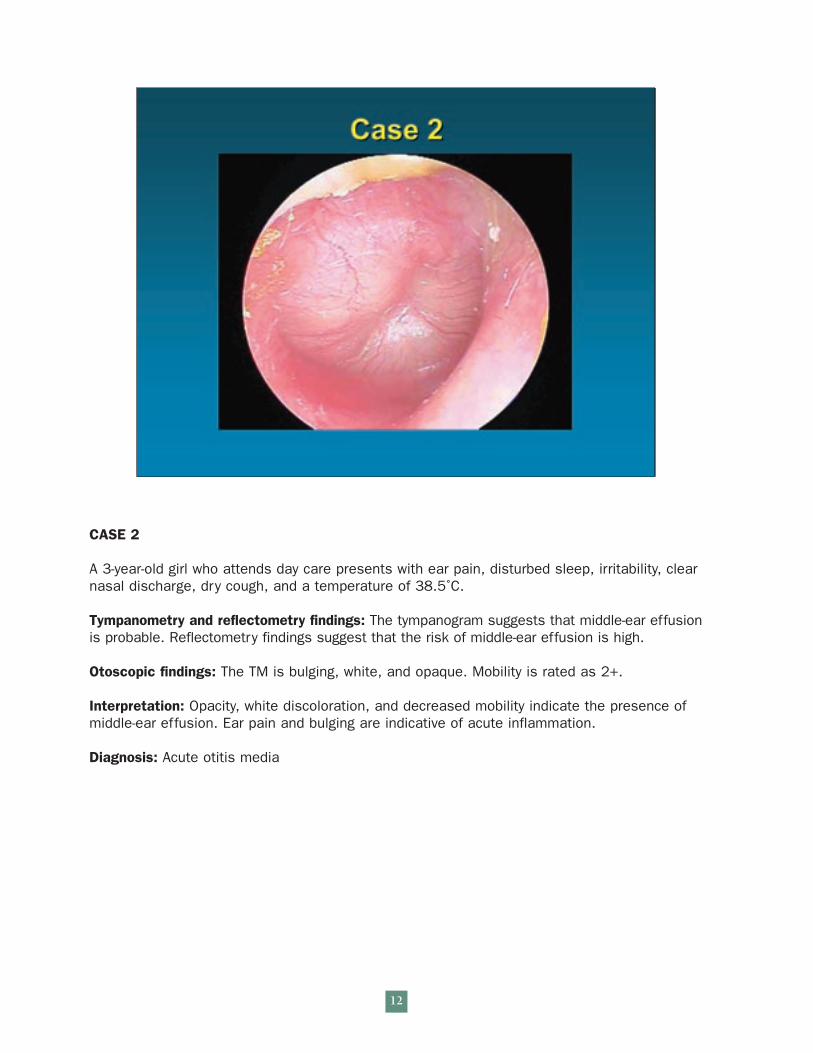
CASE 2
A 3-year-old girl who attends day care presents with ear pain, disturbed sleep, irritability, clearnasal discharge, dry cough, and a temperature of 38.5˚C.
Tympanometry and reflectometry findings: The tympanogram suggests that middle-ear effusionis probable. Reflectometry findings suggest that the risk of middle-ear effusion is high.
Otoscopic findings: The TM is bulging, white, and opaque. Mobility is rated as 2+.
Interpretation: Opacity, white discoloration, and decreased mobility indicate the presence ofmiddle-ear effusion. Ear pain and bulging are indicative of acute inflammation.
Diagnosis: Acute otitis media
12
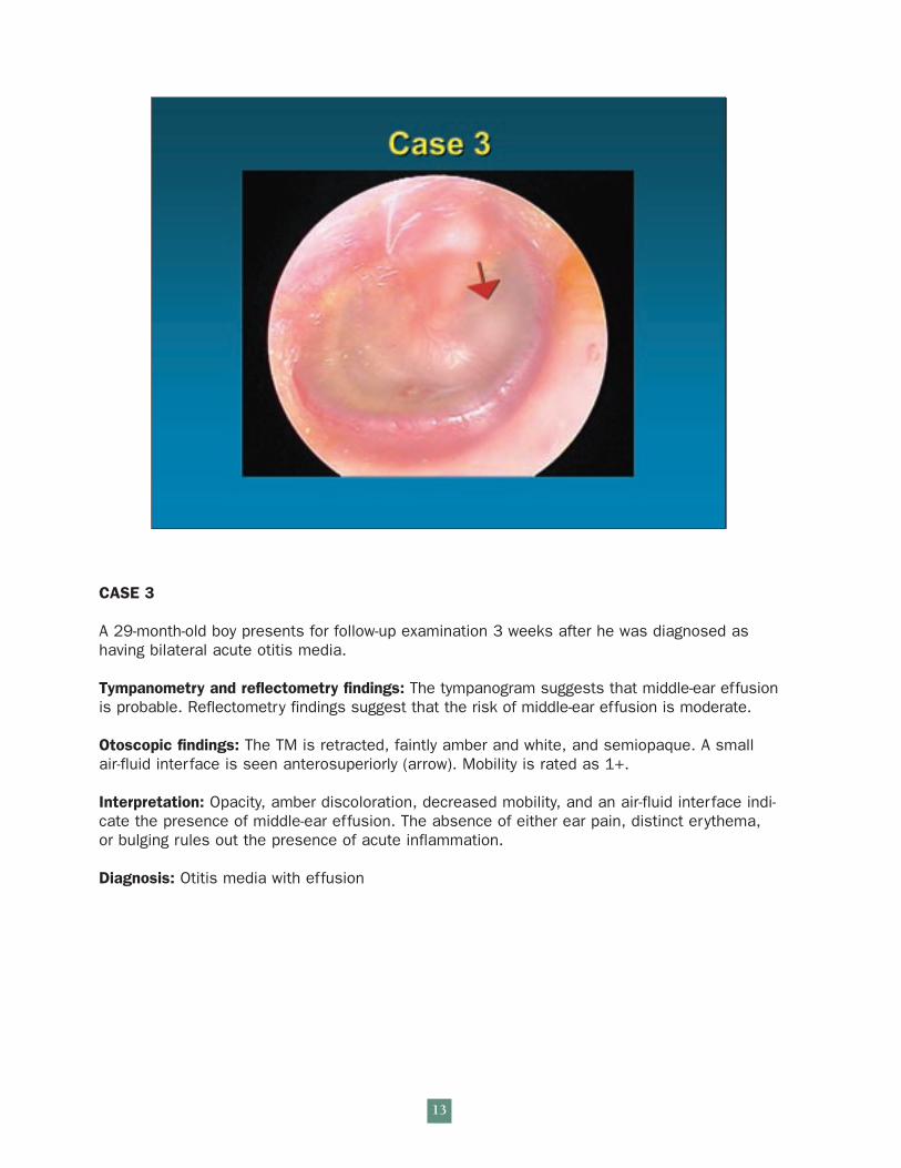
CASE 3
A 29-month-old boy presents for follow-up examination 3 weeks after he was diagnosed ashaving bilateral acute otitis media.
Tympanometry and reflectometry findings: The tympanogram suggests that middle-ear effusionis probable. Reflectometry findings suggest that the risk of middle-ear effusion is moderate.
Otoscopic findings: The TM is retracted, faintly amber and white, and semiopaque. A smallair-fluid interface is seen anterosuperiorly (arrow). Mobility is rated as 1+.
Interpretation: Opacity, amber discoloration, decreased mobility, and an air-fluid interface indi-cate the presence of middle-ear effusion. The absence of either ear pain, distinct erythema,or bulging rules out the presence of acute inflammation.
Diagnosis: Otitis media with effusion
13
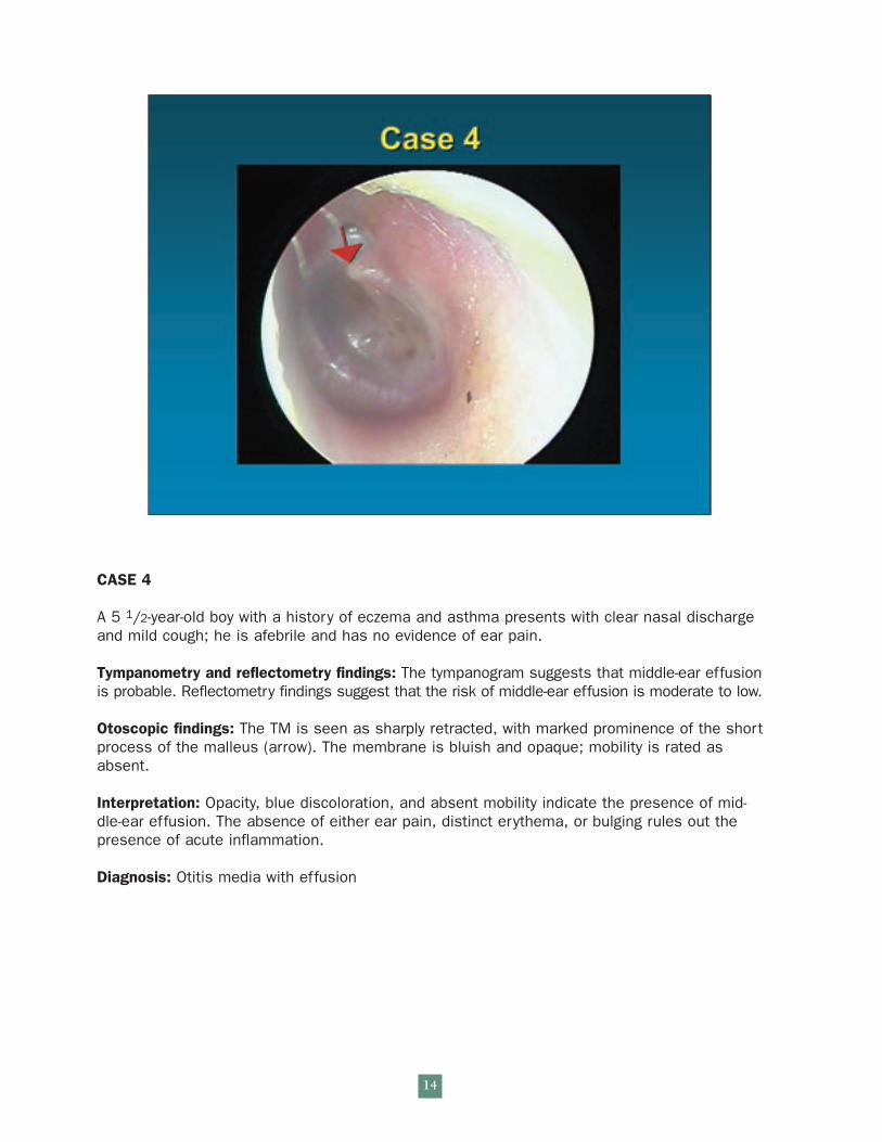
CASE 4
A 5 1/2-year-old boy with a history of eczema and asthma presents with clear nasal dischargeand mild cough; he is afebrile and has no evidence of ear pain.
Tympanometry and reflectometry findings: The tympanogram suggests that middle-ear effusionis probable. Reflectometry findings suggest that the risk of middle-ear effusion is moderate to low.
Otoscopic findings: The TM is seen as sharply retracted, with marked prominence of the shortprocess of the malleus (arrow). The membrane is bluish and opaque; mobility is rated asabsent.
Interpretation: Opacity, blue discoloration, and absent mobility indicate the presence of mid-dle-ear effusion. The absence of either ear pain, distinct erythema, or bulging rules out thepresence of acute inflammation.
Diagnosis: Otitis media with effusion
14
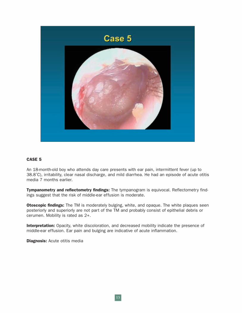
CASE 5
An 18-month-old boy who attends day care presents with ear pain, intermittent fever (up to38.8˚C), irritability, clear nasal discharge, and mild diarrhea. He had an episode of acute otitismedia 7 months earlier.
Tympanometry and reflectometry findings: The tympanogram is equivocal. Reflectometry find-ings suggest that the risk of middle-ear effusion is moderate.
Otoscopic findings: The TM is moderately bulging, white, and opaque. The white plaques seenposteriorly and superiorly are not part of the TM and probably consist of epithelial debris orcerumen. Mobility is rated as 2+.
Interpretation: Opacity, white discoloration, and decreased mobility indicate the presence ofmiddle-ear effusion. Ear pain and bulging are indicative of acute inflammation.
Diagnosis: Acute otitis media
15
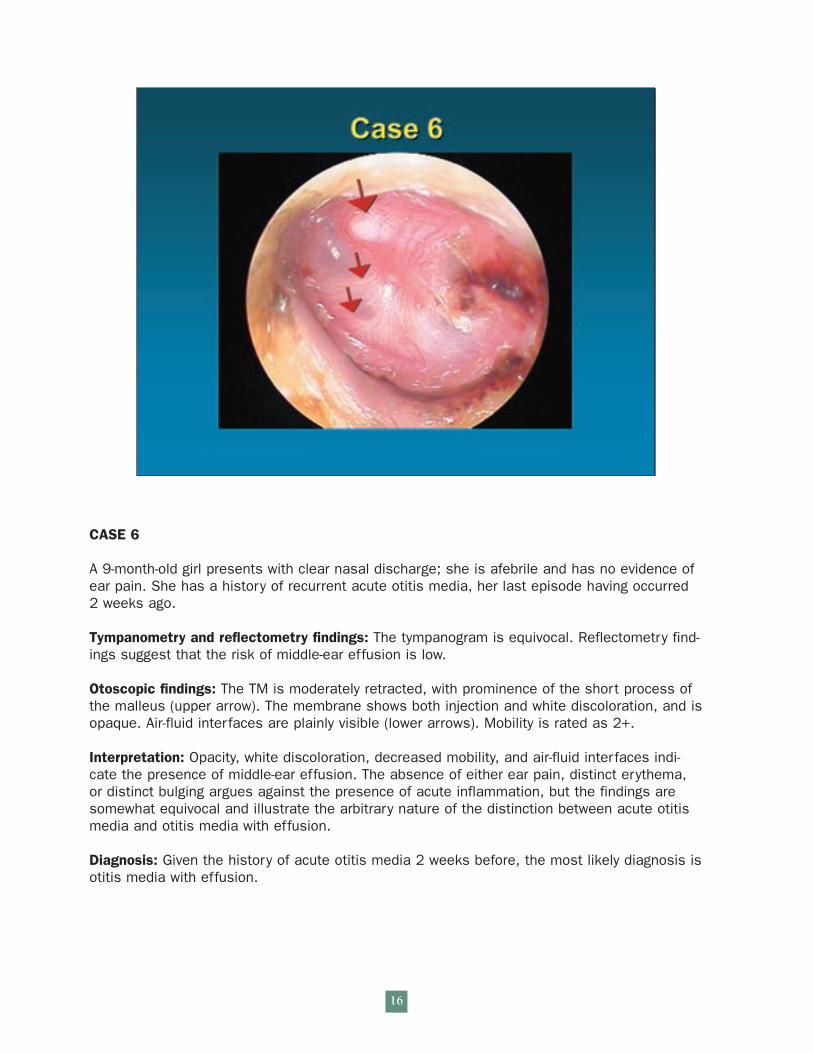
CASE 6
A 9-month-old girl presents with clear nasal discharge; she is afebrile and has no evidence ofear pain. She has a history of recurrent acute otitis media, her last episode having occurred 2 weeks ago.
Tympanometry and reflectometry findings: The tympanogram is equivocal. Reflectometry find-ings suggest that the risk of middle-ear effusion is low.
Otoscopic findings: The TM is moderately retracted, with prominence of the short process ofthe malleus (upper arrow). The membrane shows both injection and white discoloration, and isopaque. Air-fluid interfaces are plainly visible (lower arrows). Mobility is rated as 2+.
Interpretation: Opacity, white discoloration, decreased mobility, and air-fluid interfaces indi-cate the presence of middle-ear effusion. The absence of either ear pain, distinct erythema,or distinct bulging argues against the presence of acute inflammation, but the findings aresomewhat equivocal and illustrate the arbitrary nature of the distinction between acute otitismedia and otitis media with effusion.
Diagnosis: Given the history of acute otitis media 2 weeks before, the most likely diagnosis isotitis media with effusion.
16
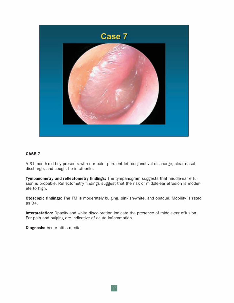
CASE 7
A 31-month-old boy presents with ear pain, purulent left conjunctival discharge, clear nasaldischarge, and cough; he is afebrile.
Tympanometry and reflectometry findings: The tympanogram suggests that middle-ear effu-sion is probable. Reflectometry findings suggest that the risk of middle-ear effusion is moder-ate to high.
Otoscopic findings: The TM is moderately bulging, pinkish-white, and opaque. Mobility is ratedas 3+.
Interpretation: Opacity and white discoloration indicate the presence of middle-ear effusion.Ear pain and bulging are indicative of acute inflammation.
Diagnosis: Acute otitis media
17
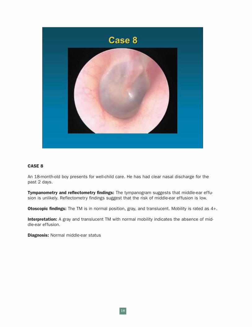
CASE 8
An 18-month-old boy presents for well-child care. He has had clear nasal discharge for thepast 2 days.
Tympanometry and reflectometry findings: The tympanogram suggests that middle-ear effu-sion is unlikely. Reflectometry findings suggest that the risk of middle-ear effusion is low.
Otoscopic findings: The TM is in normal position, gray, and translucent. Mobility is rated as 4+.
Interpretation: A gray and translucent TM with normal mobility indicates the absence of mid-dle-ear effusion.
Diagnosis: Normal middle-ear status
18
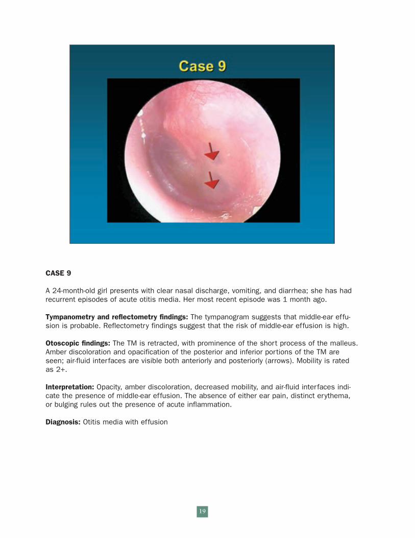
CASE 9
A 24-month-old girl presents with clear nasal discharge, vomiting, and diarrhea; she has hadrecurrent episodes of acute otitis media. Her most recent episode was 1 month ago.
Tympanometry and reflectometry findings: The tympanogram suggests that middle-ear effu-sion is probable. Reflectometry findings suggest that the risk of middle-ear effusion is high.
Otoscopic findings: The TM is retracted, with prominence of the short process of the malleus.Amber discoloration and opacification of the posterior and inferior portions of the TM areseen; air-fluid interfaces are visible both anteriorly and posteriorly (arrows). Mobility is ratedas 2+.
Interpretation: Opacity, amber discoloration, decreased mobility, and air-fluid interfaces indi-cate the presence of middle-ear effusion. The absence of either ear pain, distinct erythema,or bulging rules out the presence of acute inflammation.
Diagnosis: Otitis media with effusion
19
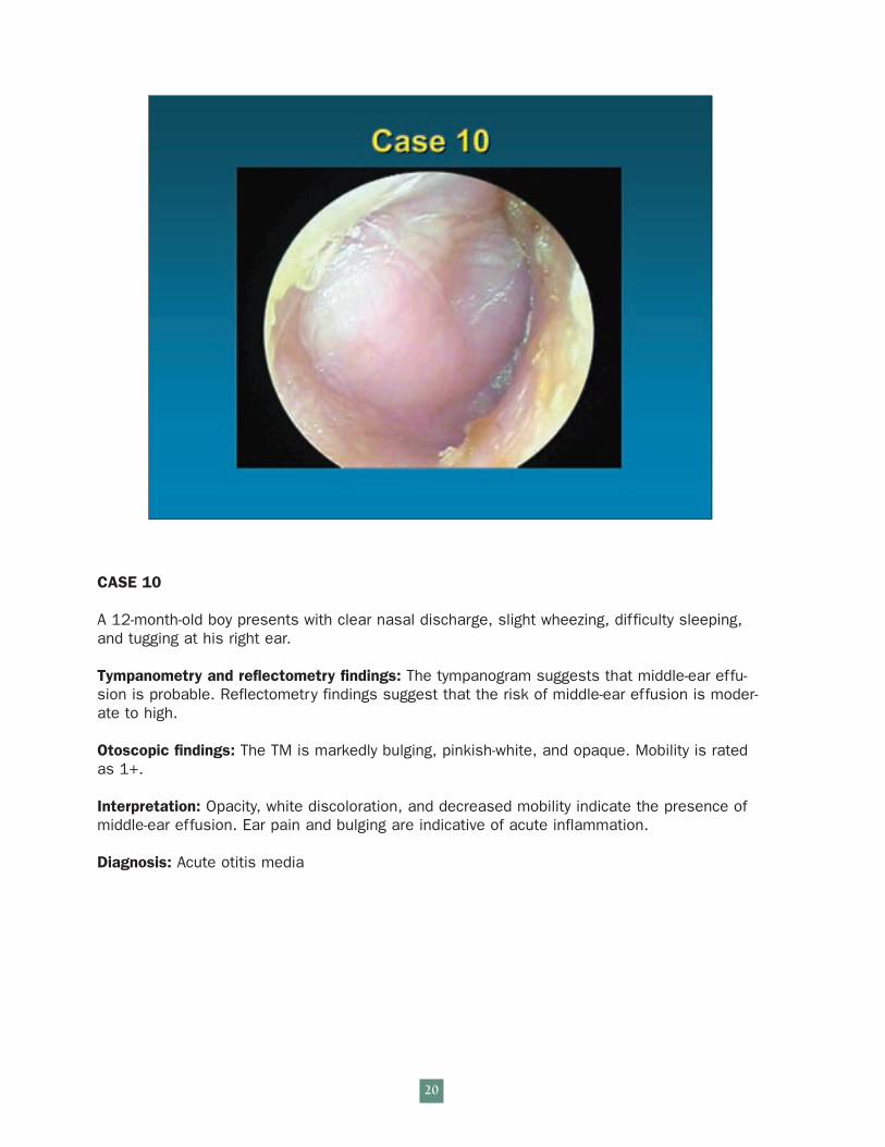
CASE 10
A 12-month-old boy presents with clear nasal discharge, slight wheezing, difficulty sleeping,and tugging at his right ear.
Tympanometry and reflectometry findings: The tympanogram suggests that middle-ear effu-sion is probable. Reflectometry findings suggest that the risk of middle-ear effusion is moder-ate to high.
Otoscopic findings: The TM is markedly bulging, pinkish-white, and opaque. Mobility is ratedas 1+.
Interpretation: Opacity, white discoloration, and decreased mobility indicate the presence ofmiddle-ear effusion. Ear pain and bulging are indicative of acute inflammation.
Diagnosis: Acute otitis media
20

CASE 11
A 9-month-old boy presents with clear nasal discharge for the past 3 days, nasal congestion,cough, and a temperature of 38.5˚C.
Tympanometry and reflectometry findings: The tympanogram suggests that middle-ear effu-sion is unlikely. Reflectometry findings suggest that the risk of middle-ear effusion is moder-ate to low.
Otoscopic findings: The TM is in normal position, pearly gray, and translucent. Mobility israted as 4+.
Interpretation: A gray and translucent TM with normal mobility indicates the absence of mid-dle-ear effusion.
Diagnosis: Normal middle-ear status
21
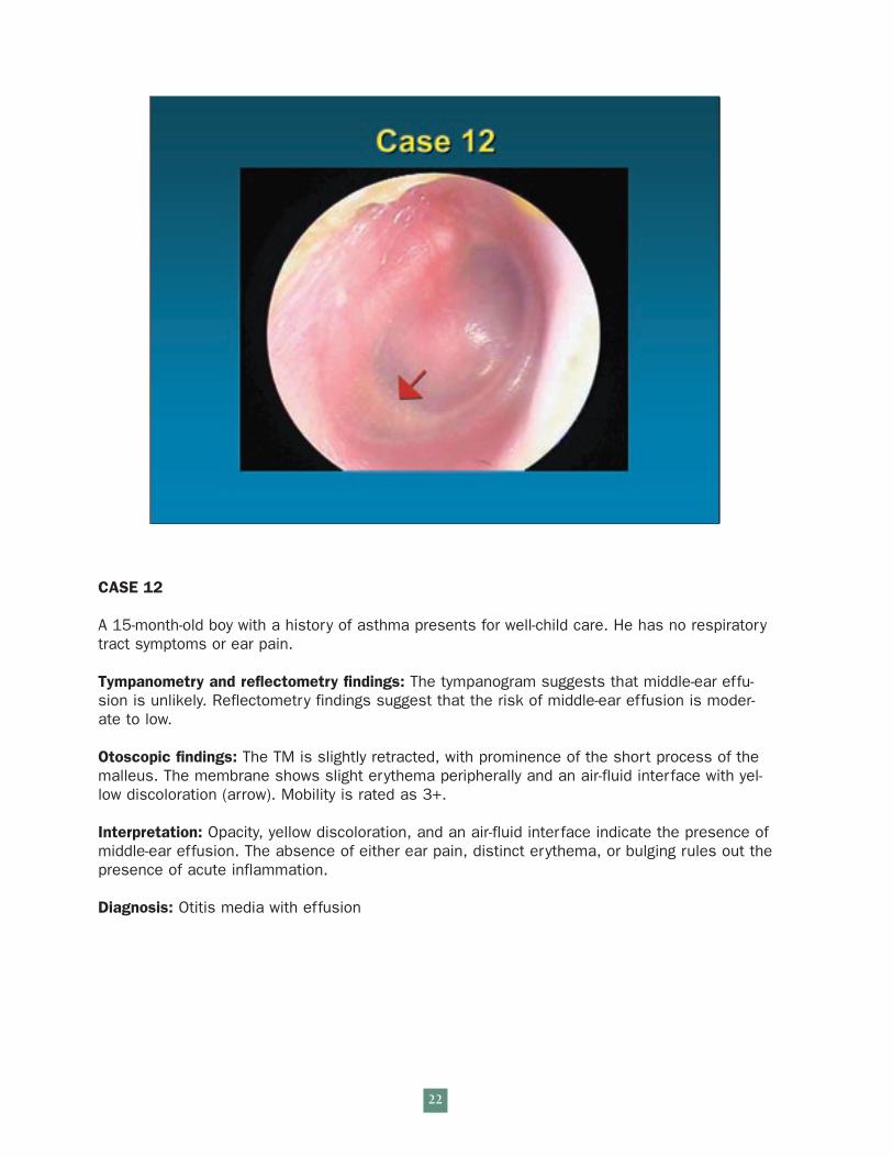
CASE 12
A 15-month-old boy with a history of asthma presents for well-child care. He has no respiratorytract symptoms or ear pain.
Tympanometry and reflectometry findings: The tympanogram suggests that middle-ear effu-sion is unlikely. Reflectometry findings suggest that the risk of middle-ear effusion is moder-ate to low.
Otoscopic findings: The TM is slightly retracted, with prominence of the short process of themalleus. The membrane shows slight erythema peripherally and an air-fluid interface with yel-low discoloration (arrow). Mobility is rated as 3+.
Interpretation: Opacity, yellow discoloration, and an air-fluid interface indicate the presence ofmiddle-ear effusion. The absence of either ear pain, distinct erythema, or bulging rules out thepresence of acute inflammation.
Diagnosis: Otitis media with effusion
22
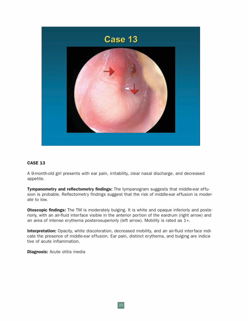
CASE 13
A 9-month-old girl presents with ear pain, irritability, clear nasal discharge, and decreasedappetite.
Tympanometry and reflectometry findings: The tympanogram suggests that middle-ear effu-sion is probable. Reflectometry findings suggest that the risk of middle-ear effusion is moder-ate to low.
Otoscopic findings: The TM is moderately bulging. It is white and opaque inferiorly and poste-riorly, with an air-fluid interface visible in the anterior portion of the eardrum (right arrow) andan area of intense erythema posterosuperiorly (left arrow). Mobility is rated as 1+.
Interpretation: Opacity, white discoloration, decreased mobility, and an air-fluid interface indi-cate the presence of middle-ear effusion. Ear pain, distinct erythema, and bulging are indica-tive of acute inflammation.
Diagnosis: Acute otitis media
23
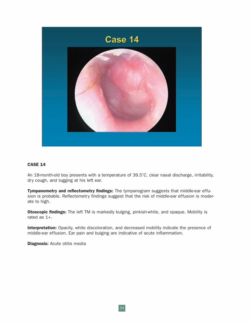
CASE 14
An 18-month-old boy presents with a temperature of 39.5˚C, clear nasal discharge, irritability,dry cough, and tugging at his left ear.
Tympanometry and reflectometry findings: The tympanogram suggests that middle-ear effu-sion is probable. Reflectometry findings suggest that the risk of middle-ear effusion is moder-ate to high.
Otoscopic findings: The left TM is markedly bulging, pinkish-white, and opaque. Mobility israted as 1+.
Interpretation: Opacity, white discoloration, and decreased mobility indicate the presence ofmiddle-ear effusion. Ear pain and bulging are indicative of acute inflammation.
Diagnosis: Acute otitis media
24
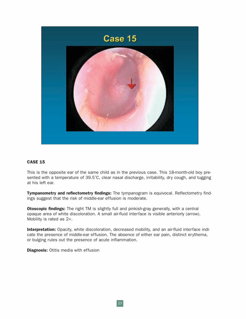
CASE 15
This is the opposite ear of the same child as in the previous case. This 18-month-old boy pre-sented with a temperature of 39.5˚C, clear nasal discharge, irritability, dry cough, and tuggingat his left ear.
Tympanometry and reflectometry findings: The tympanogram is equivocal. Reflectometry find-ings suggest that the risk of middle-ear effusion is moderate.
Otoscopic findings: The right TM is slightly full and pinkish-gray generally, with a centralopaque area of white discoloration. A small air-fluid interface is visible anteriorly (arrow).Mobility is rated as 2+.
Interpretation: Opacity, white discoloration, decreased mobility, and an air-fluid interface indi-cate the presence of middle-ear effusion. The absence of either ear pain, distinct erythema,or bulging rules out the presence of acute inflammation.
Diagnosis: Otitis media with effusion
25
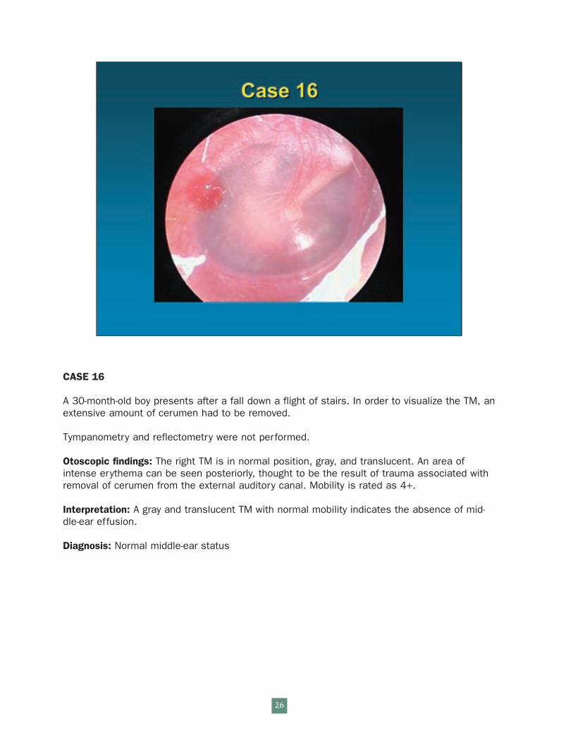
CASE 16
A 30-month-old boy presents after a fall down a flight of stairs. In order to visualize the TM, anextensive amount of cerumen had to be removed.
Tympanometry and reflectometry were not performed.
Otoscopic findings: The right TM is in normal position, gray, and translucent. An area ofintense erythema can be seen posteriorly, thought to be the result of trauma associated withremoval of cerumen from the external auditory canal. Mobility is rated as 4+.
Interpretation: A gray and translucent TM with normal mobility indicates the absence of mid-dle-ear effusion.
Diagnosis: Normal middle-ear status
26
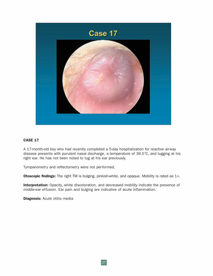
CASE 17
A 17-month-old boy who had recently completed a 5-day hospitalization for reactive airway disease presents with purulent nasal discharge, a temperature of 39.5˚C, and tugging at hisright ear. He has not been noted to tug at his ear previously.
Tympanometry and reflectometry were not performed.
Otoscopic findings: The right TM is bulging, pinkish-white, and opaque. Mobility is rated as 1+.
Interpretation: Opacity, white discoloration, and decreased mobility indicate the presence ofmiddle-ear effusion. Ear pain and bulging are indicative of acute inflammation.
Diagnosis: Acute otitis media
27
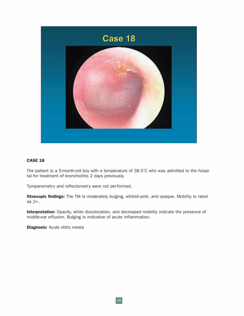
CASE 18
The patient is a 5-month-old boy with a temperature of 38.5˚C who was admitted to the hospi-tal for treatment of bronchiolitis 2 days previously.
Tympanometry and reflectometry were not performed.
Otoscopic findings: The TM is moderately bulging, whitish-pink, and opaque. Mobility is ratedas 2+.
Interpretation: Opacity, white discoloration, and decreased mobility indicate the presence ofmiddle-ear effusion. Bulging is indicative of acute inflammation.
Diagnosis: Acute otitis media
28
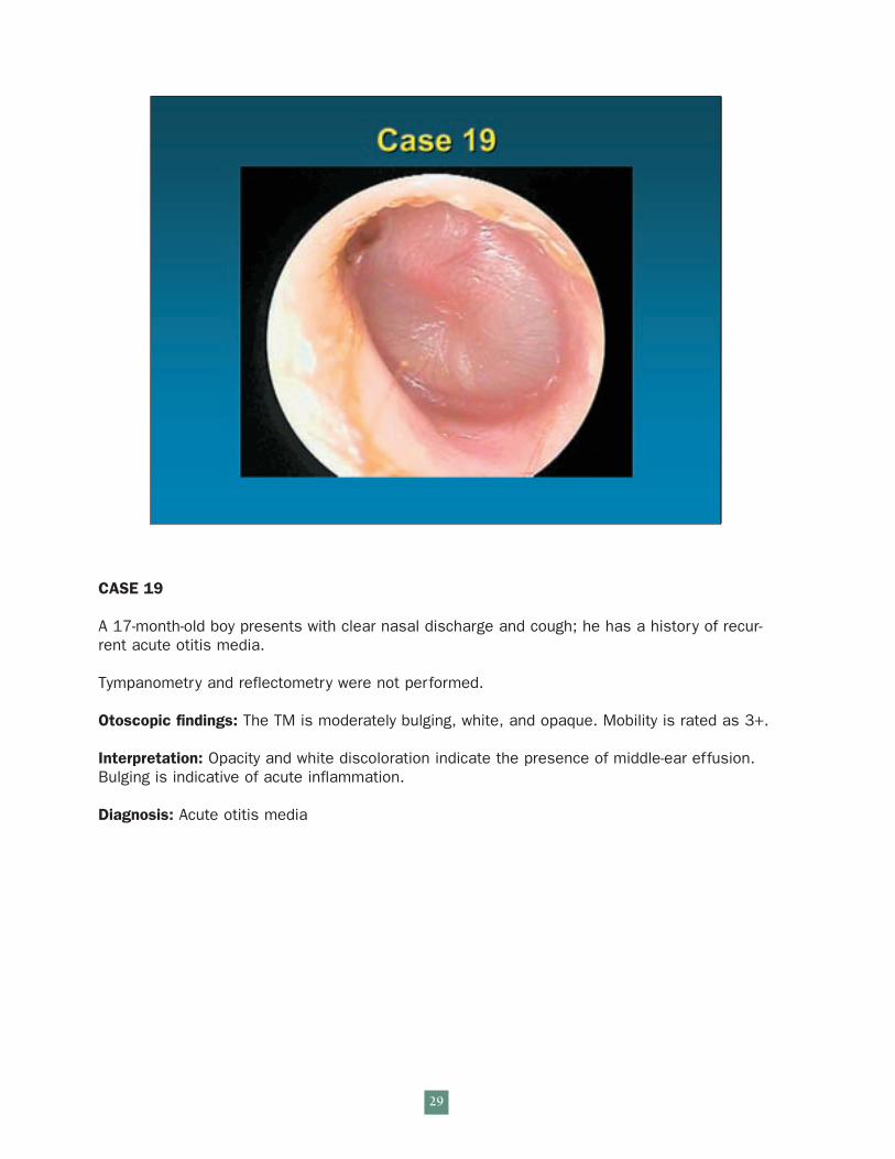
CASE 19
A 17-month-old boy presents with clear nasal discharge and cough; he has a history of recur-rent acute otitis media.
Tympanometry and reflectometry were not performed.
Otoscopic findings: The TM is moderately bulging, white, and opaque. Mobility is rated as 3+.
Interpretation: Opacity and white discoloration indicate the presence of middle-ear effusion.Bulging is indicative of acute inflammation.
Diagnosis: Acute otitis media
29
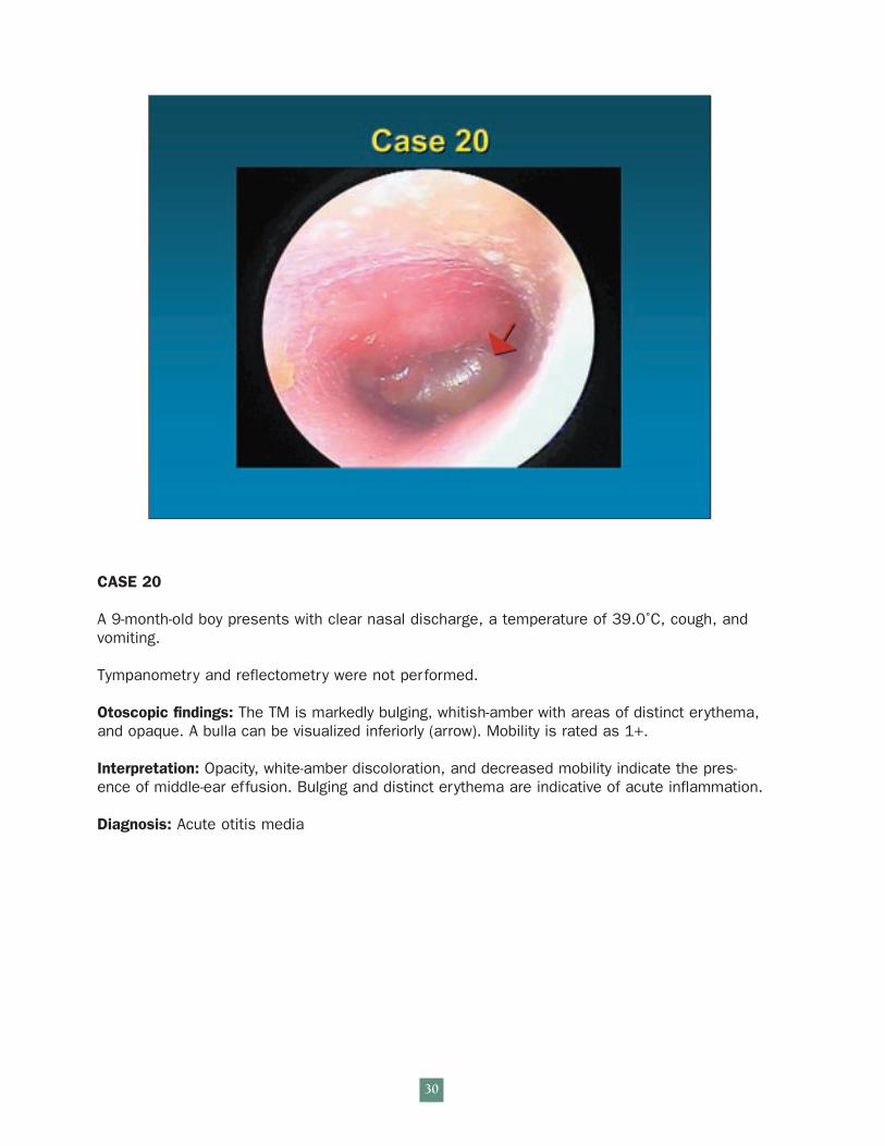
CASE 20
A 9-month-old boy presents with clear nasal discharge, a temperature of 39.0˚C, cough, andvomiting.
Tympanometry and reflectometry were not performed.
Otoscopic findings: The TM is markedly bulging, whitish-amber with areas of distinct erythema,and opaque. A bulla can be visualized inferiorly (arrow). Mobility is rated as 1+.
Interpretation: Opacity, white-amber discoloration, and decreased mobility indicate the pres-ence of middle-ear effusion. Bulging and distinct erythema are indicative of acute inflammation.
Diagnosis: Acute otitis media
30
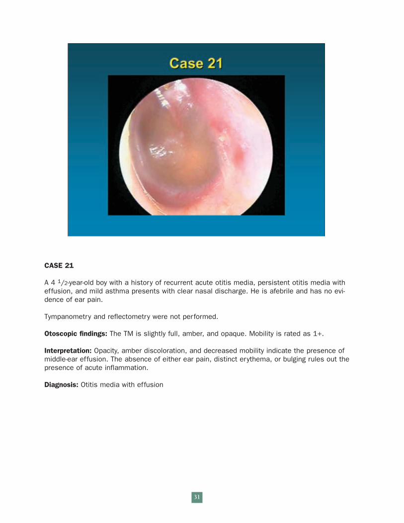
CASE 21
A 4 1/2-year-old boy with a history of recurrent acute otitis media, persistent otitis media witheffusion, and mild asthma presents with clear nasal discharge. He is afebrile and has no evi-dence of ear pain.
Tympanometry and reflectometry were not performed.
Otoscopic findings: The TM is slightly full, amber, and opaque. Mobility is rated as 1+.
Interpretation: Opacity, amber discoloration, and decreased mobility indicate the presence ofmiddle-ear effusion. The absence of either ear pain, distinct erythema, or bulging rules out thepresence of acute inflammation.
Diagnosis: Otitis media with effusion
31
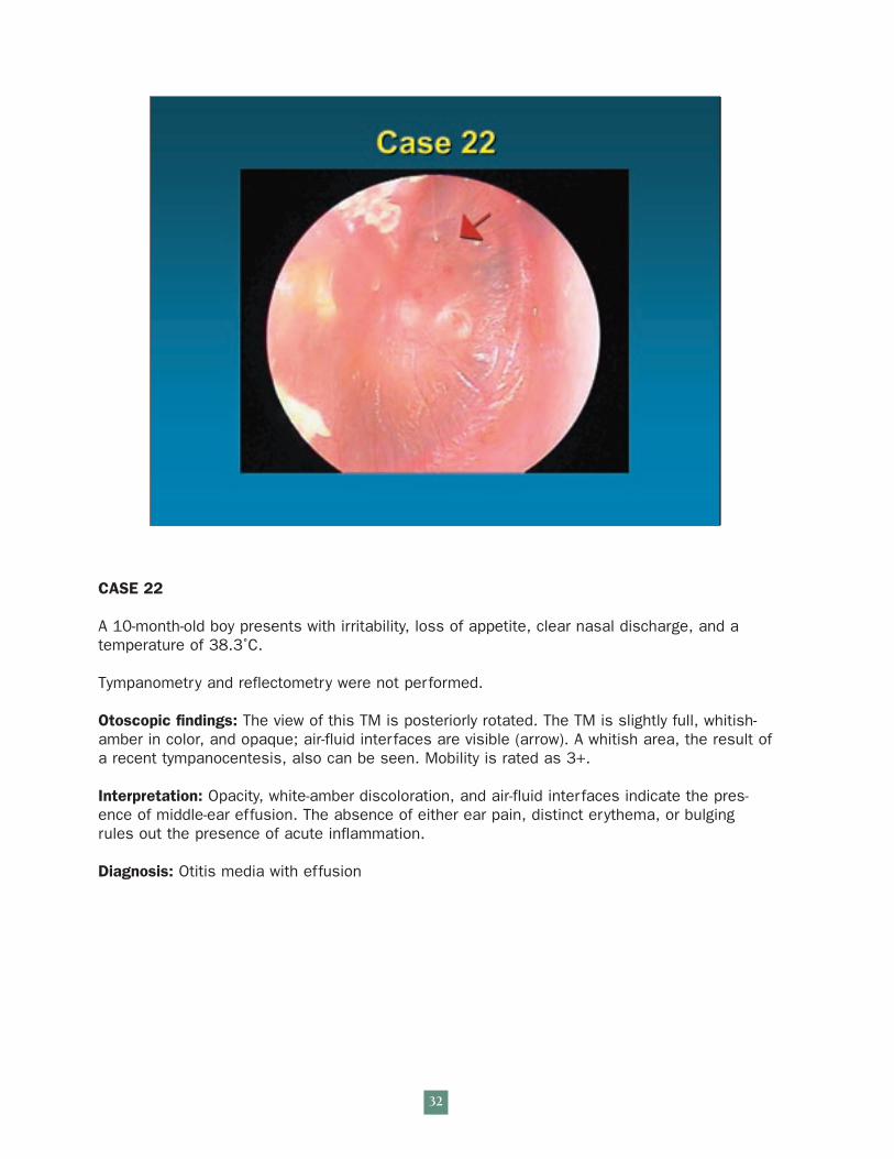
CASE 22
A 10-month-old boy presents with irritability, loss of appetite, clear nasal discharge, and atemperature of 38.3˚C.
Tympanometry and reflectometry were not performed.
Otoscopic findings: The view of this TM is posteriorly rotated. The TM is slightly full, whitish-amber in color, and opaque; air-fluid interfaces are visible (arrow). A whitish area, the result ofa recent tympanocentesis, also can be seen. Mobility is rated as 3+.
Interpretation: Opacity, white-amber discoloration, and air-fluid interfaces indicate the pres-ence of middle-ear effusion. The absence of either ear pain, distinct erythema, or bulgingrules out the presence of acute inflammation.
Diagnosis: Otitis media with effusion
32
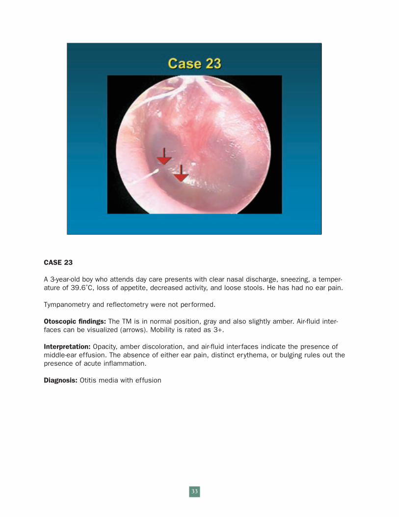
CASE 23
A 3-year-old boy who attends day care presents with clear nasal discharge, sneezing, a temper-ature of 39.6˚C, loss of appetite, decreased activity, and loose stools. He has had no ear pain.
Tympanometry and reflectometry were not performed.
Otoscopic findings: The TM is in normal position, gray and also slightly amber. Air-fluid inter-faces can be visualized (arrows). Mobility is rated as 3+.
Interpretation: Opacity, amber discoloration, and air-fluid interfaces indicate the presence ofmiddle-ear effusion. The absence of either ear pain, distinct erythema, or bulging rules out thepresence of acute inflammation.
Diagnosis: Otitis media with effusion
33
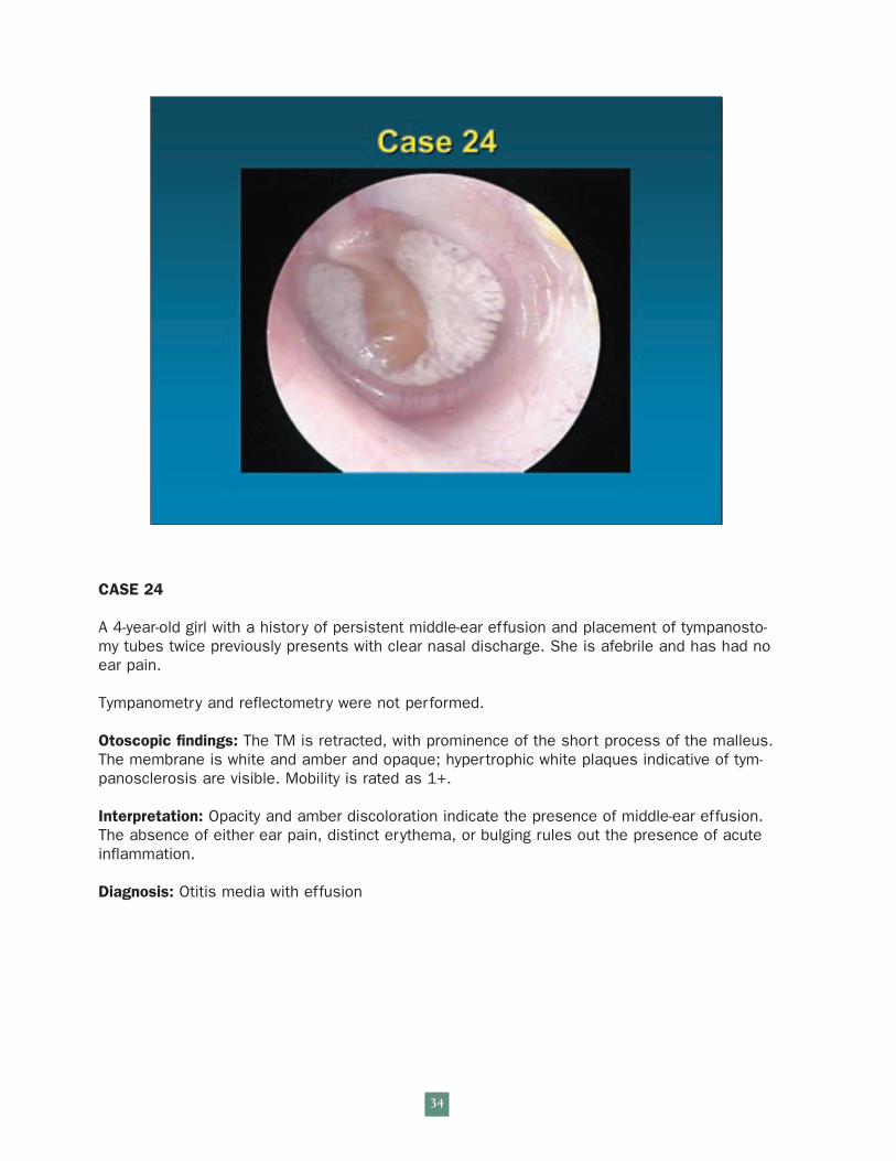
CASE 24
A 4-year-old girl with a history of persistent middle-ear effusion and placement of tympanosto-my tubes twice previously presents with clear nasal discharge. She is afebrile and has had noear pain.
Tympanometry and reflectometry were not performed.
Otoscopic findings: The TM is retracted, with prominence of the short process of the malleus.The membrane is white and amber and opaque; hypertrophic white plaques indicative of tym-panosclerosis are visible. Mobility is rated as 1+.
Interpretation: Opacity and amber discoloration indicate the presence of middle-ear effusion.The absence of either ear pain, distinct erythema, or bulging rules out the presence of acuteinflammation.
Diagnosis: Otitis media with effusion
34