Embed Size (px)
Citation preview

Copyright © 2011 Korean Neurological Association 25
Print ISSN 1738-6586 / On-line ISSN 2005-501310.3988/jcn.2011.7.1.25
ORIGINAL ARTICLEJ Clin Neurol 2011;7:25-30
Introduction
Progressive external ophthalmoplegia (PEO) is one of the most common manifestations of mitochondrial disease, either as a part of a multisystemic complex disorder or as a predominant feature of exclusive mitochondrial myopathy. Many cases of PEO are caused by the mutation of mitochondrial DNA (mtD-NA). A single heteroplasmic deletion of mtDNA is a frequent cause of sporadic PEO, being responsible for approximately 90% and 50% of cases of Kearns-Sayre syndrome and PEO,
respectively.1 In other patients who show maternal inheritance pattern, PEO is associated with various point mutations of mtDNA, of which A3243G is the most frequent.2 Yet another group of PEO patients display autosomal dominant or reces-sive inheritance patterns that are usually associated with mul-tiple mtDNA deletions or depletions that arise secondarily to nuclear gene defects.3 The responsible genes include POLG, which encodes the catalytic subunit of DNA polymerase γ; SL-C25A4, which encodes the mitochondrial adenine nucleotide translocator ANT1; C10ORF2 (also known as PEO1), which
SLC25A4 and C10ORF2 Mutations in Autosomal Dominant Progressive External Ophthalmoplegia
Kyung-Pil Park,a,b Hyang-Sook Kim,b Eun-Sook Kim,b Young-Eun Park,b,c Chang-Hoon Lee,b,d Dae-Seong Kima,b
a Department of Neurology, Pusan National University Yangsan Hospital, Yangsan, KoreabMedical Research Institute, Pusan National University School of Medicine, Yangsan, KoreacDepartments of Neurology and dPathology, Pusan National Univerdity Hospital, Busan, Korea
Received October 21, 2010Revised November 19, 2010Accepted November 19, 2010
CorrespondenceDae-Seong Kim, MDDepartment of Neurology, Pusan National University Yangsan Hospital,Beomeo-ri, Mulgeum-eup, Yangsan 626-770, Korea Tel +82-51-240-7672Fax +82-51-245-2783E-mail [email protected]
Background and PurposezzProgressive external ophthalmoplegia (PEO) with Mendelian in-heritance is a heterogeneous group of diseases associated with multiple deletions of mitochondri-al DNA (mtDNA), which results from the disturbed replication and maintenance of mtDNA sec-ondary to the mutations of nuclear genes including POLG, SLC25A4, C10ORF2, POLG2, OPA1, and RRM2B. The aim of this study was to identify the genetic defects underlying the pathology and clinical features in two Korean kindreds with autosomal dominant PEO.
MethodszzTwo pathologically proven PEO patients with a clear autosomal dominant pattern of in-heritance were selected. To exclude a large-scale rearrangement, a long-range polymerase chain re-action (PCR) was performed using DNA extracted from biopsied muscle tissue taken from each pa-tient. All coding regions and exon-intron boundaries of POLG, SLC25A4, C10ORF2, and POLG2 were amplified by PCR and directly sequenced.
ResultszzOne patient showed multiple deletions of mtDNA on long-range PCR analysis, and two known heterozygous missense mutations in SLC25A4 (p.Asp104Gly) and C10ORF2 (p.Glu479Lys) were identified in each patient. The p.Asp104Gly mutation in SLC25A4 was identified in the pa-tient with an early onset, slowly progressive, pure PEO phenotype, while the p.Glu479Lys muta-tion in C10ORF2 was identified in the other patient, with a late-onset disease and PEO plus pheno-type.
ConclusionszzTwo mutations affecting nuclear genes were identified in Korean patients with au-tosomal dominant PEO. Further studies are necessary to identify the clear pathogenetic mechanisms and establish genotype-phenotype correlations in autosomal dominant PEO. J Clin Neurol 2011;7:25-30
Key Wordszz autosomal dominant trait, progressive external ophthalmoplegia, mutation, SLC25A4, C10ORF2.

SLC25A4 and C10ORF2 Mutations in Ad-PEO
26 J Clin Neurol 2011;7:25-30
encodes T7 gene-4-like protein with intramitochondrial nucle-oid localization (TWINKLE), a mitochondrial protein that is structurally similar to the phage T7 primase/helicase; POLG2, which encodes the p55 accessory subunit of DNA polymerase γ; OPA1, which encodes optic atrophy 1; and RRM2B, which encodes the small subunit of the ribonucleotide reductase p53R2.4-8
While a few previous studies have documented a single large-scale mtDNA deletion in patients with PEO and Kearns-Sayre syndrome,9-11 there are no reported cases of PEO associated with a nuclear gene mutation in Korea. In this study we analyzed two Korean patients with autosomal dominant PEO (ad-PEO) to identify the nuclear gene defects that underlie their clinical phenotypes and pathology.
Methods
Using our muscle biopsy database, we identified ten patients with PEO in whom mitochondrial myopathy was confirmed pathologically. Among the patients, two unrelated patients who had a family history compatible with ad-PEO were selected for the study. For DNA analysis, written informed consent was ob-tained from the patients, and the study was approved by the Pusan National University Hospital Institutional Review Board. The DNA was extracted from a biopsied muscle sample from
each patient and subjected to a long-range polymerase chain reaction (PCR) protocol for the amplification of mtDNA, as described previously.12,13 After exclusion of a single large-scale rearrangement, PCR amplification of all coding exons and in-tron-exon boundaries of POLG, SLC25A4, C10ORF2, and POLG2 was performed (primer pairs and reaction conditions are available on request). Each PCR product was directly se-quenced using a BigDye terminator cycle sequencing kit (Ap-plied Biosystems, Foster City, CA, USA) and a 3730×l DNA analyzer (Applied Biosystems). The obtained chromatograms were compared with the standard DNA sequence of each gene, and the aberrant sequences were searched in The Human Gene Mutation Database (http://www.hgmd.cf.ac.uk/).14
Results
DNA analysisUpon long-PCR amplification of mtDNA, no single large-scale mtDNA deletion was observed in either patient, and multiple mtDNA deletions were observed in one patient (patient 2). Sub-sequent sequence analysis revealed heterozygous missense mu-tations in each patient: c.311A>G (p.Asp104Gly) on exon 2 of SLC25A4 in patient 1, and c.1435G>A (p.Glu479Lys) in exon 2 of C10ORF2 in patient 2 (Fig. 1). No other mutation was identified in any other gene investigated.
Fig. 1. Mutations identified in the patients. A: Patient 1: a heterozygous point muta-tion, c.311A>G (p.Asp104Gly), was iden-tified in exon 2 of SLC25A4. B: Patient 2: a heterozygous point mutation, c.1435G >A (p.Glu479Lys), was identified in exon 2 of C10ORF2.
Gly
GG T G G
G G
Gly
G GA AT
T
TC CG T
Val Asp Arg His
Arg
C G C T T T
Phe
G A G
Glr
A A
Lys
G
A CG
Asp
T GC
Leu
C CC
Pro
A
B
Park KP et al.
www.thejcn.com 27
Clinical features
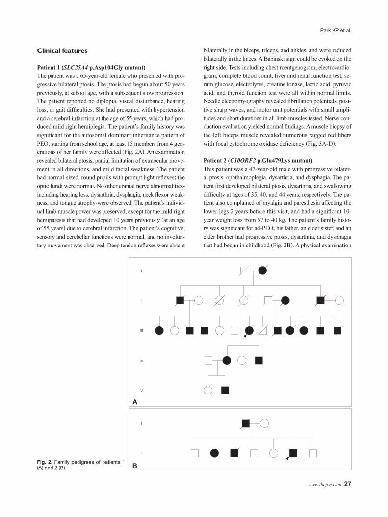
Patient1(SLC25A4p.Asp104Glymutant)The patient was a 65-year-old female who presented with pro-gressive bilateral ptosis. The ptosis had begun about 50 years previously, at school age, with a subsequent slow progression. The patient reported no diplopia, visual disturbance, hearing loss, or gait difficulties. She had presented with hypertension and a cerebral infarction at the age of 55 years, which had pro-duced mild right hemiplegia. The patient’s family history was significant for the autosomal dominant inheritance pattern of PEO; starting from school age, at least 15 members from 4 gen-erations of her family were affected (Fig. 2A). An examination revealed bilateral ptosis, partial limitation of extraocular move-ment in all directions, and mild facial weakness. The patient had normal-sized, round pupils with prompt light reflexes; the optic fundi were normal. No other cranial nerve abnormalities-including hearing loss, dysarthria, dysphagia, neck flexor weak-ness, and tongue atrophy-were observed. The patient’s individ-ual limb muscle power was preserved, except for the mild right hemiparesis that had developed 10 years previously (at an age of 55 years) due to cerebral infarction. The patient’s cognitive, sensory and cerebellar functions were normal, and no involun-tary movement was observed. Deep tendon reflexes were absent
bilaterally in the biceps, triceps, and ankles, and were reduced bilaterally in the knees. A Babinski sign could be evoked on the right side. Tests including chest roentgenogram, electrocardio-gram, complete blood count, liver and renal function test, se-rum glucose, electrolytes, creatine kinase, lactic acid, pyruvic acid, and thyroid function test were all within normal limits. Needle electromyography revealed fibrillation potentials, posi-tive sharp waves, and motor unit potentials with small ampli-tudes and short durations in all limb muscles tested. Nerve con-duction evaluation yielded normal findings. A muscle biopsy of the left biceps muscle revealed numerous ragged red fibers with focal cytochrome oxidase deficiency (Fig. 3A-D).
Patient2(C10ORF2p.Glu479Lysmutant)This patient was a 47-year-old male with progressive bilater-al ptosis, ophthalmoplegia, dysarthria, and dysphagia. The pa-tient first developed bilateral ptosis, dysarthria, and swallowing difficulty at ages of 35, 40, and 44 years, respectively. The pa-tient also complained of myalgia and paresthesia affecting the lower legs 2 years before this visit, and had a significant 10-year weight loss from 57 to 40 kg. The patient’s family histo-ry was significant for ad-PEO; his father, an elder sister, and an elder brother had progressive ptosis, dysarthria, and dysphagia that had begun in childhood (Fig. 2B). A physical examination
Fig. 2. Family pedigrees of patients 1 (A) and 2 (B).
I
I
II
II
III
IV
V
A
B

SLC25A4 and C10ORF2 Mutations in Ad-PEO
28 J Clin Neurol 2011;7:25-30
revealed that the patient had a very thin musculature, with a weight of 40 kg and a height of 170 cm. Neurological exami-nation revealed bilateral ptosis, nearly complete ophthalmople-gia, masseter and temporalis muscle atrophy, facial weakness, moderate sensorineural hearing loss with tinnitus, dysarthria with nasal voice, dysphagia, and neck flexor weakness; the op-tic fundi were normal. The patient’s limb muscle power was pre-served except for a mild elbow flexor weakness, which was grade 4 on the Medical Research Council scale. The patient had a reduced vibration sensation in the lower extremities, and hy-poactive deep tendon reflexes in the limbs. No other neurologi-cal abnormalities were observed. On laboratory examination, the patient displayed a mildly elevated serum creatine kinase level (342 U/L; normal range, 5-217 U/L). An electrocardiogram showed a nonspecific ST-segment elevation in the inferior and anterior walls. A chest roentgenogram suggested previous pul-monary tuberculosis; no evidence of cardiomegaly was observ-ed. The results of other laboratory tests including serum lactate/pyruvate, complete blood count, liver and renal functions, elec-trolytes, blood sugar, and thyroid function were unremarkable. A nerve conduction study revealed reduced compound nerve action potential amplitudes bilaterally in the sural nerves and mildly slow sensory nerve conduction velocities in the median and ulnar nerves. Needle electromyography revealed mild neu-rogenic changes in the limb muscles. The overall electrodiag-nostic findings were compatible with axonal-type sensorimotor peripheral polyneuropathy rather than myopathy. A muscle bi-opsy performed on the left biceps muscle revealed many ragged red fibers with partial deficiencies of cytochrome oxidase ac-tivities (Fig. 3E-H).
Discussion
The nature of multiple mtDNA deletions in PEO with Men-delian inheritance has been evaluated previously using single-muscle-fiber PCR analysis, which showed that each fiber has one type of deletion that is different from each cell, suggesting that each mtDNA with deletions originates in a mitochondrion and expands in postmitotic cells.15 PEO with multiple mtDNA deletions is a genetically heterogeneous entity caused by mu-tations of nuclear genes that are involved in the replication and maintenance of mtDNA. Many ad-PEO families carry a het-erozygous mutation in one of six genes (POLG, POLG2, SL-C25A4, C10ORF2, OPA1, or RRM2B), while many autosomal recessive PEO families carry POLG mutations. Previous stud-ies showed that POLG, SLC25A4, and C10ORF2 are three ma-jor genes that are mutated in ad-PEO, among which POLG is the most frequently mutated, with mutations SLC25A4 and C10ORF2 being uncommon.16 OPA1 was not analyzed in our study because the OPA1 mutant is always associated with op-tic atrophy,17 which was not a feature of our patients. Another gene, RRM2B, was not analyzed, since ad-PEO with a RRM2B mutation is considered extremely rare, and only two families with the same p.R327X mutation have been reported to date.8
SLC25A4 encodes ANT1, or ADP/ATP translocator, which is the most abundant protein in the mitochondrion. ANT is lo-cated in the inner mitochondrial membrane as a 30-kD homodi-mer, where it forms a gated pore through which ADP is ex-changed with ATP. ANT1 is one of the three isoforms of ANT that is predominantly expressed in heart and skeletal muscle.18 Mutations of ANT1 are associated exclusively with ad-PEO, and only six missense mutations have been reported thus far.14 The mutation identified in our study (p.Asp104Gly) has been
Fig. 3. Muscle biopsy findings of patients 1 (A-D) and 2 (E-H) showing multiple cytochrome-C-oxidase-negative ragged red fibers. Samples in (A) and (E) are stained with hematoxylin and eosin, those in (B) and (F) are stained with modified Gomori trichrome, those in (C) and (G) are stained for succinate dehydrogenase, and those in (D) and (H) are stained for cytochrome (C) oxidase. The white bar indicates 50 μm.

Park KP et al.
www.thejcn.com 29
reported previously in a Japanese family with a late-onset pure PEO phenotype, and has been well characterized as a patho-genic mutation.15 Although the exact mechanism by which ANT1 mutations produce multiple mtDNA deletions leading to ad-PEO is not yet clearly understood, several mechanisms in-cluding disturbed intramitochondrial deoxyATP concentration, disturbed mitochondrial permeability transition pore opening caused by structural defect of ANT1, and increased oxidative stress associated with ANT1 dysfunction have been suggested.4
C10ORF2 encodes TWINKLE; it is a primary mtDNA he-licase with nucleotide hydrolase activity and is localized to mitochondrial nucleoids.5 TWINKLE interacts functionally with the β subunit of polymerase γ,19 and is stimulated by the mitochondrial single-stranded DNA-binding protein20 to form a replication unit of mtDNA.21 Thus, mutation of C10ORF2 produces defects in both the maintenance of mtDNA copy num-ber and the integrity of mtDNA, which leads to multiple dele-tions of mtDNA and, consequently, disease. Clinically, POLG and C10ORF2 mutants share similar phenotypes including PEO, myopathy, ataxia, sensory axonal neuropathy, and Par-kinsonism, and this is probably related to the close functional interactions between polymerase γ and TWINKLE.22 More than 29 different mutations have been identified, most of which are missense mutations involving exon 1 or 2.22 The presently identified C10ORF2 mutation (p.Glu479Lys) is also located in exon 2, and was previously reported as a de novo mutation from a young Italian female with early-onset PEO and a stationary clinical course.23 However, our patient showed different clini-cal features: clear family history of autosomal dominant inher-itance, onset in the mid-thirties with clinical progression, and PEO plus other additional features including dysphagia, dysar-thria, proximal myopathy, and axonal-type sensorimotor poly-neuropathy. Although these findings may indicate the phenotyp-ic heterogeneity of this particular mutation, further accumula-tion of clinical and genetic data are needed to clarify this issue.
Two known missense mutations have been identified in this study in two Korean patients with ad-PEO: one in SLC25A4 and the other in C10ORF2. The clinical phenotype of the SL-C25A4 p.Asp104Gly mutant was similar to that described pre-viously, while the C10ORF2 p.Glu479Lys mutant phenotype was quite different. Further molecular biological and clinical genetic studies employing larger numbers of patients are need-ed to establish the clear pathogenetic mechanisms and geno-type-phenotype correlations in ad-PEO.
Conflicts of InterestThe authors have no financial conflicts of interest.
AcknowledgementsThis study was supported by a Medical Research Institute Grant (2007-7) from Pusan National University.
REFERENCES1. DiMauro S, Hirano M. Mitochondrial DNA Deletion Syndromes. In:
Pagon RA, Bird TC, Dolan CR, Stephens K. GeneReviews [Internet]. Seattle (WA): University of Washington, Seattle.
2. Mariotti C, Savarese N, Suomalainen A, Rimoldi M, Comi G, Prelle A, et al. Genotype to phenotype correlations in mitochondrial encephalo-myopathies associated with the A3243G mutation of mitochondrial DNA. J Neurol 1995;242:304-312.
3. Zeviani M, Servidei S, Gellera C, Bertini E, DiMauro S, DiDonato S. An autosomal dominant disorder with multiple deletions of mitochon-drial DNA starting at the D-loop region. Nature 1989;339:309-311.
4. Kaukonen J, Juselius JK, Tiranti V, Kyttälä A, Zeviani M, Comi GP, et al. Role of adenine nucleotide translocator 1 in mtDNA maintenance. Science 2000;289:782-785.
5. Spelbrink JN, Li FY, Tiranti V, Nikali K, Yuan QP, Tariq M, et al. Hu-man mitochondrial DNA deletions associated with mutations in the gene encoding Twinkle, a phage T7 gene 4-like protein localized in mito-chondria. Nat Genet 2001;28:223-231.
6. Van Goethem G, Dermaut B, Löfgren A, Martin JJ, Van Broeckhoven C. Mutation of POLG is associated with progressive external ophthal-moplegia characterized by mtDNA deletions. Nat Genet 2001;28:211-212.
7. Longley MJ, Clark S, Yu Wai Man C, Hudson G, Durham SE, Taylor RW, et al. Mutant POLG2 disrupts DNA polymerase gamma subunits and causes progressive external ophthalmoplegia. Am J Hum Genet 2006;78:1026-1034.
8. Tyynismaa H, Ylikallio E, Patel M, Molnar MJ, Haller RG, Suoma-lainen A. A heterozygous truncating mutation in RRM2B causes auto-somal-dominant progressive external ophthalmoplegia with multiple mtDNA deletions. Am J Hum Genet 2009;85:290-295.
9. Koh KN, Park SY, Hwang H, Chae JH, Choi JE, Kim KJ, et al. Large scale single deletion of mitochondrial DNA in chronic progressive ex-ternal ophthalmoplegia. J Korean Child Neurol Soc 2004;12:43-49.
10. Seong MW, Hwang JM, Kim JY, Ko HS, Park SS. Molecular diagno-sis for mitochondrial DNA aberrations in chronic progressive external ophthalmoplegia. J Korean Ophthalmol Soc 2005;46:323-329.
11. Kim SH, Hwang JH, Chung KW, Kim HJ, Kim JY, Park KD, et al. Ke-arns-Sayre syndrome with a large deletion in mitochondrial DNA. J Ko-rean Neurol Assoc 2006;24:260-264.
12. Goto Y, Koga Y, Horai S, Nonaka I. Chronic progressive external oph-thalmoplegia: a correlative study of mitochondrial DNA deletions and their phenotypic expression in muscle biopsies. J Neurol Sci 1990;100: 63-69.
13. Kleinle S, Wiesmann U, Superti-Furga A, Krähenbühl S, Boltshauser E, Reichen J, et al. Detection and characterization of mitochondrial DNA rearrangements in Pearson and Kearns-Sayre syndromes by long PCR. Hum Genet 1997;100:643-650.
14. Stenson PD, Mort M, Ball EV, Howells K, Phillips AD, Thomas NS, et al. The Human Gene Mutation Database: 2008 update. Genome Med 2009;1:13.
15. Komaki H, Fukazawa T, Houzen H, Yoshida K, Nonaka I, Goto Y. A novel D104G mutation in the adenine nucleotide translocator 1 gene in autosomal dominant progressive external ophthalmoplegia patients with mitochondrial DNA with multiple deletions. Ann Neurol 2002;51:645-648.
16. Agostino A, Valletta L, Chinnery PF, Ferrari G, Carrara F, Taylor RW, et al. Mutations of ANT1, Twinkle, and POLG1 in sporadic progressive external ophthalmoplegia (PEO). Neurology 2003;60:1354-1356.
17. Amati-Bonneau P, Valentino ML, Reynier P, Gallardo ME, Bornstein B, Boissière A, et al. OPA1 mutations induce mitochondrial DNA insta-bility and optic atrophy ‘plus’ phenotypes. Brain 2008;131:338-351.
18. Li K, Warner CK, Hodge JA, Minoshima S, Kudoh J, Fukuyama R, et al. A human muscle adenine nucleotide translocator gene has four ex-ons, is located on chromosome 4, and is differentially expressed. J Biol

SLC25A4 and C10ORF2 Mutations in Ad-PEO
30 J Clin Neurol 2011;7:25-30
Chem 1989;264:13998-14004.19. Farge G, Pham XH, Holmlund T, Khorostov I, Falkenberg M. The ac-
cessory subunit B of DNA polymerase gamma is required for mito-chondrial replisome function. Nucleic Acids Res 2007;35:902-911.
20. Korhonen JA, Gaspari M, Falkenberg M. TWINKLE Has 5´→3´ DNA helicase activity and is specifically stimulated by mitochondrial single-stranded DNA-binding protein. J Biol Chem 2003;278:48627-48632.
21. Korhonen JA, Pham XH, Pellegrini M, Falkenberg M. Reconstitution
of a minimal mtDNA replisome in vitro. EMBO J 2004;23:2423-2429.22. Van Hove JL, Cunningham V, Rice C, Ringel SP, Zhang Q, Chou PC,
et al. Finding twinkle in the eyes of a 71-year-old lady: a case report and review of the genotypic and phenotypic spectrum of TWINKLE-related dominant disease. Am J Med Genet A 2009;149A:861-867.
23. Virgilio R, Ronchi D, Hadjigeorgiou GM, Bordoni A, Saladino F, Mog-gio M, et al. Novel Twinkle (PEO1) gene mutations in mendelian pro-gressive external ophthalmoplegia. J Neurol 2008;255:1384-1391.