Embed Size (px)
Citation preview
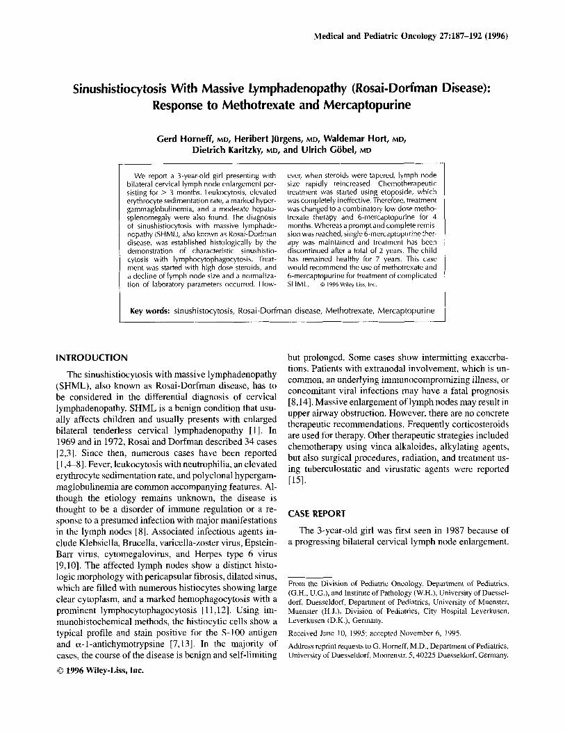
Medical and Pediatric Oncology 27: 187-192 (1996)
Sinushistiocytosis With Massive Lymphadenopathy (Rosai-Dorfman Disease): Response to Methotrexate and Mercaptopurine
Gerd Horneff, MD, Heribert Jurgens, MD, Waldemar Hort, MD,
Dietrich Karitzky, MD, and Ulrich Cobel, MD
I We report a 3-year-old girl presenting with
bilateral cervical lymph node enlargement per- sisting for > 3 months. Leukocytosis, elevated erythrocyte sedimentation rate, a marked hyper- gamrnaglobulinemia, and a moderate hepato- splenomegaly were also found. The diagnosis of sinushistiocytosis with massive lymphade- nopathy (SHML), also known as Rosai-Dorfrnan disease, was established histologically by the demonstration of characteristic sinushistio- cytosis with lymphocytophagocytosis. Treat- ment was started with high dose steroids, and a decline of lymph node size and a normaliza- tion of laboratory parameters occurred. How-
ever, when steroids were tapered, lymph node size rapidly reincreased. Chemotherapeutic treatment was started using etoposide, which was completely ineffective. Therefore, treatment was changed to a combinatory low dose metho- trexate therapy and 6-mercaptopurine for 4 months. Whereas a prompt and complete remis- sion was reached, single 6-mercaptopurine ther- apy was maintained and treatment has been discontinued after a total of 2 years. The child has remained healthy for 7 years. This case would recommend the use of methotrexate and 6-rnercaptopurine for treatment of complicated SHML. 0 1996 Wiley-Liss, Inc.
Key words: sinushistocytosis, Rosai-Dorfrnan disease, Methotrexate, Mercaptopurine
I NTROD UCTl ON
The sinushistiocytosis with massive lymphadenopathy (SHML), also known as Rosai-Dorfman disease, has to be considered in the differential diagnosis of cervical lymphadenopathy. SHML is a benign condition that usu- ally affects children and usually presents with enlarged bilateral tenderless cervical lymphadenopathy [ 11. In 1969 and in 1972, Rosai and Dorfman described 34 cases [2,3]. Since then, numerous cases have been reported [ 1,4431. Fever, leukocytosis with neutrophilia, an elevated erythrocyte sedimentation rate, and polyclonal hypergam- maglobulinemia are common accompanying features. Al- though the etiology remains unknown, the disease is thought to be a disorder of immune regulation or a re- sponse to a presumed infection with major manifestations in the lymph nodes [8]. Associated infectious agents in- clude Klebsiella, Brucella, varicella-zoster virus, Epstein- Barr virus, cytomegalovirus, and Herpes type 6 virus [9,10]. The affected lymph nodes show a distinct histo- logic morphology with pericapsular fibrosis, dilated sinus, which are filled with numerous histiocytes showing large clear cytoplasm, and a marked hemophagocytosis with a prominent lymphocytophagocytosis [ 11,121. Using im- munohistochemical methods, the histiocytic cells show a typical profile and stain positive for the S-100 antigen and a-l-antichymotrypsine [7,13]. In the majority of cases, the course of the disease is benign and self-limiting 0 1996 Wiley-Liss, Inc.
but prolonged. Some cases show intermitting exacerba- tions. Patients with extranodal involvement, which is un- common, an underlying immunocompromizing illness, or concomitant viral infections may have a fatal prognosis [8,14]. Massive enlargement of lymph nodes may result in upper airway obstruction. However, there are no concrete therapeutic recommendations. Frequently corticosteroids are used for therapy. Other therapeutic strategies included chemotherapy using vinca alkaloides, alkylating agents, but also surgical procedures, radiation, and treatment us- ing tuberculostatic and virustatic agents were reported ~ 5 1 .
CASE REPORT
The 3-year-old girl was first seen in 1987 because of a progressing bilateral cervical lymph node enlargement.
From the Division of Pediatric Oncology, Department of Pediatrics, (G.H., U.C.), and Institute of Pathology (W.H.), University of Duessel- dorf, Duesseldorf, Department of Pediatrics, University o f Muenster, Muenster (H.J.), Division of Pediatrics, City Hospital Leverkusen, Leverkusen (D.K.), Germany.
Received June 10, 1995; accepted November 6, 1995.
Address reprint requests to G. Horneff, M.D., Department of Pediatrics, University of Duesseldorf, Moorenstr. 5,40225 Duesseldorf, Germany.

188 Horneff et al.
Fig. 1. A. Lymph node with marked broadening of its capsule (arrows). HE staining, 45X. B. Lymph node with some interfollicular areas infiltrated by proliferated histiocytes. HE staining, 1 lox. C. Histiocytes with large round nuclei, prominent nucleoli, and lympho- phagocytosis (arrows). HE staining, 820X.
Her medical history was unremarkable except for several upper airway infections, otitis, and recurrent conjunctivi- tis. Sinusitis of the ethmoid cells and maxillary sinus has been treated successfully. On physical examination, bilateral enlarged submandibular lymph nodes of up to 4 cm and a moderate hepatosplenomegaly (liver 3 cm above normal range, spleen at the upper limit) were found. Laboratory examination revealed a leukocytosis of 14,500 pLI with a relative lymphopenia of lo%, an increased ESR of up to 132 m d h , a serum level of CRP of 62 mg/l, and a hypergammaglobulinemia with an elevation of all classes (IgG 26.4 g/l, IgM 3.0 g/l, IgA 3.3 g/l). All sero-
logical tests for infections carried out were negative. Chest roentgenograms, radionucleide bone scan, and bone marrow aspirate showed no abnormalities. Histopatholog- ical examination of an extirpated lymph node of the left submandibular region was performed. The lymph node architecture appeared not to be destroyed, but there was a marked fibrous thickening of the capsule (Fig. 1A). Lymphoid follicles and germinal centers were present. The interfollicular regions and the sinuses (Fig. 1B) were expanded by an infiltration of large histiocytic cells. These contain large round vesicular nuclei with one or more distinct nucleoli and an abundant pale-staining cyto-

Rosai-Dorfman Disease 189
Prednisolone
Etoposide
Methotrexate
6-Mercaptopurine
140
120
- 100
5 E E 0o Y
CT v> w 60
40
20
a 10.87 11.87 12.07 1.88 2.08 3.00 4.00 5.80 3.89 0.09 12.91 10.94
3.000
2.500
2.000 g c, 3 e. a h
1.500
- 1 .ooo
500
n
Fig. 2. Treatment and kinetics of ESR and level of immunoglobulin G in the course of the patient.
plasm frequently containing lymphocytes and plasma cells (Fig. 1C). Additionally, plasma cells were prominent in the medullary lymphoid cords and are often seen around vessels. Similar infiltrations were also seen in the cortical and paracortical region. These histological features are characteristic for the SHML.
During the following 4 months, lymph nodes increased in size. Repeated biopsy was performed, which confirmed the diagnosis. After treatment with prednisolone (2 mg/ kg body weight), rapid regression of the lymph nodes sizes was observed and hepatosplenomegaly disappeared; however, when corticosteroids were tapered, lymph node size increased again, reaching pretreatment level. A sec- ond course of high dosage corticosteroids was given fol- lowed by etoposide, frequently used for treatment of malignant histiocytosis (2 X 160 mg/m’ body surface fol- lowed by 3 X 160 mglm’ 3 weeks later). However, this treatment did not alter lymph node size or laboratory abnormalities. Consequently, a third prednisolone treat- ment was instituted together with a combinatory chemo- therapy using 6-mercaptopurine (60 mg/m2 per day) and methotrexate (12 mg/m2 weekly). Treatment and the course of laboratory parameters are outlined in Figure 2.
When a prompt and complete remission was obtained, methotrexate was stopped after 4 months and therapy with 6-mercaptopurine and low dose oral corticosteroids (5 mg/m2 every 48 hours) was maintained. Laboratory parameters normalized during the second year of treat- ment. After a total of 2 years, treatment was discontinued. There were no side effects observed, and the child has showed no further complications for 7 years.
DISCUSSION
SMHL is a histologically well defined disorder usually affecting cervical lymph nodes of children and adoles- cents. Adults and elderly people are rarely affected. The clinical presentation usually consists of massive, bilateral painless lymphadenopathy simulating a malignant pro- cess. Other lymph node regions are occasionally involved clinically or more frequently by microscopic changes only [12].
The etiology remains unknown, and the course of SMHL is often self-limiting and benign, but cases with a remitting course have been described. Since malignant transformation seems not to occur in SMHL, a fatal out-
190 Horneff et al.
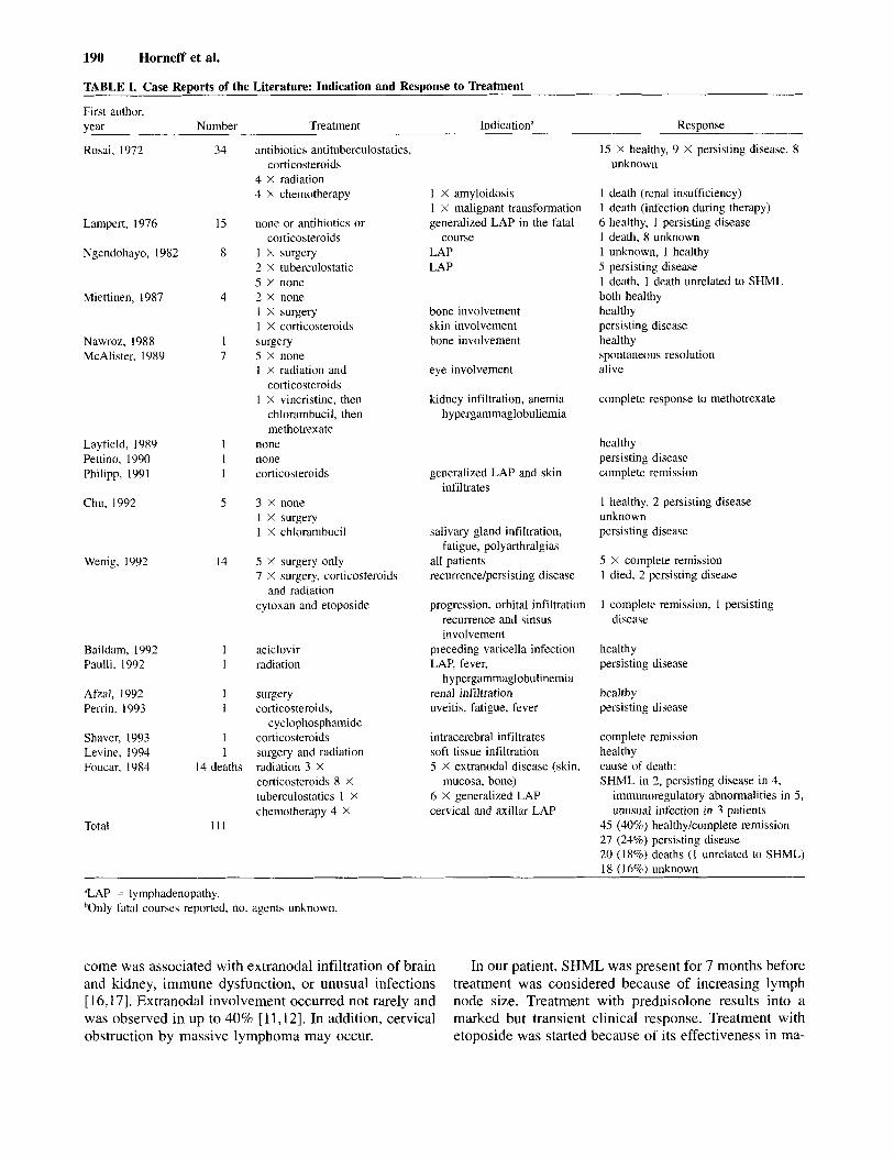
TABLE 1. Case Reports of the Literature: Indication and Response to Treatment
First author, vear Number Treatment Indication" Response
Rosai, 1972 34
Lampert, 1976 15
Ngendohayo, 1982 8
Miettinen, 1987 4
Nawroz, 1988 1 McAlister, 1989 7
Layfield, 1989 Pettino, 1990 Philipp, 1991
Chu, 1992
Wenig, 1992 14
Baildam, 1992 1 Paulli, 1992 1
Afzal, 1992 1 Perrin, 1993 1
Shaver, 1993 1 Levine, 1994 1 Foucar, 1984 I4 deaths
Total I l l
antibiotics antituberculostatics, corticosteroids
4 X radiation 4 X chemotherapy
none or antibiotics or corticosteroids
1 X surgery 2 X tuberculostatic 5 X none 2 X none I X surgery 1 X corticosteroids surgery 5 X none I X radiation and
corticosteroids 1 X vincristine, then
chlorambucil, then methotrexate
none none corticosteroids
3 X none 1 X surgery 1 X chlorambucil
5 X surgery only 7 X surgery, corticosteroids
and radiation cytoxan and etoposide
aciclovir radiation
surgery corticosteroids,
cyclophosphamide corticosteroids surgery and radiation radiation 3 X corticosteroids 8 X tuberculostatics 1 X chemotherapy 4 X
I X amyloidosis 1 X malignant transformation generalized LAP in the fatal
LAP LAP
course
bone involvement skin involvement bone involvement
eye involvement
kidney infiltration, anemia hypergammaglobuliemia
generalized LAP and skin infiltrates
salivary gland infiltration, fatigue, polyarthralgias
all patients recurrence/persisting disease
progression, orbital infiltration recurrence and sinsus involvement
preceding varicella infection LAP, fever,
hypergammaglobulinemia renal infiltration uveitis, fatigue, fever
intracerebral infiltrates soft tissue infiltration S X extranodal disease (skin,
6 X generalized LAP cervical and axillar LAP
mucosa, bone)
15 X healthy, 9 X persisting disease, 8 unknown
1 death (renal insufficiency) 1 death (infection during therapy) 6 healthy, 1 persisting disease 1 death, 8 unknown 1 unknown, I healthy 5 persisting disease 1 death, 1 death unrelated to SHML both healthy healthy persisting disease healthy spontaneous resolution alive
complete response to methotrexate
healthy persisting disease complete remission
1 healthy, 2 persisting disease unknown persisting disease
5 X complete remission 1 died, 2 persisting disease
I complete remission, 1 persisting disease
healthy persisting disease
heal thy persisting disease
complete remission healthy cause of death: SHML in 2, persisting disease in 4,
immunoregulatory abnormalities in 5 , unusual infection in 3 patients
45 (40%) healthykomplete remission 27 (24%) persisting disease 20 (18%) deaths (1 unrelated to SHML) 18 (16%) unknown
"AP = lymphadenopathy. hOnly fatal courses reported, no. agents unknown.
come was associated with extranodal infiltration of brain and kidney, immune dysfunction, or unusual infections [ 16,171. Extranodal involvement occurred not rarely and was observed in up to 40% [11,12]. In addition, cervical obstruction by massive lymphoma may occur.
In our patient, SHML was present for 7 months before treatment was considered because of increasing lymph node size. Treatment with prednisolone results into a marked but transient clinical response. Treatment with etoposide was started because of its effectiveness in ma-

Rosai-Dorfman Disease 191
(Rosai-Dorfman disease) of the head and neck. Hum Pathol 24:483492, 1991.
2. Rosai J, Dorfman RF: Sinus histiocytosis with massive lymphade- nopathy: A newly recognized benign clinicopathological entity. Arch Pathol 87:63-70, 1969.
3. Rosai J, Dorfman RF: Sinus histiocytosis with massive lymphade- nopathy: A pseudolymphoniatous benign disorder: Analysis of 34 cases. Cancer 30: 1174-1 188, 1972.
4. Ngendahayo P, Roels H, Quatacker J, Boddaert J, Ntabomuvra V, Mbonyingabo P: Sinus histiocytosis with massive lymphadenopa- thy in Rwanda: Report of eight cases with immunohistochemical and ultrastructural studies. Histopathology 7:49-64, 1982.
5. Lampert F, Lennert K: Sinus histiocytosis with massive lymphade- nopathy: Fifteen new cases. Cancer 37:783-789, 1976.
6. Foucar E, Rosai J, Dorfman RF: Sinus histiocytosis with massive lymphadenopathy. Arch Dermatol 124:1211-1216, 1988.
7. Miettinen M, Paljakka P, Haveri P, Saxen E: Sinus histiocytosis with massive lymphadenopathy: A nodal and extranodal prolifera- tion of S-100 protein positive histiocytes? Am J Clin Pathol 88:270-277, 1987.
8. Perrin C, Michiels JF, Lacour JP, Chagnon A, Fuzibet JG: Sinus histiocytosis (Rosai-Dorfman disease) clinically limited to the skin. J Cutan Pathol 20:368-374, 1993.
9. Levine PH, Jahan N, Murari P, Manak M, Jaffe ES: Detection of Human Herpesvirus 6 in tissues involved by sinus histiocytosis with massive lymphadenopathy (Rosai-Dorfman disease). J Infect Dis 166:291-295, 1992.
10. Layfield LJ: Fine needle cytologic findings in a case of sinus histiocytosis with massive lymphadenopathy (Rosai-Dorfman syn- drome). Acta Cytologica 34:767-770, 1990.
1 I . Chu P, LeBoit PE: Histologic features of cutaneous sinus histio- cytosis (Rosai-Dorfman disease): Study of cases both with and without systemic involvement. J Cutan Pathol 19:201-206, 1992.
2. McAlister WH, Herman T, Dehner LP: Sinus histiocytosis with massive lymphadenopathy (Rosai-Dorfman disease). Pediatr Ra- diol 20:425432, 1990.
3. Bonetti F, Chilosi M, Menestrina F, Scarpa A, Pelicci P-G, Amorosi E, Fiore-Donati L, Knowles I1 DM: Immunohistological analysis of Rosai-Dorfman histiocytosis: A disease of S-lW+CDl-histio- cytes. Virchows Arch A 411:129-135, 1987.
4. Foucar E, Rosai J, Dorfman RF: Sinus histiocytosis with massive lymphadenopathy: An analysis of 14 deaths occumng in a patients registry. Cancer 54: 1834-1840, 1984.
15. Baildam EM, D’Souza SW, Stevens RF: Sinus histiocytosis with massive lymphadenopathy (Rosai-Dorfman disease): Response to acylovir. JRSM 85:179-180, 1992.
16. Shaver EG, Rebsamen SL, Yachnis AT, Sutton LN: Isolated extra- nodal intracranial sinus histiocytosis in a 5-year-old boy. J Neuro- surg 79:769-773, 1993.
17. Afzal M, Baez-Giangreco A, Al Jaser AN, Onuora V: Unusual bilateral renal histiocytosis: Extranodal variant of Rosai-Dorfman disease. Arch Pathol Lab Med 1 16: 1366-1 367, 1992.
18. Philipp A, Laszig R, Werner M: Das Rosai Dorfman Syndrom.
19. Levine EA, Mandry MM: Rosai Dorfman disease of soft tissue. Surgery 115:650-652, 1994.
20. Nawroz IM, Wilson-Storey D: Sinus histiocytosis with massive lymphadenopathy (Rosai-Dorfman disease). Histopathol 14:91- 99, 1989.
21. Paulli M, Locatelli F, Kindl S, Boveri E, Facchetti F, Porta F, Rosso R, Nespoli L, Margrini U: Sinus histiocytosis with massive lymphadenopathy (Rosai-Dorfman disease): Clinico-pathological analysis of a pediatric case. Eur J Pediatr 151:672-675, 1992.
22. Paulli M, Rosso R, Kindl S, Boveri E, Marcocolo D, Chioda C, Agostini C, Magrini U, Facchetti F Immunophenotypic character- ization of the cell infiltrate in five cases of sinus histiocytosis with
HNO 40~56-581, 1992.
lignant childhood histiocytosis, but it was totally ineffec- tive. A combination therapy using corticosteroids and the antimetabolits methotrexate and 6-mercaptopurine proofed to be effective. Since thereafter the child re- mained healthy, there is no hint for an underlying immu- nodeficiency of an immunoregulatory disorder, which has been observed in other patients with complicated or recur- rent disease.
SHML generally does not by itself require chemothera- peutic treatment as shown by numerous published cases [4,5,7,12]. However, there is no information about indica- tion for treatment and there is no study of treatment efficacy available. Table I summarizes several reports in which treatment of SHML with or without extranodal manifestations has been performed. In most cases local- ized extranodal disease prompted to a primarily surgical therapy or to radiation. In several cases antibiotics or tuberculostatic agents were given without a significant response [4,5]. Corticosteroids have been tried success- fully in generalized lymphadenopathy or in a case with intracranial infiltrations [ 16-1 81. Whereas the course of SHML can be fatal or because of organ involvement or systemic manifestations, treatment with chemotherapeu- tics may be indicated and had been tried [1,8,11,12,14]. However, four patients died despite chemotherapy [ 141. In three of them (treated with either immuran, cytoxan, or methotrexate), autopsy showed ongoing SHML. One patient treated with cyclophosphamide died after an aspi- ration pneumonia, but no autopsy was performed. Cy- toxan and etoposide were used for treatment in two other patients, which was successful in one of them [ 11. Chlor- ambucil and cyclophosphamide have been tried without success in two other patients [8,11]. In a case reported by McAlister et al. [12], vincristin and chlorambucil had been tried, but disease recurred. Thereafter, methotrexate was successfully used. The analysis of 11 1 cases in the literature revealed as many patients with a complete re- mission as with persisting disease or a fatal course. Ex- cluding the data of Foucar et al. [14], who reported fatal courses only, about the half of the patients had been cured, 30% had persisting disease when reported, and 6% died; history is unknown for the remaining others.
In summary, there is only little information available to answer the question when and how patients should be treated. In extranodal or generalized manifestation or persistent disease, treatment using cytotoxic agents may be indicated when corticosteroids failed. In this case, antimetabolites should be the drug of first choice before alkylating agents or radiation are considered.
REFERENCES
1. Wenig BM, Abbondanzo SL, Childers EL, Kapadia SB, Heffner DR: Extranodal sinus histiocytosis with massive lymphadenopathy

192 Horneff et al.
massive lymphadenopathy (Rosai-Dorfman disease). Hum Pathol 231647-654, 1992.
23. Pettinato G, Manivel JC, d’Amore ESG, Petrella G: Fine needle aspiration cytology and immunocytochemical characterization of the histiocytes in sinus histiocytosis withmassive lymphadenopathy (Rosai-Dorfman syndrome). Acta Cytologica 34:77 1-777, 1990.
24. Vilde F, Arkwright S, Bonfils P, Leport C, Londero A, Vilde JL, Trotoux J: Pseudotumoral salivary location of the Rosai-Dorfman syndrome (Hemophagocytic histiocytosis. Revealing bilateral sub- maxillary and parotid involvement). Ann Oto Laryng 108:286- 291, 1991.