Embed Size (px)
Citation preview
THIEME
86 Letter to the Editor
Rosai-Dorfman Disease of the Spine Masquerading as MeningiomaVijay P. Joshi1 Prasanna Kasegaonkar2 Gauri Kelkar3 Seena Kasegaonkar4 Mrugakshi Khairnar4 Amit Agrawal5
1Department of Neurosurgery, Chandan Neurosciences Hospital, Solapur, Maharashtra, India
2Department of Neurology, Chandan Neurosciences Hospital, Solapur, Maharashtra, India
3Department of Pathology, Chandan Neurosciences Hospital, Solapur, Maharashtra, India
4Department of Radiology, Chandan Neurosciences Hospital, Solapur, Maharashtra, India
5Department of Neurosurgery, Narayana Medical College Hospital, Chinthareddypalem, Nellore, Andhra Pradesh, India
received November 13, 2016accepted after revision April 30, 2018published onlineApril 23, 2019
Address for correspondence Dr. Vijay P. Joshi, MCh, Consultant Neurosurgeon, Chandan Neurosciences Hospital, Solapur, Maharashtra, India (e-mail: [email protected]).
Indian J Neurosurg 2019;8:86–88
DOI https://doi.org/ 10.1055/s-0039-1677965 ISSN 2277-954X.
©2019 Neurological Surgeons’ Society of India
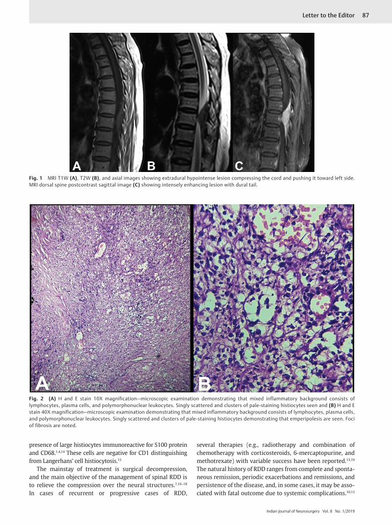
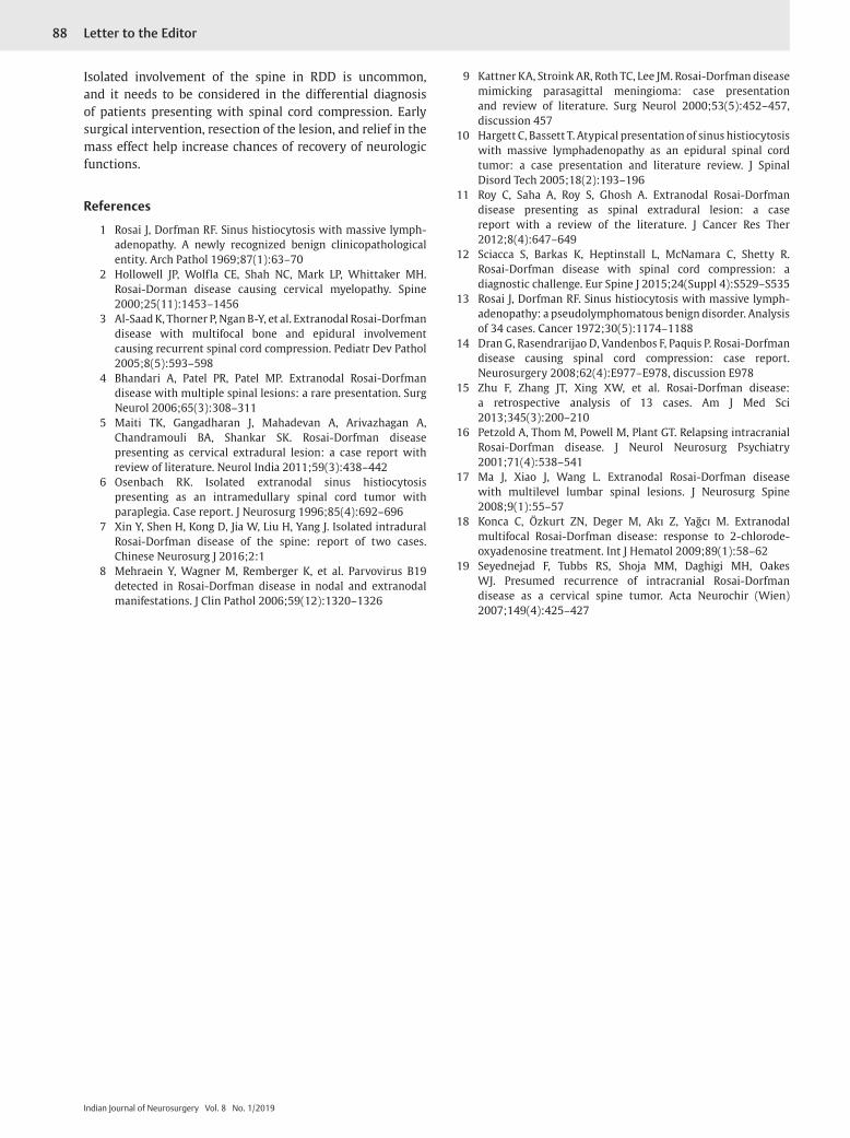
Rosai-Dorfman disease (RDD) is histiocytic proliferative disorder of idiopathic etiology and the characteristic of the disease includes sinus histiocytosis and massive lymphadenopathy.1 The central nervous system (CNS) is involved in less than 5% of the cases (75% brain and 25% spinal cord).2 Isolated involvement of the spine in RDD (without involvement of the lymph nodes) is rare with only few reported cases in literature.3–7 On imaging isolated RDD of the spine can mimic meningiomas, making its preoperative diagnosis difficult.3,4 A 65-year-old woman presented with the history of progressive onset of spastic paraparesis. There was no history of bladder or bowel involvement. On examination, the patient had grade 3 power in all muscle groups of the lower limbs; her sensory level was at D10 level. She had reduced sensations to touch and pin prick as well as decreased proprioception below this level. Axial T1-weighted (T1W) magnetic resonance imaging (MRI) of the brain showed an extradural isointense lesion compressing the cord and pushing it towards the left side. The lesion was becoming hypointense on T2-weighted (T2W) axial images, and it was enhancing after contrast administration (►Fig. 1). The patient underwent D7–D10 laminectomy. Intraoperatively the lesion was pale yellow in color, was relatively avascular, and had both intra- and extradural components. Histopathologic examination of the lesion showed features suggestive of extranodal RDD. Microscopic examination demonstrated mixed inflam-matory background consisting of lymphocytes, plasma cells, and polymorphonuclear leukocytes. Singly scattered and clusters of pale staining histiocytes demonstrating emperipolesis are seen. Foci of fibrosis are noted (►Fig. 2). The lesional histiocytes that showed emperipolesis express
S100 protein and CD30 did not reveal any R-S cells. Special stains for microorganisms were negative. Postoperatively the patient gradually improved in her paraparesis and sen-sory symptoms. On discharge, she had grade 4/5 power in all muscle groups of the lower limbs. She is doing well at 1-year follow-up.
The exact etiology of RDD is not known and it is con-sidered a benign idiopathic histiocytosis. Infectious agents (i.e., human herpes virus 6 [HHV-6] and parvovirus B19) or an abnormal immunologic response have been suggested as causative factors.4,8 Although there is no age preponder-ance for the CNS involvement in RDD, the disease usually involves male patients between 20 and 40 years of age.1,2 The systemic RDD most commonly involves the orbit, skin, respiratory tract, bones, kidneys, heart, and head and neck region, and is characterized by massive and painless cer-vical lymphadenopathy with variable symptoms of fever, leukocytosis, raised erythrocyte sedimentation rate (ESR), and weight loss.5,9,10 Involvement of the spine is uncom-mon, and usually there is absence of lymphadenopathy.11
It is difficult to make a diagnosis of isolated RDD on imaging as MRI features of this disease are almost similar to other common lesions of the spinal cord (i.e., meningiomas).4 The signal intensity of these lesions on MRI ranges from iso- to hypointensity on T1W images to iso- to hyperintensity on T2W images, and after the contrast administration, the lesions show an intense and uniform enhancement (including dural tail).12,13 Because of this, the definitive diagnoses are possible only after the histopathology (if necessary immunohistochem-istry), which will be the characteristic pattern of the disease.4,12 On histopathology, RDD is characterized by emperipolesis “phagocytosis of intact lymphocytes by macrophages” and
Published online: 23.04.2019
87Letter to the Editor
Indian Journal of Neurosurgery Vol. 8 No. 1/2019
presence of large histiocytes immunoreactive for S100 protein and CD68.1,4,14 These cells are negative for CD1 distinguishing from Langerhans’ cell histiocytosis.15
The mainstay of treatment is surgical decompression, and the main objective of the management of spinal RDD is to relieve the compression over the neural structures.7,16–18 In cases of recurrent or progressive cases of RDD,
several therapies (e.g., radiotherapy and combination of chemotherapy with corticosteroids, 6-mercaptopurine, and methotrexate) with variable success have been reported.13,19 The natural history of RDD ranges from complete and sponta-neous remission, periodic exacerbations and remissions, and persistence of the disease, and, in some cases, it may be asso-ciated with fatal outcome due to systemic complications.10,13
Fig. 1 MRI T1W (A), T2W (B), and axial images showing extradural hypointense lesion compressing the cord and pushing it toward left side. MRI dorsal spine postcontrast sagittal image (C) showing intensely enhancing lesion with dural tail.
Fig. 2 (A) H and E stain 10X magnification—microscopic examination demonstrating that mixed inflammatory background consists of lymphocytes, plasma cells, and polymorphonuclear leukocytes. Singly scattered and clusters of pale-staining histiocytes seen and (B) H and E stain 40X magnification—microscopic examination demonstrating that mixed inflammatory background consists of lymphocytes, plasma cells, and polymorphonuclear leukocytes. Singly scattered and clusters of pale-staining histiocytes demonstrating that emperipolesis are seen. Foci of fibrosis are noted.
88 Letter to the Editor
Indian Journal of Neurosurgery Vol. 8 No. 1/2019
Isolated involvement of the spine in RDD is uncommon, and it needs to be considered in the differential diagnosis of patients presenting with spinal cord compression. Early surgical intervention, resection of the lesion, and relief in the mass effect help increase chances of recovery of neurologic functions.
References
1 Rosai J, Dorfman RF. Sinus histiocytosis with massive lymph-adenopathy. A newly recognized benign clinicopathological entity. Arch Pathol 1969;87(1):63–70
2 Hollowell JP, Wolfla CE, Shah NC, Mark LP, Whittaker MH. Rosai-Dorman disease causing cervical myelopathy. Spine 2000;25(11):1453–1456
3 Al-Saad K, Thorner P, Ngan B-Y, et al. Extranodal Rosai-Dorfman disease with multifocal bone and epidural involvement causing recurrent spinal cord compression. Pediatr Dev Pathol 2005;8(5):593–598
4 Bhandari A, Patel PR, Patel MP. Extranodal Rosai-Dorfman disease with multiple spinal lesions: a rare presentation. Surg Neurol 2006;65(3):308–311
5 Maiti TK, Gangadharan J, Mahadevan A, Arivazhagan A, Chandramouli BA, Shankar SK. Rosai-Dorfman disease presenting as cervical extradural lesion: a case report with review of literature. Neurol India 2011;59(3):438–442
6 Osenbach RK. Isolated extranodal sinus histiocytosis presenting as an intramedullary spinal cord tumor with paraplegia. Case report. J Neurosurg 1996;85(4):692–696
7 Xin Y, Shen H, Kong D, Jia W, Liu H, Yang J. Isolated intradural Rosai-Dorfman disease of the spine: report of two cases. Chinese Neurosurg J 2016;2:1
8 Mehraein Y, Wagner M, Remberger K, et al. Parvovirus B19 detected in Rosai-Dorfman disease in nodal and extranodal manifestations. J Clin Pathol 2006;59(12):1320–1326
9 Kattner KA, Stroink AR, Roth TC, Lee JM. Rosai-Dorfman disease mimicking parasagittal meningioma: case presentation and review of literature. Surg Neurol 2000;53(5):452–457, discussion 457
10 Hargett C, Bassett T. Atypical presentation of sinus histiocytosis with massive lymphadenopathy as an epidural spinal cord tumor: a case presentation and literature review. J Spinal Disord Tech 2005;18(2):193–196
11 Roy C, Saha A, Roy S, Ghosh A. Extranodal Rosai-Dorfman disease presenting as spinal extradural lesion: a case report with a review of the literature. J Cancer Res Ther 2012;8(4):647–649
12 Sciacca S, Barkas K, Heptinstall L, McNamara C, Shetty R. Rosai-Dorfman disease with spinal cord compression: a diagnostic challenge. Eur Spine J 2015;24(Suppl 4):S529–S535
13 Rosai J, Dorfman RF. Sinus histiocytosis with massive lymph-adenopathy: a pseudolymphomatous benign disorder. Analysis of 34 cases. Cancer 1972;30(5):1174–1188
14 Dran G, Rasendrarijao D, Vandenbos F, Paquis P. Rosai-Dorfman disease causing spinal cord compression: case report. Neurosurgery 2008;62(4):E977–E978, discussion E978
15 Zhu F, Zhang JT, Xing XW, et al. Rosai-Dorfman disease: a retrospective analysis of 13 cases. Am J Med Sci 2013;345(3):200–210
16 Petzold A, Thom M, Powell M, Plant GT. Relapsing intracranial Rosai-Dorfman disease. J Neurol Neurosurg Psychiatry 2001;71(4):538–541
17 Ma J, Xiao J, Wang L. Extranodal Rosai-Dorfman disease with multilevel lumbar spinal lesions. J Neurosurg Spine 2008;9(1):55–57
18 Konca C, Özkurt ZN, Deger M, Akı Z, Yağcı M. Extranodalmultifocal Rosai-Dorfman disease: response to 2-chlorode-oxyadenosine treatment. Int J Hematol 2009;89(1):58–62
19 Seyednejad F, Tubbs RS, Shoja MM, Daghigi MH, Oakes WJ. Presumed recurrence of intracranial Rosai-Dorfman disease as a cervical spine tumor. Acta Neurochir (Wien) 2007;149(4):425–427


![Index [link.springer.com]978-3-642-17869-6/1.pdf · 410 Index. K Kaposi’s sarcoma, 90 ... Sarcoidosis Rosai-Dorfman disease, 335 Sarcoma, 2, ... Thalassemia, 268 Thyroglossal duct](https://img.pdfslide.us/doc/110x75/5b7c95787f8b9a9d078c2151/index-link-978-3-642-17869-61pdf-410-index-k-kaposis-sarcoma-90-.jpg)