Embed Size (px)
Citation preview
ORIGINAL CONTRIBUTION
Single-stage in situ synthesis of silver nanoparticlesin antibacterial self-assembled overlays
Aitor Urrutia & Pedro J. Rivero & Leyre Ruete &
Javier Goicoechea & Ignacio R. Matías &
Francisco J. Arregui
Received: 10 October 2011 /Revised: 22 December 2011 /Accepted: 3 January 2012 /Published online: 22 January 2012# Springer-Verlag 2012
Abstract In this work, a novel single-stage process for insitu synthesis of Ag nanoparticles (NPs) using the layer-by-layer (LbL) technique is presented. The Ag NPs wereformed into nanotextured coatings based on sequentiallyadsorbed poly(allylamine hydrochloride) (PAH) and SiO2
NPs. Such highly porous surfaces have been used in thefabrication of highly efficient ion release films for applica-tions such as antibacterial coatings. In this approach, theamino groups of the PAH acted as reducing agent and madepossible the in situ formation of the Ag NPs. This reductionreaction occurred during the LbL process as the coating wasassembled, without any further step after the fabrication andstabilization of the multilayer film. Biamminesilver nitratewas used as the Ag+ ion source during the LbL process and itwas successfully reduced to Ag NPs. All coatings were testedwith gram-positive and gram-negative bacterial cultures ofEscherichia coli, Staphylococcus aureus, and Lactobacillusdelbrueckii showing an excellent antimicrobial behavioragainst these types of bacteria (more than 99.9% of killingefficiency in all cases).
Keywords Antibacterial . Coating . Layer-by-layer (LbL)films . Silver nanoparticles . In situ synthesis
AbbreviationsNPs NanoparticlesLbL Layer-by-layerPAH Poly(allylamine hydrochloride)
BAS Biamminesilver nitrateAFM Atomic force microscopyEDX Energy dispersive X-rayTEM Transmission electron microscopySPR Surface plasmon resonance
Introduction
Microbes and bacteria are the most abundant living organ-isms, and there is always a big concern in controlling theirapparition and population on the surfaces in contact withwater, food, and tools. Consequently, the development ofhighly efficient and low-cost antibacterial surfaces has beenthe object of many research works [1, 2].
Among the wide variety of biocide agents, silver ions areof special interest because they show a broad spectrumantibiotic effect [3–6] and, at the same time, a low toxicityfor humans [7–9]. Moreover, silver ions interact with thiolgroups of some proteins, and as a result of this interaction,the replication ability of DNA molecules can be inhibited,thereby affecting the bacterial cell viability. In fact, silverhas been used for hygienic and medicinal purposes since theancient Greece [10].
Most of the approaches for the fabrication of antibacterialmaterials are based on doping some elements with silverparticles. These silver particles can be found embedded intopolymeric yarns for textile confection [11], or immobilizedinto coatings on the desired substrate (surgical instruments,etc.) [11]. The biocide effectiveness of such coatings dependson their ion releasing capability of the silver trapped in thecoating. Consequently, silver particles with a high specificsurface area have more efficient ion release mechanisms,and therefore, the antibacterial effect is enhanced. Recently,
A. Urrutia (*) : P. J. Rivero : L. Ruete : J. Goicoechea :I. R. Matías : F. J. ArreguiDepartment of Electrical and Electronic Engineering,Public University of Navarre,Campus Arrosadía,31006 Pamplona, Spaine-mail: [email protected]
Colloid Polym Sci (2012) 290:785–792DOI 10.1007/s00396-012-2591-4

the use of nanotechnology has made possible the creation ofnew nanostructured materials with extremely higher specificsurfaces that have boosted the efficiency of such antibacterialcoatings. Nanotechnology also offers the additional possibil-ity of tuning the overall properties of the coating by properlydesigning the composition and morphology of the films,opening the door to combining the biocide effect with thedesired physical properties such as optical characteristics ormechanical properties.
Some authors have reported that silver nanoparticles [12],nanorods [13], or nanotubes [14] are especially efficient anti-bacterial agents because of their large surface to volume ratio.Among other approaches, silver nanoparticles (Ag NPs) havebeen immobilized on inorganic porous hosts such as zeolites,calcium phosphate, and carbon fiber [15–17]. Moreover,silver-supported silica materials, such as silica glass [18],silica thin films [19], and silica nanoparticles [20], are alsogood candidates for antibacterial materials due to their finechemical durability and high antibacterial activity.
There are several well-known synthesis routes for Ag NPs,such as vacuum deposition using templates [21], electronbeam lithography [22], laser ablation [23], or electrodeposi-tion [24]. Nevertheless, the most used synthesis routes ofmetallic nanoparticles are wet routes based on metallic saltsolutions with further reduction and stabilization [25]. In suchtechniques, the chemical stabilization of the colloid surface isa key factor in order to get stable and well-dispersed metallicnanoparticles, and most of the times, this stabilization isachieved using surfactants and other capping molecules.
An alternative way to stabilize the nanoparticles and pre-vent their aggregation is the so-called nanoreactor synthesisroute. This technique uses the cavities or pores of mechani-cally stable structures such as aluminosilicates [26], sol–gels[27, 28], etc., as nanoparticle growth places where the Ag NPsare crystallized without aggregation. It has been demonstratedthat one useful material for the nanoreactor Ag nanoparticlessynthesis could be a layer-by-layer (LbL) coating, since insidethe coating, there are available functional groups which canact as nanoparticle synthesis seeds [29].
There are other authors that have used successfully thisapproach using the LbL technique [30]; nevertheless, all ofthem are based on a multi-stage experimental approach.Firstly, a polyelectrolyte multilayer (PEM) film is created.Afterwards, Ag+ ions are incorporated inside the polymericmatrix by ion exchange. Finally, silver ions are reduced insitu using hydrogen or other reducing agents. In other works[31], it is possible to see other modifications of the LbLstandard coating protocol in which silver nanoparticles aresynthesized using the functional groups of weak polyelec-trolytes and an additional reducing agent such as NaBH4.
In this work, a single-stage LbL process which only impliesthe addition of silver salts directly to the LbL solutions isproposed. It is important to remark that the reduction of the
silver salts occurs during the normal LbL absorption processwithout any additional step or treatment such as UV radiation,chemical reduction agents, or other techniques. This methodpresents a novel alternative to produce Ag NPs by an easierand time-efficient route and, in addition, to fabricate highlyefficient antimicrobial coatings. To our knowledge, this is thefirst time that a route to form Ag NPs during the LbL processby direct reduction is proposed.
Experimental
Materials
Poly(allylamine hydrochloride) (PAH) (Mw 56,000), silvernitrate (>99% (titration)), ammonia (25 wt.%) and LudoxTM-40 colloidal silica (SiO2, 20 nm diameter nanoparticles)were purchased from Sigma Aldrich and used as receivedwithout further purification. All aqueous solutions were pre-pared using ultrapure water with a resistivity of 18.2 MΩ cm.Standard microscopy glass slides were used as substrates.
The biamminesilver nitrate (BAS) stock solution was pre-pared by mixing AgNO3 and 25 wt.% aqueous ammonia asfollows: Firstly, a 5-mM AgNO3 water solution (212 mg ofAgNO3 in 250 mL of ultrapure water) was stirred for 2 h atroom temperature. Next, 0.83 mL of ammonia was addeddropwise to the AgNO3 solution while stirring, thus obtainingthe BAS ([Ag(NH3)2]NO3) solution.
Stock cultures of Escherichia coli and Staphylococcusaureus were purchased from ATCC (American Type CultureCollection, Manassas, VA, USA). Lactobacillus delbrueckiistock culture was obtained from CECT (The Spanish TypeCulture Collection, University of Valencia).
Fabrication of silver and silica nanocompositemultilayer films
The LbL assembly process provides the ability to placenanoparticles of different sizes throughout the multilayerfilm in a regulated manner. The deposition conditions suchas immersion time [32], solution pH [33], temperature [34],ionic strength, or concentration of the polyelectrolytes [35]allows controlling the nanoparticle aggregation. Figure 1shows a schematic view of the LbL fabrication process.
In this work, silver salts were added to the electrolytesolutions involved in the LbL process. The starting solutionswere prepared as described in earlier works [20] to achievehighly rough thin films. Awater-based solution of PAH with aconcentration of 10mM (respect to the repetitive unit) acted asthe cationic polyelectrolyte. Initially, the anionic solution wasobtained using the prepared BAS stock solution and addingSiO2 NPs to it, so that the final concentration of SiO2 NPs inthe anionic solution was 0.03 wt.%.
786 Colloid Polym Sci (2012) 290:785–792
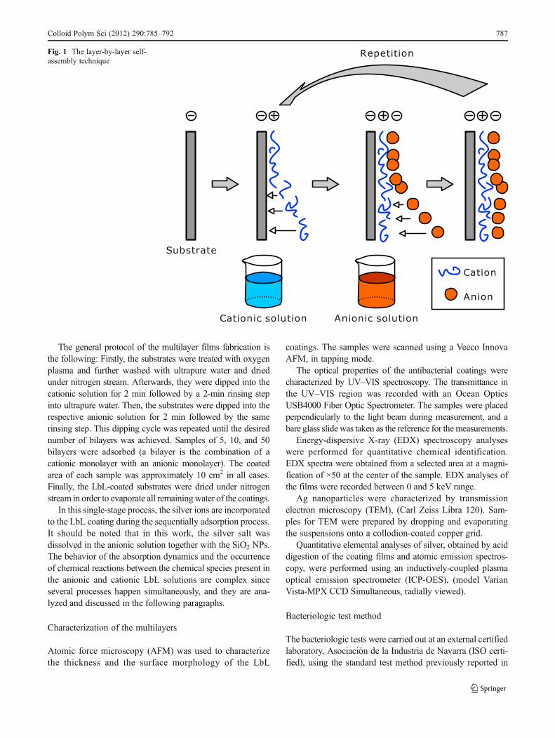
The general protocol of the multilayer films fabrication isthe following: Firstly, the substrates were treated with oxygenplasma and further washed with ultrapure water and driedunder nitrogen stream. Afterwards, they were dipped into thecationic solution for 2 min followed by a 2-min rinsing stepinto ultrapure water. Then, the substrates were dipped into therespective anionic solution for 2 min followed by the samerinsing step. This dipping cycle was repeated until the desirednumber of bilayers was achieved. Samples of 5, 10, and 50bilayers were adsorbed (a bilayer is the combination of acationic monolayer with an anionic monolayer). The coatedarea of each sample was approximately 10 cm2 in all cases.Finally, the LbL-coated substrates were dried under nitrogenstream in order to evaporate all remainingwater of the coatings.
In this single-stage process, the silver ions are incorporatedto the LbL coating during the sequentially adsorption process.It should be noted that in this work, the silver salt wasdissolved in the anionic solution together with the SiO2 NPs.The behavior of the absorption dynamics and the occurrenceof chemical reactions between the chemical species present inthe anionic and cationic LbL solutions are complex sinceseveral processes happen simultaneously, and they are ana-lyzed and discussed in the following paragraphs.
Characterization of the multilayers
Atomic force microscopy (AFM) was used to characterizethe thickness and the surface morphology of the LbL
coatings. The samples were scanned using a Veeco InnovaAFM, in tapping mode.
The optical properties of the antibacterial coatings werecharacterized by UV–VIS spectroscopy. The transmittance inthe UV–VIS region was recorded with an Ocean OpticsUSB4000 Fiber Optic Spectrometer. The samples were placedperpendicularly to the light beam during measurement, and abare glass slide was taken as the reference for themeasurements.
Energy-dispersive X-ray (EDX) spectroscopy analyseswere performed for quantitative chemical identification.EDX spectra were obtained from a selected area at a magni-fication of ×50 at the center of the sample. EDX analyses ofthe films were recorded between 0 and 5 keV range.
Ag nanoparticles were characterized by transmissionelectron microscopy (TEM), (Carl Zeiss Libra 120). Sam-ples for TEM were prepared by dropping and evaporatingthe suspensions onto a collodion-coated copper grid.
Quantitative elemental analyses of silver, obtained by aciddigestion of the coating films and atomic emission spectros-copy, were performed using an inductively-coupled plasmaoptical emission spectrometer (ICP-OES), (model VarianVista-MPX CCD Simultaneous, radially viewed).
Bacteriologic test method
The bacteriologic tests were carried out at an external certifiedlaboratory, Asociación de la Industria de Navarra (ISO certi-fied), using the standard test method previously reported in
Fig. 1 The layer-by-layer self-assembly technique
Colloid Polym Sci (2012) 290:785–792 787

other works [36, 37]. The antibacterial coatings were tested inboth gram-positive and gram-negative bacteria, more specifi-cally in E. coli (ATCC # 25922), S. aureus (ATCC # 6538P),and L. delbrueckii (CECT # 4005) cultures to observe theirantibacterial activities. To measure the effect of an antimicro-bial compound, the percentage of cell reduction between thecontrol sample and the test sample is calculated:
Cell reduction %ð Þ ¼ 1� Test Sample CFU ml=ð ÞControl CFU ml=ð Þ
� �� 100:
A sample is considered as a biocide surface if cell reductionis higher than 99% [36].
In order to take in consideration only the effect of thesilver nanoparticles in the antibacterial tests, two referenceswere used. The first reference (ref1) substrate was a bareglass slide. The second reference substrate (ref2) consistedin an LbL assembly of 50 bilayers of PAH and SiO2 withoutany silver. The experimental procedure for the fabrication ofref2 was exactly the same described in the “Fabrication ofsilver and silica nanocomposite multilayer films” section,with the only difference that the silver nitrate was not added
to the anionic solution. This was performed to distinguishany antibacterial activity from the PAH/SiO2 matrix.
Results and discussion
As it was previously reported in the “Experimental” section,LbL coatings were performed using BAS as silver ion source.The final coatings films were composed of PAH, BAS, andSiO2 NPs. In this case, as the multilayer filmwas assembled, itwas observed that the coating showed a reddish coloring,although the starting LbL solutions were colorless. As it willbe commented later, this color confirms the presence of Ag0
nanoparticles.The AFM analysis revealed that the LbL coating was
successfully built up. Figure 2 shows the surface morphologyof these coatings, having a roughness of 67 nm (root meansquare) and an average thickness of 320 nm.
Fig. 2 a AFM image of a 50bilayer coating morphologyafter drying the sample (50×50μm). b AFM image detail ofa coating nanocluster (1×1 μm)
Fig. 3 UV–VIS absorbance of the coating as the number of bilayers isincreased. Samples of 10, 20, 30, and 40 bilayers
Fig. 4 Energy-dispersive X-ray (EDX) spectroscopy analysis of the 50layer coating film
788 Colloid Polym Sci (2012) 290:785–792

The morphology of the coatings is similar to the PAH/SiO2
films reported in previous works [20, 37], showing a highspecific area. In Fig. 2b, a 1×1 μmAFM scan of the coating isshown where the surface morphology can be seen in detail.Spherical-like aggregations of SiO2 nanoparticles and poly-mer are visible, with an average diameter of approximately100 nm.
The UV–VIS absorbance spectra of the samples confirmthe existence of silver nanoparticles inside the coating [38].Figure 3 shows the UV–VIS absorbance as the number ofbilayers is increased. As it is shown in Fig. 3, the visibleabsorbance of the samples gets more intense whenmore layersare added to the LbL film, giving experimental evidence of thepresence of spontaneous chemical reactions during the LbLabsorption steps. In order to explain this phenomenon, theauthors propose an amine-assisted reduction of the silver ions,yielding metallic silver nanoparticles. In this case, the reducing
agent is the amino group of the PAH. The polycation PAH is aweak electrolyte and under the appropriate conditions theirfunctional groups can switch from ammonium to amino. Ithas been previously reported that PAH shows amino functionalgroups at pH values higher than 7 [39]. As the reaction be-tween PAH and silver occurs during the anionic immersion ofthe LbL process, the adsorbed PAH chains are exposed to thebasic medium of the BAS+SiO2NPs solution, and therefore,the reduction reaction takes place only at the surface of the LbLcoating. The Ag NPs formation is a one-electron reduction.The following reduction reaction scheme is proposed, similarlyas reported by other authors [40].
AgNO3 þ NH2R ! Ag0þNH2Rþ�þNO�
3 Scheme1ð Þ
where R is the backbone of the PAH polymer. Blanchard et al.propose in their work [40] a reducing route for chloraurateions, while in the present study the reduction affects to thebiamminesilver ions. The experimental data show that certainabsorption bands appear in the UV–VIS spectra as the LbLprocess is carried out, revealing the formation of silver nano-particles within the coating [38, 41].
The UV–VIS absorption spectra in Fig. 3 show clearly twoabsorption bands—a small peak around 385 nm and a largerand wider band with its maximum around 455 nm. It is wellknown that dispersed silver nanoparticles show a narrow andstrong absorption band with a maximum between 400 and
Fig. 5 TEM images of thecoating film. a with only SiO2
NPs. b with SiO2 and Ag NPs
Fig. 6 Image of the 50 bilayer coated substrate. The reddish color ofthe sample reveals the presence of silver nanoparticles in the coating
Table 1 Thickness, Ag wt.%, Ag vol.%, and Ag area density of thecoating films
Bilayers Coatingthickness (nm)
Ag wt.%a Agvol.%
Ag area density(μg/cm2)
5 29 25.74 3.81 0.36
10 61 26.19 3.90 0.74
20 88 26.73 4.00 1.14
30 219 28.62 4.38 3.05
40 279 30.23 4.72 4.16
50 371 32.29 5.17 6.05
aMeasured using ICP-OES
Colloid Polym Sci (2012) 290:785–792 789

410 nm because the surface plasmon resonance (SPR) cou-pling of light [38]. The SPR resonant condition is affected byfactors like the size of the nanoparticles, their aggregationdegree, the refractive index of the external medium, etc. Inthis case, the silver nanoparticles can probably have a highsize dispersion (and aggregation), and they are trapped in aPAH/SiO2 NPs matrix, therefore the SPR absorption band isshifted to 455 nm and widened. This red shift and widening ofthe SPR peak is similar as the ones measured in other works[38]. The smaller absorption peak around 380 nm is probablyrelated with multipole transitions of surface plasmon as it isalso described in [38].
In order to corroborate the presence of silver within thecoating an EDX spectroscopy analysis was performed. InFig. 4, the chemical composition of the coating is shown andthe Ag peak can be seen. The rest of the strong peaks visible inthe EDX spectrum are emitted from the substrate material asfar as the electron beam used by the EDX probe excites theatoms of the coating and also the substrate below. In this case,the substrate is a microscope glass slide, and therefore, there isa strong contribution of Si, O, Ca, Al, Mg, and Na.
Other characterization techniques were performed to con-firm the existence of the AgNPs such as TEM. Figure 5a showsthe TEM microscopic images of only silica particles, whileFig. 5b represents TEM images of the synthesized AgNPs withsilica particles. The image in Fig. 5b reveals the presence of twodifferent materials with a very different electron density. Thelighter spheres correspond to the silica nanoparticles, since theyare an isolating material, and their electron transmittance isrelatively high, while the darker particles probe the existenceof a highly conductor material which blocks the transmission of
the TEM electron beam. Such dark particles are the synthesizedAg NPs and appear mixed up with the silica nanospheres. ThisTEM characterization together with the SPR peak shown in theUV–VIS spectroscopy verifies the presence of the synthesizedAg NPs during the LbL multilayer assembly.
The visual aspect of the coating is shown in Fig. 6. Thesample had a red to brown color due to the SPR absorptionpeak of the Ag NPs in the coating. This visual aspect of thesamples is very similar to the color reported using routes forthe synthesis of nanoparticles within polymeric coatings [42].
The Ag weight ratio respect to the overall coating for 5, 10,20, 30, 40, and 50 bilayers samples (calculated by ICP-OES)are shown in Table 1. This table displays the volumetricfraction of Ag which has been calculated using the samemethod used in [29]. The density of silver has been taken as10.5 g/cm3 and the density of the polymeric phase has beentaken as 1.2 g/cm3. The amount of silver per surface unit wascalculated using the volumetric fraction and the thickness ofthe coating measured by AFM. As can be seen, the silverweight ratio increases slightly whenmore bilayers are added tothe LbL film. The silver weight ratio values are similar to othertraditional approaches [29], confirming that this new synthesisroute has an efficiency comparable to other previously reportedworks. Furthermore, this new route also enjoys the advantagethat the silver nanoparticles are synthesized during the LbLprocess making unnecessary any additional treatment.
Finally, the antibacterial activity of the coatings was stud-ied using the previously reported bacteriologic test method.The average results for 5, 10, and 50 bilayer coatings, takingthe error as the standard deviation, are represented in Table 2.According to data in Table 2, a killing efficiency higher than
Table 2 Results of killingefficiency of the multilayersmeasured in bacterialcell reduction after 24 h
Bacteria PAH/[Ag(NH3)2]NO3+SiO2 PAH+SiO2
5 bilayers 10 bilayers 50 bilayers 50 bilayers
Escherichia coli(ATCC # 25922)
99.595±0.295% 99.921±0.042% 99.980±0.009% 66.310±57.423%
Staphylococcus aureus(ATCC # 6538P)
99.987±0.012% 99.816±0.311% 99.980±0.009% 21.188±18.412%
Lactobacillus delbrueckii(CECT # 4005)
99.951±0.010% 99.956±0.011% 99.987±0.012% 58.861±23.810%
Fig. 7 Pictures of the bacteriacultures over: a ref1 substrate(bare slide). b coated substrate(50 layer PAH/[Ag(NH3)2]NO3+SiO2 coating). c ref2 substrate (50layer PAH/SiO2 coating). Noticein b that the coated side of theglass slide (left side) shows nobacteria colonies on it (whitespots). Tests performed against L.delbrueckii
790 Colloid Polym Sci (2012) 290:785–792

99% is achieved for coatings thicker than five bilayers. Thefive bilayer coatings (see Table 1) have a silver density of0.36 μg/cm2. Therefore, any Ag density higher than this valueshowed a marked biocide behavior. In all cases, the coatingsexhibited an excellent behavior against E. coli, S. aureus, andL. delbrueckii. Of these samples, only the 50 bilayer coatingsshowed a killing efficiency higher than 99.98%.
The bacteria killing efficiency can be visually appreciatedin Fig. 7, where it showed two pictures of the L. delbrueckiicell cultures over a bare glass slide (as reference substrate—Fig. 7a) and a biocide coating (Fig. 7b). It can be seen thatthe coated side of the substrate (left side of the sample)remains clean, while bacteria colonies (white spots) growsrandomly dispersed over the rest of the agar slab. Addition-ally, in Fig. 7c, a 50 bilayer PAH/SiO2 sample is shown,where bacteria also grows on the coating, thus confirmingthat the PAH killing efficiency is not high enough to obtainbiocide surfaces (killing efficiency>99%).
Conclusions
In this work, the layer-by-layer (LbL) technique was used tofabricate antibacterial coatings based on nanotexturized surfa-ces containing silver salts. The LbL coating films composedby PAH and SiO2 nanoparticles were taken as a starting pointas far as it is known that such coatings have a large specificsurface area, and consequently, they are very suitable forcatalytic and antibacterial applications. In this work, a novelsingle stage in situ synthesis of silver nanoparticles has beendemonstrated. This approach is more efficient and simplerthan the processes reported by other authors which involvedseveral stages, a first LbL coating fabrication step, followedby Ag ion loading of the LbL coating, and finally, a reductionstep using an external reducing agent, as for example, H2 orUV light. In this approach, the hypothesis of the authors is thatthe amino groups of the PAH act as reducing agent and thesynthesis of the Ag NPs occurs in situ during the LbL assem-bly, simplifying the fabrication process and saving time andchemicals.
Biamminesilver nitrate, [Ag(NH3)2]NO3, was used as asilver source. During the LbL process, it a progressive reddishcoloring of the coating was observed. The color of the overallcoating suggested that the LbL physical adsorption processoccurred simultaneously with the in situ formation of reducedsilver nanoparticles. The surface analysis revealed the forma-tion of a nanotextured coating with a root mean square rough-ness of 80 nmwhich enhances the coating interaction with thesurrounding medium, and therefore its antibacterial effect.
The overlays composed of PAH, [Ag(NH3)2]NO3 andSiO2 NPs showed a very high killing efficiency against E.coli, S. aureus, and L. delbrueckii bacteria, killing more thanthe 99.98% of them after 24 h.
This simple process for obtaining antibacterial surfacescould be used for large-area applications, or coating substrateswith complex shapes.
Acknowledgments This work was supported in part by the SpanishMinistry of Education and Science—CICYT-FEDER TEC2010-17805Research Grant and the Fuentes Dutor Foundation research grant,managed by COIINA. The authors also want to thank FIDENA foun-dation for the EDX measurements.
References
1. Mcdonnell G, Russell AD (1999) Antiseptics and disinfectants:activity, action, and resistance. Clin Microbiol Rev 12:147–179
2. Russell AD (2003) Biocide use and antibiotic resistance: the rele-vance of laboratory findings to clinical and environmental situations.Lancet Infect Dis 3:794–803
3. Klasen HJ (2000) Historical review of the use of silver in thetreatment of burns. I. Early uses. Burns 26:117–130
4. Lansdown AB (2002) Silver. I: its antibacterial properties andmechanism of action. J Wound Care 11:125–130
5. Li W-, Xie X-, Shi Q-, Zeng H-, Ou-Yang Y-Chen Y-, (2010)Antibacterial activity and mechanism of silver nanoparticles onEscherichia coli. Appl Microbiol Biotechnol 85:1115–1122
6. Bragg PD, Rainnie DJ (1974) The effect of silver ions on therespiratory chain of Escherichia coli. Can J Microbiol 20:883–889
7. Panáček A, Kolář M, Večeřová R et al (2009) Antifungal activityof silver nanoparticles against Candida spp. Biomaterials 30:6333–6340
8. Travan A, Pelillo C, Donati I et al (2009) Non-cytotoxic silvernanoparticle-polysaccharide nanocomposites with antimicrobialactivity. Biomacromolecules 10:1429–1435
9. Greulich C, Kittler S, Epple M, Muhr G, Köller M (2009) Studieson the biocompatibility and the interaction of silver nanoparticleswith human mesenchymal stem cells (hMSCs). Langenbecks ArchSurg 394:495–502
10. Chen X, Schluesener HJ (2008) Nanosilver: a nanoproduct inmedical application. Toxicol Lett 176:1–12
11. Gao Y, Cranston R (2008) Recent advances in antimicrobial treat-ments of textiles. Textil Res J 78:60–72
12. Sharma VK, Yngard RALin Y (2009) Silver nanoparticles: greensynthesis and their antimicrobial activities. Adv Colloid InterfaceSci 145:83–96
13. Sharma J, Imae T (2009) Recent advances in fabrication of aniso-tropic metallic nanostructures. J Nanosci Nanotechnol 9:19–40
14. Wang J, Wen L, Wang Z, Chen J (2006) Immobilization ofsilver on hollow silica nanospheres and nanotubes and theirantibacterial effects. Mater Chem Phys 96:90–97. doi:10.1016/j.matchemphys.2005.06.045
15. Kawashita M, Toda S, Kim H, Kokubo T, Masuda N (2003)Preparation of antibacterial silver-doped silica glass microspheres.J Biomed Mater Res A 66:266–274
16. Rivera-GarzaM, OlguínMT, García-Sosa I, Alcántara D, Rodríguez-Fuentes G (2000) Silver supported on natural Mexican zeolite as anantibacterial material. Microporous and Mesoporous Materials39:431–444
17. Park S, Jang Y (2003) Preparation and characterization of activatedcarbon fibers supported with silver metal for antibacterial behavior.J Colloid Interface Sci 261:238–243
18. Kawashita M, Tsuneyama S, Miyaji F, Kokubo T, Kozuka H,Yamamoto K (2000) Antibacterial silver-containing silica glassprepared by sol–gel method. Biomaterials 21:393–398
Colloid Polym Sci (2012) 290:785–792 791
19. Jeon H, Yi SOh S (2003) Preparation and antibacterial effects ofAg–SiO2 thin films by sol–gel method. Biomaterials 24:4921–4928
20. Bravo J, Zhai L, Wu Z, Cohen RE, Rubner MF (2007) Transparentsuperhydrophobic films based on silica nanoparticles. Langmuir23:7293–7298
21. Yonzon CR, Stuart DA, Zhang X, McFarland AD, Haynes CL,Van Duyne RP (2005) Towards advanced chemical and biologicalnanosensors—an overview. Talanta 67:438–448
22. Marqués-Hueso J, Abargues R, Canet-Ferrer J, Agouram S, ValdésJL, Martínez-Pastor JP (2010) Au-PVA nanocomposite negativeresist for one-step three-dimensional e-beam lithography. Langmuir26:2825–2830
23. Mafuné F, Kohno J, Takeda Y, Kondow T, Sawabe H (2001)Formation of gold nanoparticles by laser ablation in aqueoussolution of surfactant. J Phys Chem B 105:5114–5120
24. El-Deab MS, Sotomura T, Ohsaka T (2006) Oxygen reduction atAu nanoparticles electrodeposited on different carbon substrates.Electrochim Acta 52:1792–1798
25. Liz-Marzán LM (2004) Nanometals: formation and color. MaterialsToday 7:26–31
26. Kolesnik IV, Eliseev AA, Garshev AV, Lukashin AV, Tret’yakov YD(2004) Synthesis of silver nanoparticles in mesoporous high-aluminum aluminosilicate matrices. Russ Chem Bull 53:2496–2498
27. Rivero PJ, Urrutia A, Goicoechea J, ZamarreñoCR,Arregui FJ,MatíasIR (2011) An antibacterial coating based on a polymer/sol-gel hybridmatrix loaded with silver nanoparticles. Nanoscale Res Lett 6:1–7
28. Lee D, Rubner MF, Cohen RE (2005) Formation of nanoparticle-loaded microcapsules based on hydrogen-bonded multilayers.Chem Mater 17:1099–1105
29. Wang TC, Rubner MF, Cohen RE (2002) Polyelectrolyte multilayernanoreactors for preparing silver nanoparticle composites: controllingmetal concentration and nanoparticle size. Langmuir 18:3370–3375
30. Guo Y, Wang D, Liu S (2010) Tribological behavior of in situ Agnanoparticles/polyelectrolyte composite molecular depositionfilms. Appl Surf Sci 256:1714–1719
31. Logar M, Jančar B, Suvorov DKostanjšek R (2007) In situsynthesis of Ag nanoparticles in polyelectrolyte multilayers.Nanotechnology. doi:10.1088/0957-4484/18/32/325601
32. Raposo M, Oliveira ON Jr (1998) Adsorption mechanisms inlayer-by-layer films. Braz J Phys 28:392–404
33. Shiratori SS, Rubner MF (2000) pH-dependent thickness behaviorof sequentially adsorbed layers of weak polyelectrolytes. Macro-molecules 33:4213–4219
34. Salomäki M, Vinokurov IA, Kankare J (2005) Effect of tempera-ture on the buildup of polyelectrolyte multilayers. Langmuir21:11232–11240
35. Dubas ST, Schlenoff JB (1999) Factors controlling the growthof polyelectrolyte multilayers. Macromolecules 32:8153–8160
36. Japanese Standard Association (2000) JIS Z 2801:2000. JapaneseIndustrial Standard
37. Urrutia A, Rivero PJ, Ruete L et al (2010) An antibacterial surfacecoating composed of PAH/SiO2 nanostructurated films by layer bylayer. Phys Status Solidi C 7:2774–2777
38. Bhui DK, Bar H, Sarkar P, Sahoo GP, De SP, Misra A (2009)Synthesis and UV–vis spectroscopic study of silver nanoparticlesin aqueous SDS solution. J Mol Liq 145:33–37
39. Choi J, Rubner MF (2005) Influence of the degree of ionization onweak polyelectrolyte multilayer assembly. Macromolecules 38:116–124
40. Newman JDS, Blanchard GJ (2007) Formation and encapsulationof gold nanoparticles using a polymeric amine reducing agent.Journal of Nanoparticle Research 9:861–868
41. Vimala K, Mohan YM, Sivudu KS et al (2010) Fabrication ofporous chitosan films impregnated with silver nanoparticles: afacile approach for superior antibacterial application. Colloids SurfB Biointerfaces 76:248–258
42. Zan X, Su Z (2010) Polyelectrolyte multilayer films contain-ing silver as antibacterial coatings. Thin Solid Films 518:5478–5482
792 Colloid Polym Sci (2012) 290:785–792