Embed Size (px)
Citation preview
W ater is cheap, if not free, inmost places in the world .But during the summer of
1986, one of us (Levitt) spent half a mil-lion dollars on an amount of water thatwould scarcely wet the point of a pin.The money was not to buy the vanish-ingly small amount of water. Rather itwas to pay for the roughly two weeksof processing time on a gigantic state-of-the-art supercomputer required tocreate a model of how the water affect-ed the structure and movement of aparticular protein.
The protein was bovine pancreatictrypsin inhibitor (BPTI), which is foundin the pancreases of cattle. BPTI is a fa-vorite subject of computer modelerssimply because it is relatively small andtherefore easier to study than most oth-er proteins. It had been modeled before,by Martin Karplus of Har vard Univer -sity and his colleagues in 1977, but only“in vacuo” (as if in a vacuum)—withoutany other molecules interacting with it.No one had visualized BPTI as it reallyexists in a living cell, with thousands ofwater molecules surrounding it.
The half a million dollars tur ned outto be well spent. Not only did Levittand his colleague Ruth Sharon nd theprevious in vacuo model of BPTI to bea poor predictor of how the proteinlooked and behaved in the real world,the discovery helped to pave the wayfor computational chemists to simulatethe structures of other biological mole-cules in their native, watery environs.
Today, given the great advances incomputing technology, we can modelproteins such as BPTI and their associ -ated water molecules on a desktop com-puter in a couple of days, spending
about 80 cents for electricity . Scientistshave now simulated the aqueous (“inwater”) structures of more than 50 pro-teins and nucleic acids such as DNA.
Why is it so important to understandthe effects of water on the shapes of bi-ological molecules? Principally , becausea molecule’s structure yields clues tohow it functions, helping scientists deci-pher the biochemical interactions thatadd up to life. On a more practical lev-el, understanding the str uctures of bio-logical molecules in water may one dayhelp researchers design new drugs thatact by blocking or enhancing variousbiochemical pathways.
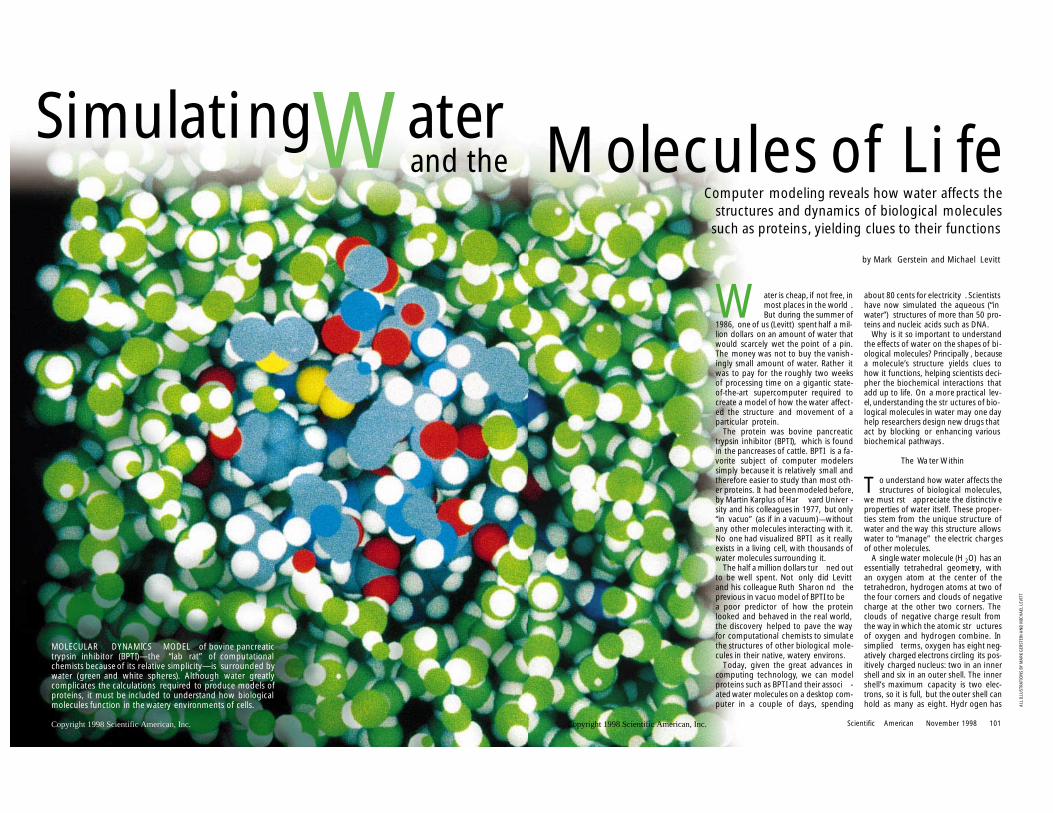
The Wa ter Within
T o understand how water affects thestructures of biological molecules,
we must rst appreciate the distinctiv eproperties of water itself. These proper-ties stem from the unique structure ofwater and the way this structure allowswater to “manage” the electric chargesof other molecules.
A single water molecule (H 2O) has anessentially tetrahedral geometry, withan oxygen atom at the center of thetetrahedron, hydrogen atoms at two ofthe four corners and clouds of negativecharge at the other two corners. Theclouds of negative charge result fromthe way in which the atomic str ucturesof oxygen and hydrogen combine. Insimplied terms, oxygen has eight neg-atively charged electrons circling its pos-itively charged nucleus: two in an innershell and six in an outer shell. The innershell’s maximum capacity is two elec-trons, so it is full, but the outer shell canhold as many as eight. Hydr ogen has
Scientific American November 1998 101
M olecules of Li feComputer modeling reveals how water affects the
structures and dynamics of biological moleculessuch as proteins, yielding clues to their functions
by Mark Gerstein and Michael Levitt
ALL
ILLU
STRA
TIO
NS
BY
MA
RK G
ERST
EIN
AN
D M
ICH
AEL
LEV
ITT
Copyright 1998 Scientific American, Inc.
SimulatingWaterand the
MOLECULAR DYNAMICS MODEL of bovine pancreatictrypsin inhibitor (BPTI)—the “lab rat” of computationalchemists because of its relative simplicity—is surrounded bywater (green and white spheres). Although water greatlycomplicates the calculations required to produce models ofproteins, it must be included to understand how biologicalmolecules function in the watery environments of cells.
Copyright 1998 Scientific American, Inc.
l MP2/CBS l TIP3P l AMOEBA
Water Dimer Structure and Energy
O-O Distance (Ång) 2.98 2.907 2.892O-O Bisector Angle (°) 57 ± 10 56.9 57.1Dimerization Energy 5.4 ± 0.7 4.98 4.95
Expt QM AMOEBA
OPLS-AAAMOEBA
ab Initio
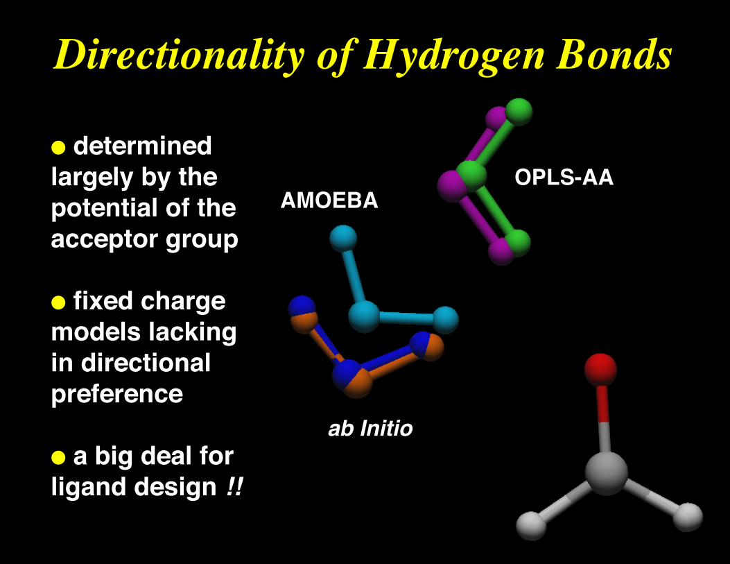
Directionality of Hydrogen Bonds
● determinedlargely by the potential of the acceptor group
● fixed chargemodels lackingin directionalpreference
● a big deal forligand design !!
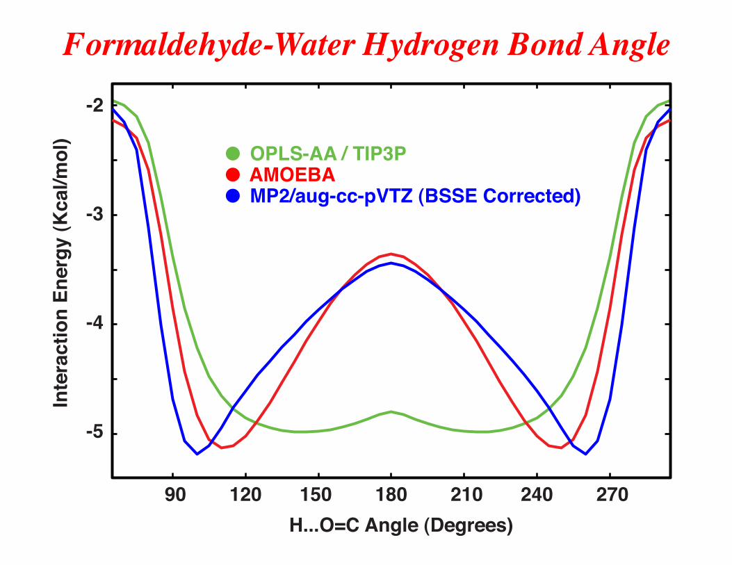
90 120 150 180 210 240 270
-2
-3
-4
-5
● OPLS-AA / TIP3P● AMOEBA● MP2/aug-cc-pVTZ (BSSE Corrected)
H...O=C Angle (Degrees)
Inte
ract
ion
Ener
gy (K
cal/m
ol)
Formaldehyde-Water Hydrogen Bond Angle
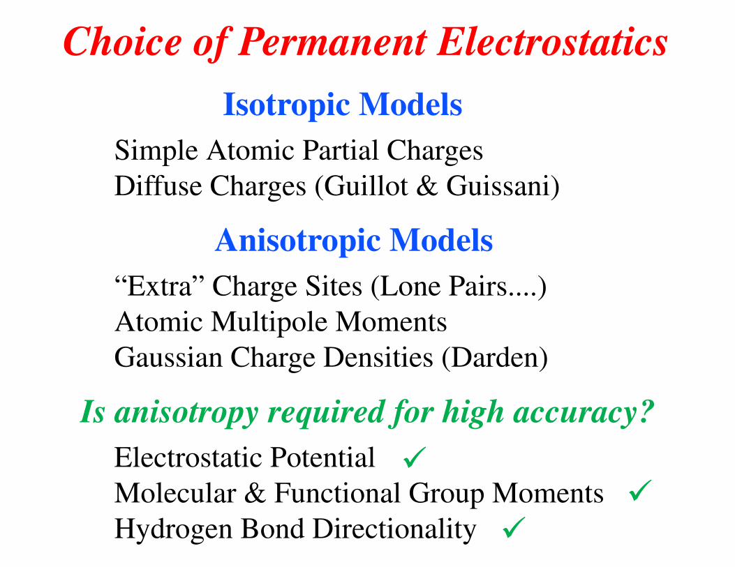
Choice of Permanent Electrostatics
Isotropic Models
Simple Atomic Partial Charges Diffuse Charges (Guillot & Guissani)
Anisotropic Models
“Extra” Charge Sites (Lone Pairs....) Atomic Multipole Moments Gaussian Charge Densities (Darden)
Is anisotropy required for high accuracy?
Electrostatic Potential Molecular & Functional Group Moments Hydrogen Bond Directionality
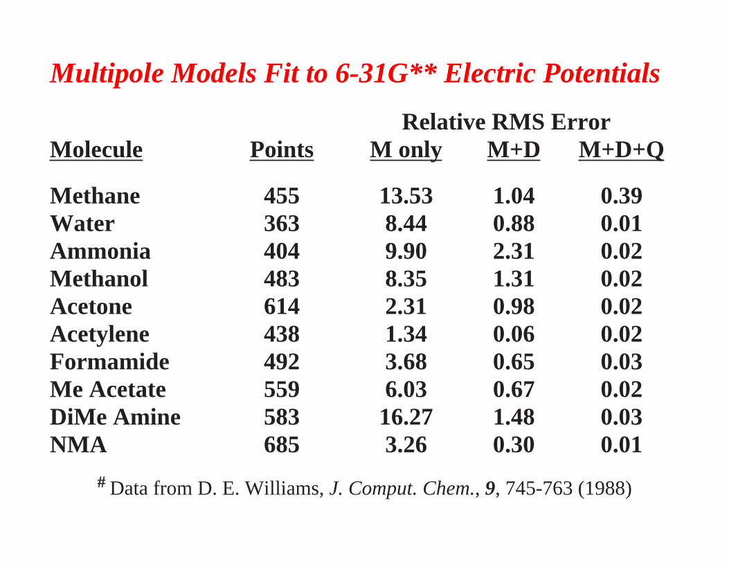
Multipole Models Fit to 6-31G** Electric Potentials
Relative RMS ErrorMolecule Points M only M+D M+D+Q
Methane 455 13.53 1.04 0.39Water 363 8.44 0.88 0.01Ammonia 404 9.90 2.31 0.02Methanol 483 8.35 1.31 0.02Acetone 614 2.31 0.98 0.02Acetylene 438 1.34 0.06 0.02Formamide 492 3.68 0.65 0.03Me Acetate 559 6.03 0.67 0.02DiMe Amine 583 16.27 1.48 0.03NMA 685 3.26 0.30 0.01
# Data from D. E. Williams, J. Comput. Chem., 9, 745-763 (1988)
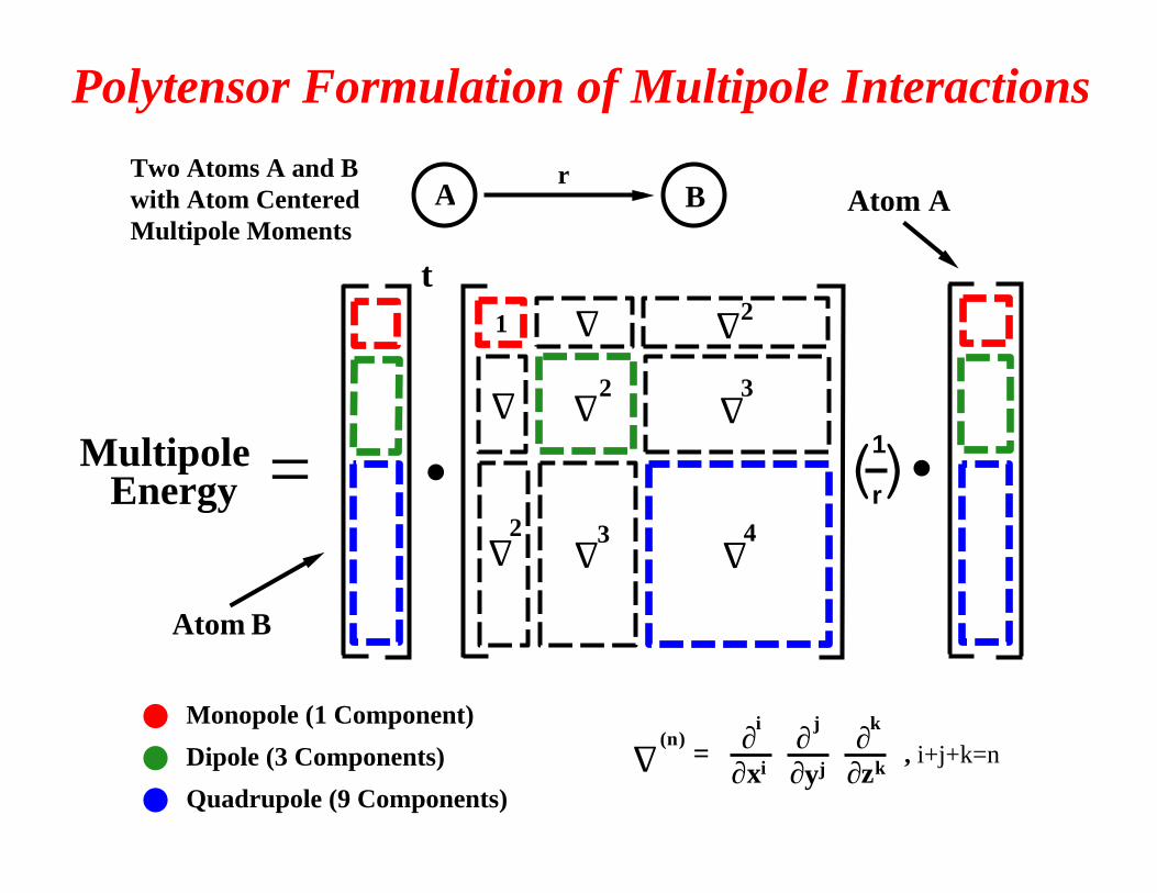
● Monopole (1 Component)
● Dipole (3 Components)
● Quadrupole (9 Components)
A B
1�r()
t
Multipole Energy =
1
∆
∆�
∆�
∆� 3
2
∆2 ∆4∆3
∆2
Atom B
Atom A
Polytensor Formulation of Multipole Interactions
● ●
Two Atoms A and Bwith Atom CenteredMultipole Moments
j k
=xi
i
zkyj, i+j+k=n
∆(n) ∂∂∂∂ ∂ ∂
r
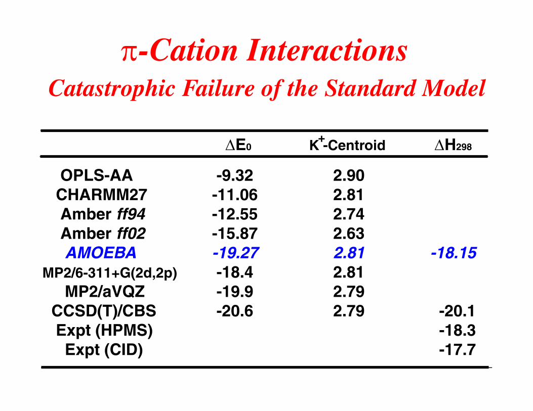
π-Cation Interactions
Catastrophic Failure of the Standard Model
∆E0 K -Centroid ∆H298
OPLS-AA -9.32 2.90 CHARMM27 -11.06 2.81 Amber ff94 -12.55 2.74 Amber ff02 -15.87 2.63 AMOEBA -19.27 2.81 -18.15MP2/6-311+G(2d,2p) -18.4 2.81 MP2/aVQZ -19.9 2.79 CCSD(T)/CBS -20.6 2.79 -20.1 Expt (HPMS) -18.3 Expt (CID) -17.7
+
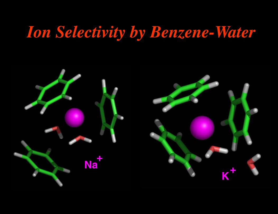
Ion Selectivity by Benzene-Water
Na+
K+
0 0 120 180 240 300 360
N-C-C=O (deg)
-0.4
-0.3
-0.2
-0.1
0
Part
ial C
harg
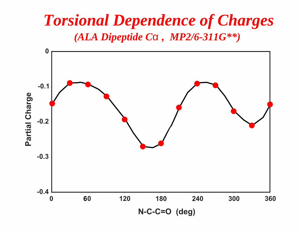
eTorsional Dependence of Charges (ALA Dipeptide Cα , MP2/6-311G**)
6
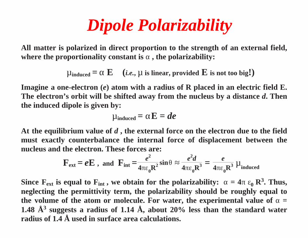
Dipole PolarizabilityAll matter is polarized in direct proportion to the strength of an external field, where the proportionality constant is α , the polarizability:
µinduced = α E (i.e., µ is linear, provided E is not too big!)
Imagine a one-electron (e) atom with a radius of R placed in an electric field E. The electron’s orbit will be shifted away from the nucleus by a distance d. Then the induced dipole is given by:
µinduced = αE = de
At the equilibrium value of d , the external force on the electron due to the field must exactly counterbalance the internal force of displacement between the nucleus and the electron. These forces are:
Fext = eE , and Fint = e2
4πε0R2 sinθ ≈ e2d
4πε0R3 = e
4πε0R3 µ
induced
Since Fext is equal to Fint , we obtain for the polarizability: α = 4π ε0 R3. Thus,
neglecting the permittivity term, the polarizability should be roughly equal to the volume of the atom or molecule. For water, the experimental value of α = 1.48 Å3 suggests a radius of 1.14 Å, about 20% less than the standard water radius of 1.4 Å used in surface area calculations.
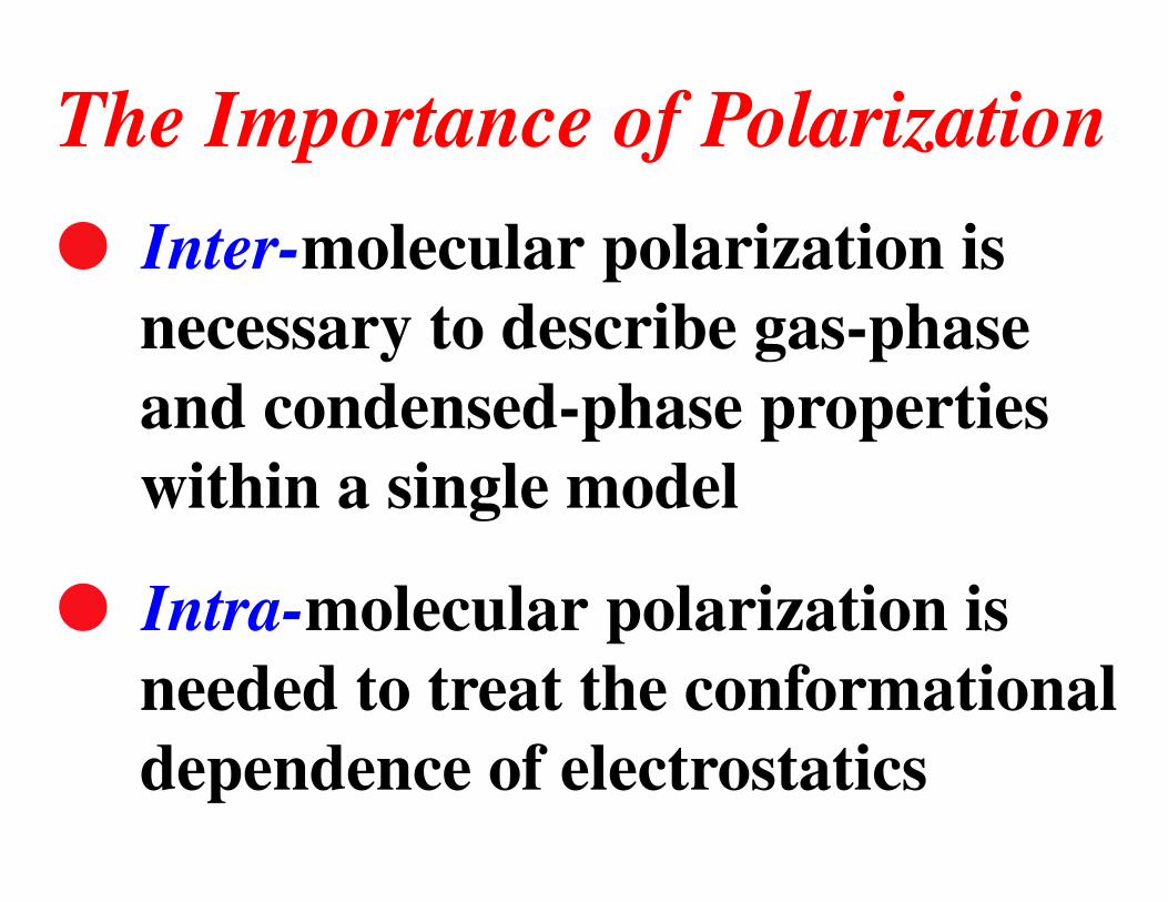
l Inter-molecular polarization is necessary to describe gas-phase and condensed-phase properties within a single model
l Intra-molecular polarization is needed to treat the conformational dependence of electrostatics
The Importance of Polarization
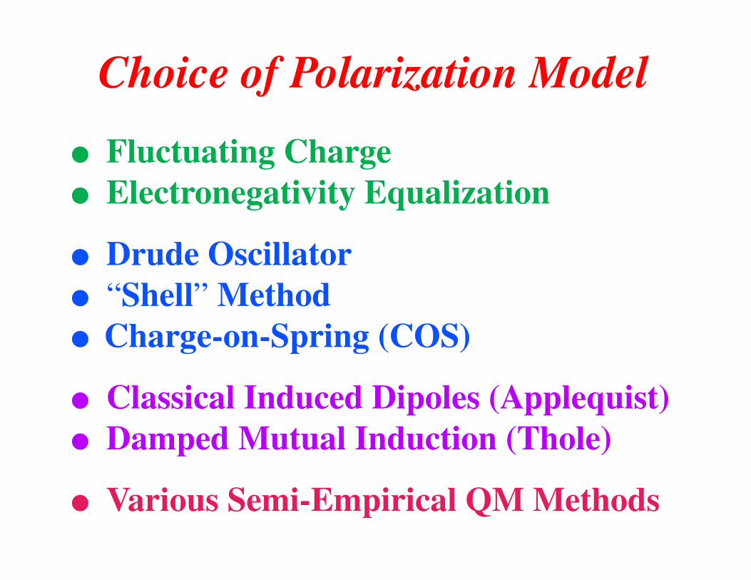
Choice of Polarization Model
● Fluctuating Charge● Electronegativity Equalization
● Drude Oscillator● “Shell” Method● Charge-on-Spring (COS)
● Classical Induced Dipoles (Applequist)● Damped Mutual Induction (Thole)
● Various Semi-Empirical QM Methods
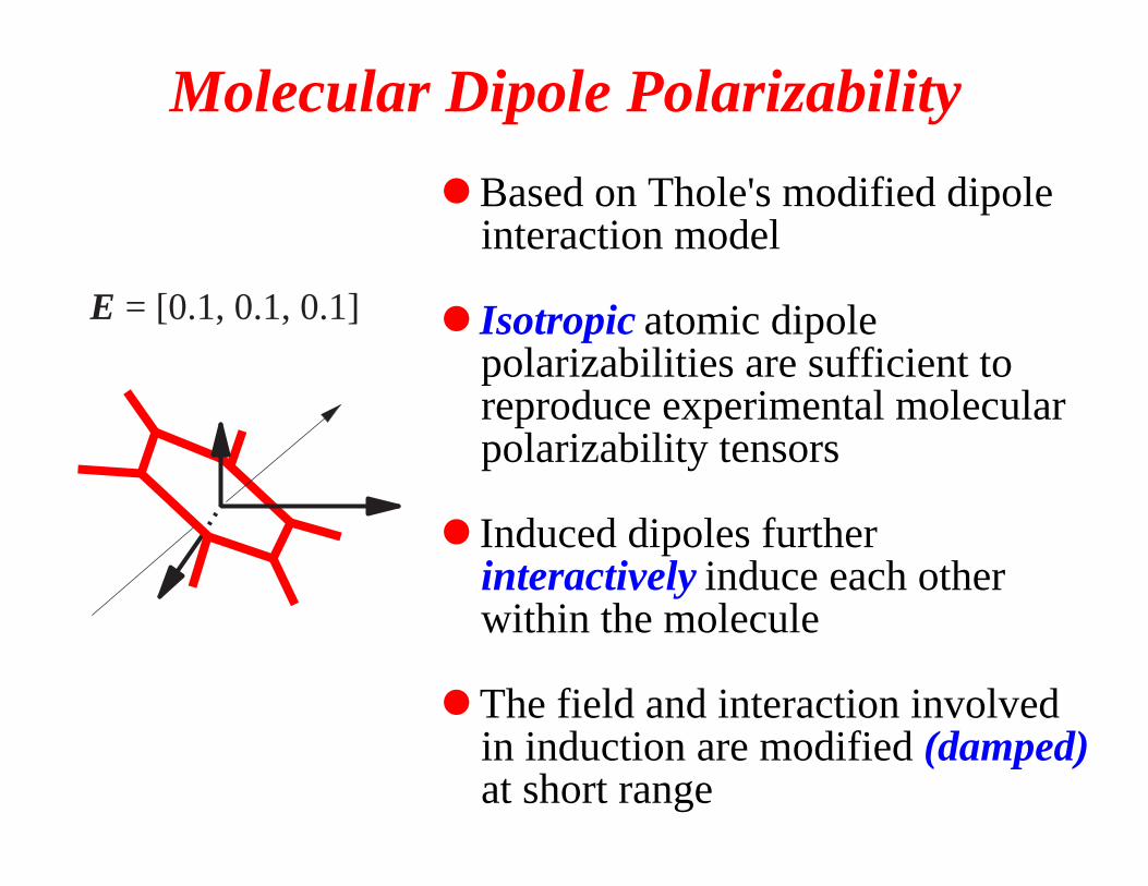
Based on Thole's modified dipole interaction model
Isotropic atomic dipole polarizabilities are sufficient to reproduce experimental molecular polarizability tensors
Induced dipoles further interactively induce each other within the molecule
The field and interaction involved in induction are modified (damped) at short range
●
●
●
●
E = [0.1, 0.1, 0.1]
Molecular Dipole Polarizability
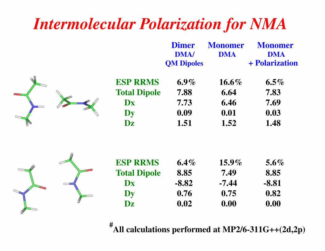
Intermolecular Polarization for NMA Dimer Monomer Monomer DMA/ DMA DMA QM Dipoles + Polarization
ESP RRMS 6.9% 16.6% 6.5%Total Dipole 7.88 6.64 7.83 Dx 7.73 6.46 7.69 Dy 0.09 0.01 0.03 Dz 1.51 1.52 1.48
ESP RRMS 6.4% 15.9% 5.6% Total Dipole 8.85 7.49 8.85 Dx -8.82 -7.44 -8.81 Dy 0.76 0.75 0.82 Dz 0.02 0.00 0.00
All calculations performed at MP2/6-311G++(2d,2p)#
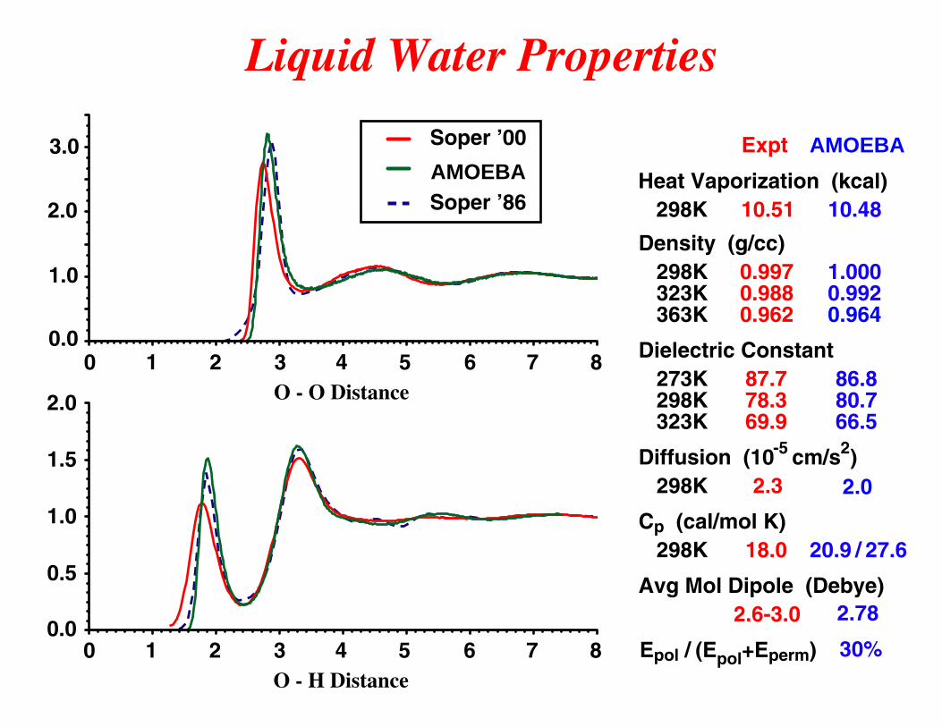
Liquid Water Properties
O - O Distance
0.0
1.0
2.0
3.0 Soper ’00
AMOEBASoper ’86
3210 4 5 6 7 8
0.0
0.5
1.0
1.5
2.0
O - H Distance3210 4 5 6 7 8
Density (g/cc)
Dielectric Constant
Diffusion (10 cm/s )
Cp (cal/mol K)
298K 323K 363K
273K 298K 323K
298K
298K
Avg Mol Dipole (Debye)
Expt
0.997 0.988 0.962
87.778.369.9
2.3
18.0
2.6-3.0
AMOEBA
1.0000.9920.964
86.880.766.5
20.9 / 27.6
2.78
30%Epol / (Epol+Eperm)
Heat Vaporization (kcal) 298K 10.51 10.48
2.0
-5 2
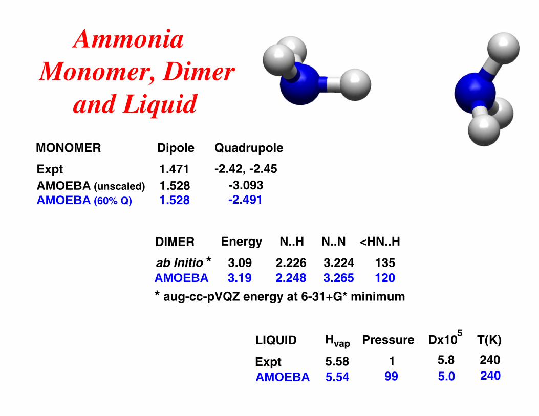
MONOMER Dipole Quadrupole
Expt 1.471 -2.42, -2.45AMOEBA (unscaled) 1.528 -3.093AMOEBA (60% Q) 1.528 -2.491
DIMER
LIQUID
Energy N..H N..N <HN..H
Hvap Pressure Dx105
T(K)
ab Initio * 3.09 2.226 3.224 135
Expt 5.58 1 5.8 240
AMOEBA 3.19 2.248 3.265 120
AMOEBA 5.54 99 5.0 240
AmmoniaMonomer, Dimer and Liquid
* aug-cc-pVQZ energy at 6-31+G* minimum
α
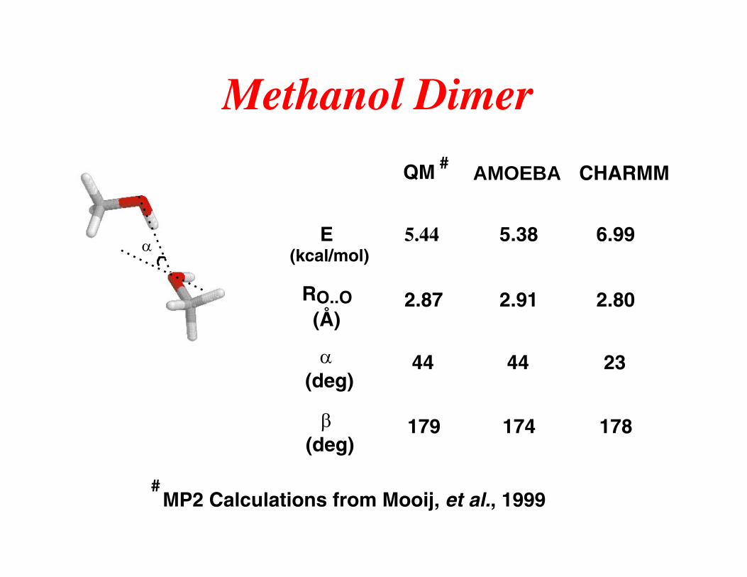
Methanol Dimer
QM # AMOEBA CHARMM
E (kcal/mol)
5.44 5.38 6.99
RO..O(Å)
2.87 2.91 2.80
α(deg)
44 44 23
β(deg)
179 174 178
# MP2 Calculations from Mooij, et al., 1999
0
1
2
3
4
5
6
Expt
AM
OE
BA
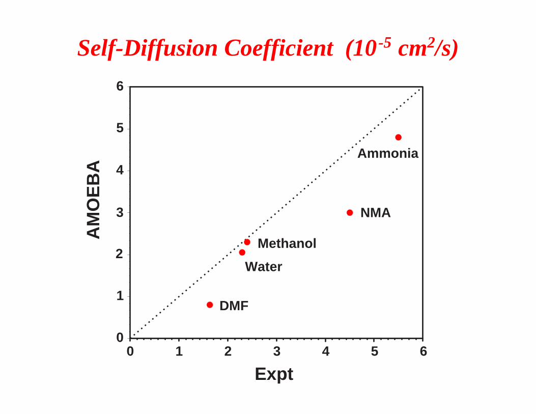
Self-Diffusion Coefficient (10 -5 cm2/s)
Water
Methanol
Ammonia
DMF
NMA
4 5 63210
AMOEBA
4
6
8
10
12
14
16E
xpt
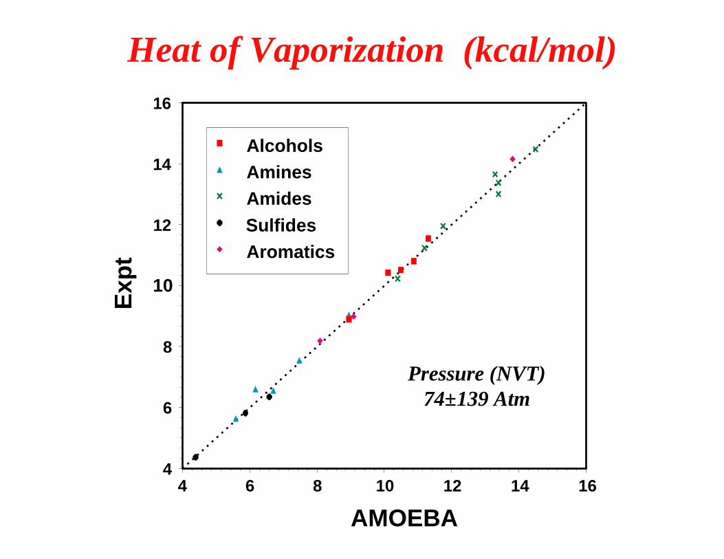
AlcoholsAminesAmidesSulfidesAromatics
Heat of Vaporization (kcal/mol)
Pressure (NVT) 74±139 Atm
16141210864
0
20
40
60
80
100D
iele
tric
Co
nst
ant
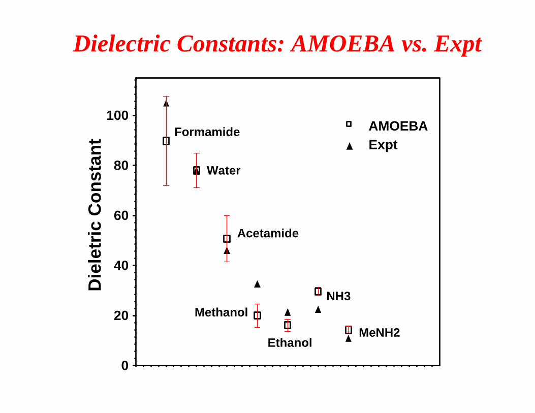
AMOEBAExpt
Dielectric Constants: AMOEBA vs. Expt
Formamide
Water
Acetamide
MethanolNH3
MeNH2Ethanol
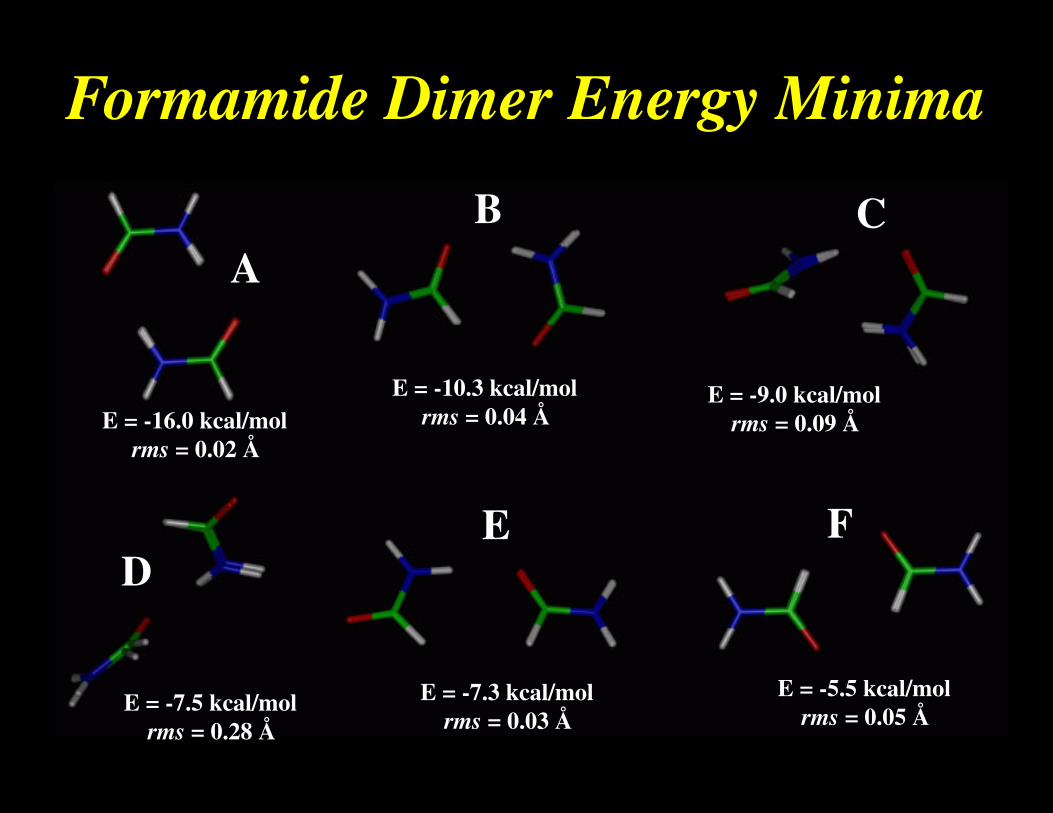
Formamide Dimer Energy Minima
AB C
DE F
E = -16.0 kcal/mol rms = 0.02 Å
E = -10.3 kcal/mol rms = 0.04 Å
E = -9.0 kcal/mol rms = 0.09 Å
E = -7.5 kcal/mol rms = 0.28 Å
E = -7.3 kcal/mol rms = 0.03 Å
E = -5.5 kcal/mol rms = 0.05 Å
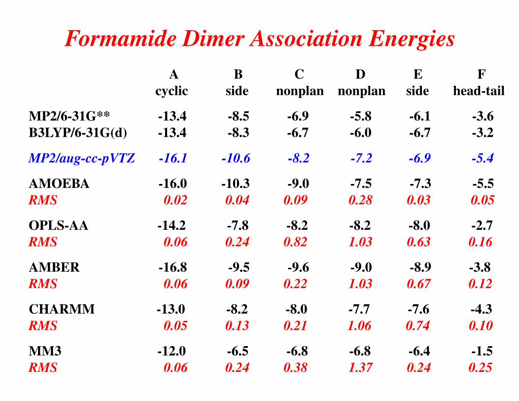
A B C D E F cyclic side nonplan nonplan side head-tail
MP2/6-31G** -13.4 -8.5 -6.9 -5.8 -6.1 -3.6B3LYP/6-31G(d) -13.4 -8.3 -6.7 -6.0 -6.7 -3.2
MP2/aug-cc-pVTZ -16.1 -10.6 -8.2 -7.2 -6.9 -5.4
AMOEBA -16.0 -10.3 -9.0 -7.5 -7.3 -5.5RMS 0.02 0.04 0.09 0.28 0.03 0.05
OPLS-AA -14.2 -7.8 -8.2 -8.2 -8.0 -2.7RMS 0.06 0.24 0.82 1.03 0.63 0.16
AMBER -16.8 -9.5 -9.6 -9.0 -8.9 -3.8RMS 0.06 0.09 0.22 1.03 0.67 0.12
CHARMM -13.0 -8.2 -8.0 -7.7 -7.6 -4.3RMS 0.05 0.13 0.21 1.06 0.74 0.10
MM3 -12.0 -6.5 -6.8 -6.8 -6.4 -1.5RMS 0.06 0.24 0.38 1.37 0.24 0.25
Formamide Dimer Association Energies
C
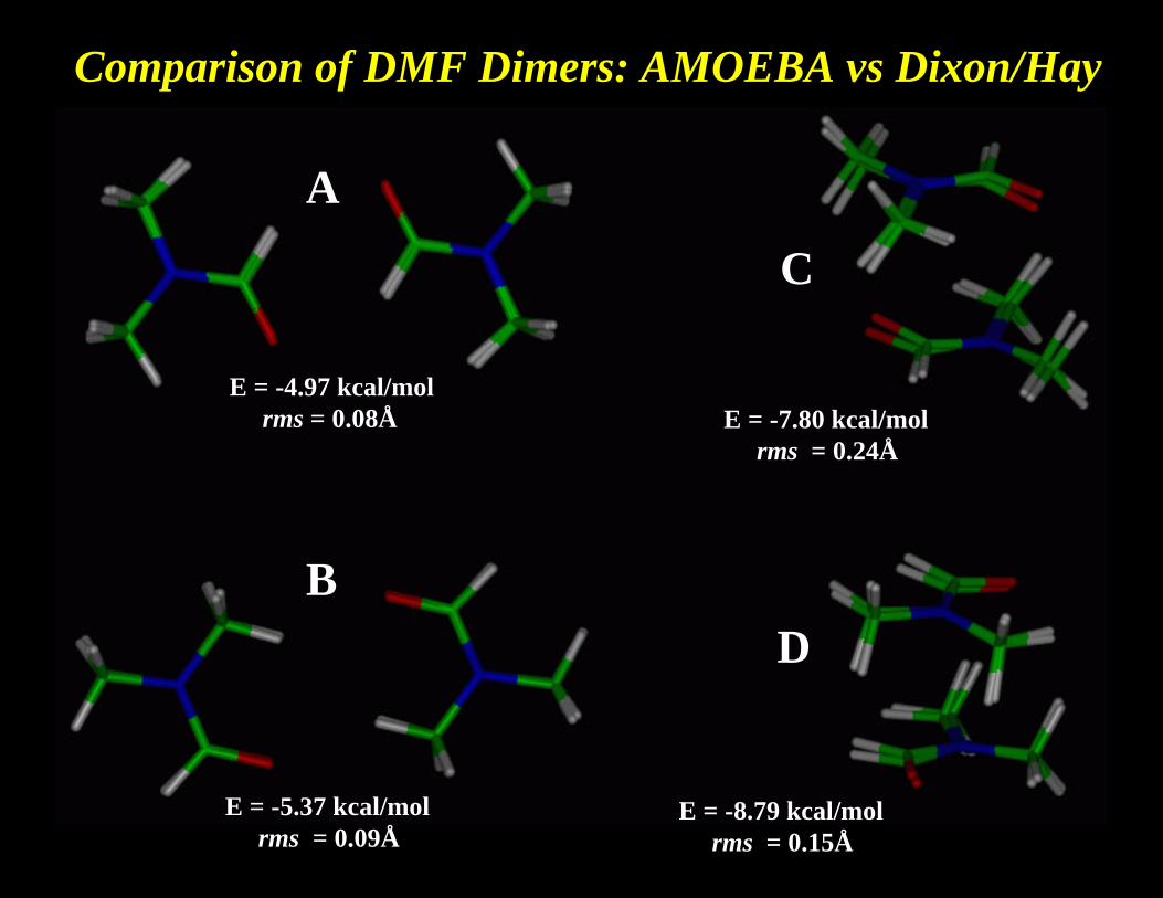
Comparison of DMF Dimers: AMOEBA vs Dixon/Hay
A
BD
E = -4.97 kcal/mol rms = 0.08Å E = -7.80 kcal/mol
rms = 0.24Å
E = -5.37 kcal/mol rms = 0.09Å
E = -8.79 kcal/mol rms = 0.15Å
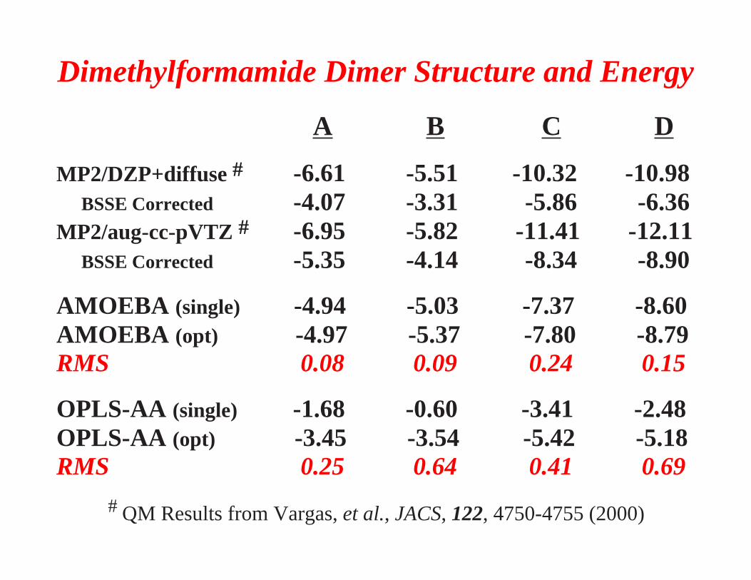
Dimethylformamide Dimer Structure and Energy
A B C D
MP2/DZP+diffuse # -6.61 -5.51 -10.32 -10.98 BSSE Corrected -4.07 -3.31 -5.86 -6.36MP2/aug-cc-pVTZ # -6.95 -5.82 -11.41 -12.11 BSSE Corrected -5.35 -4.14 -8.34 -8.90
AMOEBA (single) -4.94 -5.03 -7.37 -8.60AMOEBA (opt) -4.97 -5.37 -7.80 -8.79RMS 0.08 0.09 0.24 0.15
OPLS-AA (single) -1.68 -0.60 -3.41 -2.48OPLS-AA (opt) -3.45 -3.54 -5.42 -5.18RMS 0.25 0.64 0.41 0.69
# QM Results from Vargas, et al., JACS, 122, 4750-4755 (2000)
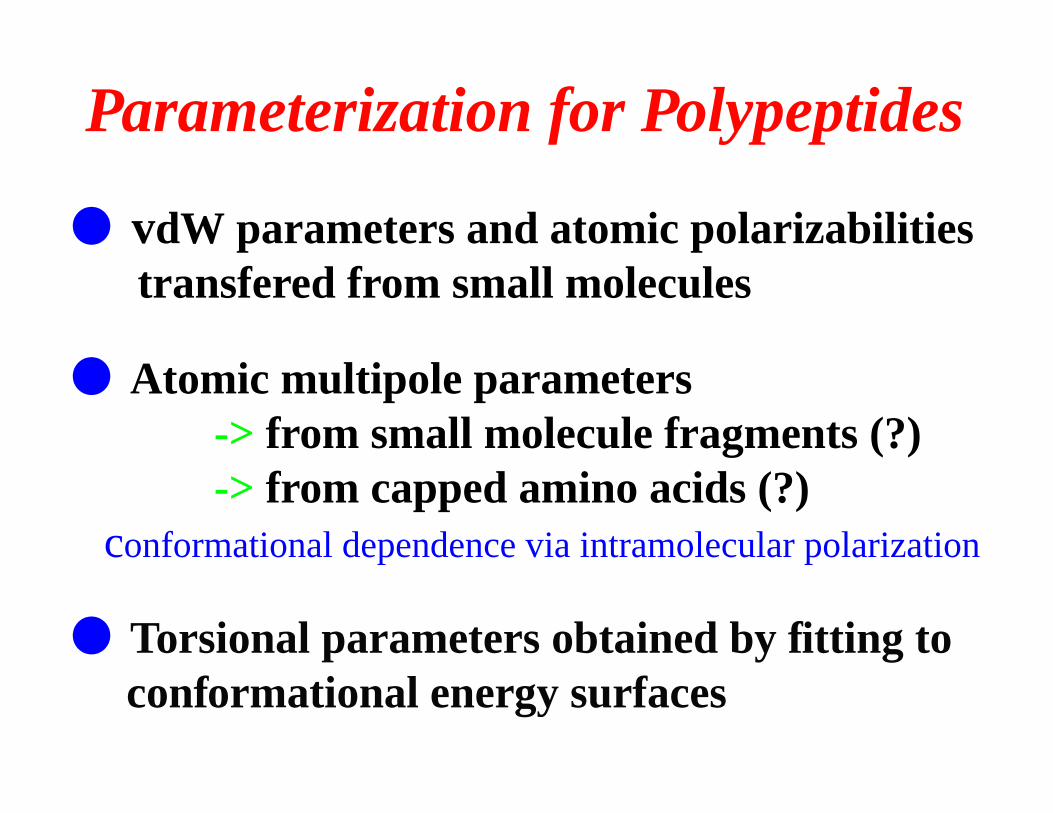
Parameterization for Polypeptides
● vdW parameters and atomic polarizabilities transfered from small molecules
● Atomic multipole parameters -> from small molecule fragments (?) -> from capped amino acids (?) conformational dependence via intramolecular polarization
● Torsional parameters obtained by fitting to conformational energy surfaces
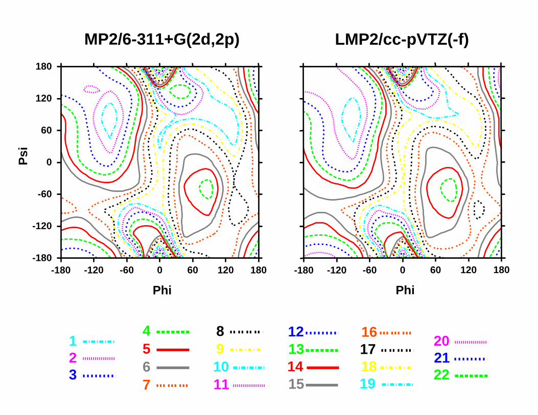
222120
19181716
15141312
111098
7654
321
LMP2/cc-pVTZ(-f)
-180 -120 -60 0 60 120 180
Phi
MP2/6-311+G(2d,2p)
-180 -120 -60 0 60 120 180
Phi
-180
-120
-60
0
60
120
180
Psi
-180
-120
-60
0
60
120
180
-180 -120 -60 0 60 120 180
Psi
Phi
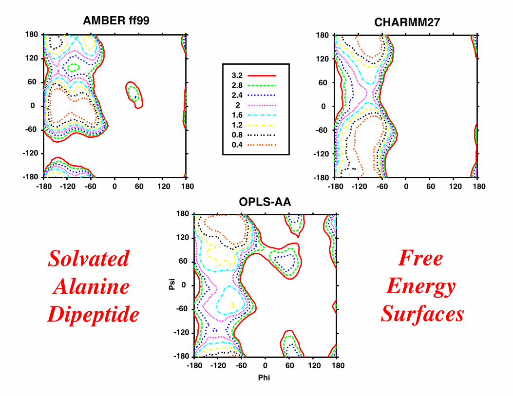
OPLS-AA
-180
-120
-60
0
60
120
180
-180 -120 -60 0 60 120 180
AMBER ff99
-180
-120
-60
0
60
120
180
-180 -120 -60 0 60 120 180
CHARMM27
3.22.82.42
1.61.20.80.4
Solvated AlanineDipeptide
Free EnergySurfaces
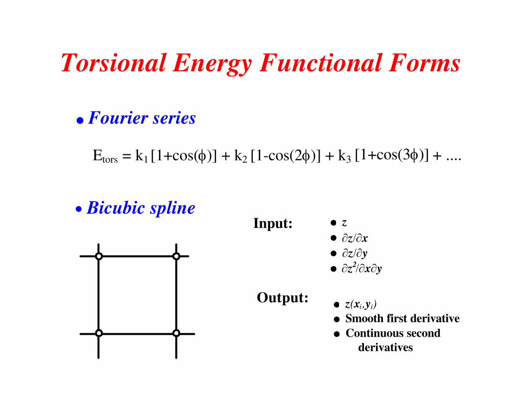
Torsional Energy Functional Forms
l Fourier series
• Bicubic spline
l z l l l
Input:
Output: l z(xi ,yi)l Smooth first derivative l Continuous second
derivatives
Etors = k1 [1+cos(φ)] + k2 [1-cos(2φ)] + k3 [1+cos(3φ)] + ....
∂z/∂x∂z/∂y∂z /∂x∂y2
3.63.22.8
2.42
1.6
1.20.80.4
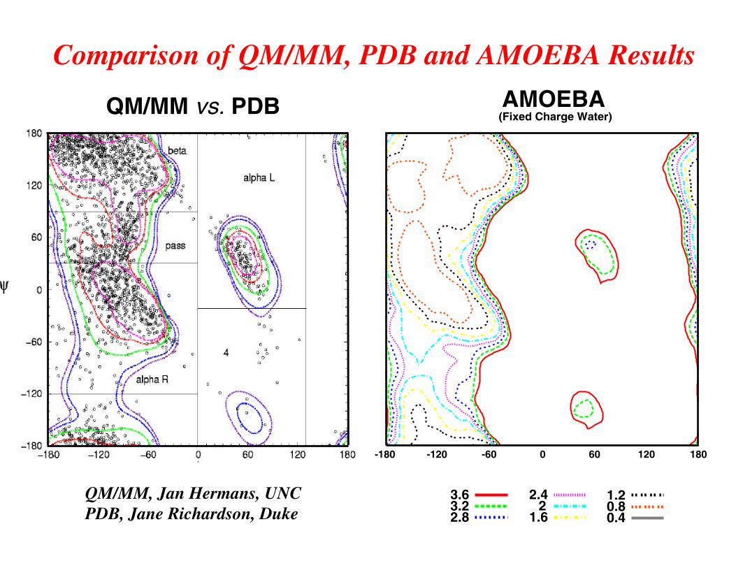
QM/MM vs. PDB AMOEBA (Fixed Charge Water)
QM/MM, Jan Hermans, UNCPDB, Jane Richardson, Duke
Comparison of QM/MM, PDB and AMOEBA Results
-180 -120 -60 0 60 120 180
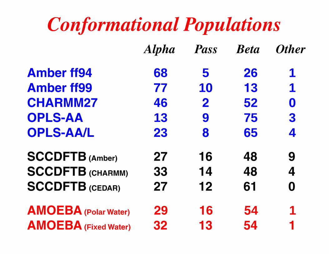
Conformational Populations Alpha Pass Beta Other
Amber ff94 68 5 26 1Amber ff99 77 10 13 1CHARMM27 46 2 52 0OPLS-AA 13 9 75 3OPLS-AA/L 23 8 65 4
SCCDFTB (Amber) 27 16 48 9SCCDFTB (CHARMM) 33 14 48 4SCCDFTB (CEDAR) 27 12 61 0
AMOEBA (Polar Water) 29 16 54 1AMOEBA (Fixed Water) 32 13 54 1
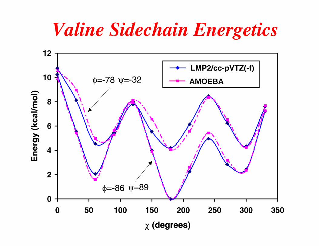
Valine Sidechain Energetics
0
2
4
6
8
10
12
0 50 100 150 200 250 300 350
χ (degrees)
En
erg
y (k
cal/m
ol)
LMP2/cc-pVTZ(-f)
AMOEBAφ=-78 ψ=-32
φ=-86 ψ=89
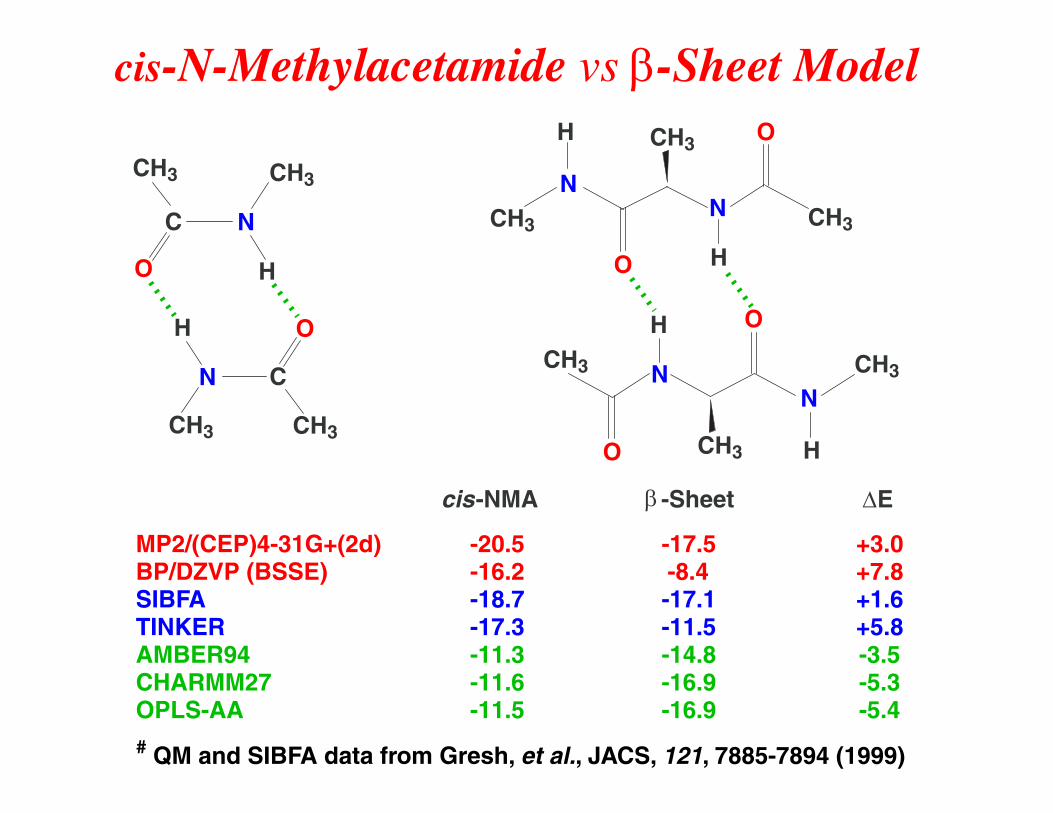
cis-N-Methylacetamide vs β-Sheet Model
cis-NMA β -Sheet ∆E
MP2/(CEP)4-31G+(2d) -20.5 -17.5 +3.0BP/DZVP (BSSE) -16.2 -8.4 +7.8SIBFA -18.7 -17.1 +1.6TINKER -17.3 -11.5 +5.8AMBER94 -11.3 -14.8 -3.5CHARMM27 -11.6 -16.9 -5.3OPLS-AA -11.5 -16.9 -5.4# QM and SIBFA data from Gresh, et al., JACS, 121, 7885-7894 (1999)
O
CH3
C N
H
CH3
O H
CH3
C N
CH3
NCH3
O
N
H
O
CH3
HCH3
NCH3
O
N
H
O
CH3
H CH3
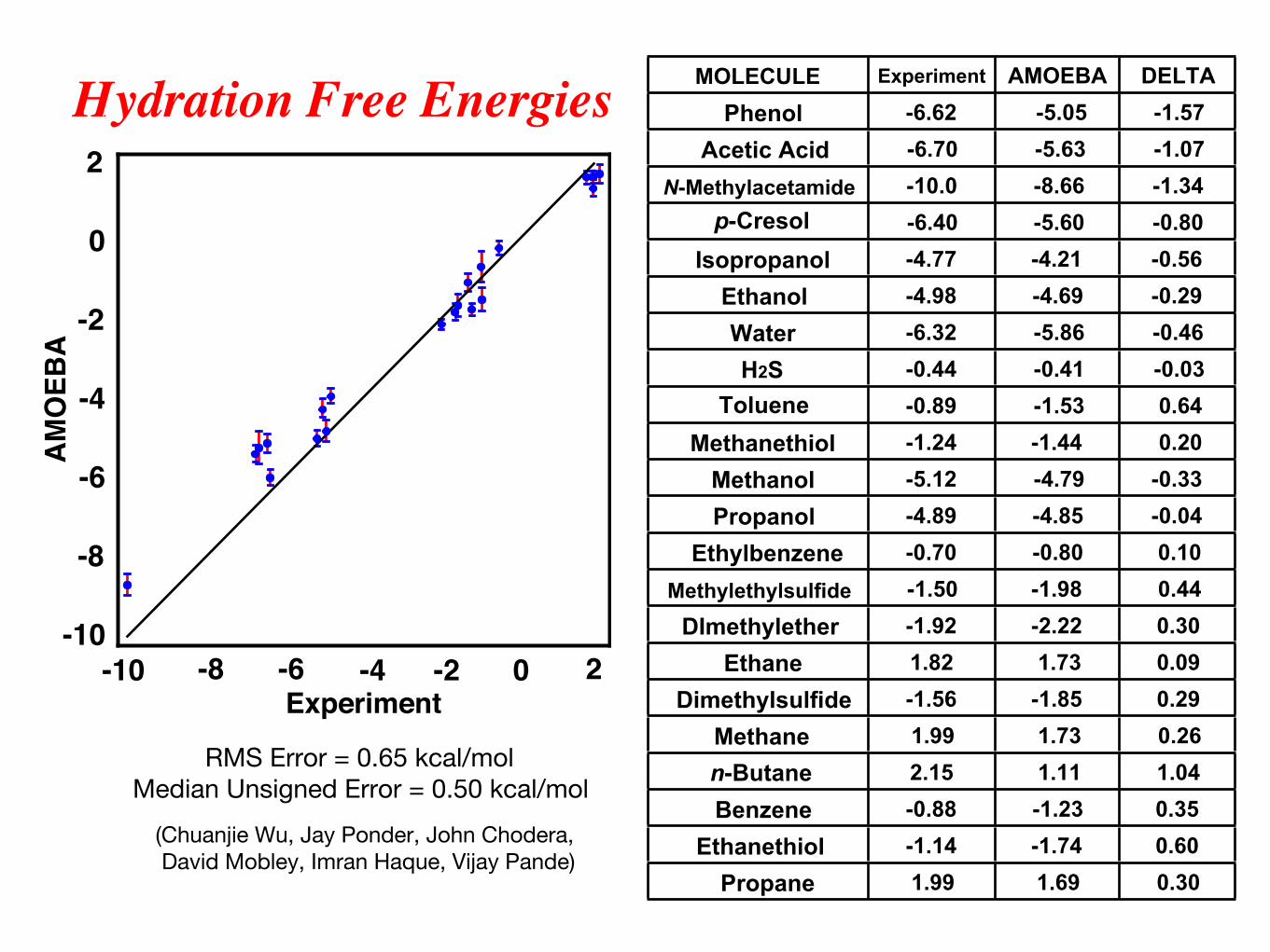
MOLECULE Experiment AMOEBA DELTAPhenol -6.62 -5.05 -1.57
Acetic Acid -6.70 -5.63 -1.07N-Methylacetamide -10.0 -8.66 -1.34
p-Cresol -6.40 -5.60 -0.80Isopropanol -4.77 -4.21 -0.56
Ethanol -4.98 -4.69 -0.29Water -6.32 -5.86 -0.46H2S -0.44 -0.41 -0.03
Toluene -0.89 -1.53 0.64Methanethiol -1.24 -1.44 0.20
Methanol -5.12 -4.79 -0.33Propanol -4.89 -4.85 -0.04
Ethylbenzene -0.70 -0.80 0.10Methylethylsulfide -1.50 -1.98 0.44
DImethylether -1.92 -2.22 0.30Ethane 1.82 1.73 0.09
Dimethylsulfide -1.56 -1.85 0.29Methane 1.99 1.73 0.26n-Butane 2.15 1.11 1.04Benzene -0.88 -1.23 0.35
Ethanethiol -1.14 -1.74 0.60Propane 1.99 1.69 0.30
-10 -8 -6 -4 -2 0 2-10
-8
-6
-4
-2
0
2
AM
OEB
A
RMS Error = 0.65 kcal/mol Median Unsigned Error = 0.50 kcal/mol
(Chuanjie Wu, Jay Ponder, John Chodera, David Mobley, Imran Haque, Vijay Pande)
Hydration Free Energies
Experiment
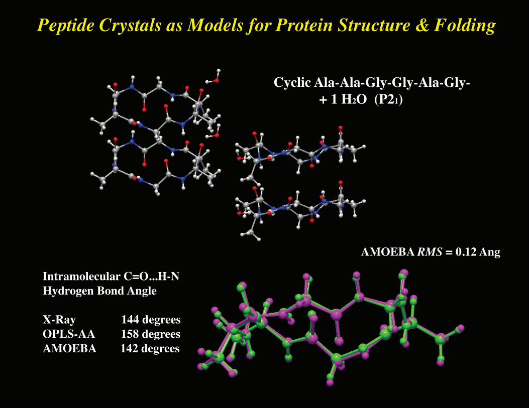
Peptide Crystals as Models for Protein Structure & Folding
Cyclic Ala-Ala-Gly-Gly-Ala-Gly- + 1 H2O (P21)
Intramolecular C=O...H-NHydrogen Bond Angle
X-Ray 144 degreesOPLS-AA 158 degreesAMOEBA 142 degrees
AMOEBA RMS = 0.12 Ang
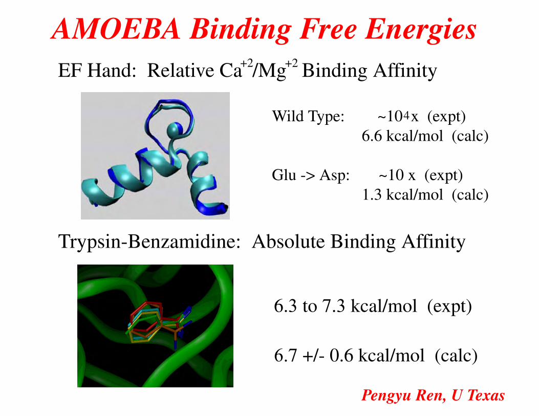
AMOEBA Binding Free EnergiesEF Hand: Relative Ca /Mg Binding Affinity+2 +2
Wild Type: ~10 x (expt) 6.6 kcal/mol (calc)
Glu -> Asp: ~10 x (expt) 1.3 kcal/mol (calc)
Trypsin-Benzamidine: Absolute Binding Affinity
6.3 to 7.3 kcal/mol (expt)
6.7 +/- 0.6 kcal/mol (calc)
4
Pengyu Ren, U Texas