Embed Size (px)
Citation preview

Page 1 of 35
Silverlon:
A Technical Review
S Thomas B.Pharm., PhD.
Medetec
January 2008

Page 2 of 35
Table of Contents
1. EXECUTIVE SUMMARY......................................................................................................................... 5
2. INTRODUCTION....................................................................................................................................... 6 2.1 Study sponsor........................................................................................................................................ 6 2.2 Aim of study .......................................................................................................................................... 6 2.3 Data reviewed........................................................................................................................................ 6 2.4 Timescales.............................................................................................................................................. 6 2.5 Conduct of review and format of report............................................................................................. 6 2.6 Limitations of the review ..................................................................................................................... 6 2.7 Statement by the author....................................................................................................................... 6
3. PRODUCT INFORMATION .................................................................................................................... 7 3.1 Product description .............................................................................................................................. 7 3.2 Product features and benefits .............................................................................................................. 7 3.3 Historical basis for the use of topical silver........................................................................................ 8 3.4 Early silver-containing dressings ....................................................................................................... 8
4. SILVERLON’S PRINCIPAL COMPETITORS.................................................................................... 10 4.1 Acticoat - Smith and Nephew ............................................................................................................ 10 4.2 Arglaes - Medline................................................................................................................................ 10 4.3 Aquacel Ag - Convatec ....................................................................................................................... 10 4.4 Avance - SSL International .............................................................................................................. 10 4.5 Calgitrol - Biomedical Technologies Inc.......................................................................................... 10 4.6 Contreet Ag – Coloplast ..................................................................................................................... 10 4.7 Contreet Hydrocolloid dressing - Coloplast ..................................................................................... 10 4.8 Silvasorb - Medline............................................................................................................................. 10
5. REVIEW OF PRECLINICAL DATA GENERATED BY THE CLIENT ......................................... 10 5.1 Antimicrobial properties.................................................................................................................... 11
5.1.1 USP Antimicrobial preservative effectiveness............................................................................... 11 5.1.2 Antimicrobial activity-dynamic test of surfaces ASTM E 2149..................................................... 12 5.1.3 Assessment of antibacterial finishes on textile materials AATCC Method 100 (24 hr) ................ 13 5.1.4 Assessment of antibacterial finishes on textile materials AATCC Test Method 100 ..................... 14 5.1.5 Dow Corning corporate test method 0923 .................................................................................... 15 5.1.6 Kirby Bauer standard antimicrobial susceptibility ....................................................................... 16
5.2 Silver ion release studies .................................................................................................................... 17 5.3 Biocompatibility studies ..................................................................................................................... 18
5.3.1 Sensitization potential ................................................................................................................... 18 5.3.2 Acute intracutaneous reactivity..................................................................................................... 18

Page 3 of 35
5.3.3 Cytotoxicity.................................................................................................................................... 18 5.3.4 Haemolysis study........................................................................................................................... 18 5.3.5 5. Muscle implantation.................................................................................................................. 18 5.3.6 Acute systemic toxicity................................................................................................................... 18
5.4 Electromotive effects ......................................................................................................................... 19
6. REVIEW OF PRECLINICAL DATA FROM OTHER SOURCES .................................................... 21 6.1 Comparison of silver dressings in an animal study ......................................................................... 21 6.2 Laboratory comparison of antimicrobial activity of silver dressings ............................................ 21
6.2.1 Silver content................................................................................................................................. 21 6.2.2 Antimicrobial activity: zone of inhibition...................................................................................... 23 6.2.3 Antimicrobial activity: challenge testing....................................................................................... 24 6.2.4 Antimicrobial activity: microbial transmission test ...................................................................... 26
7. REVIEW OF CLINICAL DATA PROVIDED BY THE CLIENT ...................................................... 27 7.1 Surgical wounds.................................................................................................................................. 27
7.1.1 Healing chronic, ischaemic infected saphenous vein harvest sites in the legs with silver plated cloth and allograft dermal regenerative matrix. .................................................................................... 27 7.1.2 Clinical case evaluation and results of Silverlon sternal dressings in cardiac surgery................ 27 7.1.3 Cardiovascular surgery assessment of Silverlon vs standard wound care methods in non-pre-complex post-operative wounds. ............................................................................................................ 27
7.2 Chronic wounds .................................................................................................................................. 28 7.2.1 Comparison of healing of lower extremity chronic wounds with silver plated cloth dressings, papain-urea-chlorophyllin ointment and hydrogel. ............................................................................... 28 7.2.2 Chronic, diabetic foot wounds with ischemia and tissue loss: use of advanced wound care techniques, to prevent further tissue loss ............................................................................................... 28
7.3 Silverlon as an adjunct to VAC therapy........................................................................................... 29 7.3.1 Silver dressings used with wound vacuum assisted closure: is there an advantage? ................... 29 7.3.2 The combined benefit of negative pressure therapy, elemental silver contact layer, & bilayered living skin equivalent in the treatment of chronic hard to heal lower extremity wounds....................... 29 7.3.3 Healing skin grafts over chronic wounds with vacuum assisted closure and silver dressings ..... 29 7.3.4 VAC with Silverlon vs VAC alone ................................................................................................. 30
7.4 Miscellaneous case studies ................................................................................................................. 30 7.4.1 Treatment of pyoderma gangrenosum with a silver contact layer ................................................ 30 7.4.2 Clinical case study treatment of coumadin tissue necrosis with silverlon .................................... 30 7.4.3 Successful treatment of a chronic venous ulcer with a silver contact dressing and compression therapy.................................................................................................................................................... 31 7.4.4 Graft vs. host disease: an innovative approach using silver contact dressing to manage grade IV skin lesions ............................................................................................................................................. 31
8. REVIEW OF PUBLISHED CLINICAL DATA .................................................................................... 32 8.1 Do silver-impregnated dressings limit infections after lumbar laminectomy? ............................. 32
9. CONCLUSIONS........................................................................................................................................ 33 9.1 Electromotive effects .......................................................................................................................... 33 9.2 Antimicrobial activity......................................................................................................................... 33

Page 4 of 35
9.3 Biocompatibility studies ..................................................................................................................... 33 9.4 Review of clinical data........................................................................................................................ 34
10. REFERENCES ........................................................................................................................................ 35

Page 5 of 35
1. EXECUTIVE SUMMARY
The principal conclusions that may be drawn from this review are as follows:
• There is a considerable body of evidence in the literature that silver ions have a profound antimicrobial effect in laboratory studies.
• There is also good evidence to show that these antimicrobial properties can be of significant benefit in preventing infection in a variety of wounds types.
• Laboratory investigations have shown that Silverlon can deliver sufficiently high concentrations of silver ions to exert an antimicrobial effect in vitro against a broad spectrum of pathogenic organisms.
• A number of scientific publications are available which describe the successful use of various brands of silver-containing dressings, but the clinical studies so far produced on Silverlon are of variable quality and individually fall far short of the standard required to provide conclusive proof of the value of this dressing in different clinical situations.
• Taken together, however, the Silverlon studies provide persuasive evidence that the dressing does possess antimicrobial properties which impacts favourably upon clinical infection rates and wound healing although this evidence would not be considered strong enough to be accepted as part of a formal systematic review.
• With the possible exception of the VAC dressing, insufficient clinical information is available to form a judgment on specific products within the range as requested by the client although it is the unsubstantiated view of the author that these may offer clinical benefits for specific applications.
• Claims made for electromotive and pain-relieving properties of the dressing although interesting have not been substantiated
• Silverlon appears to have a good safety profile, being free of toxicity and unlikely to cause adverse local effects.
• There are deficiencies in the Silverlon technical and promotional literature which should be corrected for the UK market.

Page 6 of 35
2. INTRODUCTION
2.1 Study sponsor This report was commissioned by Christian Stephenson, Product Development Manager, Lantor UK, St Helens Road, Bolton, Lancashire, BL3 3PR.
2.2 Aim of study The author was asked to review both preclinical and clinical data on the Silverlon product range to determine if the information available was sufficiently comprehensive to support new or existing medical claims for the use of the products in the treatment of different types of wounds within the UK. It was requested that particular attention be given to the negative pressure dressing, the burn contact dressing, the elastic burn wrap, the acute burn glove the calcium alginate dressing and the digidressing
2.3 Data reviewed Data was provided in electronic format and consisted of a total of forty three files although not all described clinical experience with the product. This information, which was subsequently found to have come from the company website, was supplemented by reference to other sources including publications in medical and scientific journals and data abstracted from the internet.
2.4 Timescales The documentation for review was received on January 15th, and the report was produced between January 18th and January 25th.
2.5 Conduct of review and format of report A short summary was produced of each of the documents or files reviewed which, in most instances, consists of a brief description of the methodology, and an account of the principal finding together with comments (positive or negative), upon the design, conduct and relevance of the study where appropriate. These individual critiques are included in Section 8 of this report.
2.6 Limitations of the review In many instances, the documents or files reviewed originated from the manufacturer’s website. Primary data from clinical studies or laboratory investigations was not available for examination.
2.7 Statement by the author This independent review was commissioned by the client, and produced by the author for a professional fee as a medical writer. The author therefore has no interest, financial, commercial or otherwise, in any of the products mentioned or described within the document.

Page 7 of 35
3. PRODUCT INFORMATION
3.1 Product description Silverlon is the brand name that has been applied to a number of different wound management materials, all of which share a common feature. They each consist of, or contain, a nylon fabric that has been silver-plated by means of a proprietary autocatalytic electroless chemical (reduction-oxidation) plating technique.
This technique effectively coats the entire surface of each individual fibre, resulting in a very large surface area for the release of ionic silver.
These dressings are available as wound contact materials, (normally used in conjunction with a secondary absorbent layer), island dressings, absorbent dressing pads, wound packing strips, elastic wraps, adhesive strips, adhesive bandages, tubular dressings, burn gloves, negative pressure dressings.
In order to make this review as comprehensive and as useful as possible, it was the intention of the author to produce a brief description of the composition and construction of each product in the portfolio, to facilitate understanding of how each item functions during clinical use.
Unfortunately this proved impossible to do, as the information provided in electronic form as well as that contained on the Silverlon website is extremely poor in this regard.
For example the ‘Product Description’ for Silverlon Wound Contact Dressing available at http://www.silverlon.com/pd_contact_dressing.html makes numerous claims about what the dressing does, or is claimed to do, but says virtually nothing about the product itself. It also states the ‘a special modification of a Silverlon Wound Contact Dressing, called the ‘Silverlon Peripad,’ can be applied to an external feminine napkin (peripad) for lesions of the perineal region and for application after childbirth’, but provides no indication of how this differs from the standard product.
A similar criticism can be directed most other products in the range. It is strongly suggested, therefore, that serious consideration be given to the production of individual datacards which contain basic information on the composition and construction of each product together with other relevant information in a clear and concise manner such as that found here:. http://www.dressings.org/dressings-datacards-by-alpha.html.
3.2 Product features and benefits The principal claim made for the Silverlon product range is that they combat or prevent infection in a variety of soft tissue injuries. It is suggested that this activity is dependent upon the release of positively charged silver ions, a process that is facilitated by the presence of moisture. These ions are released from the silver coated base fibres by oligodynamic action, i.e., the passive dissolution of silver into a solution.
The free silver ion is essentially immobile, but it has a marked affinity for the negatively charged chloride ion found in saline and wound exudate. It is stated that 20-30% of silver ions released from the dressing bind to chloride ions to form the very sparingly soluble silver salt. (The solubility of silver chloride in water is 0.000013 grams/litre).
Once in solution, silver ions have a broad spectrum of antimicrobial activity. Provided that sufficient silver is released by a dressing to achieve the required concentration of the active species within the wound or tissue, the structure and composition of the product itself is probably of secondary importance. From a wound healing perspective, however, these parameters may be of considerable significance as they will help determine the environment produced beneath the dressing which in turn may impact upon cell proliferation and migration (wound healing).
According to information provided by the client, however, the structure and silver coating on the Silverlon dressing may also impart some other very important but less familiar and much less well understood properties which, it is postulated, may have a profound effect upon the healing process. Specifically the dressing is claimed to influence the complex changes that occur in the electrical fields that exist within normal human skin following injury. These benefits, it is assumed, will only be delivered by a product that forms a continuous electrically conductive layer over the wound and the periwound skin.

Page 8 of 35
3.3 Historical basis for the use of topical silver Metallic silver is known to have been used for making jewellery and other ornaments for at least five thousand years1 and its antimicrobial properties were used empirically for thousands of years, long before the existence of microorganisms was first suspected. For example, Aristotle advised Alexander the Great (335 BC) to store his water in silver vessels and boil it before use.
Several excellent reviews have been published on the antimicrobial properties of silver, which include information on the mechanism of action, development of bacterial resistance, toxicity, clinical indications, and the historical background to its use2-5.
These reviews record how silver was used in the 17th century in the form of silver nitrate to ‘treat venereal buboes and chancre, to open abscesses and reduce proud flesh and sores’. They also describe how dilute solutions of silver nitrate were used in the 19th century to treat eye infections and also burns, a practice that continued well into the 20th century until this preparation was largely superseded by the use of a cream containing silver sulphadiazine formed from silver nitrate and sodium sulphadiazine.
According to Russell2, the biocidal properties of silver ions were first investigated in 1869 by Ravelin, and similar work was undertaken by Naegeli who reported in 1893 that a concentration of 0.0000001% (equivalent to 9.2x10⁻⁹ M) would kill the freshwater algae Spirogyra. He also showed that at a concentration of 0.00006% (equivalent to 5.5x10⁻⁶M), silver ions would prevent the germination of Aspergillus niger spores.
Silver ions generated by electrolysis were shown in laboratory studies to possess marked antibacterial and antifungal properties at concentrations of less than 5 µg/ml6,7. In 1978 Becker 8 described how silver ions generated in this way were used practically an as adjunctive therapy in the management of chronic osteomyelitis in fifteen wounds, twelve of which responded favourably to treatment.
In a second study9, the wounds of patients with active, chronic osteomyelitis, were surgically debrided then given daily applications of electrically activated silver dressings. Sixteen of twenty five patients (64%) of patients treated in this fashion achieved healing, but the remainder required persistent drainage or amputation.
An alternative approach to the delivery of silver ions in the prevention or treatment of orthopaedic infections was described by Spadaro10, who incorporated low concentrations of inorganic silver compounds to into polymethyl-methacrylate bone cement and showed in laboratory studies that these retained antimicrobial activity over extended periods without compromising the performance or biocompatibility of the cement.
3.4 Early silver-containing dressings The potential value of the biocidal properties of silver in other areas of wound management were soon recognised and exploited by the development of numerous experimental products. Deitch11, examined the antimicrobial activity of silver-nylon fabrics and showed them to be microbicidal in vitro against Staphylococcus. aureus, Pseudomonas. aeruginosa, and Candida albicans, and he also demonstrated that this activity could be significantly augmented by passing a weak DC current through the material, which increased the rate of release of silver ions12.
Chu13 used silver coated nylon dressings used as part of an electrical circuit in the treatment of experimental full-thickness scald injuries in a rat model which were subsequently inoculated with a lethal dose of Ps aeruginosa. When used as the anode, the silver nylon was therapeutic at currents between 0.4 and 40 microamps, but when used as a cathode, the dressing was not effective. Nylon cloth without a silver metal coating was ineffective with or without applied current, but silver nylon dressings without applied current were effective, although less so than when the dressing was used as an anode. Silver nylon without an electric current was also found to provide a protective barrier to infection if applied to the wound before the bacterial culture, leading the authors to conclude that silver nylon dressings may be a valuable antimicrobial dressing.
A silver chloride-coated nylon wound dressing possessed marked antimicrobial activity against five common wound pathogens was described by Adams14, and Ersek described how silver impregnated into aldehyde cross-linked skin15, or a porcine xenograft16, could be used to decontaminate and promoting healing of massive and chronically contaminated longstanding wounds.

Page 9 of 35
Despite the early interest in the biocidal properties of silver, the first dressing containing silver to be produced commercially was not introduced until the early 1990s. Actisorb Plus (now Actisorb Silver 220), consists of an activated charcoal cloth impregnated with a low concentration of silver and when this was first devised, the silver was included simply to kill any microorganisms that became adsorbed onto the charcoal cloth. In 1994, using a zone-inhibition technique, Furr et al.17, showed that Actisorb Plus was able to inhibit the growth of a range of different bacteria ‘in vitro’. Inhibition of this activity by the sulphydryl compound, sodium thioglycollate confirmed that this antimicrobial action was due to the release of low concentrations of silver ions from the dressing into the agar.
Originally marketed as an odour-absorbing dressing for the management of malignant and infected wounds18, early experience with the product suggested that it also appeared to have beneficial effects upon infection and healing rates of wounds such as pressure sores and leg ulcers19-22.
In the late 1990s, the clinical and commercial success of Actisorb, supported by laboratory data which confirmed the powerful antimicrobial activity of silver ions, led to the development of numerous dressings of various types, containing varying concentrations of silver in a variety of different forms. Whilst some of the new materials, like Silverlon, were developed specifically to carry and release silver in a controlled fashion, others have been produced by the relatively simple process of adding an appropriate silver compound to an existing product.

Page 10 of 35
4. SILVERLON’S PRINCIPAL COMPETITORS
4.1 Acticoat - Smith and Nephew Acticoat consists of two layers of a silver-coated, high-density polyethylene mesh, enclosing a single layer of an apertured non-woven fabric of rayon and polyester. The three components are ultrasonically welded together to maintain the integrity of the dressing in use. Silver is applied to the polyethylene mesh by a vapour deposition process – a magnetron sputtering, which results in the formation of microscopic ‘nanocrystals’ of metallic silver less than 1µm thick. A modified version, Acticoat 7, is also available which has enhanced fluid handling capability and an additional silver layer.
4.2 Arglaes - Medline Arglaes consists of a mixture of an alginate powder and an inorganic polymer containing ionic silver. In the presence of moisture the alginate absorbs liquid to form a gel and the silver complex breaks down in a controlled fashion to liberate ionic silver into the wound.
4.3 Aquacel Ag - Convatec Aquacel consists of a fleece of sodium carboxymethylcellulose fibres containing 1.2% ionic silver. In the presence of exudate, the dressing absorbs liquid to form a gel, binding sodium ions and releasing silver ions.
4.4 Avance - SSL International Avance (now discontinued) consists of a polyurethane foam-film dressing containing silver zirconium phosphate. The outer layer of Avance is resistant to water and bacterial strike-through, but permeable to water vapour and gases.
4.5 Calgitrol - Biomedical Technologies Inc Calgitrol is described by the manufacturer as a silver alginate wound dressing. It consists of an absorbent foam sheet one surface of which is coated with an alginate matrix containing ionic silver together with a ‘cleanser, moisturizer and a superabsorbent starch co-polymer’.
4.6 Contreet Ag – Coloplast Contreet is a polyurethane foam dressing that contains silver in a so-called ‘hydoactivated’ form, which is released as the foam absorbs liquid.
4.7 Contreet Hydrocolloid dressing - Coloplast The Contreet hydrocolloid dressing is based on the well-established hydrocolloid dressing technology containing silver. The dressing absorbs exudate, offers moist wound healing properties and releases silver ions. The silver is hydroactivated by the wound fluid taken up by the dressing. This mechanism ensures a sustained release of silver ions as long as the dressing takes up fluid.
4.8 Silvasorb - Medline SilvaSorb is composed of a synthetic, polyacrylate hydrophilic matrix in which is dispersed or suspended microscopic silver-containing particles. On exposure to moisture the silver is released into the wound in a controlled fashion.
5. REVIEW OF PRECLINICAL DATA GENERATED BY THE CLIENT The following studies were found on the Silverlon website but have been reproduced here in summary form as they are discussed later in this review.

Page 11 of 35
5.1 Antimicrobial properties 5.1.1 USP Antimicrobial preservative effectiveness Duplicate 20 gram portions of the test sample in 200 ml of saline were separately inoculated with the appropriate amount of suspension to achieve an approximate population of 105 -106 cells per gram of test sample. Inoculated sample aliquots were held 20-25oC throughout the course of the study. The number of colony forming units present on each sample were determined on 0,7,14,21 and 28 days after inoculation. The results are shown in Table 1.
Table 1: USP Antimicrobial preservative effectiveness
Organisms per gram of Silverlon Incubation period
S aureus P. aeruginosa E. coli C. albicans A. niger
Day 0 1.5 x 10⁵ 1.9 x 10⁵ 2.6 x 10⁵ 4.8x 10⁵ 1.2 x 10⁵
Day 7 <10 3.5 x 101 <10 <10 4.0 x 101
Day 14 0 0 0 0 0
Day 21 0 0 0 0 0
Days 28 0 0 0 0 0
Under the conditions of test, Silverlon was effective up to 28 days.

Page 12 of 35
5.1.2 Antimicrobial activity-dynamic test of surfaces ASTM E 2149 The antimicrobial activity-dynamic test of surfaces, ASTM E 2149, determines the effectiveness of Silverlon as a bactericidal antimicrobial.
This test was conducted as described in the standard using a Wound Contact Dressing 4 x 4 inches pre-wet with water, and an inoculum containing 106 cfu/ml of Ps. aeruginosa ATCC 9027 and S. aureus (MRSA).
The number of colony forming units present on each sample was determined at zero time and after 1/2 hour, 1 hour, 2 hour and 4 hours of incubation at 370C (Table 2). No viable organisms were detectable after four hours incubation under the conditions of test.
Table 2: Dynamic test of surfaces
Colony forming units/ml present after time in hours Bacterial Species
0 0.5 1 1.5 2 4
MRSA
ATCC 33591 1,500,000 210,000 3,400 2,500 120 0
Ps. aeruginosa ATCC 9027 2,400,000 0 0 0 0 0
Page 13 of 35
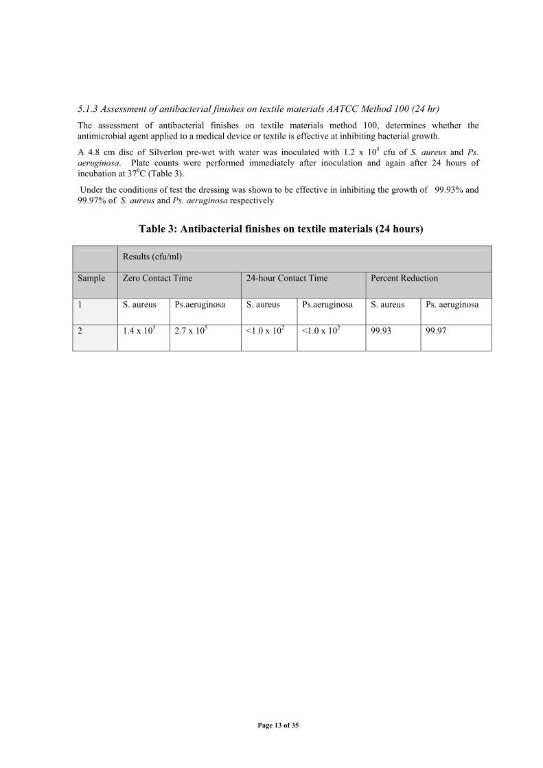
5.1.3 Assessment of antibacterial finishes on textile materials AATCC Method 100 (24 hr) The assessment of antibacterial finishes on textile materials method 100, determines whether the antimicrobial agent applied to a medical device or textile is effective at inhibiting bacterial growth.
A 4.8 cm disc of Silverlon pre-wet with water was inoculated with 1.2 x 10⁵ cfu of S. aureus and Ps. aeruginosa. Plate counts were performed immediately after inoculation and again after 24 hours of incubation at 370C (Table 3).
Under the conditions of test the dressing was shown to be effective in inhibiting the growth of 99.93% and 99.97% of S. aureus and Ps. aeruginosa respectively
Table 3: Antibacterial finishes on textile materials (24 hours)
Results (cfu/ml)
Sample Zero Contact Time 24-hour Contact Time Percent Reduction
1 S. aureus Ps.aeruginosa S. aureus Ps.aeruginosa S. aureus Ps. aeruginosa
2 1.4 x 10⁵ 2.7 x 10⁵ <1.0 x 10² <1.0 x 10² 99.93 99.97

Page 14 of 35
5.1.4 Assessment of antibacterial finishes on textile materials AATCC Test Method 100 The assessment of antibacterial finishes on textile materials method 100 determines whether the antimicrobial agent applied to a medical device or textile is capable of inhibiting bacterial growth.
A 4.8 cm disc of Silverlon pre-wet with water was inoculated with 1.2 x 10⁶ cfu of S. aureus ATCC 6538, Ps.aeruginosa ATCC 9027, Enterococcus faecalis (VRE) ATCC 51575 and S. aureus ATCC 33591.
Plate counts were performed immediately after inoculation and at 30 minutes, 2 hours and 4 hours incubation at 370C and the number of colony forming units determined and expressed as a percentage of the number present at Time 0.
Table 4: Antibacterial finishes on textile materials (4 hours)
Results in CFU/ ml (% reduction)
Time (hours)
Test Organism
Zero 0.5 2 4
S. aureus
ATCC 6538
1,700,000 5,400
(99.99)
4,000
(99.77)
3,000
(99.83)
Ps. aeruginosa
ATCC 9027
2,000,000 3,700
(99.82)
4,500
(99.78)
< 10
(99.99)
E. faecalis (VRE) ATCC 51575 1,900,000 150,000
(92.31)
5,400
(99.72)
14,000
(99.28)
S. aureus ATCC 33591 1,700,000 14,000
(99.18)
14,000
(99.18)
5,000
(99.71)
The results indicate that after four hours contact time, the dressing had reduced the number of present by >99.7% in each case.

Page 15 of 35
5.1.5 Dow Corning corporate test method 0923 Antimicrobial activity of Silverlon was evaluated by shaking a sample in 1–2 x10⁴ cfu/ml of S. aureus, ATCC 6538 and Ps. aeruginosa, ATCC 9027 suspensions for one hour. The test was carried out in duplicate and the results shown in Table 5and Table 6.
Table 5: Dow Corning test A
Test organism Organism count (cfu/ml)
Zero Time One hour % Reduction
S. aureus
ATCC 6538
9,300 <10 99.89
Ps. aeruginosa
ATCC 9027
27,000 <10 99.96
Table 6: Dow Corning test B
Test organism Organism count (cfu/ml)
Zero Time One hour % Reduction
S. aureus
ATCC 6538
10,000 <10 99.90
Ps. aeruginosa
ATCC 9027
27,000 <10 99.96
During the course of this test the number of cfu present was reduced 99.9%

Page 16 of 35
5.1.6 Kirby Bauer standard antimicrobial susceptibility
The Kirby Bauer standard antimicrobial susceptibility test determines whether an antimicrobial agent applied to a medical device or textile is able to inhibit bacterial growth.
A liquid culture of each test organism is inoculated onto the surface of a Mueller-Hinton agar plate in three different directions. The test sample, 20 mm square, is centred on the agar surface and the plate incubated at 35-370C for 16-24 hours. At the end of this period the plate is examined for the presence of inhibition of the growth of the test organisms (Table 7 and Table 8)
Table 7: Kirby Bauer test A
Test Organism: Results
(zone width-sample width)
S. aureus ATCC 33591 2 mm/l
S. aureus ATCC 6538 2 mm/l
P. aeruginosa ATCC 9027 2 mm/l
E. faecalis ATCC 51575 1mm/l
Table 8: Kirby Bauer test B
Test Organism: Results
(zone width-sample width)
S. aureus ATCC 33591 2 mm/l
S. aureus ATCC 6538 2 mm/l
P. aeruginosa ATCC 9027 2 mm/l
E. faecalis ATCC 51575 2mm/l
The Kirby Bauer test results indicate that Silverlon possess marked inhibitory activity against four very common wound pathogens two of which, S. aureus ATCC 33591 (MRSA) and E. faecalis ATCC 51575 (VRE), have multiple antibiotic resistance.

Page 17 of 35
5.2 Silver ion release studies The time-related release of silver ions from a 4 x 4 inch Silverlon Wound Contact Dressing and the 4 x 4 inch Wound Pad Dressing into tryptone soya broth at 37oC was measured by inductively coupled plasma spectroscopy and the results expressed graphically in Figure 1.
Figure 1: Release of silver ion

Page 18 of 35
5.3 Biocompatibility studies 5.3.1 Sensitization potential Silverlon fabric was tested using a standard technique to evaluate the potential for delayed dermal contact sensitization in the guinea pig. Silverlon showed no evidence of causing delayed dermal contact sensitization in this test and can therefore be regarded as hypoallergenic.
5.3.2 Acute intracutaneous reactivity Silverlon fabric was tested to evaluate the local dermal irritant effects of leachables in the rabbit. The fabric was extracted in 0.9% sodium chloride and cottonseed oil and intradermally injected into five separate sites on the right side of the back of each rabbit. No evidence of significant irritation or toxicity were detected.
5.3.3 Cytotoxicity The cytotoxicity of Silverlon was investigated in a cell culture system using standard ISO methodology. Under the conditions of this study, the test extracts showed no evidence of cell lysis or toxicity and therefore met the requirements of the test.
5.3.4 Haemolysis study The presence of any leachable chemicals within Silverlon that would cause red blood cell haemolysis was investigated in vitro using standard methodology. Under the conditions of this test, the mean haemolytic index for the test article was 4% which was therefore classed as ‘slightly haemolytic’. Given the intended use of the product this result was not considered clinically significant.
5.3.5 5. Muscle implantation This study is designed to evaluate the effects of implanting Silverlon in muscle tissue of the rabbit using standard methodology. Four pieces of Silverlon were implanted in the right paravertebral muscle of each rabbit. In the opposite muscle, four USP negative control strips were similarly implanted. Rabbits were observed daily for general health. At two weeks the rabbits were euthanatized for examination. Under the condition of this study, the macroscopic reaction was not significant as compared to the USP negative control implant material. Microscopically, the reaction was a slight irritant as compared to the USP negative control implant material. It was concluded that Silverlon fabric, when implanted into a muscle, is safe with very little tissue reactivity.
5.3.6 Acute systemic toxicity Extracts made using 0.9% sodium chloride and cottonseed oil were evaluated for systemic toxicity in accordance with standard methodology. Five mice (per extract) were weighed and than each were injected with the test extract at a dose of 50 ml/kg. Under the conditions of this study, there was no mortality or evidence of systemic toxicity from the aqueous or the oil extracts of Silverlon.

Page 19 of 35
5.4 Electromotive effects According to US Patent 7,292,762 B2, authored by Flick, healthy human skin develops an electrical potential difference across the epithelium called the transepithelial potential (TEP or epidermal battery). The TEP is generated by an active ionic transfer system involving the transportation of sodium ions through the epithelium to the internal body fluids of the animal which results in the generation of a potential difference in the order of 10 mV to 70 mV.
Wound healing is governed by many factors including the nature and extent of the injury but it is also postulated that healing may be influenced by (or indicated by) the electrical potential difference between the site and surrounding intact tissue. Wound healing studies in salamanders, and fracture healing studies in mammals have shown that healing is associated with complex changes in the local electric field which gradually returns to normal levels as the injury heals. Conversely, failure of the normal healing process, as in fracture non unions, is associated with the absence of appropriate electrical signals at the site of the injury. These currents are not insignificant, as values ranging from 10-30 µA/cm2 have been measured leaving the stump surface of children’s fingers whose tips had been accidentally amputated. This out flowing of current has also been called the “Current of Injury”.
It is generally recognized that the electromotive force (EMF) driving currents from wounds made in skin is a direct result of disruption of the transepithelial potential. When a wound is produced, an electric ‘leak’ results that short-circuits the TEP (epidermal battery) allowing the voltage to reverse at the wound surface.
Progressive measurements made laterally from the wound surface to normal periwound skin reveal that the potential across the skin is found to increase, until it reaches normal values forming a lateral voltage gradient from wounded to normal tissue. This is illustrated graphically in Figure 2, which has been abstracted from data provided by the client.
In experimental animals this lateral voltage gradient has been shown to be initially as high as 140 mV/mm, rapidly dropping by 95% within 24 hours. It is postulated that the application of a highly conductive Silverlon dressing, in contact both with the wound surface and the healthy tissue around the periphery, will alter the electrodynamic processes associated with the pathologic condition to promote healing and pain relief. This proposed effect is totally separate from the better understood and more widely accepted explanation for the antimicrobial effects of the dressing.
It is also suggested that if the silver nylon layers are separated with a non-conducting material, a capacitative field may be established by the current of injury that is present at the wound surface. The magnitude of the effect is determined by the number if intermediate layers. It was discovered in early studies that laminates having four and six layers provided some pain relief but not as effectively as dressings having eight layers.
This capacitive effect is claimed to relieve pain if applied to areas of blunt trauma such as contusions, sprains (stretched ligaments), and strains (torn muscles). The fact that the multi laminate structure provided pain relief when applied to intact skin supports the concept of an electrical field phenomena, affecting to the alteration in the electric parameters of the skin that accompanies damaged tissue beneath

Page 20 of 35
Figure 2: Graphical illustration of lateral voltage gradients

Page 21 of 35
6. REVIEW OF PRECLINICAL DATA FROM OTHER SOURCES
6.1 Comparison of silver dressings in an animal study Heggers et al., 23 compared the performance of four silver containing dressings in an animal study in which rats were given standard contact burns (20% TBSA). On day 3, the wound was excised and infected with Ps. aeruginosa and S. aureus. The animals were divided into four groups each containing five subjects whose wounds were dressed with Acticoat (Smith & Nephew), Silverlon (Argentum), Silvasorb (Medline Industries) or left undressed as controls.
The dressings remained on the wounds for 10 days when the wounds were quantitatively assessed. Mean wound counts of the control were 1.2x10⁵ for Ps. aeruginosa and 6.5x10⁵ for S. aureus.
Counts for Acticoat dressings for both organisms were 0 and 1.8x10³. For Silvasorb 0 and 6.3x10³, and for Silverlon 1.5x10⁴ and 7.4x10⁴
Acticoat and Silvasorb were both significantly lower than the control for P. aeruginosa, (P<.05) and Acticoat was significantly lower than the control for S. aureus (P<.05). Although counts for Silvasorb appear significantly lower than the controls for S. aureus, the numbers were not sufficient to be significant, however Silverlon did achieve a slight significance.
These preliminary data suggest that weekly dressing changes with these new silver dressings are feasible and economically and medically congruous.
6.2 Laboratory comparison of antimicrobial activity of silver dressings It is not unreasonable to assume that the considerable variation in the structure, composition and silver content of the dressings previously identified would impact upon their ability to release silver in sufficiently high concentrations to exert a significant antimicrobial effect either in the wound or the laboratory situation. The silver content and antimicrobial activity of the various dressings was therefore investigated in three laboratory-based studies published previously 1,24,25 Note that reference 25 contains amendments to some of the data presented in the earlier publication and also demonstrates the importance of the choice of vehicle that is used when making extract of dressings for antimicrobial assays.
Within these studies the antimicrobial properties of the dressings were tested using three different methods designed to compare various aspects of their performance. As full experimental details of these methods were published previously, only summaries of the methodology and results are provided here. Three standard organisms were used in this part of the test, a Gram-positive bacteria S. aureus (ATCC 6538P), a Gram-negative organism, E. coli (ATCC 8739), and a yeast, C. albicans (ATCC 2091).
6.2.1 Silver content Samples of each dressing were sent to Sheffield Analytical Services, a laboratory accredited by the United Kingdom Accreditation Service (UKAS) that specialises in the measurement of precious metals for assay purposes.
Within this laboratory the total extractable silver content of each dressing was determined following acid digestion of the sample using a technique called inductively coupled plasma optical emission spectroscopy (ICP-OES).
The silver content of each dressing is shown in Table 9, ranked by silver content, which indicates that major differences exist between the three products with values ranging from 546 to 1.6 mg/100cm2.

Page 22 of 35
Table 9: Silver content of dressings
Proprietary Name Ag content (mg/100cm2)
Silverlon 546
Calgitrol Ag 141
Acticoat 109
101
Contreet Ag 85*
Contreet Hydrocolloid 31.2
32.4
31.4
Aquacel Ag 8.3
Silvasorb 5.3
Actisorb Silver 220 2.9
2.4
Avance 1.6
Arglaes powder 6.87mg/gram
*Corrected value, see reference 25

Page 23 of 35
6.2.2 Antimicrobial activity: zone of inhibition Samples of each dressing were placed upon agar plates inoculated with 0.2 ml of a log-phase broth culture of each test organism. After incubation at an appropriate temperature, the plates were examined for the presence of a zone of inhibition around the perimeter of the test sample that was free of bacterial growth. If such a zone was detected the width was measured and the dressing removed from the agar and replaced upon another agar plate, seeded as before with the same microorganism. This process was repeated a maximum of seven times or until no further zone of inhibition was produced during the previous test.
The results are summarised in Table 10.
Table 10: Summary of zone of inhibition data
Test organism S. aureus E. coli C. albicans
Products that show evidence of sustained activity over two or more days
Acticoat
Aquacel Ag
Calgitrol Ag
Contreet Hydrocolloid
Silverlon
Acticoat
Calgitrol Ag
Contreet Hydrocolloid
Silverlon
Products that produce a well-defined zone of inhibition at one time interval only.
Arglaes Powder
Silvasorb
Aquacel Ag
Arglaes Powder
Acticoat
Arglaes Powder Calgitrol Ag
Contreet hydrocolloid
Silvasorb
Silverlon
Products that produce no well-defined zone of inhibition in this test.
Actisorb
Avance
Contreet Ag
Actisorb
Avance
Contreet Ag
Silvasorb
Actisorb
Aquacel Ag
Avance
Contreet Ag
Note: Because of the physical properties of Arglaes, no attempt was made to transfer the gelled powder from one agar plate to another, so only one result is reported.
This test examines the ability of a dressing to release an antimicrobial agent (in this case silver ions) from within its structure to the aqueous-based agar gel to which it is applied. To some extent this test mimics the clinical use of the product and predicts what might happen if the dressing were to be applied to the surface of a moist wound. A similar test has been used in the past to compare the antimicrobial properties of medicated tulle-type dressings 26. The process of removing the dressing from one agar plate and placing upon a second or third provides some indication of the ability of the product to deliver the antimicrobial over a sustained period.
This test has potentially serious limitations, however, for some dressings may require the presence of significant volumes of fluid (exudate) to extract or liberate a sufficient concentration of silver ions to exert

Page 24 of 35
their maximum antimicrobial effect. It is difficult to accept, however, that a product, which fails to demonstrate any measurable activity when tested in this way, could have a significant effect upon the bacterial population of an infected or heavily colonised wound.
6.2.3 Antimicrobial activity: challenge testing To portions of each dressing measuring 40 mm x 40 mm, were added 0.2 ml of a log-phase culture of each microorganism. The inoculated dressings were then incubated for 2 hours after which they were transferred into 10 ml of 0.1% peptone water (Oxoid) and vortexed to remove any viable organisms remaining in the dressings. Serial dilutions were performed in triplicate on each extract and the number of viable organisms present determined using a standard surface counting technique.
If viable organisms were recovered during this process the test was repeated as before using a 4-hour contact period then again with a 24-hour contact period. If no organisms were detected on a particular dressing after 2 hours, in subsequent tests the dressing was not extracted with peptone water but placed in 10ml of tryptone soya broth (TSB) to detect very low levels of residual contamination.
As no inactivator for silver was used during this test, there is a possibility that any remaining low concentrations of silver ions present could prevent the recovery of these organisms potentially resulting in a false negative result.
The summarised results of this test appear in Table 11. For the purpose of this report, a ‘marked antimicrobial effect’ is arbitrarily defined as a 103 reduction in the number of viable organisms present at each time interval.
It is important to note that following correspondence in the Journal of Wound Care related to the first two publications some additional work was undertaken which showed that the product Contreet was perhaps disadvantaged during the laboratory testing program as it was subsequently revealed by the manufacturer that the dressing required sodium to be presenting solution to release the silver ions from the foam.

Page 25 of 35
Table 11: Summary of microbial challenge test results
Test organism S.aureus E.coli C.albicans
Products that demonstrate marked antibacterial activity after 2 hours incubation.
Acticoat
Calgitrol Ag
Acticoat
Calgitrol Ag
Contreet Ag
Silverlon
Acticoat
Calgitrol Ag
Contreet Ag
Silverlon
Products that demonstrate marked antimicrobial activity after 4 hours incubation
Silverlon
Contreet Hydrocolloid
Aquacel Ag
Silversorb
Products that demonstrate marked antimicrobial activity after 24 hours incubation.
Actisorb
Products that demonstrate limited evidence of antimicrobial activity after 24 hours incubation.
Aquacel Ag
Contreet hydrocolloid
Contreet Ag
Silvasorb
Contreet hydrocolloid
Aquacel Ag
Silvasorb
Products that demonstrate no convincing evidence of antimicrobial activity even upon prolonged incubation
Actisorb
Avance
Avance
Actisorb
Avance

Page 26 of 35
The results of this test provide an indication of the ability of each dressing to kill or prevent the growth of predetermined numbers of bacteria applied directly to it and thus reflects what may occur when dressings are exposed to bacterial contamination either from the wound itself or from the external environment.
As with the previous method, the performance of the dressing will be influenced by the speed at which it takes up, or becomes saturated with exudate or test solution. Products such as hydrocolloids which are intended to absorb liquid over a period of many hours or days may not perform well in this method given the very short time available for fluid absorption.
6.2.4 Antimicrobial activity: microbial transmission test In this test a strip of dressing forms a bridge between two separate agar blocks in a Petri dish, one of which is sterile, the other inoculated with the test organism. This test determines the ability of bacteria to survive on the dressing surface and migrate along it from the contaminated agar to the sterile agar. A positive result in this test suggests that it is possible that microorganisms could be transported laterally out of a contaminated wound onto the surrounding skin, or potentially move in the opposite direction from the intact skin into the wound itself.
The test was carried out in triplicate, and other than the control material, the only test samples to show any evidence of microbial transfer were Actisorb and Avance. In the case of Actisorb, microbiological migration occurred on only one sample, and probably took place across along the outer nonwoven fabric outer sleeve of the dressing. No transfer occurred when only the inner core of the dressing was examined. Evidence of transmission of bacteria was clearly visible on all three samples of Avance as shown by prolific bacterial growth around the ends of the dressing on the surface of the sterile agar. No transfer of C. albicans took place with any of the samples tested including the controls, which made the tests invalid.
The results of this part of the study are consistent with those of the earlier tests in that both Acticoat and Contreet Hydrocolloid dressing not only prevented the transmission of bacteria but also produced zones of inhibition around the margin of the dressing.
The results of the tests described, demonstrate that major differences exist in the properties of the dressings examined. Perhaps not surprisingly, these appear to reflect the silver content of the products concerned as determined by chemical analysis, which varies from 1.6 to 109mg/100cm2.

Page 27 of 35
7. REVIEW OF CLINICAL DATA PROVIDED BY THE CLIENT The following abstracts have been produced from data files submitted by the client. These were principally PDF files of poster presentations or unpublished reports. Brief comments have been appended to these summaries that highlight specific issues arising from each document. A small number of clinical files have not been individually reviewed, as these contain insufficient information upon which to base a meaningful comment or simply consist of a couple of before and after images.
7.1 Surgical wounds 7.1.1 Healing chronic, ischaemic infected saphenous vein harvest sites in the legs with silver plated cloth and allograft dermal regenerative matrix. This unpublished study by Carson et al., describes the use of Silverlon in the treatment of 15 diabetic patients with limb-threatening wounds following vein graft harness. Wounds were debrided surgically followed by sharp debridement. Wound infections (n=5) were treated with systemic antibiotics. Patients with ischemia (n=15) indicated by an ankle brachial index of 0.3 to 0.8 were treated with cilostazol (n=14) and angioplasty (n=10) as bypass surgery was not appropriate. The wounds were then closed with allografts and covered with Silverlon which left in place for 3 weeks at which time another application was performed. Moisture was maintained with applications of mineral oil over the silver cloth. Ten wounds subsequently healed, three had amputations and two required flaps.
Comment: The rationale for the application of mineral oil over the silver cloth to maintain a moist environment must be questioned as this would effectively render the material hydrophobic, thereby preventing or seriously inhibiting the penetration of wound fluid and the release of silver ions from the fabric thus markedly reducing the value and effectiveness of the dressing.
7.1.2 Clinical case evaluation and results of Silverlon sternal dressings in cardiac surgery Hummel in an unpublished report dated February 2007, stated that the introduction of Silverlon in cardiac surgical practice reduced the deep sternal infection rate from 2.4% in 2003 to 2.15% in 2004 and 1.43% in 2005. During the first six months of 2006 it decreased further to 1.25%. This was equivalent to an overall reduction rate in infection of 48%. This equated to the prevention of an estimated 7 deep infections each of which from published sources is known to cost $8,000 to $42,000 to treat. This estimated savings of $56,000 to $ 294,000 compared very favourably to the additional cost of the dressings used. This was estimated to be $30,150 assuming 1.5 dressings per procedure at a cost of $25 average cost per dressing.
Comment: Assuming that the reduction in infection rates is due exclusively to the use of Silverlon and no other unrelated factor, the cost savings associated with the use of this material greatly outweigh the initial expenditure.
7.1.3 Cardiovascular surgery assessment of Silverlon vs standard wound care methods in non-pre-complex post-operative wounds. In an unpublished report, Weiss et al. described the results of a single-center, prospective, cross-sectional, randomized trial involving sixty patients in which they compared the effectiveness of Silverlon with a standard treatment in patients undergoing planned, urgent and emergent cardiovascular surgical procedures. Patients undergoing cardiovascular surgery, were randomized according to sex, age, and diabetic status. Wound care was initiated in theatre and continued throughout healing process. Wounds were cleansed with sterile water and dressed with either Silverlon or sterile 4 x 4 inch swabs and Telfa and secured with surgical tape. Compression bandages were applied to lower leg incisions. Compared with the control group, patients randomized to Silverlon treatment experienced a dramatic reduction in post-operative wound infections (12 vs 6 infections).

Page 28 of 35
Comment: It is unfortunate that the value of this study has not been fully exploited. The report is exceptionally brief, and fails to provide sufficient clinical and statistical information to facilitate meaningful interpretation of all the data although the reduction in infection rates appears impressive.
7.2 Chronic wounds 7.2.1 Comparison of healing of lower extremity chronic wounds with silver plated cloth dressings, papain-urea-chlorophyllin ointment and hydrogel. This unpublished study by Carson et al., describes the use of Silverlon in the treatment of sixty patients with chronic wounds to the legs or feet. Wounds were dressed with Silverlon moistened either with hydrogel, (Intrasite, Smith and Nephew) or papain-urea-chlorophyllin ointment, (Panafil Health point limited) or covered with hydrogel alone (no silver dressing). There were twenty patients in each group matched as to location, size of the wound, nutrition, presence or absence of diabetes, and ankle-brachial index. All patients received maintenance sharp debridement and dressing changes. Treatments were continued for six weeks or until wounds were healed. Silver dressings were applied after sharp debridement of necrotic and non-healing tissues and covered with hydrogel and a secondary porous dressing. Silver dressings were changed every five days to maintain moisture, and debridement done as needed. The silver dressing was reused once and therefore a new silver dressing applied every 10 days. Maintenance sharp debridement of non-viable tissue was done every 10 days in this group as required. Wounds allocated to papain-urea-chlorophyllin ointment and hydrogel were dressed every two days after sharp debridement, if needed, covering with paraffin gauze and porous gauze. Multi-layer compression bandages were applied where indicated (three in each in each treatment group). At the end of six weeks 15/20 wounds healed in the silver/hydrogel group, 7/20 healed in the papain-ureachlorophyllin ointment group, and 7 healed in the hydrogel group. Cellulitis occurred twice in the silver/hydrogel group, 5 times in the papain-urea-chlorophyllin group and 6 times in the hydrogel alone group. Pain averaged 3/10 throughout this course in the silver group and 5/10 in the papain-ureachlorophyllin group and hydrogel group.
Comment: The absence of a statistical analysis reduces the value of this study although the evidence suggests that the use of the silver dressing had a beneficial effect upon healing. It is unfortunate that a fourth group of patients were not included who were dressed with Silverlon alone, for this protocol cannot exclude the possibility that the application of the hydrogel actually reduced the effectiveness of the silver fabric. It is also theoretically possible that an interaction has taken place between the Silverlon and the papain ointment which has reduced the effectiveness of both. A further group of patients would be required to test this hypothesis.
7.2.2 Chronic, diabetic foot wounds with ischemia and tissue loss: use of advanced wound care techniques, to prevent further tissue loss A further unpublished study, Carson et al., described the treatment of twenty-five insulin dependent diabetic patients with chronic foot and ankle wounds that had previously been dressed with a variety of modalities. All patients had previously been offered amputation and had refused. Fifteen patients were successfully treated using a specialized atherectomy catheter to open up occluded tibial arteries. All patients also received medication for their peripheral arterial disease. The local wound treatment consisted of allografts (average of three per patient). These were dressed with moist silver dressings and left in place for 2-4 weeks with periodic moistening of the fabric to maintain a moist environment. The silver dressings were changed every two weeks. Fifteen of the twenty-five patients previously assigned to a major amputation emerged from this treatment with a functional foot which remains patent for 6-24 months, the current follow-up time for this group. Six patients required a major amputation but at a lower level than previously planned. There were no episodes of sepsis. Two patients died (causes not directly related to wound) and two had a major amputation (above knee) as originally planned. The deaths and major amputations occurred in patients without renal failure and these patients also had successful opening of their tibial arteries with indices of 0.8 and 0.72 respectively. The use of the treatment protocol described was considered to dramatically improve the limb salvage and function of this special group of ischemic, diabetic patients with pre-existing tissue loss.

Page 29 of 35
Comment: The impressive results achieved with these highly vulnerable patients clearly demonstrate the value of the surgical procedures described. Unfortunately, in the absence of a control group in which patients were treated in exactly the same fashion but had their allografts dressed with a different primary dressing, it is not possible to draw any firm conclusions about the contribution made by the silver dressing.
7.3 Silverlon as an adjunct to VAC therapy 7.3.1 Silver dressings used with wound vacuum assisted closure: is there an advantage? Rodriguez et al. described the use of Silverlon dressings and vacuum assisted closure (VAC) in the treatment of 80 patients with chronic wounds associated with tissue defects of the legs and feet. In 40 patients a piece of Silverlon was laid on to the wound bed and the VAC dressing was placed on top and secured as normal. In the remaining patients the VAC dressing was applied over an inert knitted viscose dressing. All the wounds included in the study had previously failed to heal with standard moist wound healing treatments and had been present for two months or more. They also had a prior history of one or more episodes of sepsis and a history of colonization by methicillin resistant staphylococcus aureus, vancomycin resistant enterococcus or both. The average wound volume in the silver group was 12 cc and 10 cc in the non silver dressing group. Early in the study it was found that vacuum dressings over the silver fabric configuration could be changed every five days rather than the usual three and that the silver fabric could be use for ten days before being discarded. Wounds were treated for three months or until healed. Thirty-five of the wounds in the silver group healed were healed at three months compared with only 20 patients healed in the non-silver group. No infections occurred during treatment in the silver group, but 6 occurred in the non-silver group. The authors concluded that the silver dressings, which could be reused for 10-15 days with VAC, appeared to facilitate faster healing than comparable wounds similarly treated without silver. Less frequent dressing changes and fewer infectious complications also provided clinical medical and economic advantages.
Comment: The potentially very valuable study appears to demonstrate that the use of Silverlon and VAC therapy together produced improve healing rates compared with VAC therapy alone in this specific group of patients. It is unfortunate that the study was not randomised and that some statistical analysis was not conducted on the results which would have increased the value of the findings.
7.3.2 The combined benefit of negative pressure therapy, elemental silver contact layer, & bilayered living skin equivalent in the treatment of chronic hard to heal lower extremity wounds. Van Gils et al. used Silverlon in conjunction with a bilayered living skin equivalent (Apligraf, Novartis) and negative pressure therapy (NPT) in the treatment of six patients with chronic lower extremity wounds. To prepare the wound bed for grafting, NPT was applied over a Silverlon dressing for an average of 14 days, after which the patient received an application of LSE, and the wound was once again was dressed with Silverlon and NPT for a further 6-7 days. Using this treatment all six wounds successfully healed. The authors concluded that the combination of N.P.T, elemental silver contact dressing, and L.S.E. in colonized hard to heal chronic wounds produces ‘an accelerated wound healing potential.’
Comment: Despite the excellent clinical results achieved, given the non-comparative nature of the study it is not possible to assess the contribution that the Silverlon dressing made to the treatment outcome.
7.3.3 Healing skin grafts over chronic wounds with vacuum assisted closure and silver dressings Following extensive use of Vacuum Assisted Closure (VAC) over a period of number of years, Carson et al. began to use the technique as a primary dressing for skin grafts in close proximity to chronic wounds, following dehiscence after primary repair, skin grafts over acute wounds in areas with difficult contours and areas with frequent motion such as knees and ankles and mediastinal areas. Some of these wounds, perhaps as many as 10%, became infected which is not remarkable for chronic wounds. For this reason the authors chose to use Silverlon between the VAC sponge and the graft instead of an inert mesh fabric to prevent and control infection. The silver fabric also served as a protective layer between the graft and the sponge and allowed easy removal of the VAC dressing without disturbing the graft. One hundred consecutive patients with chronic wounds of the legs and trunk had their wounds treated in this way, ninety seven of whom

Page 30 of 35
progressed to satisfactory healing and closure. The three patients who failed to heal did not appear to have infections - inability to heal appeared to result from a failure of the vascular attachment of the grafts as a result of the patient/caregiver inadvertently disconnecting the VAC for long periods (over 1 hour) which lead to maceration of the area. All wounds healed with subsequent grafts. The authors concluded that VAC and silver fabric together provide a very effective dressing for skin grafts over chronic wounds.
Comment: Despite the excellent clinical results achieved, given the non-comparative nature of the study it is not possible to assess the contribution that the Silverlon dressing made to the treatment outcome.
7.3.4 VAC with Silverlon vs VAC alone This single case study reported by Carson provides very persuasive visual evidence of a beneficial effect resulting from the use of VAC and Silverlon dressing. A male patient with a large infected found following a failed abdominoplasty had half his very extensive wound dressed with Silverlon and VAC, the other half with VAC alone. After five days that portion of the wound containing the silver dressing had clearly made significantly more progress towards healing, containing 40% more granulation tissue. After 3 weeks that portion of the wound dressed with Silverlon had healed completely but the side with VAC therapy alone, although improved, had not achieved complete healing.
Comment: This study provides very impressive evidence for the value of combining Silverlon dressings and VAC therapy in this type of wound.
7.4 Miscellaneous case studies 7.4.1 Treatment of pyoderma gangrenosum with a silver contact layer This unpublished case study describes the use of Silverlon in the treatment of a wound that originated from a cat bite which deteriorated and was subsequently diagnosed as pyoderma gangrenosum. The wound was treated with the silver dressing and a four layer bandage and healed within 10 weeks.
Comment: No indication was provided of basis of diagnosis, or any information on the use of additional therapies such as systemic or topical steroids. Images provided appear to show staining around the wound commonly associated with venous ulceration.
7.4.2 Clinical case study treatment of coumadin tissue necrosis with silverlon This case history by Rogers, describes the successful use of Silverlon in the treatment of bilateral multiple small areas of skin loss on the lower legs believed to be associated with coumadin use and known clinically as coumadin–induced tissue necrosis. Conventional treatments had proved ineffective so Silverlon treatment was tried, applying the dressing to one leg only, the other continued to be dressed with conventional dressings and thereby acted as a control. In less than two weeks all the wounds dressed with Silverlon had healed, but the wounds on the other leg remain unchanged. Silverlon was then applied to the second leg and within a week all these had healed also. Both legs remained healed after one month.
Comment: The results of this simple study are impressive and raise the possibility that the silver dressing may have an effect upon healing that is divorced from its antimicrobial effect.

Page 31 of 35
7.4.3 Successful treatment of a chronic venous ulcer with a silver contact dressing and compression therapy In this case report, Publicover, described the use of Silverlon and compression therapy (Unna boot) in the treatment of a 53 year old male with diabetes, peripheral vascular disease, and a longstanding non-healing venous ulcer associated intractable oedema. The wounds were cleansed and debrided as appropriate and a Silverlon dressing applied moistened with sterile water. This was covered with a secondary dressing and compression therapy applied. Dressings were changed twice weekly for five weeks then weekly until closure which was achieved in 17 weeks.
Comment: The use of Silverlon and compression successfully healed a wound that had previously remained open for about 6 years.
7.4.4 Graft vs. host disease: an innovative approach using silver contact dressing to manage grade IV skin lesions Graft vs Host Disease (GVHD) is a condition experienced by patients who undergo bone marrow or stem cell transplants. It most commonly presents in the acute form as a maculopapular rash, plaques and/or desquamation which may cause itching or pain. As the disease progresses, the rash may become confluent and involve the entire body. Lichenoid or schleradermatous changes are characteristic of “chronic” GVHD. In severe cases, potentially life threatening bullous lesions (epidermal necrolysis) can occur. Selekof et al. described how a home made Silverlon ‘vest’ was used to treat a 23 year old female with recurrent acute myelogenous leukemia (AML) who had recently undergone her second allogenic stem cell transplant. This patient presented with extreme pain, in a very agitated state. Following administration of high doses of narcotics, examination revealed the presence of multiple open bullae on her face, back neck and chest as well as erythema multiforme. Initial treatments with convention materials proved unsuccessful so an alternative regimen was devised which consisted of gently cleansing of the open bullae with warm sterile water followed by the application of a vest fashioned from Silverlon fabric and stretch netting by the patient’s mother. This garment was worn for 3 day intervals and moistened with warm sterile water. Within 24 hours, the patient’s pain was reduced, and within a few days the wounds began to close. Within 11 days the treatment was discontinued.
Comment: A novel approach to the treatment of a distressing and painful condition in which the use of Silverlon appears to have contributed positively to the healing process.

Page 32 of 35
8. REVIEW OF PUBLISHED CLINICAL DATA A review of the published literature conducted through ‘Pub Med’
8.1 Do silver-impregnated dressings limit infections after lumbar laminectomy? Epstein 27 retrospectively reviewed treatment outcomes for 234 patients following lumbar laminectomy with instrumented fusion. The wounds of the first 128 patients were dressed postoperatively with iodine or alcohol-based swab and dry gauze. The second group had their wounds dressed with Silverlon dressings for the first 2 weeks after surgery. Three of 128 patients receiving conventional dressings developed deep postoperative wound infections confirmed by culture confirmed. All were successfully managed with 6 weeks of postoperative antibiotics, and none required secondary surgery. A further eleven patients developed superficial infection/irritation seven of whom required oral antibiotics alone and 4 were referred to plastic surgeons for superficial wound revision. Of the 106 patients treated with Silverlon, none experienced any deep or superficial wound infections or irritation. Other clinical, surgical, and outcome data were comparable for both groups The authors concluded that although the number of cases in each series was small, there appeared to be a positive trend toward a reduction in postoperative wound infection using the silver dressing for this indication.
Comment: The results of this study are impressive and certainly appear to support the proposition that the use of Silverlon as described reduces the incidence of postoperative wound infection for this group of patients, however, once again the value of the data is somewhat reduced because the study was time-based and not a randomised control trial. Also no statistical analysis of the results has been performed. Furthermore, although considered highly unlikely, there remains the possibility that other confounding factors might have existed which could have influenced the outcome of the investigation. These could include changes in personnel or facilities etc.

Page 33 of 35
9. CONCLUSIONS The principal conclusions that may be drawn from this review are as follows.
9.1 Electromotive effects The data provided on the proposed electromotive and pain relieving properties of the Silverlon dressing fabric are intriguing and appear to have some scientific basis. Nevertheless it is considered that more detailed research and extensive testing, including well conducted randomized controlled trials, is required to validate these effects in order to support formal claims of this nature
9.2 Antimicrobial activity The scientific and wound management literature contains of wealth of data which strongly supports the proposition that silver ions have a potent antimicrobial effect and for this reason numerous wound dressings have been introduced which are claimed to inhibit the growth of bacteria in the dressing, and the wound to which is applied.
The rate and extent of the release of silver ions from the various products available depends upon several factors including the chemical form, the total concentration and the distribution or location within the product, but provided sufficiently high concentrations of silver ions are obtained within the local wound environment all dressings which are capable of achieving this threshold level are likely to prove to be broadly equivalent in terms of their ability to control or prevent infection irrespective of their structure and composition. This does not mean however that all these dressings will necessarily perform equally well in the provision of a moist (but not macerated) local wound environment.
Although considerable information is available on the minimum concentration of silver ions required to prevent the growth of a range of pathogenic organisms, much less data are available on the maximum values which should be delivered as high concentrations of silver are known to be toxic to mammalian cells. For this reason, it is toxicity issues around the release of silver (silver dumping) that are most likely to generate debate between clinicians or commercial companies, rather than discussions about the minimum concentrations of silver required within a wound to exert an antibacterial effect.
Because the rate of silver ion release is so heavily dependant upon the nature of the formulation, it is not possible to use the total silver content of a number of dressings as a basis upon which to make comparisons of their likely clinical efficacy.
For example, Silverlon contains a massive amount of silver in relative terms, but as this is present in the metallic form, in percentage terms, the production of silver ions is relatively slow compared with products in which the silver is present in the form of a salt.
For this reason, the manufacturers of Silverlon have commissioned a number of laboratory studies which demonstrate that Silverlon is capable of releasing sufficiently high concentrations of silver ions to exert a significant antimicrobial effect over a sustained period. It is argued, however, that some of these tests such as the antimicrobial activity-dynamic test of surfaces (ASTM E 2149), and the assessment of antibacterial finishes on textile materials, are of little relevance to a surgical dressing.
The USP antimicrobial preservative test, which is also of little clinical relevance to a wound dressing, does confirm that silver ions are released from the fabric in sufficient quantities to kill microorganisms in suspension, as does the Dow Corning corporate test method 0923.
The Kirby Bauer standard antimicrobial susceptibility test, like other zone of inhibition test systems is of more relevance for a product that is to be applied to a moist surface colonized or infected with bacteria.
9.3 Biocompatibility studies The results of the biocompatibility studies, as reported on the website, suggest that Silverlon has a good safety profile, being free of cellular or systematic toxicity and unlikely to induce adverse skin reactions. The results of the haemolysis study and implantation study further confirm the safety of the product as the mild haemolytic effect is considered to be irrelevant given the nature of the product and its use as a topical wound dressing.

Page 34 of 35
9.4 Review of clinical data The individual reports of clinical studies specifically related to Silverlon that were examined during the course of this review were, with one or two exceptions, of generally poor quality. In a number of cases the study protocols were flawed, the studies were often non-comparative, there was generally no statistical analysis of the data, patient numbers were small, and in some instances the study reports were too brief for a detailed examination. The evidence supporting the use of Silverlon in venous ulcers was particularly weak. None of the articles examined would meet the requirements of a systematic review.
Nevertheless, taken together these data provided a persuasive body of evidence which suggests that the use of the Silverlon product range can impact favourably upon the prevention and control of infection and the promotion of wound healing, particularly when used as an adjunct to vacuum assisted closure.
No clinical evidence was seen which supported the proposed electromotive effects, or of the ability to relieve pain when applied to intact skin. In one instance, an article on the website dated February 2004, made reference to an on-going study involving the use of Silverlon and DC current in the treatment of donor sites and split thickness skin grafts after burn injury, but despite the fact that completion was forecast in 2-3 months, this study is now 3-4 years old and no final data appears to be available
As part of the brief that was provided by the client prior to the commencement of this review, it was requested that particular attention be given to the negative pressure dressing, the burn contact dressing, the elastic burn wrap, the acute burn glove the calcium alginate dressing and the digidressing.
Unfortunately, with the possible exception of the negative pressure dressing, insufficient data was available on the individual products upon which to draw any evidence-based conclusions. Nevertheless, it is the unsubstantiated personal opinion of the author that the benefits of the silver plated nylon yarns identified both in the laboratory studies supported by the results of the limited clinical investigations are likely to contribute favourably to the performance of these more specialized products. Once again, clinical studies will be required to confirm this supposition.
Elsewhere in this review, it has been suggested that, from an antimicrobial perspective at least, provided that a sufficiently high concentration of silver ions is delivered to the wound to exceed the MIC value for the organisms present and that this concentration is below the levels required to cause toxicity, it may not matter too much about the composition of the product responsible for the silver ion production. This means that publications which describe the use of other silver dressings may possibly be able to be used as secondary supporting data provided that their use is limited strictly to the discussion of antimicrobial effects.
Dr S Thomas
Medical Device Consultant Medetec™ http://www.medetec.co.uk/

Page 35 of 35
10. REFERENCES 1. Thomas S, McCubbin P. An in vitro analysis of the antimicrobial properties of 10 silver-containing dressings. Journal of Wound Care 2003;12(8):305-8. 2. Russell AD, Hugo WB. Antimicrobial activity and action of silver. In: Ellis GP, Luscombe DK, eds. Progress in Medinical Chemistry: Elsevier Science, 1994: 351-369. 3. Lansdown AB. Silver. I: Its antibacterial properties and mechanism of action. Journal of Wound Care 2002;11(4):125-30. 4. Lansdown AB. Silver. 2: Toxicity in mammals and how its products aid wound repair. Journal of Wound Care 2002;11(5):173-7. 5. White RJ. An historical overview of the use of silver in modern wound management. British Journal of Nursing (15 silver supplement) 2002;10:3-8. 6. Spadaro JA, Berger TJ, Barranco SD, Chapin SE, Becker RO. Antibacterial effects of silver electrodes with weak direct current. Antimicrob Agents Chemother 1974;6(5):637-42. 7. Berger TJ, Spadaro JA, Bierman R, Chapin SE, Becker RO. Antifungal properties of electrically generated metallic ions. Antimicrob Agents Chemother 1976;10(5):856-60. 8. Becker RO, Spadaro JA. Treatment of orthopaedic infections with electrically generated silver ions. A preliminary report. J Bone Joint Surg Am 1978;60(7):871-81. 9. Webster DA, Spadaro JA, Becker RO, Kramer S. Silver anode treatment of chronic osteomyelitis. Clin Orthop 1981(161):105-14. 10. Spadaro JA, Webster DA, Becker RO. Silver polymethyl methacrylate antibacterial bone cement. Clin Orthop Relat Res 1979(143):266-70. 11. Deitch EA, Marino AA, Gillespie TE, Albright JA. Silver-nylon: a new antimicrobial agent. Antimicrob Agents Chemother 1983;23(3):356-9. 12. Deitch EA, Marino AA, Malakanok V, Albright JA. Silver nylon cloth: in vitro and in vivo evaluation of antimicrobial activity. J Trauma 1987;27(3):301-4. 13. Chu CS, McManus AT, Pruitt BA, Jr., Mason AD, Jr. Therapeutic effects of silver nylon dressings with weak direct current on Pseudomonas aeruginosa-infected burn wounds. J Trauma 1988;28(10):1488-92. 14. Adams AP, Santschi EM, Mellencamp MA. Antibacterial properties of a silver chloride-coated nylon wound dressing. Vet Surg 1999;28(4):219-25. 15. Ersek RA, Denton DR. Cross-linked silver-impregnated skin for burn wound management. J Burn Care Rehabil 1988;9(5):476-81. 16. Ersek RA, Navarro JA. Maximizing wound healing with silver-impregnated porcine xenograft. Todays OR Nurse 1990;12(12):4-9. 17. Furr JR, Russell AD, Turner TD, Andrews A. Antibacterial activity of Actisorb Plus, Actisorb and silver nitrate. J. Hospital Infection 1994;27(3):201-208. 18. Thomas S, Fisher B, Fram PJ, Waring MJ. Odour-absorbing dressings. Journal of Wound Care 1998;7(5):246-50. 19. Mulligan CM, Bragg AJD, O'Toole OB. A controlled comparative trial of Actisorb activated charcoal cloth dressings in the community. British Journal of Clinical Practice 1986;9(1):145-8. 20. Milward PA. Comparing treatment for leg ulcers. Nursing Times 1991;87(13):70-72. 21. Cassino R, Ricci E, Amione P. Treatment of 150 infected chronic skin lesions with Actisorb - A review. Poster Presentation, Centro Vulnologico Italiano, Turin, Italy, 2002. 22. Stadler R. Treatment of chronic wounds with Actisorb. 2002. 23. Heggers J, Goodheart RE, Washington J, et al. Therapeutic efficacy of three silver dressings in an infected animal model. J Burn Care Rehabil 2005;26(1):53-6. 24. Thomas S, McCubbin P. A comparison of the antimicrobial effects of four silver-containing dressings on three organisms. Journal of Wound Care 2003;12(3):101-107. 25. Thomas S, Ashman P. In-vitro testing of silver containing dressings. Journal of Wound Care 2004;13(9):392-393. 26. Thomas S. An experimental evaluation of a chlorhexidine medicated tulle gras dressing, (letter). J. Hosp. Infect. 1982;3:399-400. 27. Epstein NE. Do silver-impregnated dressings limit infections after lumbar laminectomy with instrumented fusion? Surg Neurol 2007;68(5):483-5; discussion 485.