Embed Size (px)
Citation preview
www.elsevier.com/locate/ynimg
NeuroImage 32 (2006) 445 – 456
Sex differences in mental rotation: Top–down versus
bottom–up processing
Tracy Butler,a,* Julianne Imperato-McGinley,b,1 Hong Pan,a Daniel Voyer,c Juan Cordero,b
Yuan-Shan Zhu,b Emily Stern,a,1 and David Silbersweiga,1
aFunctional Neuroimaging Laboratory, Department of Psychiatry, Weill Medical College of Cornell University, Box 140, 1300 York Avenue, New York,
NY 10021, USAbDepartment of Endocrinology, Weill Medical College of Cornell University, New York, NY 10021, USAcDepartment of Psychology, University of New Brunswick Fredericton, NB, Canada
Received 24 October 2005; revised 22 February 2006; accepted 4 March 2006
Available online 22 May 2006
Functional MRI during performance of a validated mental rotation
task was used to assess a neurobiological basis for sex differences in
visuospatial processing. Between-sex group analysis demonstrated
greater activity in women than in men in dorsalmedial prefrontal
and other high-order heteromodal association cortices, suggesting
women performed mental rotation in an effortful, ‘‘top–down’’
fashion. In contrast, men activated primary sensory cortices as well
as regions involved in implicit learning (basal ganglia) and mental
imagery (precuneus), consistent with a more automatic, ‘‘bottom–
up’’ strategy. Functional connectivity analysis in association with a
measure of behavioral performance showed that, in men (but not
women), accurate performance was associated with deactivation of
parieto-insular vestibular cortex (PIVC) as part of a visual –
vestibular network. Automatic evocation by men to a greater extent
than women of this network during mental rotation may represent
an effective, unconscious, bottom–up neural strategy which could
reasonably account for men’s traditional visuospatial performance
advantage.
D 2006 Elsevier Inc. All rights reserved.
Introduction
The biological basis for possible cognitive differences
between men and women remains a topic of great interest
and controversy. Mental rotation is a visuospatial task which
gives rise to robust sex-based differences in performance, with
men performing more accurately than women on average
(Voyer et al., 1995). It therefore serves as a valuable probe for
investigating the neurobiological underpinnings of sex differ-
ences in cognition. Several prior studies aimed at determining
1053-8119/$ - see front matter D 2006 Elsevier Inc. All rights reserved.
doi:10.1016/j.neuroimage.2006.03.030
* Corresponding author. Fax: +1 212 746 5818.
E-mail address: [email protected] (T. Butler).1 Senior authors.
Available online on ScienceDirect (www.sciencedirect.com).
the neural basis for this male advantage have shown that men
and women both activate regions of prefrontal, parietal, and
temporal–occipital regions during mental rotation, with no
(Dietrich et al., 2001; Tagaris et al., 1996; Unterrainer et al.,
2000) or variable (Fink et al., 2003; Jordan et al., 2002;
Seurinck et al., 2004; Thomsen et al., 2000) between-sex
differences, though greater activity in frontal brain regions has
been detected in women in a majority of studies (Seurinck et
al., 2004; Thomsen et al., 2000; Weiss et al., 2003). One
limitation of these prior studies, likely to account in part for
discrepant results, is the use of variable, study-specific mental
rotation paradigms created by each set of investigators for use
during fMRI scanning. None of these study-specific tasks gave
rise to significant male behavioral performance advantages in
scanned subjects (perhaps due to small sample sizes typical of
neuroimaging studies), and none were known to do so in
larger populations, calling into question the usefulness of
results in explaining a neural basis for this advantage. We
therefore utilized a computerized mental rotation task optimized
for use during fMRI scanning, which we have previously
shown to be a valid measure of mental rotation abilities and to
produce the expected pattern of sex-based performance in a
large sample of normal subjects (Voyer et al., 2006), to
investigate the neural basis for sex differences in mental
rotation performance. In addition to standard between-sex
group analysis of imaging data, we incorporated performance
accuracy at each level of task difficulty into correlational
analyses in order to assess contributions of sex, level of
behavioral performance, and interaction effects to fMRI results,
in accordance with studies emphasizing the importance of this
approach (Seurinck et al., 2004; Shelton and Gabrieli, 2004;
Tagaris et al., 1996; Unterrainer et al., 2000). Functional
connectivity analyses were performed in order to explore brain
networks engaged during accurate mental rotation, and how
they might differ by sex.
T. Butler et al. / NeuroImage 32 (2006) 445–456446
Materials and methods
Subjects
32 healthy subjects underwent fMRI scanning as part of the
present study, which was approved by the New York Presby-
terian Hospital–Weill/Cornell Institutional Review Board. Ac-
ceptable data were obtained from 25 subjects: 13 women (mean
age 28.6, std 7.5) and 12 men (mean age 30.1, std 5.9). All
subjects were strongly right-handed according to the Edinburgh
Handedness Inventory, free from medical, neurological and
psychiatric disease, and taking no medications. Reasons for
excluding subjects from final analyses were MRI ghosting
artifact greater than 5% (one woman, two men); head movement
greater than 1/3 of a voxel based on examination of realignment
parameters (two women, one man); and failure to perform the
task in the scanner (one man).
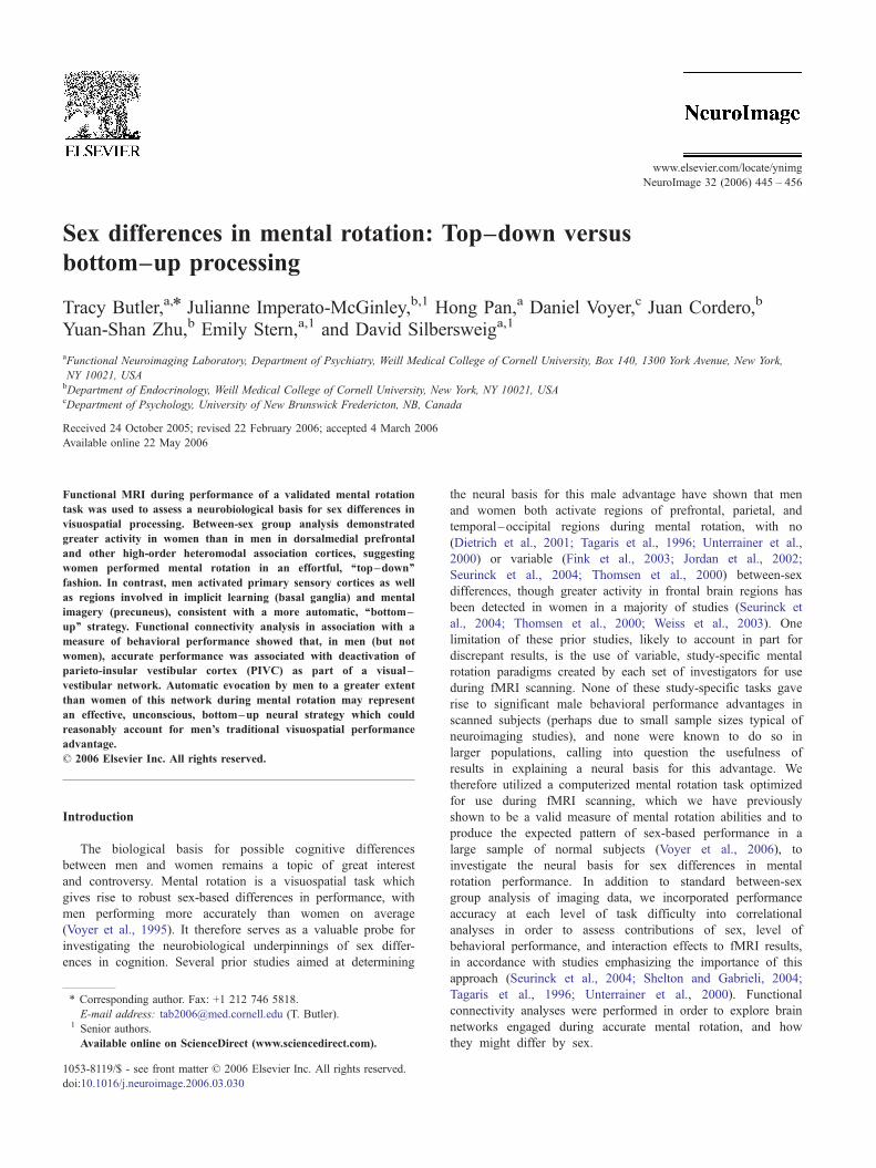
Stimuli
Redrawn versions of Shepard and Metzler original cube
figure (Peters et al., 1995) were presented in pairs (Fig. 1).
Figures were white on a black background. Stimuli pairs were
either the same but rotated with respect to one another (‘‘same’’
trials), or they were mirror images of each other (‘‘different’’
trials). Stimuli were rotated with respect to one another around
their vertical axis. Presentation of stimuli in the scanner was
controlled by the Integrated Functional Imaging System (IFIS;
MRI Devices Inc. Gainesville, FL) by means of Eprime software
(Psychology Software Tools Inc., Pittsburgh, PA). Each pair of
figures subtended a visual angle of approximately 6.3- horizon-
tally and 2.5- vertically as displayed on an MRI-compatible
LCD screen attached to the scanner’s head coil.
Experimental conditions
The Rotate activation conditions consisted of pairs of figures
which were either identical (same) or mirror images (different)
Fig. 1. Examples of stimuli pairs derived from Shepar
rotated by 40-, 80-, 120- or 160- with respect to one another.
Subjects were instructed to mentally rotate figures into alignment in
order to decide if they were the same or different.
The Compare activation condition consisted of pairs of
figures which were either identical or mirror images which
were not rotated (i.e., rotated 0-) with respect to one another.
Subjects were instructed that in this condition, there was no
need to try to rotate figures into alignment. This condition
controlled for visual properties of the stimuli, the saccades and
evaluation process required to reach a same or different decision,
and the motor act of pressing a button.
Practice
Immediately preceding scanning, to familiarize subjects with
the task and ensure they understood instructions, subjects
performed a brief (approximately 5 min) practice version of the
task on a computer. They were required to attain at least 60%
correct in the easiest Rotate condition, in which figures were
rotated 40- with respect to one another, as well as in the Compare
condition. No subject required more than two practice runs to
reach this criterion.
Experimental task
In the context of a block design paradigm, stimuli pairs at
each angle of rotation (including the 0- compare condition) were
grouped into blocks of five trials each. Each trial lasted 7.5 s,
resulting in blocks lasting 37.5 s. Each trial consisted of an
orienting signal (a fixation cross in the center of the screen) for
500 ms, followed by a pair of stimuli presented for 7 s. Subjects
responded to stimuli by pressing a button with their right index
finger for a same response or right middle finger for a different
response. Response accuracy and latency were recorded. Stimuli
remained on-screen even when a response was produced faster
than 7 s (in order to equalize visual stimulation across
conditions and subjects). A dot appeared for 250 ms upon a
subject’s response, to indicate to the subject that the button press
d and Metzler (1971) used in the present study.

T. Butler et al. / NeuroImage 32 (2006) 445–456 447
had been recorded. If subjects did not respond within 7 s, the
next trial began. Within each block of five trials, there were
three same trials and two different trials presented in pseudo-
random order.
An epoch (run) consisted of four 37.5-s active blocks, each
followed by a 24-s rest block during which subjects looked at
the center of the screen, which was marked by a dash. There
were four Rotate epochs and three Compare epochs. Within each
Rotate epoch, there was one block for each of the four angles of
rotation. This resulted in 4 blocks (20 trials) of stimuli at each
of the four angle of rotation, for a total of 16 Rotate blocks (80
trials.) Within each Compare epoch, all four active blocks were
composed of unrotated pairs of stimuli, resulting in a total of 12
Compare blocks (60 trials.) The rationale for separating Rotate
and Compare trials into different epochs was to distinguish these
two conditions and to allow for a reminder of task instructions
prior to stimuli presentation in order to ensure that subjects did
not mistakenly engage in mental rotation during the compare
condition (as had been noted in preliminary testing). Thus, the
word ‘‘rotate’’ appeared on the screen immediately before each
Rotate epoch, and the word ‘‘compare’’ appeared on the screen
immediately before each Compare epoch.
The order of epochs and of blocks within epochs was the same
for each subject, and was counterbalanced to control for systematic
bias due to time and/or order effects.
After scanning, subjects completed a questionnaire about their
experience.
Behavioral data analysis
Average accuracy (proportion of correct responses) and
reaction time (RT) were calculated for each subject at each
angle of rotation (0-, 40-, 80-, 120-, 160-). Rotate trials to
which a subject responded in less than 250 ms were excluded
from analysis. Accuracy was calculated both with and without
omitted trials (when no subject response was recorded.) RT was
calculated only for correct trials. Using SAS (Cary, NC, USA),
separate two-way repeated measures analyses of variance
examined the dependent measures of accuracy and RT, with
sex as the between-subjects factor and degree of rotation as the
within-subjects factor. Results at the P < 0.05 threshold were
considered significant.
Image acquisition
Image data were acquired on one of two identical GE Signa
3 T MRI scanner (max gradient strength 40 mT/m, max gradient
slew rate 150 T/m/s; General Electric Company, Waukesha, WI)
using blood oxygen level-dependent (BOLD) fMRI. Approxi-
mately equal numbers of men and women were scanned on each
scanner (6 women, 6 men on scanner #1; 7 women, 6 men on
Table 1
Mean accuracy (ACC) and reaction time (RT) in ms in women (n = 13) and men
Compare (0-) 40- 80-
ACC (std) Women 0.99 (0.02) 0.83 (0.14) 0.74
Men 1.0 (0.01) 0.92 (0.13) 0.82
RT (std) Women 1336.4 (270.6) 3531.4 (512.2) 3980.7
Men 1398.3 (492.6) 3762.8 (697.9) 4224.9
Trials in which no response was recorded, or a response with RT <250 ms was reco
scanner #2). After shimming to maximize homogeneity, a series of
fMRI scans was collected using gradient echo echo-planar imaging
(EPI) (TR-2000; TE-30; flip angle = 70-; FoV = 240 mm; 27 slices;
5 mm thickness with 1 mm inter-slice space; matrix = 64 � 64).
Images were acquired over the whole brain parallel to the AC–
PC plane. The first six volumes of each epoch were discarded. A
reference T1-weighted anatomical image with the same slice
placement and thickness and a matrix of 256 � 256 was
acquired immediately preceding the EPI acquisition. A high-
resolution T1-weighted anatomical image was acquired using a
spoiled gradient (SPGR) sequence with a resolution of 0.9375 �0.9375 � 1.5 mm3.
Image processing and data analysis
Image processing performed within a customized Statistical
Parametric Mapping (SPM) 99 software package (Frackowiack
et al., 2004) included manual AC–PC re-orientation of all
anatomical and EPI images; realignment of functional EPI
images based on intracranial voxels to correct for slight head
movement between scans; co-registration of functional EPI
images to corresponding high-resolution anatomical image for
each subject; stereotactic normalization to the standardized
coordinate space of Talairach and Tournoux (Montreal Neuro-
logical Institute, MNI average of 152 T1 brain scans) based on
the high-resolution anatomical image; and spatial smoothing of
the normalized EPI images with an isotropic Gaussian kernel
(FWHM = 7.5 mm).
A voxel-by-voxel univariate multiple linear regression model
at the subject level determined the extent to which each voxel’s
activity correlated with the principal regressor, which consisted
of stimulus onset times (onset of active blocks) convolved with
a prototypical hemodynamic response function. The temporal
global fluctuation estimated as the mean intensity within brain
region of each volume was removed through proportional
scaling. The first order temporal derivative of the principal
regressor (to compensate for slight latency differences in
individual hemodynamic response from the prototypical re-
sponse), temporal global fluctuation, realignment parameters,
and scanning periods were incorporated as covariates of no
interest. This first-level analysis resulted in a set of contrast
images of condition-specific effects for each subject, which were
entered into second-level random effects analyses to best
account for inter-subject variability and allow population-based
inferences to be drawn (McGonigle et al., 2000).
Voxel-by-voxel univariate group-level random effects analy-
ses examined within-group between-condition effects (Rotate
versus Compare in men and women separately), between-group
within-condition effects (women versus men during Rotate) and
interaction effects using these contrast images, with subject age
and scanner entered as covariates of no interest in an ANCOVA
(n = 12) for each experimental condition
120- 160- Average rotation
(0.18) 0.76 (0.13) 0.68 (0.17) 0.75 (0.16)
(0.18) 0.86 (0.12). 0.72 (0.18) 0.83 (0.17)
(519.5) 4045.9 (638.9) 4289.7 (708.8) 3961.9 (645.0)
(733.1) 4240.3 (653.7) 4226.0 (600.9) 4113.5 (682.8)
rded, were excluded from analysis. RTwas calculated only for correct trials.

Table 2
Brain regions differentially active during mental rotation in men and women
Volume (mm3) x y z Punc PFDRcorr z score
Greater in women
L dorsalmedial prefrontal cortex/superior frontal gyrus (BA 8) 594 �9 27 51 <0.0001 0.01 4.01
R dorsalmedial prefrontal cortex/superior frontal gyrus (BA 8)a 162 15 30 54 <0.0001 0.011 3.41
L inferior occipito-temporal cortex (area LOC) 27 �39 �69 �9 0.001 0.012 3.29
R temporal pole (BA 21) 27 54 9 �30 0.001 0.012 3.15
Greater in men
R postcentral gyrus (BA 5) 810 24 �42 66 <0.0001 0.005 �4.19L ventral globus pallidus 270 �15 �3 �6 <0.0001 0.006 �3.87L medial parietal/paracentral lobule (BA 5) 189 �15 �36 51 <0.0001 0.006 �3.77L postcentral gyrus (BA 3) 81 �18 �42 66 <0.0001 0.006 �3.34L precuneus 162 �9 �69 51 <0.0001 0.006 �3.3Bilateral peri-midbrain 81 �3 �36 3 0.001 0.006 �3.22a This is the only cluster that in part reflects differences in deactivation (see text for explanation).
T. Butler et al. / NeuroImage 32 (2006) 445–456448
setting. Regionally specific results were considered statistically
significant if the P value was less than 0.01 for the F statistic of
the ANCOVA and the peak voxel-wise P value was less than
0.001 for the effect-specific T statistic and survived correction
over the whole brain for false discovery rate at P < 0.05
(Genovese et al., 2002).
To identify brain areas most active in association with
accurate performance of the mental rotation task in men, in
women, and in men as compared to women, a voxel-by-voxel
univariate correlational analysis was performed using a multiple
regression model in an ANCOVA setting, with each subject’s
contrast image from each Rotate condition (40-, 80-, 120-,160-) as the independent variable, the z-transformed percentage
of correct responses in the corresponding condition as the
regressor of interest, and the subject factor, subject age, and
scanner as covariates of no interest. For this supplemental
analysis, results were considered statistically significant if the P
value was less than 0.01 for the F statistic; voxel-wise P <
0.0001; and cluster size was greater than three voxels (81 mm3).
To identify brain networks associated with accurate mental
rotation performance in men and women, a voxel-by-voxel
univariate correlational analysis was performed using a
multiple regression model in an ANCOVA setting, with each
subject’s contrast image from each Rotate condition as the
independent variable, the condition-specific activation level in
‘‘seed’’ voxels (identified through the above-described accuracy
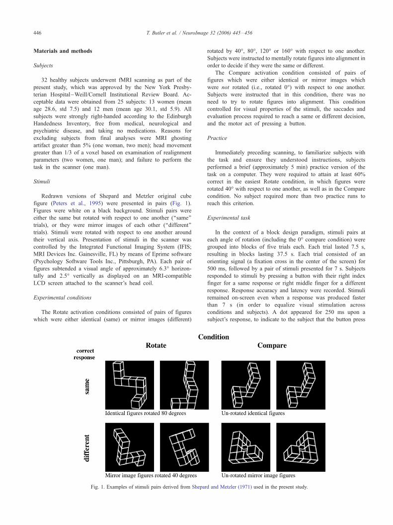
Fig. 2. Areas of significantly different activation between men and women perfor
greater activity in men in blue/purple. Note greater activity in women in bilateral
occipital cortex (near angular gyrus), and left temporal–occipital cortex (area LOC
left ventral basal ganglia. BOLD fMRI results are displayed as a T-map at a PUNC
sections [left = left, right = right] selected to illustrate areas of sex differences.
correlational analysis) in the corresponding condition as the
regressor of interest, and the subject factor, subject age, and
scanner as covariates of no interest. Again, for these
supplemental analyses, results were considered statistically
significant at a stringent threshold of P < 0.05 corrected for
multiple comparisons over the entire brain, with cluster size
greater than six voxels (162 mm3).
Results
Behavioral results
For both accuracy and RT, there was an expected main effect of
degree of rotation, with greater degree of rotation associated with
lower accuracy [F(4,92) = 29.10, P < 0.0001] and longer RT
[F(4,92) = 240.5, P < 0.0001] in both men and women. There was
a weak trend towards a main effect of sex on performance
accuracy, with men performing non-significantly better than
women [F(1,23) = 2.04, P = 0.17], though this trend was not
apparent when omitted trials were counted as incorrect and
included in the analysis [F(1,23) = 0.29, P = 0.59]. There was
no main effect of sex on RT [F(1,23) = 0.48, P = 0.5], and no
interaction between degree of rotation and sex for either accuracy
[F(4,92) = 0.81, P = 0.52] or RT [F(4,92) = 0.71, P = 0.6]. See
Table 1 for mean accuracy and reaction times.
ming mental rotation. Greater activity in women is displayed in yellow/red;
dorsalmedial prefrontal cortex, left temporal pole, right temporal–parieto-
). Note greater activity in men in bilateral postcentral gyri, precuneus, and
< 0.01 threshold (for illustration purposes), overlaid onto canonical T1 axial
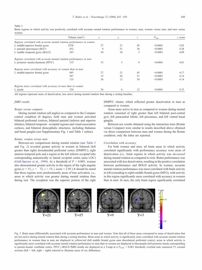
Table 3
Brain regions in which activity was positively correlated with accurate mental rotation performance in women, men, women versus men, and men versus
women
Volume (mm3) x y z Punc z score
Regions correlated with accurate mental rotation performance in women
L middle/superior frontal gyrus 2538 �27 21 45 <0.0001 5.02
L parietal (precuneus) (BA7) 432 �9 �51 36 <0.0001 4.28
L middle temporal gyrus (BA22) 243 �54 �36 3 <0.0001 4.09
Regions correlated with accurate mental rotation performance in men
L posterior insula/claustrum (PIVC) 135 �33 �3 9 <0.0001 4.01
Regions more correlated with accuracy in women than in men
L middle/superior frontal gyrus 945 �27 21 45 <0.0001 4.17
567 �15 36 51 <0.0001 4.14
135 �36 12 54 <0.0001 4.14
Regions more correlated with accuracy in men than in women
L insula 324 �36 6 12 <0.0001 �4.33All regions represent areas of deactivation, less active during mental rotation than during a resting baseline.
T. Butler et al. / NeuroImage 32 (2006) 445–456 449
fMRI results
Rotate versus compare
During mental rotation (all angles) as compared to the Compare
control condition (0 degree), both men and women activated
bilateral prefrontal cortices, bilateral parietal (inferior and superior
lobules), bilateral temporal–occipital regions and visual association
cortices, and bilateral diencephalic structures, including thalamus
and basal ganglia (see Supplementary Fig. 1 and Table 1 online).
Rotate, women versus men
Between-sex comparisons during mental rotation (see Table 2
and Fig. 2) revealed greater activity in women in bilateral (left
greater than right) dorsalmedial prefrontal cortex (DMPFC), right
anterior temporal pole and a region in the left inferior occipital lobe
corresponding anatomically to lateral occipital cortex (area LOC)
(Grill-Spector et al., 1999). At a threshold of P < 0.005, women
also demonstrated greater activity in the region of the right angular
gyrus (x = 51, y = �72, z = 15; z score = 2.95.) It should be noted
that these regions were predominantly areas of true activation, i.e.,
areas in which activity was greater during mental rotation than
during rest. The exception was the superior portion of the right
Fig. 3. Brain areas differentially associated with accurate performance in men and
are less active during mental rotation than during a resting baseline. Brain areas in
performance in women than in men are displayed in yellow/red (left middle fr
significantly more correlated with accurate mental rotation performance in men tha
to parieto-insular vestibular cortex, PIVC.) BOLD fMRI results are displayed as
sections [left = left, right = right] selected to illustrate areas of sex differences.
DMPFC cluster, which reflected greater deactivation in men as
compared to women.
Areas more active in men as compared to women during mental
rotation consisted of right greater than left bilateral post-central
gyri, left paracentral lobule, left precuneus, and left ventral basal
ganglia.
Between-sex results obtained using the interaction term (Rotate
versus Compare) were similar to results described above obtained
via direct comparison between men and women during the Rotate
condition; only the latter are reported.
Correlation with accuracy
For both women and men, all brain areas in which activity
correlated significantly with performance accuracy were areas of
deactivation (i.e., brain regions in which activity was decreased
during mental rotation as compared to rest). Better performance was
associated with less deactivation, resulting in the positive correlation
between performance and BOLD activity. In women, accurate
mental rotation performance was most correlated with brain activity
in left (extending to right) middle frontal gyrus (MFG), with activity
in this region significantly more correlated with accuracy in women
than in men. In men, the only brain region significantly correlated
women. Note that all of these areas correspond to areas of deactivation that
which activity is significantly more correlated with accurate mental rotation
ontal gyrus near dorsolateral prefrontal cortex); areas in which activity is
n in women are displayed in blue/purple (left posterior insula corresponding
a T-map at a PUNC < 0.001 threshold, overlaid onto canonical T1 coronal

T. Butler et al. / NeuroImage 32 (2006) 445–456450
with accurate performance was left posterior insula bordering on
claustrum. Activity in this region was significantly more correlated
with accuracy in men than in women, with the point of maximal sex
difference located slightly anteriorly. This posterior insula/claustrum
region appears to correspond anatomically to the cortical projections
of the vestibular system, referred to as parieto-insular vestibular
cortex (PIVC) (Brandt and Dieterich, 1999; Guldin and Grusser,
1998) and further discussed below. PIVC correlation with accurate
performance inmenwas bilateral, with weaker right-sided activation
also detected (x = 33, y = �12, z = 6; z score = 3.11). Table 3 lists
brain regions significantly associated with accurate mental rotation
performance in men, women, in women as compared to men, and in
men as compared to women. Fig. 3 shows brain areas differentially
associated with accurate performance in men and women.
Examination of sex- and condition-specific activity in these two
regions found to be most associated with accurate performance in
women and men (left MFG and left PIVC, respectively) shows
decreased activity in both of these regions during mental rotation
as compared to a resting baseline, as noted above (Figs. 4A, B).
Increasing task difficulty (operationalized by increasing angle of
rotation) was associated with further linear decreases in left MFG
activity in women (but not men) and further linear decreases in left
PIVC activity in men (but not women).
Fig. 4. (A) Bar plot of sex- and condition-specific activity in left posterior insula co
9), the region most correlated with accurate mental rotation performance in men.
significantly below baseline activity for all mental rotation conditions), with further
in men (gray arrow), but not women. (B) Bar plot of sex- and condition-specific ac
most correlated with accurate mental rotation performance in women. Note that M
MFG activity associated with increasing angle of rotation in women (gray arrow)
standard error.
Functional connectivity analyses
The two brain regions found to be most associated with
accurate performance in women and men (left MFG and left PIVC,
respectively) were used as seed voxels in separate functional
connectivity analyses to inform interpretation of results and to
identify brain networks differentially associated with accurate
mental rotation performance in men and women.
Left MFG functional connectivity
Brain regions identified as correlating positively with left
MFG activity during mental rotation are listed in Table 4 and
depicted in Supplementary Fig. 2 online. Networks were similar
for both men and women and included bilateral frontal regions,
bilateral angular gyri, bilateral temporal association cortices, and
most prominently, bilateral posterior cingulate. In women,
positive correlation also identified left pulvinar of the thalamus
and left cerebellum. In men, positive correlation identified dorsal
anterior cingulate, right parahippocampal gyrus, and right
postcentral gyrus. In general, these regions appear to correspond
to a ‘‘default’’ brain network previously shown to be active when
subjects are not engaged in any particular task (Greicius and
Menon, 2004; Raichle et al., 2001). Brain regions inversely
rresponding to parieto-insular vestibular cortex (PIVC; x = �33, y = �3, z =Note that PIVC is deactivated during mental rotation (i.e., PIVC activity is
linear decrease in PIVC activity associated with increasing angle of rotation
tivity in left middle frontal gyrus (MFG; x = �27, y = 21, z = 45), the region
FG is also deactivated during mental rotation, with further linear decrease in
, but not men. Zero corresponds to a resting baseline. Vertical bars indicate
Table 4
Brain regions in women and in men functionally connected (positive correlation) to left middle frontal gyrus (MFG; x = �27, y = 21, z = 45), the region most
correlated with accurate mental rotation performance in women
Region in women Volume
(mm3)
x y z z
score
Pcorr Region in men Volume
(mm3)
x y z z score Pcorr
Frontal
Seed voxel and
surrounding b/l
frontal regions
50,868 �27 21 45 >8 <0.0001 Seed voxel and
surrounding L
frontal region
11,502 �27 21 45 >8 <0.0001
L mid/superior
frontal gyrus
2052
378
�36�18
12
48
27
42
6.77
5.31
<0.0001
0.002
R middle
frontal gyrus
324 27 9 45 5.18 0.003
R dorsal anterior
cingulate
1215 18 24 42 6.75 <0.001
Temporal
R middle temporal
gyrus
1755 63 �36 �3 >8 <0.0001 R middle
temporal gyrus
594 48 �60 6 5.95 <0.0001
378 51 0 �10 6.18 <0.0001
162 66 �51 0 5.21 0.003
L mid/superior
temporal gyrus
1836 �69 �18 �12 7.62 <0.0001 L middle
temporal gyrus
4833 �54 �57 18 7.14 <0.0001
1539 �57 �39 0 7.61 <0.0001 3294 �48 �45 0 6.87 <0.0001
R posterior
parahippocampal
gyrus
3942 36 �48 �6 6.89 <0.0001
Posterior cingulate
b/l posterior cingulate 24,948 �6 �39 39 >8 <0.0001 L posterior
cingulate
729 �15 �51 27 5.43 0.001
Parietal
L inferior parietal lobule 7317 �39 �66 39 >8 <0.0001 L parietal 648 �18 �5 64 5.53 0.001
405 �30 �27 8 5.74 <0.0001
R angular gyrus 3942 48 �60 33 >8 <0.0001 R parietal,
incl. angular
gyrus
486 51 �75 30 6.01 <0.0001
297 42 �72 54 6.32 <0.0001
Subcortical
L thalamus (pulvinar) 621 �9 �27 6 5.8 <0.0001
L cerebellum (uvula) 1080 �3 �63 �39 6.56 <0.0001
Brain networks are similar across sex, and appear to correspond to a ‘‘default’’ brain network (Raichle et al., 2001). Corrected P values are based on a
search volume of 65,730 voxels in women, 61,595 voxels in men. [Brain regions inversely correlated with left MFG can be found in Supplementary
Table 3 online].
T. Butler et al. / NeuroImage 32 (2006) 445–456 451
correlated with left MFG activity in women and men are listed in
Supplementary Table 2 online.
Left PIVC functional connectivity
Brain regions identified as correlating inversely with left PIVC in
men and women are presented in Table 5. In contrast to similar MFG-
correlated networks in men and women, PIVC-correlated networks
differed markedly by sex, in particular for the inverse correlation, as
shown in Fig. 5. In men, left PIVC inversely correlated activity was
extensive and consisted of a large-scale network of visuospatial-related
brain regions including most prominently bilateral parieto-occipital
association cortex (precuneus, BA 7). Activity inversely correlatedwith
PIVC was also present in men in left parahippocampal gyrus, bilateral
post-central gyri, and anterior and posterior cingulate.
In women, inverse correlation with activity in left PIVC was
relatively sparse and was most prominent in bilateral frontal regions,
with scattered foci of inversely correlated activity also present in
bilateral temporal and temporal–occipital association cortices and right
precuneus. Brain regions positively correlated with left PIVC activity in
women and men are listed in Supplementary Table 3 online.
Discussion
Behavioral findings
Behavioral results show the expected increasing reaction time
and decreasing accuracy with increasing angle of rotation
originally described by Shepard and Metzler (1971), confirming
that subjects were appropriately engaged in the task. Despite
slightly better mental rotation performance by men (men: average
83% correct; women: average 75% correct), there were no sig-
nificant sex differences in accuracy or reaction time. Thus, the sex-
specific neural profiles identified using fMRI were detected in a
setting of no significant difference in performance. The absence of

Table 5
Brain regions in men and in women functionally connected (inverse correlation) to left posterior insula (corresponding to parieto-insular vestibular cortex
[PIVC]; x = �33, y = �3, z = 9), the region most correlated with accurate mental rotation performance in men. Corrected P values are based on search
volume = 61,595 voxels in men, 65,730 voxels in women
Brain region Volume (mm3) x y z z score Pcorr
Men
Bilateral parieto-occipital (precuneus, BA7) 17,739 21 �75 51 <�8 <0.0001
R inferior occipital gyrus (BA 18) 1296 27 �84 12 �6.57 <0.0001
L parahippocampal gyrus 1269 �30 �45 �12 �6.54 <0.0001
1161 �30 �27 �21 �6.52 <0.0001
L midbrain/diencephalon 540 �9 �6 �6 �6.11 <0.0001
L postcentral gyrus 1377 �36 �39 60 �5.96 <0.0001
R postcentral gyrus 162 30 �45 69 �5.77 <0.0001
R rostral anterior cingulate (BA 32) 648 9 42 15 �5.59 <0.0001
L posterior hippocampus 351 �27 �45 3 �5.54 0.001
R posterior cingulate 837 6 �54 9 �5.53 0.001
L inferior occipital gyrus 324 �30 �78 �6 �5.31 0.002
R occipital (lingual gyrus) 162 3 �69 �3 �5.21 0.003
Women
R fusiform gyrus 999 24 �81 �21 �6.00 <0.0001
L inf. temporal gyrus 405 �54 �75 �6 �5.84 <0.0001
Bilateral superior medial frontal gyri 567 0 42 54 �5.53 0.001
R precuneus 297 15 �60 �45 �5.43 0.001
R periventricular (near post. corpus callosum) 648 15 �36 18 �5.32 0.002
R middle frontal gyrus 405 51 33 33 �5.25 0.002
Brain regions positively correlated with left PIVC can be found in Supplementary Table 2 online.
T. Butler et al. / NeuroImage 32 (2006) 445–456452
in-scanner behavioral sex differences may be due to low statistical
power for the behavioral data, since prior extensive testing has
shown the mental rotation task used in the present study to be a
valid measure of mental rotation abilities which does in fact give
rise to the expected male performance advantage when applied to a
larger population (Voyer et al., 2006).
fMRI findings
Rotate versus compare
Although this report focuses on sex differences, it should be
noted that mental rotation activated widespread, bilateral prefron-
tal, frontal, parietal, occipitotemporal, and occipital cortical regions
in both men and women (see Supplementary Fig. 1 and Table 1
online), in agreement with multiple prior mental rotation studies
(e.g., Carpenter et al., 1999; Cohen et al., 1996). Contrary to early
electrophysiologic and behavioral studies, but in agreement with
many fMRI studies of mental rotation (e.g., Cohen et al., 1996), we
found no marked asymmetry of cortical activation during mental
rotation.
Subcortical structures active in both men and women during
mental rotation in the present study included thalamus and basal
ganglia. Although subcortical activity during mental rotation has
received little prior attention, possibly due to an original focus
(theoretical or due to scanning limitations) on cortical regions and
laterality issues, caudate activation during mental rotation has been
reported previously (Alivisatos and Petrides, 1996). Patients with
basal ganglia damage have been shown to have specific deficits in
the ability to perform mental rotation (Harris et al., 2002; Lawrence
et al., 2000). Discrete cortical–basal ganglia– thalamocortical
circuits (Alexander et al., 1986) are known to mediate a variety
of cognitive and emotional behaviors, including voluntary eye
movements and visuospatial learning and memory (Packard and
Knowlton, 2002) relevant to mental rotation.
Rotate, women versus men
Between-sex comparisons between men and women per-
forming mental rotation (Table 2 and Fig. 2) revealed significantly
greater activity in women in left greater than right dorsal medial
prefrontal cortex—a region strongly implicated in top–down,
effortful cognitive processing (Frith and Dolan, 1996) including
decision making (Turk et al., 2004) and spatial working memory
(McCarthy et al., 1994). Greater frontal activity in women is in
broad accord with the majority of prior functional neuroimaging
studies of mental rotation demonstrating sex differences (Seurinck
et al., 2004; Thomsen et al., 2000; Weiss et al., 2003).
There was greater activity in women in a region of left
temporal–occipital association cortex during mental rotation. This
finding is in close agreement with two prior fMRI studies of sex
differences in mental rotation, one of which found greater activity
in women in a left temporal–occipital region overlapping the
cluster detected in the present study (Jordan et al., 2002), while
another reported greater temporal–occipital activity in women in
the right hemisphere (Weiss et al., 2003). This temporal–occipital
region appears to correspond anatomically to area LOC, a region
that mediates object recognition (Grill-Spector et al., 1999). In the
present study, both men and women activated LOC bilaterally
during mental rotation. Based on the idea that the amount of neural
activity that a given cognitive process engenders is dependent on
the computational demand that the task imposes (Just et al., 1996),
greater intensity of LOC activation in women can perhaps be
considered indicative of more effortful, less automatic recognition
of complex three-dimensional objects from two-dimensional line
drawings. In support of this explanation, the traditional male
performance advantage for mental rotation can be abolished when
the task uses actual objects (constructed from wooden blocks)
rather than line drawings (McWilliams et al., 1997, though also see
Robert and Chevrier, 2003). Future functional neuroimaging
studies could examine sex differences during mental rotation of

Fig. 5. Network of brain regions active in inverse correlation with activity
in left parieto-insular vestibular cortex (PIVC; x = �33, y = �3, z = 9)
during mental rotation in men (green) and women (red). Results are
displayed at a threshold of P < 0.05, corrected for multiple comparisons
over the whole brain. Note extensive activation of parietal–occipital
cortices in men, but not women.
T. Butler et al. / NeuroImage 32 (2006) 445–456 453
stimuli varying systematically in three-dimensional depth cues
(e.g., shading, shadowing) to verify this speculation.
At a more liberal statistical threshold, women showed greater
activity than men in posterior right middle temporal gyrus just
inferior to angular gyrus (x = 51, y = �72, z = 15), a region of
heteromodal association cortex considered essential for binding
visual and other information into a coherent spatial representation
necessary for the adaptive deployment of spatial attention
(Mesulam, 2000). As with area LOC, greater activity in this
region in women may relate to possible greater effort required to
integrate complex visual information into a representation condu-
cive to efficient image manipulation. It should be noted, however,
that lack of effort quantification renders these explanations
speculative.
In contrast to women’s greater cortical activity in regions
associated with conscious, effortful processing, men showed
greater activity in basal ganglia, which has been implicated in
automatic, effortless task performance (Rauch et al., 1997; Salmon
and Butters, 1995).
Greater activation in men was also present in left precuneus,
corresponding to Brodmann area (BA) 7. The precuneus has been
called ‘‘the mind’s eye’’ for its role in memory-related visual
imagery (Fletcher et al., 1995). Specific precuneus activation was
recently demonstrated in association with improved performance
of a motor learning task (Oishi et al., 2005). Greater precuneus
activity in men during mental rotation may indicate sex differences
in the use of visual imagery to facilitate task performance. In
addition, as further discussed below, the precuneus appears to play
a key role in coordinating interactions between visuospatial cortical
regions and cortical regions responsible for vestibular processing
(Brandt and Dieterich, 1999), and in the present study was found
via functional connectivity analysis to be a prominent node in a
visual–vestibular network of brain regions active in men during
accurate mental rotation performance.
Men showed greater activity than women in bilateral post-
central gyri. This finding is in accord with a previous report
demonstrating greater sensorimotor activity in men during mental
rotation (Jordan et al., 2002), in which it was suggested that men
may take a more concrete, ‘‘hands on’’ approach to mental rotation.
In addition, this region may be involved in vestibular processing
(Guldin and Grusser, 1998).
Considered broadly, patterns of sex-specific activity obtained
through standard between-sex group analysis suggest that women
rely more upon frontal and other high-order heteromodal associ-
ation cortices to perform mental rotation in an effortful, ‘‘top–
down’’ fashion. In contrast, men appear to take a more ‘‘bottom–
up’’ approach, with less dorsal prefrontal involvement, consistent
with the notion of ‘‘flow’’ by which less frontal control of task
performance allows primary/early sensory brain regions to interact
most effectively with subcortical structures to facilitate accurate,
effortless task performance (Dietrich, 2004).
Correlational and connectivity analyses
To integrate behavioral data into image analyses, correlational
analyses were performed in which each subject’s fMRI data from
each Rotate condition (40-, 80-, 120-, 160-) was correlated with
his or her level of performance in that condition. By incorporating
precise, empiric information about a subject’s actual performance
at a given point during scanning (rather than information about
what that subject should have been doing based on paradigm
design), such correlational methods complement and extend
traditional categorical analyses.
Brain regions correlated with accurate performance in men
By this correlational analysis, a key, novel finding of the
present study is that accurate mental rotation performance in men,
but not women, was associated with deactivation of a region of left
posterior insula/claustrum which appears to correspond anatomi-
cally to parieto-insular vestibular cortex (PIVC), the cortical
projections of the vestibular system. It should be emphasized that
PIVC was less active during mental rotation as compared to the
control condition or to rest, highlighting the increasingly acknowl-
edged importance of considering task-related deactivations when
interpreting functional imaging results (Greicius et al., 2003;
McKiernan et al., 2003; Raichle et al., 2001). PIVC has been
identified recently in humans via functional imaging performed
during caloric or electrical stimulation of the peripheral vestibular
apparatus in the ear (Bottini et al., 1994; Brandt and Dieterich,
1999; Lobel et al., 1998). In men, level of putative PIVC
deactivation was exactly proportional to angle of rotation, strongly
supporting the involvement of this region in the mental rotation
process (see Fig. 4A). Based on this pattern of PIVC activity, as
well as on results from functional connectivity analysis discussed
below, we believe this stepwise deactivation of PIVC in correlation
with stepwise increases in angular disparity reflects activity of a
visual–vestibular reciprocally inhibitory brain network (Brandt et
al., 1998; Brandt and Dieterich, 1999; Deutschlander et al., 2002)
T. Butler et al. / NeuroImage 32 (2006) 445–456454
intimately involved (in men) in the neural process of mental
rotation. Women also deactivated the left PIVC region during
mental rotation, but unlike men, showed no clear pattern with
respect to task difficulty or level of performance.
Analysis of PIVC functional connectivity (Table 5 and Fig. 5)
indicated that, in men (much more so than in women), decreased
PIVC activity was associated with increased activity in a network
of visual regions composed most prominently of bilateral parietal–
occipital visual association cortices (precuneus, BA 7). An
interpretation for this mental rotation-associated pattern of activity
in men comes from recent work demonstrating a relationship of
reciprocal inhibition between cortical regions responsible for
vestibular and visual processing: deactivation of visual regions
occurs during vestibular stimulation (Deutschlander et al., 2002;
Wenzel et al., 1996), while deactivation of PIVC has been
demonstrated when visual regions are activated during visually
induced perceived self movement in the absence of actual body
movement, a phenomenon known as vection (Brandt et al., 1998;
Brandt and Dieterich, 1999; Deutschlander et al., 2002; Kleinsch-
midt et al., 2002). By minimizing mismatch between visual and
vestibular input, this reciprocally inhibitory relationship between
visually responsive and vestibularly responsive cortex is consid-
ered to allow perception of motion to be based on the most reliable
sensory source, perhaps serving to minimize such unpleasant
consequences as motion sickness. Interestingly, susceptibility to
motion sickness has been shown to vary inversely with spatial
ability in men (Levine and Stern, 2002) and to be more common in
women than in men (Turner and Griffin, 1999, though see Cheung
and Hofer, 2002), perhaps suggesting sex differences in the nature
of these visual–vestibular connections.
In the context of the present study, deactivation of vestibular
cortex in proportion to angle of rotation suggests that, for men,
mental rotation of three-dimensional visual stimuli may in a sense
be neurally similar to visually induced perceived self-motion.
Given that no subjects reported such a sensation or engaging in
imagined self-rotation as a task strategy (based on a post-scan
questionnaire), a plausible explanation for findings is that male
subjects automatically and unconsciously activated brain regions
involved in egocentric visuomotor transformations during mental
rotation, and that activation of these regions was associated with
accurate performance. Sex-based differences in utilization of this
visual–vestibular network consisting of activated and deactivated
cortical sensory processing nodes could reasonably contribute to
sex-based differences in mental rotation performance.
This first demonstration of an apparent role for the cortical
projections of the vestibular system in mental rotation sheds light
on prior behavioral findings. Present results provide a mechanistic
explanation for how actual movement (active or passive) around an
array of objects can improve visuospatial performance (Wang and
Simons, 1999), and why egocentric strategies (imagined self-
movement around an object) are in general more effective than
allocentric strategies (imagined object rotation) (Wraga et al.,
1999). From an evolutionary perspective, egocentric transforma-
tions have been considered a more ‘‘natural’’ ability, since
organisms have been walking around objects to inspect them for
millions of years, while revolving objects remain rare (Wraga et al.,
1999). It makes sense, then, that the vestibular system – a
phylogenetically conserved neural system active during actual
circumnavigation around objects – should play a role in accurately
imagined object rotations. An explanation for why women appear
not to recruit the vestibular system during performance of mental
rotation will require additional study but may relate to prior
findings of sex differences in the use of egocentric versus
allocentric strategies for spatial memory (Burgess et al., 2001).
Present results are also relevant to understanding how such factors
as weightlessness (Leone, 1998; Matsakis et al., 1993) and head tilt
(Corballis et al., 1978) affect visuospatial functioning. In strong
support of a role for the vestibular system in mental rotation, direct
vestibular stimulation has recently been shown to specifically
disrupt performance of tasks requiring mental imagery, including
mental rotation (Mast et al., 2005).
Brain regions correlated with accurate performance in women
In women, the region most associated with accurate perfor-
mance was a portion of left MFG in the region of dorsolateral
prefrontal cortex (DLPFC). Activity in this region was decreased
(as compared to a resting baseline) in both men and women under
all active experimental conditions. In women, it decreased further
with decreasing accuracy (and increasing angle of rotation; see Fig.
4B). Based on the well-known role of DLPFC in working memory,
increased activity in DLPFC during mental rotation – a task
requiring mental maintenance and manipulation of complex
images – might have been predicted. In accordance with this
prediction, and in close agreement with prior studies of spatial
working memory (e.g., D’Esposito et al., 1998), a more inferior
portion of right MFG/DLPFC corresponding to BA 9 was in fact
activated by both men and women during mental rotation (men
Rotate versus rest: x = 45, y = 12, z = 30, z score = 3.68; women
Rotate versus rest: x = 48, y = 24, z = 33, z score = 3.76). The
region of left MFG deactivation during mental rotation in the
present study was located dorsal/superior (z = 45) to this probable
spatial working memory-related region of MFG activation and
appears instead to overlap with MFG activity detected in studies of
the brain’s default network (Greicius et al., 2003; Raichle et al.,
2001). As a node in this default network, this region of DLPFC has
been shown to be more active at rest than during performance of
any specific cognitive activation task, providing an explanation for
the initially unexpected finding that this region was less active
during the Rotate (and Compare) conditions than at baseline in the
present study. In support of this explanation, functional connec-
tivity analyses (Table 4 and Supplementary Fig. 2 online) showed
that left MFG activity co-varied with almost the entire default
network as described in prior studies (Greicius et al., 2003; Raichle
et al., 2001) in both men and women (though more prominent in
women). In this context, identification of left MFG as the region
most associated with accuracy in women can perhaps be
understood as reflecting effort and/or cognitive demands, with
greater task demands associated with both suppression of default
network activity and with poor performance, though further
investigation would be needed to confirm this notion.
Conclusion
In sum, using a validated mental rotation task in association
with fMRI, we have demonstrated distinct, sex-based patterns of
neural activity during mental rotation. Between-sex group analysis
revealed greater activity in females of heteromodal cortical regions
involved in top–down, effortful maintenance in working memory
of visuospatial transformations and calculations, including dorsal
medial prefrontal cortex. In contrast, men appear to take a more

T. Butler et al. / NeuroImage 32 (2006) 445–456 455
bottom–up approach, demonstrating greater activity in brain
regions such as precuneus and basal ganglia involved in more
automatic processing. A mechanistic explanation for why men and
women may take these different approaches was provided by
functional connectivity analysis, which showed that men, but not
women, engaged a large-scale network composed of reciprocally
inhibitory visuospatial and vestibular brain regions when they
performed mental rotation accurately. Accurate mental rotation
performance in women was most associated with deactivation of a
‘‘default network’’ of brain regions, which may be a marker of task
demands and effort rather than a specific processing mode.
Automatic evocation by men to a greater extent than women of a
visual–vestibular network during mental rotation may represent an
unconscious, bottom–up neural strategy which could reasonably
account for their traditional performance advantage, and help
explain why women appear to rely upon brain regions implicated
in more effortful, top–down processing. Future functional neuro-
imaging studies could utilize actual vestibular stimulation to
confirm this new finding of vestibular cortex inhibition during
mental rotation and further investigate sex differences in this
network.
Acknowledgments
We are grateful to Danny Q. Chen, Oliver Tuescher, Wolfgang
Engelien, Cristina Sison and Xun Liu for their help with this
project. We thank Michael Peters for sharing his re-drawn figures
with us.
This research was supported by NIH RO1 MH0646 and by the
General Clinical Research Center at the Weill Medical College of
Cornell University, NIH/NCRR M01 RR00047.
Appendix A. Supplementary data
Supplementary data associated with this article can be found, in
the online version, at doi:10.1016/j.neuroimage.2006.03.030.
References
Alexander, G.E., DeLong, M.R., Strick, P.L., 1986. Parallel organization of
functionally segregated circuits linking basal ganglia and cortex. Annu.
Rev. Neurosci. 9, 357–381.
Alivisatos, B., Petrides, M., 1996. Functional activation of the human brain
during mental rotation. Neuropsychologia 35, 111–118.
Bottini, G., Sterzi, R., Paulesu, E., Vallar, G., Cappa, S.F., Erminio, F.,
Passingham, R.E., Frith, C.D., Frackowiak, R.S., 1994. Identification of
the central vestibular projections in man: a positron emission tomog-
raphy activation study. Exp. Brain Res. 99, 164–169.
Brandt, T., Dieterich, M., 1999. The vestibular cortex. Its locations,
functions, and disorders. Ann. N. Y. Acad. Sci. 871, 293–312.
Brandt, T., Bartenstein, P., Janek, A., Dieterich, M., 1998. Reciprocal
inhibitory visual–vestibular interaction. Visual motion stimulation
deactivates the parieto-insular vestibular cortex. Brain 121 (Pt. 9),
1749–1758.
Burgess, N., Maguire, E.A., Spiers, H.J., O’Keefe, J., 2001. A temporopar-
ietal and prefrontal network for retrieving the spatial context of lifelike
events. NeuroImage 14, 439–453.
Carpenter, P.A., Just, M.A., Keller, T.A., Eddy, W., Thulborn, K., 1999.
Graded functional activation in the visuospatial system with the amount
of task demand. J. Cogn. Neurosci. 11, 9–24.
Cheung, B., Hofer, K., 2002. Lack of gender difference in motion
sickness induced by vestibular Coriolis cross-coupling. J. Vestib. Res.
12, 191–200.
Cohen, M.S., Kosslyn, S.M., Breiter, H.C., DiGirolamo, G.J., Thompson,
W.L., Anderson, A.K., Brookheimer, S.Y., Rosen, B.R., Belliveau, J.W.,
1996. Changes in cortical activity during mental rotation. A mapping
study using functional MRI. Brain 119 (Pt. 1), 89–100.
Corballis, M., Nagourney, B.A., Shetzer, L.I., Stefanatos, G., 1978. Mental
rotation under head tilt: factors influencing the location of the subjective
reference frame. Percept. Psychophys. 24, 263–273.
D’Esposito, M., Aguirre, G.K., Zarahn, E., Ballard, D., Shin, R.K., Lease,
J., 1998. Functional MRI studies of spatial and nonspatial working
memory. Brain Research Cognitive Brain Research 7, 1–13.
Deutschlander, A., Bense, S., Stephan, T., Schwaiger, M., Brandt, T.,
Dieterich, M., 2002. Sensory system interactions during simulta-
neous vestibular and visual stimulation in PET. Hum. Brain Mapp.
16, 92–103.
Dietrich, A., 2004. Neurocognitive mechanisms underlying the experience
of flow. Conscious Cogn. 13, 746–761.
Dietrich, T., Krings, T., Neulen, J., Willmes, K., Erberich, S., Thron, A.,
Sturm, W., 2001. Effects of blood estrogen level on cortical activation
patterns during cognitive activation as measured by functional MRI.
NeuroImage 13, 425–432.
Fink, G.R., Marshall, J.C., Weiss, P.H., Stephan, T., Grefkes, C., Shah,
N.J., Zilles, K., Dieterich, M., 2003. Performing allocentric visuo-
spatial judgments with induced distortion of the egocentric reference
frame: an fMRI study with clinical implications. NeuroImage 20,
1505–1517.
Fletcher, P., Frith, C.D., Baker, S.C., Shallice, T., Frackowiak, R.S., Dolan,
R.J., 1995. The mind’s eye-precuneus activation in memory-related
imagery. NeuroImage 2, 195–200.
Frackowiack, R.S.J., Friston, K.J., Frith, C.D., R.J., D., Price, C.J., Zeki, S.,
Ashburner, J., Penny, W., 2004. Human Brain Function, 2nd edRElsevier Academic Press.
Frith, C., Dolan, R., 1996. The role of the prefrontal cortex in higher
cognitive functions. Cogn. Brain Res. 5, 175–181.
Genovese, C.R., Lazar, N.A., Nichols, T., 2002. Thresholding of statistical
maps in functional neuroimaging using the false discovery rate.
NeuroImage 15, 870–878.
Greicius, M.D., Menon, V., 2004. Default-mode activity during a passive
sensory task: uncoupled from deactivation but impacting activation.
J. Cogn. Neurosci. 16, 1484–1492.
Greicius, M.D., Krasnow, B., Reiss, A.L., Menon, V., 2003. Functional
connectivity in the resting brain: a network analysis of the default mode
hypothesis. Proc. Natl. Acad. Sci. U. S. A. 100, 253–258.
Grill-Spector, K., Kushnir, T., Edelman, S., Avidan, G., Itzchak, Y.,
Malach, R., 1999. Differential processing of objects under various
viewing conditions in the human lateral occipital complex. Neuron
24, 187–203.
Guldin, W.O., Grusser, O.J., 1998. Is there a vestibular cortex? Trends
Neurosci. 21, 254–259.
Harris, I.M., Harris, J.A., Caine, D., 2002. Mental-rotation deficits
following damage to the right basal ganglia. Neuropsychology 16,
524–537.
Jordan, K., Wustenberg, T., Heinze, H.J., Peters, M., Jancke, L., 2002.
Women and men exhibit different cortical activation patterns during
mental rotation tasks. Neuropsychologia 40, 2397–2408.
Just, M.A., Carpenter, P.A., Keller, T.A., Eddy, W.F., Thulborn, K.R., 1996.
Brain activation modulated by sentence comprehension. Science 274,
114–116.
Kleinschmidt, A., Thilo, K.V., Buchel, C., Gresty, M.A., Bronstein, A.M.,
Frackowiak, R.S., 2002. Neural correlates of visual–motion perception
as object- or self-motion. NeuroImage 16, 873–882.
Lawrence, A.D., Watkins, L.H., Sahakian, B.J., Hodges, J.R., Robbins,
T.W., 2000. Visual object and visuospatial cognition in Huntington’s
disease: implications for information processing in corticostriatal
circuits. Brain 123 (Pt. 7), 1349–1364.
T. Butler et al. / NeuroImage 32 (2006) 445–456456
Leone, G., 1998. The effect of gravity on human recognition of disoriented
objects. Brain Res. Rev. 28, 203–214.
Levine, M.E., Stern, R.M., 2002. Spatial task performance, sex differences,
and motion sickness susceptibility. Percept. Mot. Skills 95, 425–431.
Lobel, E., Kleine, J.F., Bihan, D.L., Leroy-Willig, A., Berthoz, A., 1998.
Functional MRI of galvanic vestibular stimulation. J. Neurophysiol. 80,
2699–2709.
Mast, F.W., Merfeld, D.M., Kosslyn, S.M., 2005. Visual mental imagery
during caloric vestibular stimulation. Neuropsychologia.
Matsakis, Y., Lipshits, M., Gurfinkel, V., Berthoz, A., 1993. Effects of
prolonged weightlessness on mental rotation of three-dimensional
objects. Exp. Brain Res. 94, 152–162.
McCarthy, G., Blamire, A.M., Puce, A., Nobre, A.C., Bloch, G., Hyder, F.,
Goldman-Rakic, P., Shulman, R.G., 1994. Functional magnetic reso-
nance imaging of human prefrontal cortex activation during a spatial
working memory task. Proc. Natl. Acad. Sci. U. S. A. 91, 8690–8694.
McGonigle, D.J., Howseman, A.M., Athwal, B.S., Friston, K.J.,
Frackowiak, R.S., Holmes, A.P., 2000. Variability in fMRI: an
examination of intersession differences. NeuroImage 11, 708–734.
McKiernan, K.A., Kaufman, J.N., Kucera-Thompson, J., Binder, J.R., 2003.
A parametric manipulation of factors affecting task-induced deactivation
in functional neuroimaging. J. Cogn. Neurosci. 15, 394–408.
McWilliams, W., Hamilton, C.J., Muncer, S.J., 1997. On mental rotation in
three dimensions. Percept. Mot. Skills 85, 297–298.
Mesulam, M., 2000. Principles of Behavioral and Cognitive Neurology, 2nd
edR Oxford University Press, New York.
Oishi, K., Toma, K., Bagarinao, E.T., Matsuo, K., Nakai, T., Chihara, K.,
Fukuyama, H., 2005. Activation of the precuneus is related to reduced
reaction time in serial reaction time tasks. Neuroscience Research
Supplement 52, 37–45.
Packard, M.G., Knowlton, B.J., 2002. Learning and memory functions of
the basal ganglia. Annu. Rev. Neurosci. 25, 563–593.
Peters, M., Laeng, B., Latham, K., Jackson, M., Zaiyouna, R., Richardson, C.,
1995. A redrawn Vandenberg and Kuse mental rotations test: different
versions and factors that affect performance. Brain Cogn. 28, 39–58.
Raichle, M.E., MacLeod, A.M., Snyder, A.Z., Powers, W.J., Gusnard, D.A.,
Shulman, G.L., 2001. A default mode of brain function. Proc. Natl.
Acad. Sci. U. S. A. 98, 676–682.
Rauch, S.L., Whalen, P.J., Savage, C.R., Curran, T., Kendrick, A., Brown,
H.D., Bush, G., Breiter, H.C., Rosen, B.R., 1997. Striatal recruitment
during an implicit sequence learning task as measured by functional
magnetic resonance imaging. Hum. Brain Mapp. 5, 124–132.
Robert, M., Chevrier, E., 2003. Does men’s advantage in mental rotation
persist when real three-dimensional objects are either felt or seen? Mem.
Cogn. 31, 1136–1145.
Salmon, D.P., Butters, N., 1995. Neurobiology of skill and habit learning.
Curr. Opin. Neurobiol. 5, 184–190.
Seurinck, R., Vingerhoets, G., de Lange, F.P., Achten, E., 2004. Does
egocentric mental rotation elicit sex differences? NeuroImage 23,
1440–1449.
Shelton, A.L., Gabrieli, J.D., 2004. Neural correlates of individual differ-
ences in spatial learning strategies. Neuropsychology 18, 442–449.
Shepard, R.N., Metzler, J., 1971. Mental rotation of three-dimensional
objects. Science 171, 701–703.
Tagaris, G.A., Kim, S.G., Strupp, J.P., Andersen, P., Ugurbil, K.,
Georgopoulos, A.P., 1996. Quantitative relations between parietal
activation and performance in mental rotation. NeuroReport 7,
773–776.
Thomsen, T., Hugdahl, K., Ersland, L., Barndon, R., Lundervold, A.,
Smievoll, A.I., Roscher, B.E., Sundberg, H., 2000. Functional magnetic
resonance imaging (fMRI) study of sex differences in a mental rotation
task. Med. Sci. Monit. 6, 1186–1196.
Turk, D.J., Banfield, J.F., Walling, B.R., Heatherton, T.F., Grafton, S.T.,
Handy, T.C., Gazzaniga, M.S., Macrae, C.N., 2004. From facial cue to
dinner for two: the neural substrates of personal choice. NeuroImage 22,
1281–1290.
Turner, M., Griffin, M.J., 1999. Motion sickness in public road
transport: passenger behavior and susceptibility. Ergonomics 42,
444–461.
Unterrainer, J., Wranek, U., Staffen, W., Gruber, T., Ladurner, G., 2000.
Lateralized cognitive visuospatial processing: is it primarily gender-
related or due to quality of performance? A HMPAO-SPECT study.
Neuropsychobiology 41, 95–101.
Voyer, D., Voyer, S., Bryden, M.P., 1995. Magnitude of sex differences in
spatial abilities: a meta-analysis and consideration of critical variables.
Psychol. Bull. 117, 250–270.
Voyer, D., Butler, T., Cordero, J., Brake, B., Silbersweig, D., Stern, E.,
Imperato-McGinley, J., 2006. The relation between computerized and
paper-and-pencil mental rotation tasks: a validation study. J. Clin. Exp.
Neuropsychol.
Wang, R.F., Simons, D.J., 1999. Active and passive scene recognition
across views. Cognition 70, 191–210.
Weiss, E., Siedentopf, C.M., Hofer, A., Deisenhammer, E.A., Hoptman,
M.J., Kremser, C., Golaszewski, S., Felber, S., Fleischhacker, W.W.,
Delazer, M., 2003. Sex differences in brain activation pattern during
a visuospatial cognitive task: a functional magnetic resonance
imaging study in healthy volunteers. Neurosci. Lett. 344, 169–172.
Wenzel, R., Bartenstein, P., Dieterich, M., Danek, A., Weindl, A.,
Minoshima, S., Ziegler, S., Schwaiger, M., Brandt, T., 1996. Deactiva-
tion of human visual cortex during involuntary ocular oscillations. A
PET activation study. Brain 119 (Pt. 1), 101–110.
Wraga, M., Creem, S.H., Proffitt, D.R., 1999. The influence of spatial
reference frames on imagined object- and viewer rotations. Acta
Psychol. (Amst.) 102, 247–264.