Embed Size (px)
Citation preview
�������� ����� ��
Sequence and comparative analysis of Leuconostoc dairy bacteriophages
Witold Kot, Lars H. Hansen, Horst Neve, Karin Hammer, Susanne Ja-cobsen, Per D. Pedersen, Søren J. Sørensen, Knut J. Heller, Finn K. Vogensen
PII: S0168-1605(14)00056-7DOI: doi: 10.1016/j.ijfoodmicro.2014.01.019Reference: FOOD 6439
To appear in: International Journal of Food Microbiology
Received date: 14 October 2013Revised date: 24 January 2014Accepted date: 29 January 2014
Please cite this article as: Kot, Witold, Hansen, Lars H., Neve, Horst, Hammer, Karin,Jacobsen, Susanne, Pedersen, Per D., Sørensen, Søren J., Heller, Knut J., Vogensen, FinnK., Sequence and comparative analysis of Leuconostoc dairy bacteriophages, InternationalJournal of Food Microbiology (2014), doi: 10.1016/j.ijfoodmicro.2014.01.019
This is a PDF file of an unedited manuscript that has been accepted for publication.As a service to our customers we are providing this early version of the manuscript.The manuscript will undergo copyediting, typesetting, and review of the resulting proofbefore it is published in its final form. Please note that during the production processerrors may be discovered which could affect the content, and all legal disclaimers thatapply to the journal pertain.
ACC
EPTE
D M
ANU
SCR
IPT
ACCEPTED MANUSCRIPT
1
Title:
Sequence and comparative analysis of Leuconostoc dairy bacteriophages
Authors:
Witold Kota, Lars H. Hansen
b,c, Horst Neve
d, Karin Hammer
e, Susanne Jacobsen
e, Per
D. Pedersenf, Søren J. Sørensen
b, Knut J. Heller
d and †Finn K. Vogensen
a.
Affiliations:
aDepartment of Food Science, Faculty of Science, University of Copenhagen,
Rolighedsvej 30, DK-1958 Frederiksberg, Denmark
bDepartment of Biology, Faculty of Science, University of Copenhagen,
Universitetsparken 15, DK-2100 København Ø, Denmark.
cDepartment of Environmental Science, Aarhus University, Frederiksborgvej 399,
Roskilde, Denmark
dDepartment of Microbiology and Biotechnology, Max Rubner-Institut, Hermann-
Weigmann-Straße 1, D-24103 Kiel, Germany.
eCenter for Systems Microbiology, Department of Systems Biology, Technical
University of Denmark, DK-2800 Kgs. Lyngby, Denmark.
fClerici-Sacco Group, Via Manzoni 29, I-22071 Cadorago, Italy.
†Corresponding author: Finn K. Vogensen
email: [email protected],
phone: +45 353 33211
Permanent address: Department of Food Science, Faculty of Science, University of
Copenhagen, Rolighedsvej 30, DK-1958 Frederiksberg, Denmark
ACC
EPTE
D M
ANU
SCR
IPT
ACCEPTED MANUSCRIPT
2
Abstract
Bacteriophages attacking Leuconostoc species may significantly influence the quality
of the final product. There is however limited knowledge of this group of phages in
the literature. We have determined the complete genome sequences of nine
Leuconostoc bacteriophages virulent to either Leuconostoc mesenteroides or
Leuconostoc pseudomesenteroides strains. The phages have dsDNA genomes with
sizes ranging from 25.7 to 28.4 kb. Comparative genomics analysis helped classifying
the 9 phages into two classes, which correlates with the host species. High percentage
of similarity within the classes on both nucleotide and protein level was observed.
Genome comparison also revealed very high conservation of the overall genomic
organization between the classes. The genes were organized in functional modules
responsible for replication, packaging, head and tail morphogenesis, cell lysis and
regulation and modification, respectively. No lysogeny modules were detected. To
our knowledge this report provides the first comparative genomic work done on
Leuconostoc dairy phages.
Keywords: bacteriophages, lactic acid bacteria, Leuconostoc, comparative genomics
ACC
EPTE
D M
ANU
SCR
IPT
ACCEPTED MANUSCRIPT
3
1. Introduction
Phages cause large problems in dairy industry, resulting in significant losses during
production (Brøndsted et al., 2001; Lubbers et al., 1995; Moineau et al., 2002). Until
now, most work regarding dairy phages attacking mesophilic starter cultures has
focused on phages of Lactococcus lactis (Moineau et al., 2002). However, recent
reports suggest that a number of the commercial starter cultures might be affected by
Leuconostoc (Ln.) phages (Kleppen et al., 2012). Leuconostoc species are minor
components of L- (Leuconostoc strains as flavor producers) and DL- (Leuconostoc
and Lactococcus lactis subsp. lactis biovar. diacetylactis strains as flavor producers)
mesophilic starter cultures (Høier et al., 2010). Despite only being a minor component
of a starter, Leuconostoc species are responsible for producing a variety of flavor
compounds that are important for the final quality of various dairy products (Parente
and Cogan, 2004). Three Leuconostoc species have been reported as components of
dairy starters. The majority of isolates has been classified either as Ln. mesenteroides
or Ln. pseudomesenteroides. More rarely, members of Ln. lactis have been isolated
from the dairy environment (Zamfir et al., 2006). As Leuconostoc species are
marginally responsible for the acidification process during cheese-making and
because these bacteria are present in much lower numbers than Lactococcus lactis,
phage attack on Leuconostoc strains remains undetected by standard acidification
tests (Davey et al., 1995). A drop in number of Leuconostoc cells, due to phage attack
during fermentation, may change the concentration of certain flavor compounds. Due
to the heterofermentative nature of Leuconostoc eye formation of the cheese may also
be compromised (Atamer et al., 2011; Hemme and Foucaud-Scheunemann, 2004).
In addition to being present in cheese, the same Leuconostoc species can be found in
other fermented dairy products such as butter, sour cream, villi, and buttermilk
ACC
EPTE
D M
ANU
SCR
IPT
ACCEPTED MANUSCRIPT
4
(Atamer et al., 2011; Johansen and Kibenich, 1992; Nieto-Arribas et al., 2010; Olsen
et al., 2007). Some of these Leuconostoc species are also associated with fermentation
of plant-derived foods e.g. kimchi or sauerkraut and feeds e.g. silage
(Johanningsmeier et al., 2007; Jung et al., 2011; Yang et al., 2010).
The first report regarding Leuconostoc dairy phage was communicated in 1946 by
Mosimann and Ritter (Mosimann and Ritter, 1946). For many years characterization
of such isolates were however limited to electron microscopy analysis of morphology
and to host-range (Neve et al., 1988; Shin and Sato, 1979; Sozzi et al., 1978). Few
reports have characterized Leuconostoc dairy phages at the molecular level and these
studies have been restricted to DNA hybridization experiments (Davey et al., 1995).
In 2011, characterization of the thermal resistance of 77 Leuconostoc phages isolated
from dairy products was communicated (Atamer et al., 2011). Recently, some of us
were involved in further characterization of these isolates, which resulted in a
classification of lytic dairy Leuconostoc phages based on DNA:DNA hybridization,
host-range and morphology (Ali et al., 2013).
Today there are three full genomic sequences of Leuconostoc phages present in public
databases. In 2010, Lu et al. determined and analyzed the full genomic sequence of
the lytic Ln. mesenteroides phage Φ1-A4 isolated from a sauerkraut fermentation (Lu
et al., 2010). Shortly after, the sequence of the temperate Ln. pseudomesenteroides
phage ΦMH1 from a UV-induced bacterial lysate from kimchii was determined (Jang
et al., 2010). In 2012, the first sequence of the lytic Leuconostoc phage ΦLmd1
isolated from a dairy product was published (Kleppen et al., 2012).
In the present study we determined the genomic sequences of nine phages of
Leuconostoc isolated in relation to a product defect (e.g. diminished eye formation or
absence of mold growth in blue cheeses) from a several European locations. We
ACC
EPTE
D M
ANU
SCR
IPT
ACCEPTED MANUSCRIPT
5
performed the comparative genomic of the sequenced phages. In addition, we tested
the phylogenetic relationship with other known LAB phages. The aim of this work
was to provide insights into dairy Leuconostoc phages population and evaluate their
diversity.
2. Materials and Methods
2.1 Phages, bacterial strains and media
The strains used in this study are listed in Table 1. The material for phage isolation
was obtained from different European geographic locations (Table 2). Species
designation of Leuconostoc strains was based on sequencing of a nearly complete 16S
rRNA gene using universal primers 27F and 1492R (Macrogen Europe, Netherlands)
followed by BLAST (Basic Local Alignment Search Tool) in the NCBI database
(National Center of Biotechnology Information). Strains were propagated on MRS
agar plates or in MRS broth (Difco, Sparks, USA) at 28oC, aerobically and for 16
hours. For phage propagation MRS was supplemented with 10 mM CaCl2 (MRS-Ca).
Phage host-range and phage titers were determined by spotting 10 μl of serial
dilutions of phage solution on bacterial lawns in MRS-Ca top agarose (MRS-Ca broth,
0,8% agarose) on MRS-Ca agar plates and incubated overnight at 28oC.
2.2 Phage preparation and DNA isolation
Phage lysates were performed essentially as described for λ phage by Sambrook and
Russell (Sambrook and Russell, 2001). Briefly, log-phase host cells were infected
with the corresponding phage with a low multiplicity of infection (0.01), left at 28oC
until complete lysis occurred. Cellular debris was removed by centrifugation for 10
min at 11,000 x g. Phage particles were precipitated with 10% PEG6000 (Merck) for
12 to 16 h and after centrifugation at 11,000 x g resuspended in SM buffer (100 mM
ACC
EPTE
D M
ANU
SCR
IPT
ACCEPTED MANUSCRIPT
6
sodium chloride, 10 mM magnesium sulfate, 50 mM Tris [pH 7.5], and 0.01%
gelatin). Phages were purified by two-step centrifugation in CsCl gradients
(Sambrook and Russell, 2001). The first centrifugation was a block gradient
centrifugation for 2 h at 22,000 rpm using Beckman SW28 rotor followed by a second
equilibrium centrifugation at 38,000 rpm for 22 h using Beckman SW55Ti rotor.
Phage DNA was isolated from dialyzed phage solution using phenol-chloroform
extraction as described by Sambrook and Russell for phage λ (Sambrook and Russell,
2001). The phages were stored at high titer in the CsCl solution at 4oC.
2.3 Electron microscopy
Drops (10 µl) of purified phages taken from CsCl gradients were placed for 15-min
on Millipore MF filter membrane discs (type VSWP 0.025 µm, Merck, Darmstadt,
Germany) floating on SM-buffer. After micro-dialysis, an ultra-thin carbon film was
transferred in a drop of phage solution diluted 1:50 with SM-buffer and was incubated
for 10 min for phage adsorption. The carbon film was washed twice in demineralized
water and stained for 30 s with 2% (w/v) uranyl acetate (Agar Scientific, Stansted,
United Kingdom). Stained carbon films were transferred onto 400-mesh copper grids
(Agar Scientific) and examined with a Tecnai 10 transmission electron microscope
(FEI, Eindhoven, The Netherlands) at an acceleration voltage of 80 kV. Phages were
photographed with a Megaview G2 CCD camera (Olympus SIS, Münster, Germany).
2.4 Library construction, sequencing and assembly of sequences
In all cases DNA from CsCl purified phages was used for library construction.
Library preparation and sequencing were done using standard protocols as
recommended by the manufacturers. Two different approaches were used to
determine the complete genome sequences of the phages. Eight phages were
sequenced using the 454 Roche Titanium platform (Life Sciences, Branford, USA)
ACC
EPTE
D M
ANU
SCR
IPT
ACCEPTED MANUSCRIPT
7
These phages were sequenced as part of tagged pools of unrelated phages, built as
MID-tagged Rapid libraries and sequenced in one region (half a picotitre plate) using
the GS FLX Titanium Sequencing Kit XLR70. One phage, P793, was sequenced as
96 base reads using the Illumina HighSeq2000 (Illumina, San Diego, USA) platform,
again as part of a pool of unrelated phages, tagged with an index as part of one lane of
the flowcell. Custom indexing primers were used to build libraries as described earlier
(Kampmann et al., 2011). Reads were assembled into contigs using CLC Genomics
Workbench 5.0.1 (CLC bio, Aarhus, Denmark). The assembly process was confirmed
by PCRs (Table 3). In order to obtain sequences of the cos-sites, primers flanking the
cos-region were designed (Table 3). Ligation was performed prior to PCR using T4
ligase (New England Biolabs, Ipswich, USA) according to the protocol recommended
by manufacturer. After ligation and amplification by PCR, fragments were sequenced
using Sanger sequencing. Additional Sanger sequencing of isolated-linear phage
DNA was performed using the same primers in order to analyze the cos-site region.
All Sanger sequencing for verification and cos-site determination were performed at
Macrogen (Macrogen Europe, the Netherlands) using customized primers (Table 3).
2.5 Sequence analysis
The obtained sequences were subjected to a two-stage ORF prediction process. First,
sequences were analyzed using the Genmark.hmm program (Besemer and
Borodovsky, 1999) and afterwards additional manual check was performed.
Additional criteria were taken into consideration during manual check i.e. the
presence of a convincing potential Shine–Dalgarno sequence with homology to the
consensus AGGAGG (Mahanivong et al., 2001) in a close distance upstream from the
most convincing initiation codon (preferably ATG but also GTG or TTG).
Alternatively, in absence of a potential ribosomal binding site, the initiation codon
ACC
EPTE
D M
ANU
SCR
IPT
ACCEPTED MANUSCRIPT
8
could be placed closely to the putative stop codon of the preceding gene giving a
possibility for translational coupling (Brøndsted et al., 2001; Lubbers et al., 1995).
Predicted ORFs were analyzed using a combination of blastp and psi-blast algorithms
on the NCBI non-redundant protein sequences database. Translated ORFs were
analyzed for Pfam (Protein Family) domains using the full Pfam database with a
maximum E-value of 1.0 using CLC Main Workbench 6.6.2 (CLC bio, Aarhus,
Denmark). Genome comparison was calculated using blastn and tblastx algorithm
(BLAST 2.2.26+). Tblastx comparison was visualized using Easyfig 2.1 software
(Sullivan et al., 2011) with the following blast options: minimum alignment length of
50 bp, maximum E-value of 0.0001 and minimal identity value of 30%. The
phylogenetic analysis of LAB phages was calculated using Geneious 6.1.2 with the
matrix cost 5.0/-3.0.
2.6 Analysis of structural proteins
CsCl-purified phages (approx. 1011
pfu/ml) were dialyzed against water and mixed
with loading buffer (final concentrations: 50 mM Tris-HCl, 3% SDS, 13% sucrose,
0,1 M DTT, 0,2 mg/ml bromophenol blue) and boiled for 10 min. Phage structural
proteins were separated on a gradient 10-20% SDS-PAGE gel (RunBlue, Expedeon,
UK). Gel bands were manually excised and subjected to in-gel tryptic digestion
essentially as described before (Zhang et al., 2007). Briefly, gel bands were de-stained
in 40 % ethanol and dehydrated in 100% acetonitrile. Bands were rehydrated in 10
mM NH4HCO3 and digested with 12.5 ng µl-1
trypsin (Promega, porcine sequencing
grade) on ice for 45 min. The digests were diluted five-fold with 10 mM NH4HCO3
and incubated at 37° C for 16h. The supernatant was removed from gel and stored at –
20° C until analysis.
ACC
EPTE
D M
ANU
SCR
IPT
ACCEPTED MANUSCRIPT
9
Samples were added on an AnchorchipTM
(Bruker-Daltonics, Bremen, Germany) as
described before (Zhang et al., 2007). Mass determinations were obtained by an
Ultraflex II MALDI-TOF mass spectrometer (Bruker-Daltonics, Bremen, Germany).
Spectra were externally calibrated using a tryptic digest of -lactoglobulin. The
obtained spectra were analysed using Flex-Analysis 3.0.96 and Biotools 3.1 software
(Bruker-Daltonics, Bremen, Germany) before searching at in-house MASCOT
(Matrix Science, Boston, USA) server against translated ORFs from sequenced
Leuconostoc phages.
2.7 Genome accession numbers
The GenBank accession numbers for the nucleotide sequences are KC013021-
KC013029.
3. Results and discussion
3.1 General description of phages
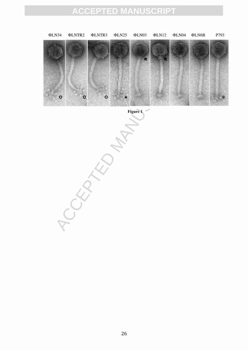
The morphology of the phages was determined by transmission electron microscopy
(TEM). The obtained micrographs showed that all tested phages have a long, non-
contractive tail and an isometric head thus belonging to the B1 morphotype of the
Siphoviridae family within the order Caudovirales (Ackermann and DuBow, 1987).
Furthermore, phages could be classified into 5 morphotypes according to the
classification proposed previously (Ali et al., 2013). Phages ΦLN34, ΦLNTR2 and
ΦLNTR3 belong to morphotype Ia (with defined, globular appendices), phage
ΦLN25 belongs to morphotype Ib (with defined, y-shaped appendices). Phages
ΦLN03 and ΦLN12 can be classified into morphotype IIa (lack of appendices,
presence of the neck passage structure (NPS)). Phages ΦLN04 and ΦLN6B belong to
ACC
EPTE
D M
ANU
SCR
IPT
ACCEPTED MANUSCRIPT
10
morphotype IIb (lack of appendices and NPS). The phage P793 is the only member of
IIc morphotype (undefined base plate appendices, no NPS) (Figure 1).
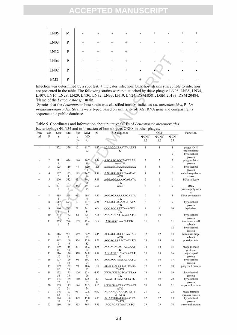
The host range of phages was determined against strains of Ln. mesenteroides (15
strains), Ln. pseudomesenteroides (7 strains) or Ln. lactis (3 strains). The phages
ΦLN25, ΦLN34, ΦLNTR2 and ΦLNTR3 propagated exclusively on Ln.
mesenteroides strains, while the phages ΦLN03, ΦLN04, ΦLN12, P793 and ΦLN6B
only propagated on Ln. pseudomesenteriodes strains (Table 4). None of the phages
formed plaques on Ln. lactis strains. The phages revealed four different host-range
patterns; two were exclusive for Ln. mesenteroides strains and two were exclusive for
Ln. pseudomesenteroides. None of the patterns were overlapping. Limited number of
hosts and high conservation of host-range patterns in Leuconostoc phages has been
observed before (Atamer et al., 2011) and could be partially explained by a small
diversity of Leuconostoc strains found in starter cultures (Johansen and Kibenich,
1992; Nieto-Arribas et al., 2010) or broad host-ranges of the receptor binding protein.
The relatively narrow diversity of the Leuconostoc host strains susceptible to the
phages of this study was suggested by rep-PCR (data not shown).
3.2 Genomic organization of Leuconostoc phages
The phages have a dsDNA genome with sizes ranging from 25.7 to 28.4 kb. The
genomic G+C content was in range from 36.0% in phage ΦLN34 to 36.8% in phage
ΦLN03, which is close to the G+C content of 37.7% found in Ln. mesenteroides
ATCC 8293 (Makarova et al., 2006).
The 9 sequenced phages can be divided in two classes that differ greatly in terms of
nucleotide sequence between classes but are conserved within the class. Class I is
constituted of phages attacking Ln. mesenteroides and class II are phages attacking Ln.
pseudomesenteroides. The high conservation regarding the genomic sequence and the
ACC
EPTE
D M
ANU
SCR
IPT
ACCEPTED MANUSCRIPT
11
host-range patterns is noteworthy, taking into consideration that phages were isolated
from different geographic locations (Table 2). Bioinformatic analysis revealed
presence of 38-42 potential ORFs per genome. ORFs were named with consecutive
numbers starting from the first predicted ORF in the closest proximity to the left cos-
site (cosL) of the genome.
The putative functions of the genes, based on the similarities to already known
sequences, are listed in Table 5 for Ln. mesenteroides phages and in Table 6 for Ln.
pseudomesenteroides phages. Restriction patterns on ligated and linear phage DNA
suggested that the phages utilize cos-type packaging system (data not shown).
Comparison between Sanger reads on amplified-ligated and isolated-linear phage
DNA indeed revealed the presence of 3’ overhang cos-sites. Sequence of the
conserved 12 nt cos-sites of the 4 Ln. mesenteroides phages was determined
(CGGTTAGTAGTA). The cos sequence was shorter than 22-nucleotide cos-site
reported for phage Φ1-A4 (Lu et al., 2010) however the beginning of the Φ1-A4 cos-
site (GGTTAATAGTAGTCTTTTTTAA) share high similarity with the sequence of
the newly sequenced Ln. mesenteroides phages. The 13 nt cos-sites of the 5 Ln.
pseudomesenteroides phages (TCGTGCAATAGTA) were also conserved and
identical to the first 13 nt of phage ΦLmd1 (TCGTGCAATAGTAGGCG
TTTTAA)(Kleppen et al., 2012).
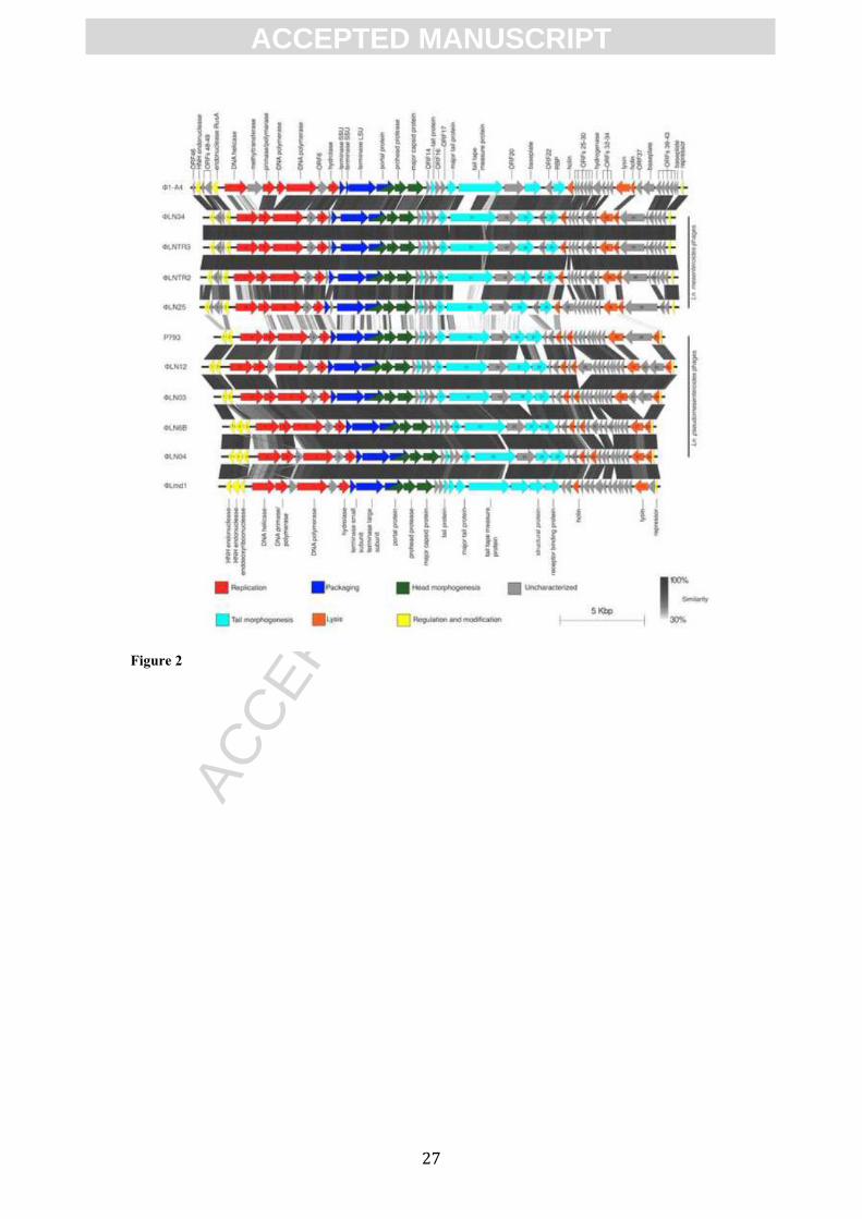
3.3 Comparative genomics
The overall composition of the modules in both classes was very similar to the ones
from Ln. mesenteroides phages Φ1-A4 and ΦLmd1, however Φ1-A4 is more related
to Ln. mesenteroides phages while ΦLmd1 clusters with Ln. pseudomesenteroides
phages group (Figure 2). The similarities to the temperate phage ΦMH1 were limited,
indicating that temperate Ln. pseudomesenteroides phages are not the source of the
ACC
EPTE
D M
ANU
SCR
IPT
ACCEPTED MANUSCRIPT
12
lytic Ln. pseudomesenteroides phages. Though, Blastp analysis of all putative
gpORFs from ΦMH1 versus all gpORFs of the newly sequenced phages resulted in 7
unique hits of E-value below 0.01. The similarities were found within gpORF28 from
ΦLN12 and a putative methylase from ΦMH1 (e-value 1.80e-85), putative baseplate
(e-value, 1.64e-36), large terminase (e-value, 2.74e-18), TMP (e-value, 1.68e-10) and
putative endonuclease (e-value, 2.39e-6) and two hypothetical proteins without
predicted function.
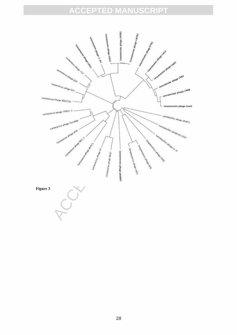
The whole genome comparison with several LAB phages revealed that phages of Ln.
mesenteroides and Ln. pseudomesenteroides form separate clusters (Figure 3).
Five functional modules specific for replication, packaging, morphogenesis, cell lysis
and regulation/modification were identified in all phage genomes. No lysogeny
modules were detected. Comparative genome analysis showed high percentage of
similarity within the classes on both nucleotide and protein level. High level of
conservation within classes is present especially in the replication, packaging and
structural module (Figure 2). Similarities at the nucleotide level were limited between
the two different classes (data not shown). This was also previously shown by
DNA:DNA hybridizations and sequencing of the mtp gene and flanking regions (Ali
et al., 2013).
3.4 Genetic diversity of Leuconostoc phages
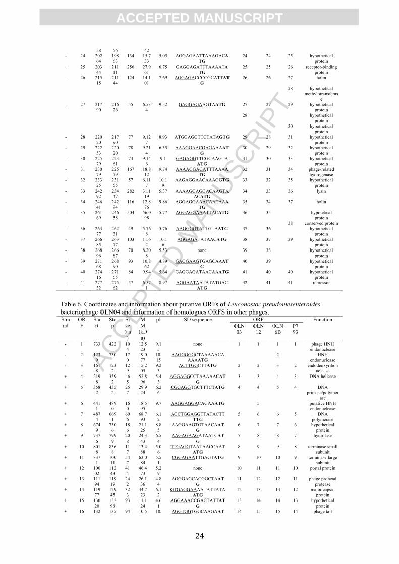
The core genome of the analyzed Ln. mesenteroides phages consisted of 36 ORF out
of total pool of 47 ORFs. In case of Ln. pseudomesenteroides phages homologs of 37
ORFs were detected in all the phages out of the total pool of 44 ORFs (table 5 and 6,
respectively).
In the Ln. mesenteroides ΦLN25 phage there is a putative gene (ORF12) between the
small terminase subunit gene (ORF11) and the large terminase subunit gene (ORF13).
ACC
EPTE
D M
ANU
SCR
IPT
ACCEPTED MANUSCRIPT
13
An additional gene located between the terminase subunits was recently reported for a
number of 936 phages (Castro-Nallar et al., 2012). Two different versions of putative
lysin were detected in Ln. mesenteroides phages. In phage ΦLN25 the putative lysin
showed 98% similarity to the amidase from phage Φ1-A4. In phages ΦLN34,
ΦLNTR2 and ΦLNTR3 the lysin exhibited high similarity to the lysin from
Leuconostoc citreum KM20 (e-value, 6.47e-52). The two types of lysin did not show
any significant nucleotide similarity with each other.
The transmission electron micrographs showed that two of the Ln.
pseudomesenteroides phages, ΦLN03 and ΦLN12 had a distinct neck passage
structure (NPS) (Figure 1). NPS genes are commonly found in lactococcal phages
belonging to the P335, 936 and c2 phages species and are part of their structural
module (Brøndsted et al., 2001; Høier et al., 2010; Rousseau and Moineau, 2009).
Although the putative structural module is highly conserved in the Ln.
pseudomesenteroides phages, two possible locations for the NPS determinant were
identified by comparative genomics. The first putative location was detected as a 573
bp long in-frame insertion located in ORF20 and ORF21 in phages ΦLN03 and
ΦLN12, respectively. The second putative NPS determinant was located in close
proximity to the right cos-site of phage ΦLN03 and ΦLN12. It consisted of ORF36
and ORF37 in phage ΦLN03 and showed high similarity to an insertion element from
Lactobacillus delbrueckii phage LL-K (e-values, 4.11e-10 and 4.11e-50, respectively)
(Forsman and Alatossava, 1993). Further experiments are necessary in order to
specify the actual NPS determinant. Apparently, this NPS is not involved in host-
range interactions as phages lacking these structures (i.e. ΦLN04 and ΦLN6B) had
the same host range as phages ΦLN03 and ΦLN012.
In the sequenced Leuconostoc phages two putative methyltransferase genes could be
ACC
EPTE
D M
ANU
SCR
IPT
ACCEPTED MANUSCRIPT
14
found. One of them was encoded by ORF28 in ΦLN12 and was found only in this
phage. The gene product showed significant similarity (e-value, 1.84e-143) to a
putative DNA methyltransferase from bacteriophage Φ1-A4, however it was located
differently. In phage Φ1-A4 this methyltransferase was located in the replication
module while in phage ΦLN12 it was placed next to the putative lytic enzyme. This
gene also showed high similarity with a putative methylase from temperate
Leuconostoc phage ΦMH1 (e-value, 1.72e-16). The second putative methyltransferase
was gpORF28 of phage ΦLN25. It was also located next to the putative lysis module
and had significant similarity (e-value, 9.41e-172) to the methyltransferase of a type I
restriction-modification system from Ln. lactis KCTC 3528. DNA methyltransferases
are sometimes incorporated to the phage genome as a strategy of overcoming hosts
restriction modification system (Labrie et al., 2010).
In Ln. pseudomesenteroides phages ΦLN04 and ΦLN12 an additional gene coding for
the HNH endonuclease (ORF6 and ORF5, respectively) located between genes coding
for DNA primase and DNA polymerase was detected. A related endonuclease is
encoded by ORF6 from the ΦLmd1 phage (e-value, 1.89e-89) (Kleppen et al., 2012).
HNH endonucleases have been reported to mobilize their own reading frames by
generating DNA breaks at specific sites, activity of homing endonucleases may lead
to site-specific recombination and may result in insertion, deletion, mutation or
correction of DNA sequence (Stoddard, 2011). The majority of the identified putative
endonucleases were clustered together in close proximity to the left cos-site of the
phage genomes, being part of the regulation/modification module.
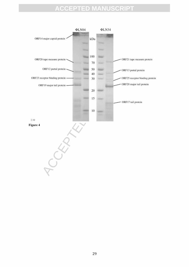
3.5 Protein identification
Two phages were selected for protein identification using mass spectrometry; phage
ΦLN34 as the Ln. mesenteroides phages representative and ΦLN04 as Ln.
ACC
EPTE
D M
ANU
SCR
IPT
ACCEPTED MANUSCRIPT
15
pseudomesenteriodes phages representative. SDS-PAGE profiles of selected phages
revealed 6 and 5 major bands respectively (Figure 4). Obtained results from peptide
mass fingerprints and MS/MS analysis allowed identification of the majority of the
structural proteins of the two phages. The largest predicted protein with 92,1 kDa in
ΦLN34 (ORF21) and actual size of 75 kDa in SDS-PAGE gel was identified as a tape
measure protein (TMP). This may suggest that TMP is being processed prior to
incorporation into the phage particle. Proteolytic cleavage of the C-terminal part of
TMP has been recorded before in phages including the lactococcal phage Tuc2009
(Mc Grath et al., 2006). The second largest band in both phages (46,4 kDa in ΦLN04
and 42,9 kDa in ΦLN34) was identified as putative portal protein (ORF12 and ORF13,
respectively).
The third band in both of the phages was identified as a receptor binding protein
(RBP). The function of this gene was recently verified in Ln. pseudomesenteroides
phages (Kot et al., 2013). RBP of ΦLN04 (ORF23, app. 30 kDa) seems to be slightly
bigger than the RBP of ΦLN34 (ORF25, app. 28 kDa) and can be explained by
considerably different shape in structures observed on the micrographs of those
phages (Figure 1).
Identification of an approx. 21 kDa protein band in phage ΦLN04 suggested that this
protein was the major tail protein (ORF19). This finding was supported by the large
amount of the protein present in SDS-PAGE gel (Figure 4).
It proved to be difficult to obtain significant matches of the smallest structural
proteins. The smallest identified protein was a 12,5 kDa protein band from phage
ΦLN34, which showed high similarity to a putative tail protein (ORF17).
It was not possible to identify the putative head protein among bands excised from the
SDS-PAGE gel in any of the phages, however it was possible to get a match for the
ACC
EPTE
D M
ANU
SCR
IPT
ACCEPTED MANUSCRIPT
16
putative major capsid protein (ORF14ΦLN04) when analyzing the protein material that
failed to enter the gel. This could suggest that capsids of phage ΦLN04 undergo a
wholesale head crosslinking similar to phage HK97 (Jang et al., 2010; Popa et al.,
1991).
4. Conclusions
Nine dairy Leuconostoc phages were characterized and sequenced. Phages can be
divided into two different genotypes. The sequenced bacteriophages exhibit four
different host patterns, two for Ln. mesenteroides and two for Ln.
pseudomesenteroides. High conservation within genotypes and host range pattern is
notable taking into consideration different locations from which phages were obtained.
Phages of Ln. mesenteroides cluster together with the Ln. mesenteroides phage ΦA1-
4, while Ln. pseudomesenteroides phages cluster together with the Ln. mesenteroides
subsp. dextranicum phage ΦLmd1.
To our knowledge this report provides the first comparative genomic work performed
on phages lytic to the Leuconostoc species. The phages are highly conserved within
their classes both on nucleotide and protein level. Additionally, similarities on protein
level were present between the classes. The sequenced phages had analogous,
conserved genetic organization suggesting close evolutionary distance between them.
Genome comparison between the sequenced phages provided additional information
that may result in deeper understanding of phage genetics and evolutionary
mechanisms occurring in phages.
5. Acknowledgments
Witold Kot is the recipient of a PhD scholarship from the University of Copenhagen,
Denmark. We thank Clerici-Sacco Group, Cadorago, Italy for the financial support
for the phage sequencing. Anne Blicher is thanked for technical assistance with mass

ACC
EPTE
D M
ANU
SCR
IPT
ACCEPTED MANUSCRIPT
17
spectrometric analysis. The Danish Center for Advanced Food Studies contributed to
the Bruker Ultraflex II mass spectrometer.
References
Ackermann, H.W., DuBow, M.S., 1987. Viruses of Prokaryotes 480. Ali, Y., Kot, W., Atamer, Z., Hinrichs, J., Vogensen, F.K., Heller, K.J., Neve, H., 2013.
Classification of lytic bacteriophages attacking dairy Leuconostoc starter strains. Applied and Environmental Microbiology 79, 3628-3636.
Atamer, Z., Ali, Y., Neve, H., Heller, K.J., Hinrichs, J., 2011. Thermal resistance of bacteriophages attacking flavour-producing dairy Leuconostoc starter cultures. International Dairy Journal 21, 327–334.
Besemer, J., Borodovsky, M., 1999. Heuristic approach to deriving models for gene finding. Nucleic Acids Res 27, 3911–3920.
Brøndsted, L., Østergaard, S., Pedersen, M.M., Hammer, K., Vogensen, F.K., 2001. Analysis of the Complete DNA Sequence of the Temperate Bacteriophage TP901-1: Evolution, Structure, and Genome Organization of Lactococcal Bacteriophages. Virology 283, 17–17.
Castro-Nallar, E., Chen, H., Gladman, S., Moore, S.C., Seemann, T., Powell, I.B., Hillier, A., Crandall, K.A., Chandry, P.S., 2012. Population Genomics and Phylogeography of an Australian Dairy Factory Derived Lytic Bacteriophage. Genome Biology and Evolution 4, 382–393.
Cogan, T., 1993. Cheese: chemistry, physics and microbiology. Cheese: chemistry. Davey, G.P., Ward, L.J.H., Brown, J.C.S., 1995. Characterisation of four Leuconostoc
bacteriophages isolated from dairy fermentations. FEMS Microbiol. Lett. 128, 21–25.
Dicks, L., Fantuzzi, L., Gonzales, F.C., Toit, Du, M., Dellaglio, F., 1993. Leuconostoc argentinum sp. nov., Isolated From Argentine Raw Milk. Int. J. Syst. Bacteriol. 43, 347–351.
Forsman, P., Alatossava, T., 1993. Repeated sequences and the sites of genome rearrangements in bacteriophages of Lactobacillus delbrueckii subsp. lactis. Archives of virology 137, 43–54.
Garvie, E.I., Zezula, V., Hill, V.A., 1974. Guanine plus cytosine content of the deoxyribonucleic acid of the leuconostocs and some heterofermentative lactobacilli. Int. J. Syst. Bacteriol. 24, 248–251.
Hemme, D., Foucaud-Scheunemann, C., 2004. Leuconostoc, characteristics, use in dairy technology and prospects in functional foods. International Dairy Journal 14, 467–494.
Høier, E., Janzen, T., Rattray, F., Sørensen, K., Børsting, M.W., Brockmann, E., Johansen, E., 2010. Technology of Cheesemaking. Wiley-Blackwell, Oxford, UK.
Jang, S.H., Hwang, M.H., Chang, H.-I., 2010. Complete genome sequence of ΦMH1, a Leuconostoc temperate phage. Archives of virology 155, 1883–1885.
Johanningsmeier, S., McFeeters, R.F., Fleming, H.P., Thompson, R.L., 2007. Effects of Leuconostoc mesenteroides starter culture on fermentation of cabbage with
ACC
EPTE
D M
ANU
SCR
IPT
ACCEPTED MANUSCRIPT
18
reduced salt concentrations. J Food Sci 72, 166–172. Johansen, E., Kibenich, A., 1992. Characterization of Leuconostoc Isolates From
Commercial Mixed Strain Mesophilic Starter Cultures. Journal of Dairy Science 75, 1186–1191.
Jung, J.Y., Lee, S.H., Kim, J.M., Park, M.S., Bae, J.W., Hahn, Y., Madsen, E.L., Jeon, C.O., 2011. Metagenomic Analysis of Kimchi, a Traditional Korean Fermented Food. Applied and Environmental Microbiology 77, 2264–2274.
Kampmann, M.-L., Fordyce, S.L., Avila-Arcos, M.C., Rasmussen, M., Willerslev, E., Nielsen, L.P., Gilbert, M.T.P., 2011. A simple method for the parallel deep sequencing of full influenza A genomes. CORD Conference Proceedings 178, 243–248.
Kandler, O., 1970. Amino acid sequence of the murein and taxonomy of the genera Lactobacillus, Bifidobacterium, Leuconostoc and Pediococcus. Int. J. Syst. Bacteriol. 20, 491–507.
Kleppen, H.P., Nes, I.F., Holo, H., 2012. Characterization of a Leuconostoc Bacteriophage Infecting Flavor Producers of Cheese Starter Cultures. Applied and Environmental Microbiology 78, 6769–6772.
Kot, W., Hammer, K., Neve, H., Vogensen, F.K., 2013. Identification of the Receptor-Binding Protein in lytic Leuconostoc pseudomesenteroides Bacteriophages. Applied and Environmental Microbiology 79, 3311-3314.
Labrie, S.J., Samson, J.E., Moineau, S., 2010. Bacteriophage resistance mechanisms. Nature Reviews Microbiology 8, 317–327.
Lu, Z., Altermann, E., Breidt, F., Kozyavkin, S., 2010. Sequence Analysis of Leuconostoc mesenteroides Bacteriophage 1-A4 Isolated from an Industrial Vegetable Fermentation. Applied and Environmental Microbiology 76, 1955–1966.
Lubbers, M.W., Waterfield, N.R., Beresford, T.P., Le Page, R.W., Jarvis, A.W., 1995. Sequencing and analysis of the prolate-headed lactococcal bacteriophage c2 genome and identification of the structural genes. Applied and Environmental Microbiology 61, 4348–4356.
Mahanivong, C., Boyce, J., Davidson, B., Hillier, A., 2001. Sequence analysis and molecular characterization of the Lactococcus lactis temperate bacteriophage BK5-T. Applied and Environmental Microbiology 67, 3564–3576.
Makarova, K., Slesarev, A., Wolf, Y., Sorokin, A., Mirkin, B., Koonin, E., Pavlov, A., Pavlova, N., Karamychev, V., Polouchine, N., Shakhova, V., Grigoriev, I., Lou, Y., Rohksar, D., Lucas, S., Huang, K., Goodstein, D.M., Hawkins, T., Plengvidhya, V., Welker, D., Hughes, J., Goh, Y., Benson, A., Baldwin, K., Lee, J.H., Diaz-Muniz, I., Dosti, B., Smeianov, V., Wechter, W., Barabote, R., Lorca, G., Altermann, E., Barrangou, R., Ganesan, B., Xie, Y., Rawsthorne, H., Tamir, D., Parker, C., Breidt, F., Broadbent, J., Hutkins, R., O'Sullivan, D., Steele, J., Unlu, G., Saier, M., Klaenhammer, T., Richardson, P., Kozyavkin, S., Weimer, B., Mills, D., 2006. Comparative genomics of the lactic acid bacteria. Proc. Natl. Acad. Sci. USA 103, 15611–15616.
Mc Grath, S., Neve, H., Seegers, J.F.M.L., Eijlander, R., Vegge, C.S., Brøndsted, L., Heller, K.J., Fitzgerald, G.F., Vogensen, F.K., van Sinderen, D., 2006. Anatomy of a Lactococcal Phage Tail. J. Bacteriol. 188, 3972–3982.
Moineau, S., Tremblay, D., Labrie, S., 2002. Phages of lactic acid bacteria: from genomics to industrial applications. ASM News 68, 388–393.
Mosimann, W., Ritter, W., 1946. Bacteriophages as cause of loss of aroma in

ACC
EPTE
D M
ANU
SCR
IPT
ACCEPTED MANUSCRIPT
19
butter cultures (Bakteriophagen als Ursache von Aromaschwund in Rahmsäuerungskulturen). Schweizerische Milchzeitung 72, 211–212.
Neve, H., Lilischkis, R., Teuber, M., 1988. Characterisation of a virulent bacteriophage of Leuconostoc mesenteroides subsp. cremoris. Kieler Milchwirtschaftliche Forschungsberichte 40, 205–215.
Nieto-Arribas, P., Sesena, S., Poveda, J.M., Palop, L., Cabezas, L., 2010. Genotypic and technological characterization of Leuconostoc isolates to be used as adjunct starters in Manchego cheese manufacture. Food Microbiology 27, 85–93.
Olsen, K.N., Brockmann, E., Molin, S., 2007. Quantification of Leuconostoc populations in mixed dairy starter cultures using fluorescence in situ hybridization. J. Appl. Microbiol. 103, 855–863.
Parente, E., Cogan, T.M., 2004. Starter cultures: General aspects, in: Patrick F Fox Timothy M Cogan and Timothy P Guinee, P.L.H.M. (Ed.), Cheese: Chemistry, Physics and Microbiology, Cheese Chemistry, Physics and Microbiology. Academic Press, pp. 123–147.
Popa, M.P., McKelvey, T.A., Hempel, J., Hendrix, R.W., 1991. Bacteriophage HK97 structure: wholesale covalent cross-linking between the major head shell subunits. Journal of Virology 65, 3227–3237.
Rousseau, G.M., Moineau, S., 2009. Evolution of Lactococcus lactis Phages within a Cheese Factory. Applied and Environmental Microbiology 75, 5336–5344.
Sambrook, J., Russell, D.W., 2001. Molecular cloning. CSHL Press. Shin, C., Sato, Y., 1979. Isolation of Leuconostoc Bacteriophages from Dairy
Products. Japanese journal of zootechnical science 50 (6), 419–422. Sozzi, T., Poulin, J.M., Maret, R., Pousaz, R., 1978. Isolation of a Bacteriophage of
Leuconostoc mesenteroides from Dairy Products. J. Appl. Microbiol. 44, 159–161.
Stoddard, B.L., 2011. Homing Endonucleases: From Microbial Genetic Invaders to Reagents for Targeted DNA Modification. Structure 19, 7–15.
Sullivan, M.J., Petty, N.K., Beatson, S.A., 2011. Easyfig: a genome comparison visualiser. Bioinformatics 27, 1009–1010.
Yang, J., Cao, Y., Cai, Y., Terada, F., 2010. Natural populations of lactic acid bacteria isolated from vegetable residues and silage fermentation. Journal of Dairy Science 93, 3136–3145.
Zamfir, M., Vancanneyt, M., Makras, L., Vaningelgem, F., Lefebvre, K., Pot, B., Swings, J., De Vuyst, L., 2006. Biodiversity of lactic acid bacteria in Romanian dairy products. Systematic and Applied Microbiology 29, 487–495.
Zhang, X., Shi, L., Shu, S., Wang, Y., Zhao, K., Xu, N., Liu, S., Roepstorff, P., 2007. An improved method of sample preparation on AnchorChip™ targets for MALDI-MS and MS/MS and its application in the liver proteome project. Proteomics 7, 2340–2349.
ACC
EPTE
D M
ANU
SCR
IPT
ACCEPTED MANUSCRIPT
20
Figure Captions:
Figure 1. Transmission electron micrographs of phages. Phages were negatively
stained with 2% uranyl acetate. Morphological details are indicated as follows:
globular base plate appendices (), y-shaped base plate appendices (�), neck passage
structure (), undefined, fluffy base plate appendices ().
Figure 2. Schematic representation of the genomic comparison of Leuconostoc phages.
Genomes are indicated with thick, black lines. Arrow represents putative protein. The
color of the arrows indicate the putative function of the gene product and the number
indicates the number of the ORF. Each genome was compared only with the
succesive genome. Gray shading connecting two genomes corresponds to the
similarity level obtain from tblastx comparison. All genomes are alligned according to
the position of their cos-sites.
Figure 3. Phylogenetic tree of a total of 29 LAB infecting phages. The distance matrix
and visualization was performed in Geneious 6.1.2. Phages sequenced in this study
are indicated with the bold font. Other phages of Leuconostoc are indicated with bold,
italic font.
Figure 4. Purified proteins from Ln. pseudomesenteroides ΦLN04 and Ln.
mesenteroides ΦLN34 phages. Inner lanes show the protein marker (PageRuler™
Unstained Broad Range Protein Ladder, Fermentas). Protein standard is indicated in
kilodalton (kDa).
ACC
EPTE
D M
ANU
SCR
IPT
ACCEPTED MANUSCRIPT
21
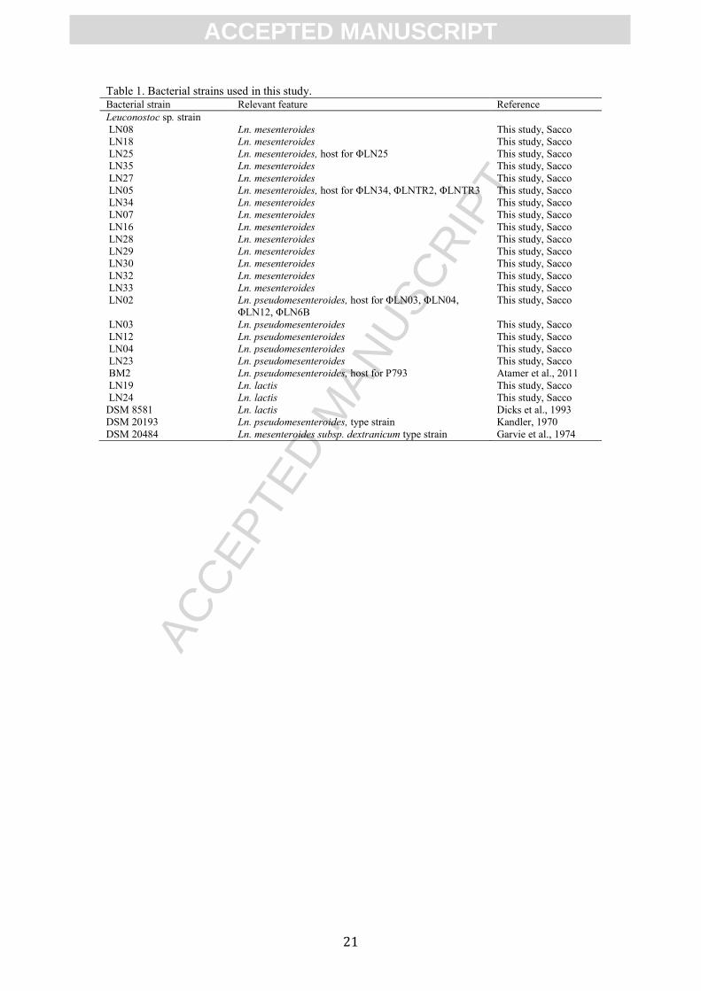
Table 1. Bacterial strains used in this study. Bacterial strain Relevant feature Reference
Leuconostoc sp. strain
LN08 Ln. mesenteroides This study, Sacco
LN18 Ln. mesenteroides This study, Sacco
LN25 Ln. mesenteroides, host for ΦLN25 This study, Sacco
LN35 Ln. mesenteroides This study, Sacco
LN27 Ln. mesenteroides This study, Sacco
LN05 Ln. mesenteroides, host for ΦLN34, ΦLNTR2, ΦLNTR3 This study, Sacco
LN34 Ln. mesenteroides This study, Sacco
LN07 Ln. mesenteroides This study, Sacco
LN16 Ln. mesenteroides This study, Sacco
LN28 Ln. mesenteroides This study, Sacco
LN29 Ln. mesenteroides This study, Sacco
LN30 Ln. mesenteroides This study, Sacco
LN32 Ln. mesenteroides This study, Sacco
LN33 Ln. mesenteroides This study, Sacco
LN02 Ln. pseudomesenteroides, host for ΦLN03, ΦLN04,
ΦLN12, ΦLN6B
This study, Sacco
LN03 Ln. pseudomesenteroides This study, Sacco
LN12 Ln. pseudomesenteroides This study, Sacco
LN04 Ln. pseudomesenteroides This study, Sacco
LN23 Ln. pseudomesenteroides This study, Sacco
BM2 Ln. pseudomesenteroides, host for P793 Atamer et al., 2011
LN19 Ln. lactis This study, Sacco
LN24
DSM 8581
DSM 20193
DSM 20484
Ln. lactis
Ln. lactis
Ln. pseudomesenteroides, type strain Ln. mesenteroides subsp. dextranicum type strain
This study, Sacco
Dicks et al., 1993
Kandler, 1970
Garvie et al., 1974
ACC
EPTE
D M
ANU
SCR
IPT
ACCEPTED MANUSCRIPT
22
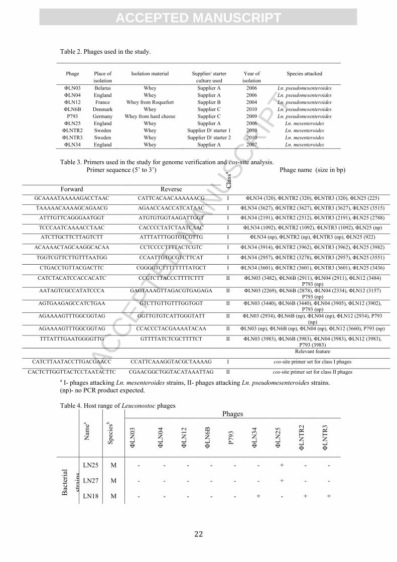
Table 2. Phages used in the study.
Phage Place of
isolation
Isolation material Supplier/ starter
culture used
Year of
isolation
Species attacked
ΦLN03 Belarus Whey Supplier A 2006 Ln. pseudomesenteroides
ΦLN04 England Whey Supplier A 2006 Ln. pseudomesenteroides
ΦLN12 France Whey from Roquefort Supplier B 2004 Ln. pseudomesenteroides
ΦLN6B Denmark Whey Supplier C 2010 Ln. pseudomesenteroides
P793 Germany Whey from hard cheese Supplier C 2009 Ln. pseudomesenteroides
ΦLN25 England Whey Supplier A 2006 Ln. mesenteroides
ΦLNTR2 Sweden Whey Supplier D/ starter 1 2010 Ln. mesenteroides
ΦLNTR3 Sweden Whey Supplier D/ starter 2 2010 Ln. mesenteroides
ΦLN34 England Whey Supplier A 2007 Ln. mesenteroides
Table 3. Primers used in the study for genome verification and cos-site analysis.
Primer sequence (5’ to 3’)
Cla
ssa Phage name (size in bp)
Forward Reverse
GCAAAATAAAAAGACCTAAC CATTCACAACAAAAAACG I ΦLN34 (320), ΦLNTR2 (320), ΦLNTR3 (320), ΦLN25 (225)
TAAAAACAAAAGCAGAACG AGAACCAACCATCATAAC I ΦLN34 (3627), ΦLNTR2 (3627), ΦLNTR3 (3627), ΦLN25 (3515)
ATTTGTTCAGGGAATGGT ATGTGTGGTAAGATTGGT I ΦLN34 (2191), ΦLNTR2 (2512), ΦLNTR3 (2191), ΦLN25 (2788)
TCCCAATCAAAACCTAAC CACCCCTATCTAATCAAC I ΦLN34 (1092), ΦLNTR2 (1092), ΦLNTR3 (1092), ΦLN25 (np)
ATCTTGCTTCTTAGTCTT ATTTATTTGGTGTCGTTG I ΦLN34 (np), ΦLNTR2 (np), ΦLNTR3 (np), ΦLN25 (922)
ACAAAACTAGCAAGGCACAA CCTCCCCTTTTACTCGTC I ΦLN34 (3914), ΦLNTR2 (3962), ΦLNTR3 (3962), ΦLN25 (3982)
TGGTCGTTCTTGTTTAATGG CCAATTGTGCGTCTTCAT I ΦLN34 (2957), ΦLNTR2 (3278), ΦLNTR3 (2957), ΦLN25 (3551)
CTGACCTGTTACGACTTC CGGGGTCTTTTTTTTATGCT I ΦLN34 (3601), ΦLNTR2 (3601), ΦLNTR3 (3601), ΦLN25 (3436)
CATCTACATCCACCACATC CCGTCTTACCCTTTTCTTT II ΦLN03 (3482), ΦLN6B (2911), ΦLN04 (2911), ΦLN12 (3484) P793 (np)
AATAGTCGCCATATCCCA GAGTAAAGTTAGACGTGAGAGA II ΦLN03 (2269), ΦLN6B (2878), ΦLN04 (2334), ΦLN12 (3157)
P793 (np)
AGTGAAGAGCCATCTGAA GTCTTGTTGTTTGGTGGT II ΦLN03 (3440), ΦLN6B (3440), ΦLN04 (3905), ΦLN12 (3902), P793 (np)
AGAAAAGTTTGGCGGTAG GGTTGTGTCATTGGGTATT II ΦLN03 (2934), ΦLN6B (np), ΦLN04 (np), ΦLN12 (2934), P793
(np)
AGAAAAGTTTGGCGGTAG CCACCCTACGAAAATACAA II ΦLN03 (np), ΦLN6B (np), ΦLN04 (np), ΦLN12 (3660), P793 (np)
TTTATTTGAATGGGGTTG GTTTTATCTCGCTTTTCT II ΦLN03 (3983), ΦLN6B (3983), ΦLN04 (3983), ΦLN12 (3983), P793 (3983)
Relevant feature
CATCTTAATACCTTGACGAACC CCATTCAAAGGTACGCTAAAAG I cos-site primer set for class I phages
CACTCTTGGTTACTCCTAATACTTC CGAACGGCTGGTACATAAATTAG II cos-site primer set for class II phages
a I- phages attacking Ln. mesenteroides strains, II- phages attacking Ln. pseudomesenteroides strains.
(np)- no PCR product expected.
Table 4. Host range of Leuconostoc phages
Nam
ea
Sp
ecie
sb
Phages
ΦL
N0
3
ΦL
N0
4
ΦL
N1
2
ΦL
N6
B
P7
93
ΦL
N3
4
ΦL
N2
5
ΦL
NT
R2
ΦL
NT
R3
Bac
teri
al
stra
ins
LN25 M - - - - - - + - -
LN27 M - - - - - - + - -
LN18 M - - - - - + - + +
ACC
EPTE
D M
ANU
SCR
IPT
ACCEPTED MANUSCRIPT
23
LN05 M - - - - - + - + +
LN03 P + + + + - - - - -
LN12 P + + + + - - - - -
LN04 P + + + + - - - - -
LN02 P + + + + - - - - -
BM2 P - - - - + - - - -
Infection was determined by a spot test, + indicates infection. Only host strains susceptible to infection
are presented in the table. The following strains were not attacked by these phages: LN08, LN35, LN34,
LN07, LN16, LN28, LN29, LN30, LN32, LN33, LN19, LN24, DSM 8581, DSM 20193, DSM 20484. aName of the Leuconostoc sp. strain.
bSpecies that the Leuconostoc host strain was classified into. M indicates Ln. mesenteroides, P- Ln.
pseudomesenteroides. Strains were typed based on similarity of 16S rRNA gene and comparing its
sequence to a public database.
Table 5. Coordinates and information about putative ORFs of Leuconostoc mesenteroides
bacteriophage ΦLN34 and information of homologues ORFS in other phages. Stra
nd
OR
F
Star
t
Sto
p
Siz
e
(aa)
MM
(kD
a)
pI SD sequence ORF Function
ΦLNT
R2
ΦLNT
R3
ΦLN
25
- 1 672 370 101 11.7
22
8.47 ACAAGGATAATTAATAT
G
1 1 1 phage HNH
endonuclease
2 hypothetical
protein
- 2 111
1
674 146 16.7
88
8.54 AAGAAGAGGTACTAAA
AAATG
2 2 3 phage-related
protein
- 3 125
4
110
8
49 6.20
4
11.6
1
AGGAGCGAAGAAGAAA
TG
3 3 4 hypothetical
protein
- 4 162
5
125
1
125 14.9
86
8.92 AACAGGAGGGTAACAT
ATG
4 4 5 endodeoxyribonu
clease
+ 5 200
7
332
0
438 50.5
95
5.89 AGGAGGAAAACAGATA
TG
5 5 6 DNA helicase
+ 6 331
7
407
2
252 29.1
75
6.51 none 6 6 7 DNA
primase/polymera
se
+ 7 415
1
598
6
612 69.8
6
7.57 AGGAGAAAAAAGATTA
TG
7 7 8 DNA polymerase
+ 8 617
6
674
8
191 21.7
65
5.26 ATAAGGAGAACATATA
TG
8 8 9 hypothetical
protein
+ 9 680
1
743
3
211 24.1
84
6.3 GGGAGGAATTAAAGTA
TG
9 9 10 hydrolase
+ 10 744
5
762
7
61 7.31
7
7.16 AGGAGGATTGACTATG 10 10 hypothetical
protein
+ 11 763
6
796
2
109 12.4
88
5.5 ATGAGGTAATATATG 11 11 11 terminase small
subunit
12 hypothetical
protein
+ 12 816
6
981
2
549 62.9
99
5.45 ACGAGGAGGGTAATAG
ATG
12 12 13 terminase large
subunit
+ 13 982
5
109
46
374 42.9
82
5.21 AGGAGAAAACTATATG 13 13 14 portal protein
+ 14 109
06
115
98
231 25.2
52
4.78 AGGAGACACTACGAAT
G
14 14 15 phage prohead
protease
+ 15 116
50
126
03
318 34.8
75
5.59 AGGAGACCTATAATAT
G
15 15 16 major capsid
protein
+ 16 127
18
129
90
91 10.3
94
4.77 AGGAGGTGACACAATG 16 16 17 hypothetical
protein
+ 17 129
80
132
58
93 10.6
92
10.4 AGAGGAGGCGATCAGA
TATG
17 17 18 phage tail protein
+ 18 132
58
135
75
106 12.4
79
4.92 GGGAGGTAGTCATTTAA
TG
18 18 19 hypothetical
protein
+ 19 135
72
139
01
110 12.5
07
11.3
8
AGGTGTTAATATTATG 19 19 20 hypothetical
protein
+ 20 139
50
145
31
194 21.3
54
5.13 AGGAGAATTAATCAATT
ATG
20 20 21 major tail protein
+ 21 146
63
173
95
911 92.8
83
9.92 AGAAAGGAAATGTATT
ATATG
21 21 22 phage tail tape
measure protein
+ 22 174
59
186
55
399 45.8
22
5.03 AGAATGGAGGAAATTA
TATG
22 22 23 hypothetical
protein
+ 23 186 196 333 36.8 5.35 AGGAGATTAATCATG 23 23 24 structural protein
ACC
EPTE
D M
ANU
SCR
IPT
ACCEPTED MANUSCRIPT
24
58 56 42
- 24 202
64
198
63
134 15.7
33
5.05 AGGAGAATTAAAGACA
TG
24 24 25 hypothetical
protein
+ 25 203
44
211
11
256 27.9
61
6.75 GAGGAGATTTAAAATA
TG
25 25 26 receptor-binding
protein
- 26 215
15
211
44
124 14.1
01
7.69 AGGAGACCCCGCATTAT
G
26 26 27 holin
28 hypothetical
methylotransferas
e
- 27 217
90
216
26
55 6.53
4
9.52 GAGGAGAAGTAATG 27 27 29 hypothetical
protein
28 hypothetical
protein
30 hypothetical
protein
- 28 220
20
217
90
77 9.12
7
8.93 ATGGAGGTTCTATAGTG 29 28 31 hypothetical
protein
- 29 222
53
220
20
78 9.21
4
6.35 AAAGGAACGAGAAAAT
G
30 29 32 hypothetical
protein
- 30 225
79
223
61
73 9.14
6
9.1 GAGAGGTTCGCAAGTA
ATG
31 30 33 hypothetical
protein
- 31 230
79
225
79
167 18.8
12
9.74 AAAAGGAGATTTAAAA
TG
32 31 34 phage-related
hydrogenase
- 32 233
25
231
55
57 6.11
7
10.1
9
AAGAGGAACAAACGTG 33 32 35 hypothetical
protein
- 33 242
92
234
47
282 31.1
19
5.37 AAAAGGAGGACAAGTA
ACATG
34 33 36 lysin
- 34 246
41
242
94
116 12.8
76
9.86 AGGAGGAAACAATAAA
TG
35 34 37 holin
- 35 261
69
246
58
504 56.0
98
5.77 AGGAGGAAATTACATG 36 35 hypotetical
protein
38 conserved protein
- 36 263
77
262
31
49 5.76
8
5.76 AAGGGGTATTGTAATG 37 36 hypothetical
protein
- 37 266
85
263
77
103 11.6
2
10.1
6
AGGAGATATAACATG 38 37 39 hypothetical
protein
- 38 268
96
266
87
70 8.20
8
5.53 none 39 38 hypothetical
protein
- 39 271
68
268
90
93 10.8
62
4.89 GAGGAAGTGAGCAAAT
G
40 39 hypothetical
protein
- 40 274
16
271
65
84 9.94 5.64 GAGGAGATAACAAATG 41 40 40 hypothetical
protein
- 41 277
32
275
62
57 6.57
1
8.97 AGGAATAATATATGAC
ATG
42 41 41 repressor
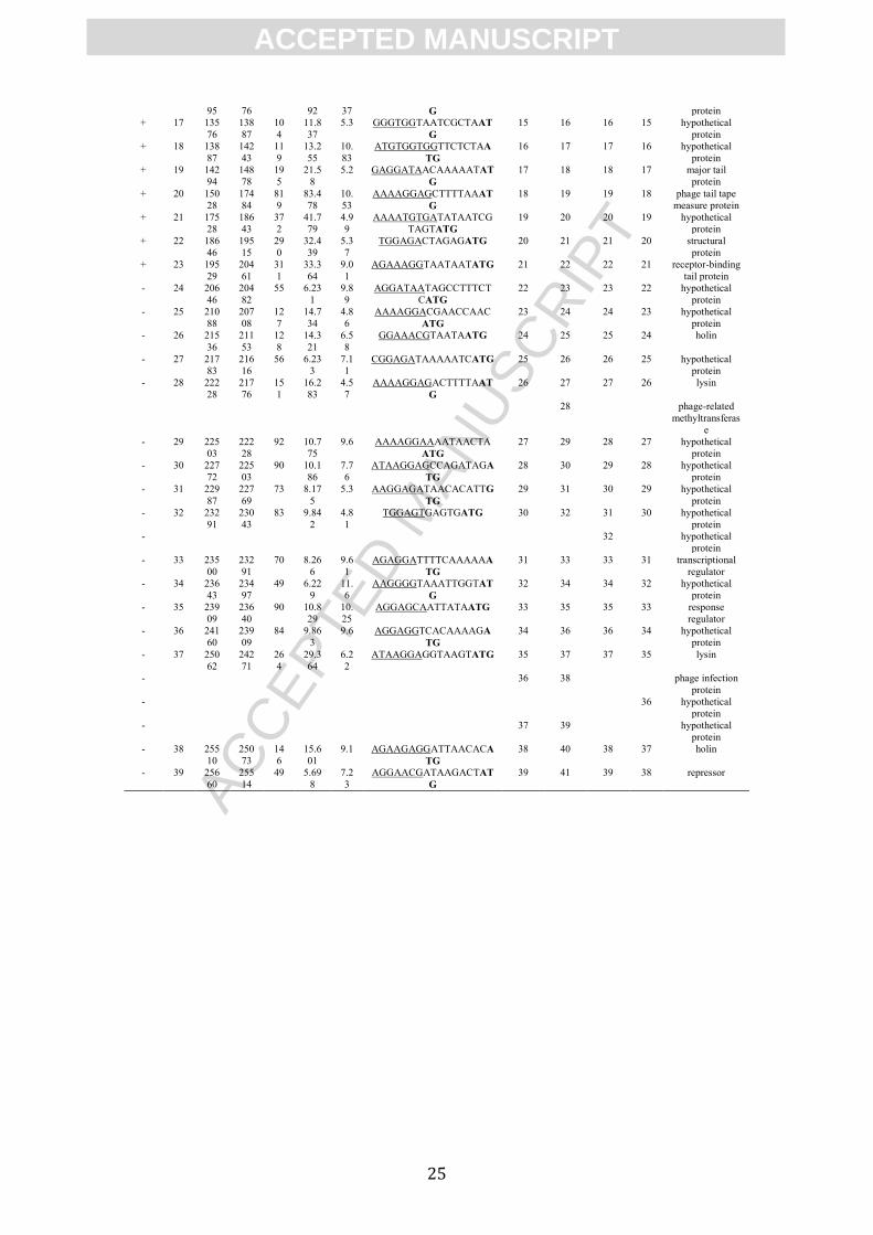
Table 6. Coordinates and information about putative ORFs of Leuconostoc pseudomesenteroides
bacteriophage ΦLN04 and information of homologues ORFS in other phages. Strand
ORF
Start
Stop
Size
(aa
)
MM
(kD
a)
pI SD sequence ORF Function
ΦLN03
ΦLN12
ΦLN6B
P793
- 1 733 422 10
4
12.5
23
9.1
5
none 1 1 1 1 phage HNH
endonuclease
- 2 123
9
730 17
0
19.0
77
10.
15
AAGGGGGCTAAAAACA
AAAATG
2 HNH
endonuclease
- 3 161
8
123
2
12
9
15.2
05
9.2
3
ACTTGGCTTATG 2 2 3 2 endodeoxyribon
uclease
+ 4 219
8
359
2
46
5
52.8
96
5.4
3
AGGAGGCCTAAAAACAT
G
3 3 4 3 DNA helicase
+ 5 358
2
435
2
25
7
29.9
24
6.2
6
CGGAGGTGCTTTCTATG 4 4 5 4 DNA
primase/polymer
ase
+ 6 441
1
489
0
16
0
18.5
95
9.7 AAGGAGGACAGAAATG 5 putative HNH
endonuclease
+ 7 487
4
669
1
60
6
68.7
93
6.1
2
AGCTGGAGGTTATACTT
TTG
5 6 6 5 DNA
polymerase
+ 8 674
9
730
6
18
6
21.1
25
8.8
5
AAGGAAGTGTAACAAT
G
6 7 7 6 hypothetical
protein
+ 9 737
6
799
9
20
8
24.3
43
6.5
4
AAGAGAAGATAATCAT
G
7 8 8 7 hydrolase
+ 10 801
8
836
8
11
7
13.4
88
5.0
6
TTGAGGTAATAACCAAT
ATG
8 9 9 8 terminase small
subunit
+ 11 837
1
100
11
54
7
63.0
84
5.5
1
CGGAGAATTGAGTATG 9 10 10 9 terminase large
subunit
+ 12 100
02
112
43
41
4
46.4
73
5.2
9
none 10 11 11 10 portal protein
+ 13 111
94
119
19
24
2
26.1
36
4.8
4
AGGGAGCACGGCTAAT
G
11 12 12 11 phage prohead
protease
+ 14 119
77
129
45
32
3
34.7
23
6.1
2
GTGAGGAAAATATTATA
ATG
12 13 13 12 major capsid
protein
+ 15 130
20
132
98
93 11.1
24
4.6
1
AGGAAACCGACTATTAT
G
13 14 14 13 hypothetical
protein
+ 16 132 135 94 10.5 10. AGGTGGTGGCAAGAAT 14 15 15 14 phage tail
ACC
EPTE
D M
ANU
SCR
IPT
ACCEPTED MANUSCRIPT
25
95 76 92 37 G protein
+ 17 135
76
138
87
10
4
11.8
37
5.3 GGGTGGTAATCGCTAAT
G
15 16 16 15 hypothetical
protein
+ 18 138
87
142
43
11
9
13.2
55
10.
83
ATGTGGTGGTTCTCTAA
TG
16 17 17 16 hypothetical
protein
+ 19 142
94
148
78
19
5
21.5
8
5.2 GAGGATAACAAAAATAT
G
17 18 18 17 major tail
protein
+ 20 150
28
174
84
81
9
83.4
78
10.
53
AAAAGGAGCTTTTAAAT
G
18 19 19 18 phage tail tape
measure protein
+ 21 175
28
186
43
37
2
41.7
79
4.9
9
AAAATGTGATATAATCG
TAGTATG
19 20 20 19 hypothetical
protein
+ 22 186
46
195
15
29
0
32.4
39
5.3
7
TGGAGACTAGAGATG 20 21 21 20 structural
protein
+ 23 195
29
204
61
31
1
33.3
64
9.0
1
AGAAAGGTAATAATATG 21 22 22 21 receptor-binding
tail protein
- 24 206
46
204
82
55 6.23
1
9.8
9
AGGATAATAGCCTTTCT
CATG
22 23 23 22 hypothetical
protein
- 25 210
88
207
08
12
7
14.7
34
4.8
6
AAAAGGACGAACCAAC
ATG
23 24 24 23 hypothetical
protein
- 26 215
36
211
53
12
8
14.3
21
6.5
8
GGAAACGTAATAATG 24 25 25 24 holin
- 27 217
83
216
16
56 6.23
3
7.1
1
CGGAGATAAAAATCATG 25 26 26 25 hypothetical
protein
- 28 222
28
217
76
15
1
16.2
83
4.5
7
AAAAGGAGACTTTTAAT
G
26 27 27 26 lysin
28 phage-related
methyltransferas
e
- 29 225
03
222
28
92 10.7
75
9.6 AAAAGGAAAATAACTA
ATG
27 29 28 27 hypothetical
protein
- 30 227
72
225
03
90 10.1
86
7.7
6
ATAAGGAGCCAGATAGA
TG
28 30 29 28 hypothetical
protein
- 31 229
87
227
69
73 8.17
5
5.3 AAGGAGATAACACATTG
TG
29 31 30 29 hypothetical
protein
- 32 232
91
230
43
83 9.84
2
4.8
1
TGGAGTGAGTGATG 30 32 31 30 hypothetical
protein
- 32 hypothetical
protein
- 33 235
00
232
91
70 8.26
6
9.6
1
AGAGGATTTTCAAAAAA
TG
31 33 33 31 transcriptional
regulator
- 34 236
43
234
97
49 6.22
9
11.
6
AAGGGGTAAATTGGTAT
G
32 34 34 32 hypothetical
protein
- 35 239
09
236
40
90 10.8
29
10.
25
AGGAGCAATTATAATG 33 35 35 33 response
regulator
- 36 241
60
239
09
84 9.86
3
9.6 AGGAGGTCACAAAAGA
TG
34 36 36 34 hypothetical
protein
- 37 250
62
242
71
26
4
29.3
64
6.2
2
ATAAGGAGGTAAGTATG 35 37 37 35 lysin
- 36 38 phage infection
protein
- 36 hypothetical
protein
- 37 39 hypothetical
protein
- 38 255
10
250
73
14
6
15.6
01
9.1 AGAAGAGGATTAACACA
TG
38 40 38 37 holin
- 39 256
60
255
14
49 5.69
8
7.2
3
AGGAACGATAAGACTAT
G
39 41 39 38 repressor
ACC
EPTE
D M
ANU
SCR
IPT
ACCEPTED MANUSCRIPT
26
Figure 1
ACC
EPTE
D M
ANU
SCR
IPT
ACCEPTED MANUSCRIPT
27
Figure 2
ACC
EPTE
D M
ANU
SCR
IPT
ACCEPTED MANUSCRIPT
28
Figure 3
ACC
EPTE
D M
ANU
SCR
IPT
ACCEPTED MANUSCRIPT
29
Figure 4
ACC
EPTE
D M
ANU
SCR
IPT
ACCEPTED MANUSCRIPT
30
Research Highlights:
Nine dairy Leuconostoc phages were characterized and sequenced
Phages were isolated in relation to a fermentation problem
Sequenced phages can be grouped in two classes that correlate with the host
species
Comparative genomic work revealed high conservation within the classes