Embed Size (px)
Citation preview
1
Title: Safety, Pharmacokinetics and Pharmacodynamics of a Humanized anti- Semaphorin 4D
Antibody, in a First-In-Human Study of Patients with Advanced Solid Tumors
Authors: Amita Patnaik1, Glen J. Weiss2, John E. Leonard3, Drew Warren Rasco1, Jasgit C.
Sachdev4, Terrence L. Fisher3, Laurie A. Winter3, Christine Reilly3, Robert B. Parker3, Danielle
Mutz1, Lisa Blaydorn2, Anthony W. Tolcher1, Maurice Zauderer3 and Ramesh K. Ramanathan4
Authors’ Affiliations: South Texas Accelerated Research Therapeutics (START) Center for
Cancer Care, San Antonio, TX1; Cancer Treatment Centers of America, Goodyear, AZ2 ,
Vaccinex, Inc, Rochester, NY3, Virginia G. Piper Cancer Center at Scottsdale Healthcare/TGen,
Scottsdale, AZ4.
Author Current Addresses: Ramesh K. Ramanathan- Mayo Clinic, 13400 E Shea Blvd,
Scottsdale, AZ; Glen J Weiss and Lisa Blaydorn – Cancer Treatment Centers of America, 14200
W Celebrate Way, Goodyear, AZ
Running Title: Safety of VX15/2503 in Patients with Advanced Solid Tumors.
Keywords: VX15/2503; anti-Semaphorin 4D; Humanized IgG4; tumor microenvironment;
Phase 1.
Financial Support: This phase 1 study was funded solely by Vaccinex, Inc., resources.
Corresponding Author: John E Leonard, PhD Vaccinex, Inc
1895 Mt Hope Ave Rochester, NY 14620 (C) 760-815-5960 (F) 585 271 2765 [email protected]
Research. on January 31, 2018. © 2015 American Association for Cancerclincancerres.aacrjournals.org Downloaded from
Author manuscripts have been peer reviewed and accepted for publication but have not yet been edited. Author Manuscript Published OnlineFirst on October 7, 2015; DOI: 10.1158/1078-0432.CCR-15-0431
2
Disclosure of Potential Conflicts of Interest: Vaccinex, Inc., a private Delaware corporation,
has patent rights based on inventions described in this publication and is developing a humanized
anti-SEMA4D antibody for clinical use. T.L. Fisher, C. Reilly, L. A. Winter, R.B. Parker, M.
Zauderer, and J. E. Leonard are employees of or have ownership interest in Vaccinex, Inc. Julie
Deardorff, Ph.D, assisted in writing the manuscript and received funding from Vaccinex, Inc.
The other authors declare no conflict of interest. None of the other authors has any relationships
that they believe could be construed as resulting in an actual, potential, or perceived conflict of
interest with regard to this manuscript.
Acknowledgements
We thank Dr. Gil Price, MD, for clinical support, Jennifer Seils for technical support, PPD, Inc.,
Wilmington, NC, for data management, and Julie Deardorff, Ph.D, for assistance in writing this
manuscript. We also thank the research staff at South Texas Accelerated Research Therapeutics
(START) Center for Cancer Care, San Antonio, TX, and The Virginia G. Piper Cancer Center at
Scottsdale Healthcare/TGen, Scottsdale, AZ and the patients who participated in this study and
made possible the clinical evaluation of VX15/2503.
Word Totals
Translational Relevance - 129
Abstract – 228
Intro - 499
M/M - 1840
Results - 1781
Discussion – 987
Manuscript Total – 5107
Research. on January 31, 2018. © 2015 American Association for Cancerclincancerres.aacrjournals.org Downloaded from
Author manuscripts have been peer reviewed and accepted for publication but have not yet been edited. Author Manuscript Published OnlineFirst on October 7, 2015; DOI: 10.1158/1078-0432.CCR-15-0431

3
Translational Relevance
Tumor growth depends on dynamic interactions within the tumor microenvironment (TME), and
anti-tumorigenic immune activity is often inhibited due to restricted entry of effectors into the
TME. SEMA4D is expressed by tumor cells and leukocytes infiltrating the tumor stroma,
promotes tumor proliferation and metastasis, and regulates cellular adhesion and motility of cells
of the immune and vascular systems. The humanized IgG4 anti-SEMA4D antibody
(VX15/2503) was well tolerated in a first-in-human study enrolling advanced solid tumor
patients, with19% (8/42) of patients exhibiting stable disease for ≥16 weeks. Preclinical studies
suggest that combining VX15/2503 with immune modulating agents such as CTLA4 or
PD1/PDL1 may improve the clinical activity of immune checkpoint blockade inhibitors and
other immune enhancing therapies. Thus, combination therapy studies enrolling subjects with
select tumor types are planned.
Research. on January 31, 2018. © 2015 American Association for Cancerclincancerres.aacrjournals.org Downloaded from
Author manuscripts have been peer reviewed and accepted for publication but have not yet been edited. Author Manuscript Published OnlineFirst on October 7, 2015; DOI: 10.1158/1078-0432.CCR-15-0431

4
Abstract
Purpose:
Study objectives included evaluating the safety, tolerability, pharmacokinetics (PK),
pharmacodynamics (PD), and anti-tumor activity of VX15/2503 in advanced solid tumor
patients.
Experimental design
Weekly IV doses were administered on a 28-day cycle. Safety, immunogenicity, PK, efficacy,
T-cell membrane-associated SEMA4D (cSEMA4D) expression and saturation, soluble SEMA4D
(sSEMA4D) serum levels, and serum biomarker levels were evaluated.
Results
Forty-two patients were enrolled into 7 sequential cohorts and an expansion cohort (20 mg/kg).
VX15/2503 was well tolerated. Treatment-related adverse events were primarily Grade 1 or 2
and included nausea (14.3%) and fatigue (11.9%); arthralgia, decreased appetite, infusion-related
reaction, and pyrexia were each 7.3%. One pancreatic cancer patient (15 mg/kg) experienced a
Grade 3 dose-limiting toxicity; elevated γ-glutamyl transferase. Complete cSEMA4D saturation
was generally observed at serum antibody concentrations ≥0.3 µg/mL, resulting in decreased
cSEMA4D expression. Soluble SEMA4D levels increased with dose and infusion number.
Neutralizing anti-VX15/2503 antibodies led to treatment discontinuation for one patient.
VX15/2503 Cmax and AUC generally increased with dose and dose number. One patient
(20 mg/kg) experienced a partial response; 19 patients (45.2%) exhibited SD for ≥8 weeks and
Research. on January 31, 2018. © 2015 American Association for Cancerclincancerres.aacrjournals.org Downloaded from
Author manuscripts have been peer reviewed and accepted for publication but have not yet been edited. Author Manuscript Published OnlineFirst on October 7, 2015; DOI: 10.1158/1078-0432.CCR-15-0431

5
eight (19%) had SD for ≥16 weeks. Subjects with elevated B /T lymphocytes exhibited longer
progression-free survival.
Conclusions
VX15/2503 was well tolerated and produced expected PD effects. The correlation between
immune cell levels at baseline and PFS is consistent with an immune-mediated mechanism of
action. Future investigations will be in combination with immunomodulatory agents.
Research. on January 31, 2018. © 2015 American Association for Cancerclincancerres.aacrjournals.org Downloaded from
Author manuscripts have been peer reviewed and accepted for publication but have not yet been edited. Author Manuscript Published OnlineFirst on October 7, 2015; DOI: 10.1158/1078-0432.CCR-15-0431

6
Introduction
The tumor microenvironment (TME), comprised of interactions between proliferating neoplastic
cells and stromal components, including endothelial cells, leukocytes, fibroblasts and
extracellular matrix (ECM) proteins, is critical for tumor growth (1, 2). The anti-tumorigenic
effects of the immune system, promoted by infiltrating Th1 and cytotoxic T lymphoctyes, is
often inhibited due to restricted entry of effectors into the TME (3). Recently developed
immunotherapies that target regulatory pathways of these effectors have demonstrated durable
clinical responses; examples include ipilimumab, an antibody targeting the CTLA-4/B7 pathway,
and pembrolizumab and nivolumab, antibodies targeting the PD1/PDL1 pathway, which
modulates T cell activity (4). The combination of such immunotherapies or other
immunomodulatory agents with a complementary therapy allowing increased infiltration of
immune effectors into the TME may significantly increase response rates.
VX15/2503 is a humanized IgG4 monoclonal antibody (MAb) that binds specifically to
semaphorin 4D (SEMA4D; CD100), which is widely expressed on leukocytes infiltrating into
the tumor stroma as well as on many tumors, and regulates the inflammatory milieu within the
TME (5,6). Originally defined as an axonal-guidance factor, SEMA4D is a member of the
semaphorin family of proteins that play important roles in physiological processes affecting
tumor progression, immune cell regulation, and vascular growth (5, 7, 8). Activation of
PLEXIN- B1, the high-affinity SEMA4D receptor, induces tumor cell proliferation and
migration, and activation and migration of endothelial cells (9-11). Stimulation of two other
known SEMA4D receptors, PLEXIN B-2 (intermediate affinity; expressed on keratinocytes and
other cells) and CD72 (low-affinity; expressed in lymphoid tissue), appears to be involved in
epithelial wound repair (12) and regulation of B-cell responses (13), respectively.
Research. on January 31, 2018. © 2015 American Association for Cancerclincancerres.aacrjournals.org Downloaded from
Author manuscripts have been peer reviewed and accepted for publication but have not yet been edited. Author Manuscript Published OnlineFirst on October 7, 2015; DOI: 10.1158/1078-0432.CCR-15-0431
7
SEMA4D has both a cellular, membrane-bound form (cSEMA4D) as well as a biologically
active soluble ligand (sSEMA4D) generated by cleavage of the cellular form (13).
Immunohistochemical analysis of SEMA4D demonstrated that both the cellular and soluble
forms are overexpressed on several tumor types including breast, pancreatic, colon, ovarian,
urogenital, and head and neck (14), with overexpression correlating with poor prognosis in
sarcomas and pancreatic cancer (6, 15, 16).
In vitro, VX15/2503 neutralized both cellular and soluble forms of SEMA4D and blocked its
binding to its receptors. In murine tumor models, the murine anti-SEMA4D progenitor of
VX15/2503, MAb 67-2, reduced the growth of syngeneic tumors resulting in responses that were
durable and promoted immunologic memory (17). Furthermore, these studies demonstrated that
MAb 67-2 neutralized tumor-expressed SEMA4D, shifted the balance of M1 and M2
macrophage toward a pro-inflammatory, anti-tumorigenic M1 response and increased the
recruitment and activation of cytotoxic T lymphocytes into the tumor (17); the observed
correlation between immune cell levels and progression-free survival in the present is consistent
with these immune-mediated mechanisms. Nonclinical toxicology studies of VX15/2503
performed in rats and cynomolgus macaques demonstrated that the antibody produced no
toxicities of clinical consequence, was not immunosuppressive, and that its half-life increased
with dose level (18, 19). The Phase 1 study reported here is a first-in-human, open-label,
multiple-dose, dose-escalation study that evaluated the safety, tolerability, PK and PD of
VX15/2503 in patients with refractory solid tumors.
Research. on January 31, 2018. © 2015 American Association for Cancerclincancerres.aacrjournals.org Downloaded from
Author manuscripts have been peer reviewed and accepted for publication but have not yet been edited. Author Manuscript Published OnlineFirst on October 7, 2015; DOI: 10.1158/1078-0432.CCR-15-0431
8
Materials and Methods
Study Drug
VX15/2503 is a humanized IgG4 monoclonal antibody (MAb) generated from the murine anti-
SEMA4D antibody MAb 67-2 (18); it contains a hinge mutation to prevent in vivo Fab arm
recombination (20). VX15/2503 was produced in culture using a proprietary CHO cell line
constructed as previously described (18). The sequences of the inserted retrovector protein
coding regions were verified by analysis and the production cells were tested according to Good
Manufacturing Practice guidelines. The expressed antibody was purified using standard
techniques and formulated at approximately 20 mg/mL in a sodium acetate buffer, pH 5.4. The
vialed antibody was stable for >36 months when stored at 5 ± 3ºC (18).
Study Design
This Phase 1 study was a two-center, nonrandomized, open-label, multiple-dose, dose-escalation
and dose-expansion study of VX15/2503 in adult patients with advanced solid tumors, relapsed
or refractory to standard treatment, for which no curative therapy was available. The primary
objectives were to evaluate the safety and tolerability of VX15/2503 (including the maximum
tolerated dose [MTD]); secondary and exploratory objectives included assessments of
immunogenicity, pharmacokinetics, pharmacodynamics, anti-tumor activity, and serum
biomarker levels. VX15/2503 was administered intravenously, weekly (ie, Days 1, 8, 15, and
22) on a 28-day cycle. Seven dose levels were evaluated: 0.3, 1, 3, 6, 9, 15, and 20 mg/kg. The
starting dose of 0.3 mg/kg was derived by determining the human equivalent dose from the rat
(most sensitive species) no observed adverse effect level (100 mg/kg) (18) and then applying a
Research. on January 31, 2018. © 2015 American Association for Cancerclincancerres.aacrjournals.org Downloaded from
Author manuscripts have been peer reviewed and accepted for publication but have not yet been edited. Author Manuscript Published OnlineFirst on October 7, 2015; DOI: 10.1158/1078-0432.CCR-15-0431

9
cumulative safety factor of 100. Dose escalation proceeded using a standard 3+3 scheme with
the stipulation that patients in the enrolled population must have completed at least 1 cycle of
treatment. The MTD was defined as the highest dose level with DLTs in <33% of the patients in
the cohort. A 20 mg/kg expansion cohort was planned if no MTD was reached during dose-
escalation.
The study initially enrolled subjects under a sentinel dose strategy. The first 7 patients (all
patients in Cohort 1 and the first 3 in Cohort 2) were treated with an initial dose on Day-14 and
followed for 14 days; the patients then advanced to weekly dosing on Day 1. As no safety
signals were observed, this regimen was eliminated by protocol amendment commencing with
patient 8 (Cohort 2).
Delays in patient treatment in cycle 1 due to toxicity were allowed at the discretion of the
Investigator and Sponsor. The patient was allowed to continue treatment at the next lower dose
level after the toxicity recovered to baseline or Grade 1. No intrapatient dose escalation was
allowed.
Following completion of Cycle 1, patients who did not experience a DLT, had stable disease and
continued to meet all eligibility criteria could receive additional cycles of VX15/2503 at the
same dose level, at the discretion of the Investigator and Sponsor. Additional cycles were
repeated without interruption, except as necessary for hematologic or nonhematologic toxicity
due to any reason (up to 14 days), or until disease progression or other unacceptable toxicity
occurred.
Institutional Review Board approvals for the study protocol, amendments and informed consent
documents were obtained prior to study initiation; study procedures were conducted in
Research. on January 31, 2018. © 2015 American Association for Cancerclincancerres.aacrjournals.org Downloaded from
Author manuscripts have been peer reviewed and accepted for publication but have not yet been edited. Author Manuscript Published OnlineFirst on October 7, 2015; DOI: 10.1158/1078-0432.CCR-15-0431
10
accordance with the Declaration of Helsinki. The ClinicalTrials.gov identifier was
NCT01313065.
Inclusion Criteria
Men and women ≥18 years old with histologically or cytologically confirmed advanced solid
tumors, relapsed or refractory to standard treatment, and who demonstrated progressive disease
prior to entry were eligible if they had the following: measureable disease as defined by
Response Evaluation Criteria in Solid Tumors (RECIST) 1.1; a life expectancy of ≥3 months per
Investigator assessment; an Eastern Cooperative Oncology Group performance status of ≤2; and
adequate renal, and hepatic function. Patients of reproductive potential must have been willing
to use a medically acceptable method of contraception throughout the study period and for at
least 4 weeks after the last dose of VX15/2503. Patients in the expansion cohort (20 mg/kg)
additionally must have had one of the following characteristics: a diagnosis of a pancreatic
neuroendocrine tumor or soft tissue sarcoma, bone metastasis, or an advanced solid tumor with a
T-cell count ≥1500 cells/µL or a B-cell count ≥250 cells/µL at Screening.
Exclusion Criteria
Exclusion criteria included the following: received treatment with antineoplastic agents within 3
weeks of the start of therapy; received treatment with an investigational agent, hematopoietic
growth factor support, or oral or parenteral corticosteroids at >10 mg/day of prednisolone or
equivalent within 4 weeks of the start of therapy; was on concurrent antineoplastic therapy with
the exception of continuing luteinizing hormone-releasing hormone agonist/antagonist therapy
for patients with castrate-resistant prostate cancer; required systemic immunosuppressive
therapy; had untreated brain metastases, central nervous system tumor involvement, a previous
Research. on January 31, 2018. © 2015 American Association for Cancerclincancerres.aacrjournals.org Downloaded from
Author manuscripts have been peer reviewed and accepted for publication but have not yet been edited. Author Manuscript Published OnlineFirst on October 7, 2015; DOI: 10.1158/1078-0432.CCR-15-0431
11
diagnosis of autoimmune disease, an infection requiring parenteral antibiotic therapy or causing
fever within 1 week of the start of therapy; a hepatitis B or C or human immunodeficiency virus
(HIV) infection; clinically significant cardiac disease; sensitivity to VX15/2503 or the
ingredients or excipients of VX15/2503; or other intercurrent illness or condition, including
alcohol or drug abuse, which could impact the patient’s compliance with or ability to complete
the study. Women may not have been breastfeeding or pregnant and must have had a negative
pregnancy test within 3 days of the start of therapy.
Safety Assessments
Safety evaluations were performed throughout the study for all patients who received
VX15/2503 (Safety Population). Evaluations included periodic physical examination as well as
vital sign measurements, clinical laboratory testing (hematology, PT, aPTT, INR, serum
chemistry, and complete urinalysis), and monitoring for adverse events. Cycle 1 assessments
were generally performed at each weekly infusion, with adverse event monitoring also conducted
between days 1 and 8; 12-lead electrocardiograms (ECG) were performed less frequently.
Assessments conducted for cycle 2 and beyond were performed on the day of infusion.
The detection of human anti-VX15/2503 antibodies (HAHA) in sera was performed using a
validated ELISA based on the method published by Bourdage (21); assays were performed by
Covance Laboratories, Indianapolis, IN. Blood samples were collected at pre-dose on Days 1
and 15 of all cycles (and at Day -14 for the sentinel patients only), at end-of treatment (EOT),
and on Days 8 and 28 during follow-up. High-titer responses were defined empirically as those
serum samples requiring dilution of 1:100 or greater.
Research. on January 31, 2018. © 2015 American Association for Cancerclincancerres.aacrjournals.org Downloaded from
Author manuscripts have been peer reviewed and accepted for publication but have not yet been edited. Author Manuscript Published OnlineFirst on October 7, 2015; DOI: 10.1158/1078-0432.CCR-15-0431
12
Samples for immunophenotypic analysis of T cells (total, helper, or cytotoxic), B cells, and
natural killer (NK) cells were also collected on Day 1 (pre-dose, 4 hours after the start of the
infusion, and 24 hours after the start of the infusion), Days 8, 15, and 22 of Cycle 1; pre-dose on
Day 1 of subsequent cycles; the end-of-treatment (EOT); and Follow-Up Days 15 and 28. Cell
levels were measured by flow cytometric analysis (Covance Laboratories) using Multitest TBNK
reagents (Becton Dickinson [BD]).
Adverse Events and Definition of Dose-Limiting Toxicity
All adverse events were recorded and designated a grade according to the Common Terminology
Criteria for Adverse Events (CTCAE), version 4.03; events were classified as unrelated or
possibly, probably, or definitely related to treatment. Dose-limiting toxicities were defined as
any ≥Grade 3 hematologic, nonhematologic, or laboratory toxicity that was not definitely related
to the underlying disease reported during Cycle 1.
Pharmacokinetics
All patients who received at least one dose of VX15/2503 and who provided at least one end-of-
infusion (EOI) and post-infusion blood sample were evaluated for PK. In Cycle 1, for the first
and last infusions (Days 1 and 22, or Days -14 and 22 for the sentinel patients only), samples
were collected pre-dose and the following times after the start of infusion: 1 hour, EOI, and 4, 8,
24, 48, and 96 hours; samples were also collected at 168 and 240 hours for Cohort 1 and 2
patients, respectively. For Study Days 8 and 15 in Cycle 1 and dosing days in subsequent cycles,
blood samples were collected at pre-dose and EOI. Additional samples were to be collected at
Research. on January 31, 2018. © 2015 American Association for Cancerclincancerres.aacrjournals.org Downloaded from
Author manuscripts have been peer reviewed and accepted for publication but have not yet been edited. Author Manuscript Published OnlineFirst on October 7, 2015; DOI: 10.1158/1078-0432.CCR-15-0431

13
the EOT visit and on days 8, 15 and 28 post-EOT. A validated ELISA was used to measure
VX15/2503 serum concentrations (cf Supplementary Materials and Methods).
The serum concentration-time data for VX15/2503 were analyzed by noncompartmental analysis
using WinNonlin® software (Pharsight Corporation). Standard descriptive statistics were used to
summarize serum VX15/2503 concentration data; standard PK parameters (Cmax; CL; Tmax; and
effective half-life, etc.) were determined by cohort and time point. Analyses of AUC and Cmax to
assess dose proportionality were performed as previously described (22).
Pharmacodynamics and Biomarkers
Pharmacodynamic (PD) evaluations were performed on all patients who received at least one
dose of VX15/2503 and who provided at least one post-infusion blood sample. Validated
assessments included cSEMA4D saturation and expression levels on circulating T lymphocytes,
and serum sSEMA4D levels, as described (18); refer also to Supplementary Materials and
Methods. Blood samples were collected pre-dose and EOI on Days 1 and 15 of all cycles (and at
Day -14 of Cycle 1 for sentinel patients only) for cSEMA4D analysis and on the same schedule
as PK sample collection (except for no collection on Day 3 of Cycle 1) for sSEMA4D analysis.
Assessment of changes in tumor immunohistology was planned but acquisition of primary tumor
samples was voluntary and none of the enrolled subjects consented for tumor biopsy.
Validated assays to determine serum levels of the soluble growth factors vascular endothelial
growth factor (VEGF), hepatocyte growth factor (HGF), placental growth factor (PLGF), and
soluble MET, and the tumor biomarkers chromogranin A (23), bone-specific alkaline
phosphatase and urine N-telopeptide (24) were performed by Covance Laboratories for samples
Research. on January 31, 2018. © 2015 American Association for Cancerclincancerres.aacrjournals.org Downloaded from
Author manuscripts have been peer reviewed and accepted for publication but have not yet been edited. Author Manuscript Published OnlineFirst on October 7, 2015; DOI: 10.1158/1078-0432.CCR-15-0431

14
from expansion cohort patients with pancreatic neuroendocrine tumors or bone metastases,
respectively. Samples were collected at Screening, and for Cycle 1 and even-numbered cycles at
pre-dose on Day 1 (and on Day-14 of Cycle 1 for the sentinel patients only) and the end of cycle
(Days 25 to 28), as well as at EOT, and on Day 28 during follow-up.
Evaluation of Tumor Response
Computed tomography (CT) or magnetic resonance imaging (MRI) was used to assess antitumor
activity according to RECIST 1.1; assessments were performed at Screening and at the end
(Days 25 to 28) of every even-numbered cycle during treatment, and at end of treatment. For
patients with bone metastases, 18F-NaF or 18F-FDG positron emission tomography (PET) scans
were also performed.
Objective response rate (confirmed complete response, confirmed partial response) and
progression-free survival (PFS) were determined, as was duration of stable disease, defined as
the time between the date of first dose of study drug and the earliest of the date of assessment of
disease progression, withdrawal from the study, death, or censoring.
The determination of the number of normalized leukocytes and the assessment of correlations
with PFS are described in Supplementary Materials and Methods.
Statistical Analysis
Demographic, safety, PK, PD, efficacy, and analytical data were summarized using standard
methods (ie, n, mean, geometric mean, standard error [SE], standard deviation [SD], coefficient
of variation [CV], median, minimum, maximum, 95% confidence interval [CI], and the 25th and
Research. on January 31, 2018. © 2015 American Association for Cancerclincancerres.aacrjournals.org Downloaded from
Author manuscripts have been peer reviewed and accepted for publication but have not yet been edited. Author Manuscript Published OnlineFirst on October 7, 2015; DOI: 10.1158/1078-0432.CCR-15-0431

15
75th percentiles) for continuous variables. Categorical variables were summarized using
frequency counts and percentages. Some analyses were performed by cohort and time point;
patients in Cohorts 7 and 8 were grouped together as both cohorts received 20 mg/kg
VX15/2503. Duration of stable disease and PFS were summarized using Kaplan-Meier methods,
including quartiles of duration and probability of maintenance of stable disease at selected time
points.
Results
Patient Demographics and Baseline Characteristics
A total of 42 patients with advanced refractory solid tumors were enrolled at two study sites from
May 17, 2011 to October 10, 2013; the last study visit was on January 7, 2014. Thirty-four
patients were enrolled during the dose-escalation phase (Cohorts 1 to 7) and 8 during the
expansion phase (Cohort 8). All patients received at least 1 weekly infusion of the intended dose
of VX15/2503: 0.3 mg/kg (N=4, Cohort 1), 1 mg/kg (N=4, Cohort 2), 3 mg/kg (N=3, Cohort 3),
6 mg/kg (N=4, Cohort 4), 9 mg/kg (N=4, Cohort 5), 15 mg/kg (N=8, Cohort 6), and 20 mg/kg
(N=15, Cohort 7/8). Study discontinuation was primarily due to disease progression (69.0%).
The longest duration on study, including follow up, was 385 days (breast carcinoma; 15 mg/kg;
Cohort 6).
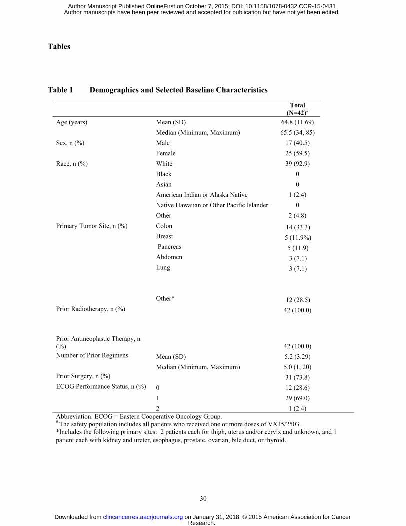
Patient demographics and baseline characteristics are summarized in Table 1 and were similar
across cohorts. Most enrolled patients were white, female, and had an ECOG status of 1 or 2 at
study entry. The most common primary tumor sites were colon (33.3%), breast (11.9%) and
pancreas (11.9%); lung (11.9%), liver and lymph node (7.1%) were the most common sites of
metastatic disease. All patients had undergone prior antineoplastic therapy with a mean of 5.2
Research. on January 31, 2018. © 2015 American Association for Cancerclincancerres.aacrjournals.org Downloaded from
Author manuscripts have been peer reviewed and accepted for publication but have not yet been edited. Author Manuscript Published OnlineFirst on October 7, 2015; DOI: 10.1158/1078-0432.CCR-15-0431
16
treatments (range: 1 to 20); most had prior radiotherapy (61.9%) and surgery (73.8%). The mean
time since last treatment with antineoplastic agents was 3.0 months (range: 1 to 31 months).
Safety and Tolerability
VX15/2503 treatment was well tolerated with the highest dose administered being 20 mg/kg; no
MTD was determined. No dose-related safety trends regarding incidence or severity were
observed. Twelve deaths, none treatment related, occurred during the study; 7 deaths were due
to disease progression and 5 were due to unknown causes. Patients received a mean of 2.9
cycles, (range: 1 to 14 cycles). The median duration of exposure was 73.4 days (range: 1 to 372
days) with a median number of doses and total dose administered of 8.0 doses (mean: 11; range:
1 to 54 doses) and 5411.6 mg (mean: 9987.7 mg; range: 44.0 to 64,800 mg), respectively.
Sixty-nine Treatment-Emergent Adverse Events (TEAEs) were considered possibly, probably or
definitely related to VX15/2503; 11 of these reported by 6 patients (14.3%) were definitely
related. As summarized in Table 2, 26 patients experienced treatment-related TEAEs, with the
most common being nausea (6/42, 14.3%) and fatigue (5/42, 11.9%). Six patients (14.3%)
experienced a TEAE that led to study discontinuation, with malignant neoplasm progression
being the only event reported in >1 patient (2/42, 4.8%). All treatment-related events were
Grade 1 or 2 except a Grade 3 γ-glutamyltransferase [GGT] elevation which was the sole DLT; it
was considered severe and possibly treatment related. This DLT was experienced at week three
by a pancreatic cancer patient (Cohort 6, 15 mg/kg) with progressive liver metastases. The
patient presented with a Grade 1 elevation at baseline; treatment was delayed at Week 4 due to
the DLT and the patient was discontinued from the study in week 5 (progressive disease). This
Research. on January 31, 2018. © 2015 American Association for Cancerclincancerres.aacrjournals.org Downloaded from
Author manuscripts have been peer reviewed and accepted for publication but have not yet been edited. Author Manuscript Published OnlineFirst on October 7, 2015; DOI: 10.1158/1078-0432.CCR-15-0431

17
DLT resulted in enrollment of 5 additional patients into Cohort 6; two discontinued in cycle 1
due to disease progression and no subsequent DLTs occurred in the remaining three patients.
Thus dose escalation proceeded to Cohort 7 (20 mg/kg).
A Grade 2 infusion-related reaction was experienced by patient 202004 in Cohort 2 (1.0 mg/kg)
who exhibited increasing HAHA titers from post-infusion 1 through the fourth and final weekly
infusion. Although this subject had received prior MAb therapy, no HAHA was evident at
baseline. This patient was discontinued because the HAHA response was neutralizing,
accelerating VX15/2503 clearance and reducing antibody-mediated PD effects. A high titer
response (>1:100 dilution) was observed in 5 other patients (11.9%) and a low-titer response in
18 (42.9%); neutralizing antibodies effects were not observed in these patients. However, one of
these 23 patients (Cohort 6; 15 mg/kg) experienced transient infusion-related reactions during
doses 2 and 3 that correlated temporally with the HAHA response. This subject received one
additional VX15/2503 infusion at a reduced rate with no infusion-related reaction.
Flow cytometric analyses of lymphocyte levels and lymphocyte subsets including T cells (total,
helper, or cytotoxic), B and NK cells showed no effects on these various populations following
weekly administration of VX15/2503, regardless of dose level.
Pharmacokinetics
PK samples collected after the initial antibody dose for the first 7 patients covered the time-
period from 0 to 168 or 240 hours, while those for all other subjects were collected through 96
hours; day 22 samples for all patients treated with four weekly doses were collected through 96
hours following the fourth dose. Using these data the effective half-life (t1/2 eff) values for weekly
Research. on January 31, 2018. © 2015 American Association for Cancerclincancerres.aacrjournals.org Downloaded from
Author manuscripts have been peer reviewed and accepted for publication but have not yet been edited. Author Manuscript Published OnlineFirst on October 7, 2015; DOI: 10.1158/1078-0432.CCR-15-0431

18
doses of VX15/2503 administered to subjects at all dose levels ranged from 2.7 to 6.9 days; t1/2 eff
values at the three highest dose levels ranged between 4.3 and 5.2 days.
Following the first administration of VX15/2503, the Cmax and total exposure (AUC0-168) ranged
from 3 µg/mL to 288 µg/mL and 109 µg*hr/mL to 26,023 µg*hr/mL across escalating dose
groups, respectively. Following 4 or 5 weekly doses of antibody, the Cmax and AUC0-168 ranged
from 3 µg/mL to 411 µg/mL and 131 µg*hr/mL to 38,351 µg*hr/mL, respectively. The Cmax and
AUC of VX15/2503 increased with dose level as shown in Fig.1. Data analyses showed that
increases in both Cmax and AUC were slightly higher than dose proportional as the 95%
confidence interval values for the slope terms were slightly greater than one (not shown). The
median Tmax values ranged from 2 to 4 hours across all dose levels. The accumulation ratio
values ranged from 1.0 to 2.0.
Pharmacodynamics and Clinical Biomarkers
Expected PD effects associated with the binding of VX15/2503 to cellular and soluble SEMA4D
were generally consistent with results from previous in vitro and in vivo studies (18). Weekly
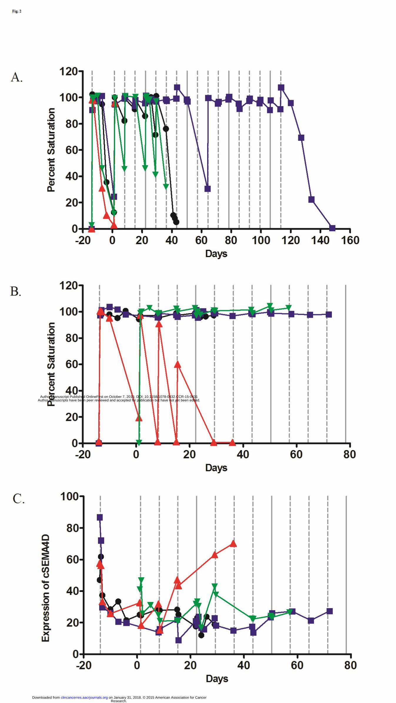
doses of VX15/2503 produced repeated transient cSEMA4D saturation for two of four patients
in the 0.3 mg/kg cohort (Fig 2A) (subjects 101001 and 201002); the third subject (101002)
exhibited continued maximal saturation. The fourth subject in this cohort (101003) received
only two infusions before discontinuing (disease progression). Cellular SEMA4D saturation
declined following cessation of treatment and antibody clearance from the periphery; saturation
values returned to baseline when VX15/2503 serum concentrations fell below the saturation
threshold of approximately ≤0.3 µg/mL (cf Fig. 2A, subjects 101001 and 101002).
Research. on January 31, 2018. © 2015 American Association for Cancerclincancerres.aacrjournals.org Downloaded from
Author manuscripts have been peer reviewed and accepted for publication but have not yet been edited. Author Manuscript Published OnlineFirst on October 7, 2015; DOI: 10.1158/1078-0432.CCR-15-0431
19
Fig. 2B shows that administration of doses of ≥1 mg/kg VX15/2503 produced complete,
sustained cSEMA4D saturation post-infusion for patients 102004, 202003 and 102005; these
cSEMA4D saturation data are representative of patients treated at higher VX15/2503 dose levels.
However, patient 202004, who developed a neutralizing HAHA response (cf Safety and
Tolerability discussion, above), exhibited declining cSEMA4D saturation values, attesting to the
increasing immune response following each infusion.
Fig. 2C shows that peripheral T cell cSEMA4D expression declined within 24 hours post-first
infusion due to internalization of the antibody/receptor complex; data shown are for cohort 2
patients 102004, 202003, 202004 and 102005 treated with 1 mg/kg of VX15/2503. Results at
higher VX15/2503 dose levels were similar. A 60% reduction of cSEMA4D expression was
generally sustained during treatment intervals of up to 1 year (not shown). Cellular SEMA4D
expression remained suppressed until VX15/2503 serum levels declined following cessation of
dosing, allowing cSEMA4D levels to return to baseline (not shown). Similar results were
observed following a neutralizing anti-VX15/2503 immune response; see Cohort 2 patient
202004 (Fig. 2C) whose cSEMA4D levels rose to baseline.
Levels of total sSEMA4D (free and antibody-complexed ligand) in sera increased with dose
level and with infusion number at a given dose level, consistent with the expected increased
half-life of the VX15/2503-soluble SEMA4D complex versus that of the free soluble receptor (cf
9.0 mg/kg patients; Supplementary Fig. S1). Levels appeared to reach steady-state after 8 to 10
weekly doses (Patients 105009 and 205011); patients dosed for shorter periods did not reach
steady-state. Total sSEMA4D levels declined after treatment cessation and antibody clearance
(Supplementary Fig. S1), as illustrated by patients 105009, 205011 and 205012, who received
their last dose on Days 330, 170 and 50, respectively. These patients’ SEMA4D levels were
Research. on January 31, 2018. © 2015 American Association for Cancerclincancerres.aacrjournals.org Downloaded from
Author manuscripts have been peer reviewed and accepted for publication but have not yet been edited. Author Manuscript Published OnlineFirst on October 7, 2015; DOI: 10.1158/1078-0432.CCR-15-0431

20
analyzed ≥ 20 days after EOT and data show sSEMA4D levels approaching baseline for patient
105009. Similar results were obtained for other patients (not shown).
No pharmacologic effects of VX15/2503 administration were observed on the levels of serum
VEGF, PLGF, MET, or HGF as their levels were unaffected by dose level or dose number (data
not shown). Similarly, an absence of pharmacologic effects was noted for chromogranin A,
bone-specific alkaline phosphatase and urine N-telopeptide assessed for expansion cohort
patients with pancreatic neuroendocrine tumors or bone metastases, respectively (not shown).
Anti-Tumor Activity
Patient PFS by cohort and by tumor type are shown in Fig.3; tumor diagnosis and reasons for
patient discontinuation are also shown. No complete responses were observed. The median
duration of stable disease and the duration of PFS were 7.82 weeks (range: 0.57 to 54.8 weeks).
One of 15 patients treated at 20 mg/kg (papillary thyroid cancer) experienced a partial clinical
response of 334 days (47.7 weeks) that continued at study exit (physician decision; study Day
337). A hilar lymph node in this subject decreased from 1.5 cm to 0.8 cm, representing a 53.3%
reduction. This patient had received 8 different prior therapies before entering this study. The
longest period between prior treatment regimens was 37 months (between regimens one and
two).
Nineteen patients (45.2%) exhibited no evidence of disease progression for at least 8 weeks
(Fig.3). Of these, 13 patients (31%) exhibited no evidence of disease progression for between 8
and 16 weeks, and 6 patients (14.3%), 5 of whom were in the ≥6 mg/kg cohorts, also exhibited
no evidence of disease progression for ≥16 weeks. The longest durations of stable disease were
observed for patients with colorectal (9 mg/kg; 48 weeks) and breast cancer (15 mg/kg; 55
Research. on January 31, 2018. © 2015 American Association for Cancerclincancerres.aacrjournals.org Downloaded from
Author manuscripts have been peer reviewed and accepted for publication but have not yet been edited. Author Manuscript Published OnlineFirst on October 7, 2015; DOI: 10.1158/1078-0432.CCR-15-0431

21
weeks). These patients had received, respectively, three and 6 prior therapies; their respective
longest prior periods of stable disease were, 37 and 28 months.
Although assessed lymphocyte levels remained generally unchanged from baseline during
treatment with VX15/2503, the normalized baseline number of B cells plus T cells for patients in
the 9.0 to 20 mg/kg cohorts exhibited a significant correlation (Spearman’s rank coefficient rs =
0.6133; p≤0.001) with their PFS duration (Fig. 4). Weaker but significant (p≤0.03) correlations
were observed for baseline values of absolute lymphocytes (rs = 0.4195), T cells (rs = 0.4315),
and B cells (rs = 0.4256). Three patients with PFS of 48 to 55 weeks and four patients who
exhibited the absence of disease progression for at least 16 weeks all had relatively high T- or B-
cells at baseline (T-cells ≥1200/µL; B-cells ≥250/µL); these subjects were treated with
VX15/2503 doses of 6 mg/kg or higher [one patient in cohort 1 with SD of > 18 weeks exhibited
baseline lymphocyte levels below these values].
Discussion
VX15/2503 was well tolerated in advanced solid tumor patients when administered as a weekly
IV infusion. The highest planned dose of 20 mg/kg was employed and no MTD was determined.
No dose-related safety trends regarding adverse event incidence or severity were observed. The
most common (>10% incidence) treatment-related adverse events were nausea and fatigue. All
treatment-related adverse events were Grade 1 or 2 except the sole reported DLT, a Grade 3 GGT
elevation experienced in a 15 mg/kg cohort patient with progressive liver metastases who had
elevated GGT levels at baseline. After further review the DLT was considered only temporally
related with VX15/2503 administration and more likely caused by underlying disease.
Research. on January 31, 2018. © 2015 American Association for Cancerclincancerres.aacrjournals.org Downloaded from
Author manuscripts have been peer reviewed and accepted for publication but have not yet been edited. Author Manuscript Published OnlineFirst on October 7, 2015; DOI: 10.1158/1078-0432.CCR-15-0431
22
Twenty-three of the patients developed anti-VX15/2503 antibody responses, with a response titer
> 100 detected in six patients (14.3%). Two patients [one with a high-titer response (Cohort 2)
and one with a low-titer response (Cohort 5)] developed Grade 2 infusion-related reactions that
were temporally related with the HAHA response. Only the Cohort 2 subject produced a
neutralizing infusion reaction, which not only cleared administered antibody from the periphery
but also neutralized VX15/2503-mediated effects on cSEMA4D saturation and expression, and
reduced sSEMA4D levels. These findings resulted in treatment termination and study
discontinuation for this patient.
Pharmacologic effects were similar to those observed in primates treated at similar VX15/2503
dose levels (18). Lymphocyte levels generally remained unchanged from baseline, regardless of
antibody dose level or number. The threshold for cSEMA4D saturation of human T cells was
estimated to be approximately 0.3 µg/mL, similar to that determined for primate T cells (0.5
µg/mL) (18). Furthermore, the expression of cSEMA4D on T lymphocytes declined for all
patients during treatment; similar receptor internalization has been reported for CD20 on B
lymphocytes (25). Cellular SEMA4D levels in fresh tumor specimens were not assessed as no
patients consented to biopsies.
Levels of total sSEMA4D present in the sera of treated patients increased with dose level and
with infusion number (Supplementary Fig. S1), consistent with the increased half-life of the
VX15/2503-sSEMA4D complex versus that of the soluble ligand. Similar results were reported
for secreted VEGF (26). While sSEMA4D levels declined following treatment cessation, study
discontinuation after EOT generally precluded following patients’ sSEMA4D levels return to
baseline. Data from cohort 1 or 2 patients did show normalization of sSEMA4D levels, however
Research. on January 31, 2018. © 2015 American Association for Cancerclincancerres.aacrjournals.org Downloaded from
Author manuscripts have been peer reviewed and accepted for publication but have not yet been edited. Author Manuscript Published OnlineFirst on October 7, 2015; DOI: 10.1158/1078-0432.CCR-15-0431
23
(not shown). Finally, no serum biomarkers were identified as the levels of these proteins
remained unchanged with VX15/2503 treatment.
This study was not designed to collect extensive PK data; nonetheless sufficient samples
allowing for complete PK analyses were obtained from the first seven patients (0.3 and 1.0
mg/kg dose cohorts) and PK parameters were estimated for the remaining patients using data
derived from the limited samples available. Thus the effective half-life estimates of 2.7 to 6.9
days for the dose range of 3 to 20 mg/kg VX15/2503 may not reflect the true half-life since these
values were determined with samples collected over the more limited time period of 0 to 96
hours. The Cmax and AUC0-168 values, determined following multiple weekly doses of
VX15/2503, were reliably determined and exhibited a slightly greater than dose proportional
increase with increasing dose level. Finally, little or no accumulation occurred after multiple
weekly doses of VX15/2503 because of the low accumulation ratio values.
Exploratory evaluations of efficacy included anti-tumor activity and PFS. Nineteen patients
(45.2%) exhibited no evidence of disease progression for at least 8 weeks and 8 (19%) showed a
similar absence of disease progression for at least 16 weeks. One patient with papillary thyroid
cancer (20 mg/kg cohort) achieved a partial response of 47.7 weeks that was ongoing at study
exit. This patient along with the 2 other patients with the longest PFS (48-55 weeks; all in the 9
to 20 mg/kg cohorts) and 4 patients who had stable disease for ≥16 weeks (all in the 6 mg/kg
cohorts and above) all had relatively high T- or B-cells at baseline.
The observed strong correlation between the normalized baseline number of B cells plus T cells
and PFS for patients in the 9.0 to 20 mg/kg cohorts suggested that these heavily pre-treated
patients entered the study with a more robust immune potential despite their prior therapy.
Research. on January 31, 2018. © 2015 American Association for Cancerclincancerres.aacrjournals.org Downloaded from
Author manuscripts have been peer reviewed and accepted for publication but have not yet been edited. Author Manuscript Published OnlineFirst on October 7, 2015; DOI: 10.1158/1078-0432.CCR-15-0431

24
Nevertheless, this elevated immune potential was not of itself sufficient to inhibit tumor growth
as all patients had tumors that were progressing at the time they enrolled in the trial. Tumor
stabilization and extended PFS in these patients was only induced following treatment with
VX15/2503. This is consistent with preclinical studies in which striking immune-mediated anti-
tumor effects were observed in tumor-bearing mice treated with the murine anti-SEMA4D
antibody MAb 67-2 alone or in combination with checkpoint inhibitors (17). These preclinical
studies demonstrated that neutralization by anti-SEMA4D antibody of SEMA4D residing at the
tumor margin allowed enhanced trafficking of tumoricidal macrophage and activated
CD8+lymphocytes into the tumor microenvironment. The neutralization of SEMA4D thus
facilitated the infiltration of immune cells into the tumor microenvironment, thereby engendering
an effective anti-tumor response. Although patients in the present study did not volunteer fresh
biopsies to permit examination of SEMA4D expression and immune cell distribution in tumor, a
recently published report characterized a similar gradient of SEMA4D expression in human
colorectal carcinoma (27).
The findings from this first-in-human study support the further investigation of VX15/2503 for
the treatment of advanced refractory solid tumors, as it was well tolerated at all doses evaluated,
demonstrated expected PD effects, and 45% of patients exhibited the absence of disease
progression for at least 8 weeks. Targeting SEMA4D represents a novel therapeutic strategy to
promote immune infiltration into tumor that is complementary to the mechanism of actions of
other immunomodulatory therapies. Administration of VX15/2503 in combination with immune
enhancing therapies such as checkpoint blockade inhibitors or vaccines that enhance overall
immune response but do not affect ability to penetrate tumor may result in increased efficacy.
Research. on January 31, 2018. © 2015 American Association for Cancerclincancerres.aacrjournals.org Downloaded from
Author manuscripts have been peer reviewed and accepted for publication but have not yet been edited. Author Manuscript Published OnlineFirst on October 7, 2015; DOI: 10.1158/1078-0432.CCR-15-0431

25
Future studies in selected tumor types will utilize VX15/2503 in combination with other
immunomodulatory agents.
Research. on January 31, 2018. © 2015 American Association for Cancerclincancerres.aacrjournals.org Downloaded from
Author manuscripts have been peer reviewed and accepted for publication but have not yet been edited. Author Manuscript Published OnlineFirst on October 7, 2015; DOI: 10.1158/1078-0432.CCR-15-0431

26
References
1. Peranzoni E, Rivas-Caicedo A, Bougherara H, Salmon H, and Donnadieu E. Positive and
negative influence of the matrix architecture. Cell Mol Life Sci 2013;70:4431-48.
2. Carmeliet P. Angiogenesis in health and disease. Nat Med 2003;9:653-60.
3. Atreya I, Neurath MF. Immune cells in colorectal cancer: Prognostic relevance and
therapeutic strategies. Expert Rev Anticancer Ther 2008;8:561-72.
4. Lizée G, Overwijk WW, Radvanyi L, Gao J, Sharma P, and Hwu P. Harnessing the power of
the immune system to target cancer. Ann RevMed 2013;64:71-90.
5. Delaire S, Billard C, Tordjman R, Chédotal A, Elhabazi A, Bensussan A, et al. Biological
activity of soluble CD100: Soluble CD100, similarly to H-SemaIII, inhibits immune cell
migration. J Immunol 2001;166:4348-54.
6. Ch’ng ES and Kumanogoh A. Roles of SEMA4D and plexin B1in tumor progression. Molec
Can 2010;9:251-60.
7. Giraudon P, Vincent P, Vuaillat C, Verlaeten O, Cartier L, Marie-Cardine A, et al.
Semaphorin CD100 from activated T lymphocytes induces process extension collapse in
oligodendrocytes and death of immature neural cells. J Immunol 2004;172:1246-55.
8. Giraudon P, Vincent P, and Vuaillat C. T-cells in neuronal injury and repair: semaphorins
and related T-cell signals. Neuro Mol Med 2005;7:207-16.
9. Worzfield T, Swiercs JM, Looso M, Straub BK, Sivaraj KK and Offermanns S. ErbB-2
signals through plexin-B1 to promote breast cancer metastasis. J Clin Invest 2012;122:1296-
05.
10. Giordano S, Corso S, Conrotto P, Artigiani S, Gilestro G, Barberis D, et al. The semaphorin
4D receptor controls invasive growth by coupling with Met. Nat Cell Biol 2002;4:720-24.
Research. on January 31, 2018. © 2015 American Association for Cancerclincancerres.aacrjournals.org Downloaded from
Author manuscripts have been peer reviewed and accepted for publication but have not yet been edited. Author Manuscript Published OnlineFirst on October 7, 2015; DOI: 10.1158/1078-0432.CCR-15-0431
27
11. Conrotto P, Valdembri SC, Serini G, Tamagnone L, Comoglio PM, Bussolino F, et al.
Sema4D induces angiogenesis through Met recruitment by Plexin B1. Blood 2005;105:4321-
29.
12. Witherden DA, Watanabe M, Garijo O, Rieder SE, Sarkisyan G, Cronin SJF, et al. The
CD100 receptor interacts with its plexin B2 ligand to regulate epidermal γδ T cell function.
Immunity 2012;37:314–25.
13. Hall KT, Boumsell L, Schultze JL, Boussiotis VA, Dorfman DM, Cardoso, AA, et al.
Human CD100, a novel leukocyte semaphorin that promotes B-cell aggregation and
differentiation. Proc Natl Acad Sci USA 1996;93:11780-85.
14. Basile JR, Castilho RM, Williams VP, and Gutkind JS. Semaphorin 4D provides a link
between axon guidance processes and tumor-induced angiogenesis. Proc Natl Acad Sci USA
2006;103:9017-22.
15. Campos M, Campos SG, Ribeiro GG, Eguchi FC, Da Silva SR, De Oliveira CZ, et al. Ki-67
and CD100 immunohistochemical expression is associated with local recurrence and poor
prognosis in soft tissue sarcomas, respectively. Oncol Lett 2013;5:1527-35.
16. Kato S, Kubota K, Shimamura T, Shinohara Y, Kobayashi N, Watanabe S, et al. Semaphorin
4D, a lymphocyte semaphorin, enhancestumor cell motility through binding its receptor,
plexinB1, in pancreatic cancer. Can Sci 2011;102:2029-37.
17. Evans E., Jonason A, Bussler H, Torno, S, Veeraraghavan J, Reilly C, et al. Antibody
blockade of Semaphorin 4D promotes immune infiltration into tumor and enhances response
to other immunomodulatory therapies. Can Immunol Res January 22, 2015; DOI:
10.1158/2326-6066.CIR-14-0171.
Research. on January 31, 2018. © 2015 American Association for Cancerclincancerres.aacrjournals.org Downloaded from
Author manuscripts have been peer reviewed and accepted for publication but have not yet been edited. Author Manuscript Published OnlineFirst on October 7, 2015; DOI: 10.1158/1078-0432.CCR-15-0431

28
18. Leonard JE, Fisher TE, Winter L, Cornelius C, Seils J, Reilly C, et al. Nonclinical Safety
Evaluation of VX15/2503: a Humanized IgG4 Anti-SEMA4D Antibody. Molec Can Therap
2015; 14; 964–72.
19. Smith E, Jonason A, Reilly C, Veeraraghavan J, Fisher T, Doherty M, et al. SEMA4D
compromises blood-brain barrier, activates microglia and inhibits remyelination in
neurodegenerative disease. Neurobiol of Disease 2015;73:254-68.
20. Labrijn AF, Buijsse AO, van den Bremer ET, Verwilligen AY, Bleeker WK, Thorpe SJ, et al.
Therapeutic IgG4 antibodies engage in Fab-arm exchange with endogenous human IgG4 in
vivo. Nat Biotechnol 2009;27:767-71.
21. Bourdage J, Cook C, Farrington DL, Chain JS, and Konrad RJ. An affinity capture elution
(ACE) assay for detection of anti-drug antibody to monoclonal antibody therapeutics in the
presence of high levels of drug. J Immunol Meth 2007;327:10-17.
22. Smith BP, Vandenhende FR, DeSante KA, Farid NA, Welch PA, Callaghan JT, et al.
Confidence interval criteria for assessment of dose proportionality. Pharm Res.
2000;17:1278-83.
23. Strossberg JR, Weber JM, Feldman M, Coppola D, Meredith K and Kvols LK. Prognostic
validity of the American Joint Committee on cancer staging classification for midgut
neuroendocrine tumors. J Clin Onc 2013;31:420-25.
24. Bergmann P, Body J-J, Boonen S, Boutsen Y, Devogelaer J-P, Goemaere S, et al. Evidence-
based guidelines for the use of biochemical markers of bone turnover in the selection and
monitoring of bisphosponate treatment in osteoporosis: a consensus document of the Belgian
Bone Club. Int J Clin Pract 2009;63:19-26
Research. on January 31, 2018. © 2015 American Association for Cancerclincancerres.aacrjournals.org Downloaded from
Author manuscripts have been peer reviewed and accepted for publication but have not yet been edited. Author Manuscript Published OnlineFirst on October 7, 2015; DOI: 10.1158/1078-0432.CCR-15-0431
29
25. Beers SA, French RR, Chan HTC, Lim SH, Jarrett TC, Vidal RM, et al. Antigenic
modulation limits the efficacy of anti-CD20 antibodies: implications for antibody selection.
Blood 2010;24: 5191-01.
26. Gordon MS, Margolin K, Talpaz M, Sledge GW Jr, Holmgren E, Stalter S, et al. Phase I
safety and pharmacokinetic study of recombinant human anti-vascular endothelial growth
factor in patients with advanced cancer. J Clin Oncol 2001;19:843-50.
27. Jin-Shen Wang, Chang-Qing Jing, Ke-Shu Shan, Yue-Zhi Chen, Xiao-Bo Guo, Zhi-Xin Cao,
et al. Semaphorin 4D and hypoxia-inducible factor-1α overexpression is related to prognosis
in colorectal carcinoma. World J Gastroenterol 2015; 21: 2191-98.
Research. on January 31, 2018. © 2015 American Association for Cancerclincancerres.aacrjournals.org Downloaded from
Author manuscripts have been peer reviewed and accepted for publication but have not yet been edited. Author Manuscript Published OnlineFirst on October 7, 2015; DOI: 10.1158/1078-0432.CCR-15-0431
30
Tables
Table 1 Demographics and Selected Baseline Characteristics
Total (N=42)#
Age (years) Mean (SD) 64.8 (11.69) Median (Minimum, Maximum) 65.5 (34, 85) Sex, n (%) Male 17 (40.5) Female 25 (59.5) Race, n (%) White 39 (92.9) Black 0 Asian 0 American Indian or Alaska Native 1 (2.4) Native Hawaiian or Other Pacific Islander 0 Other 2 (4.8) Primary Tumor Site, n (%) Colon 14 (33.3) Breast 5 (11.9%) Pancreas 5 (11.9) Abdomen 3 (7.1) Lung 3 (7.1) Other* 12 (28.5) Prior Radiotherapy, n (%) 42 (100.0) Prior Antineoplastic Therapy, n (%) 42 (100.0) Number of Prior Regimens Mean (SD) 5.2 (3.29) Median (Minimum, Maximum) 5.0 (1, 20) Prior Surgery, n (%) 31 (73.8) ECOG Performance Status, n (%) 0 12 (28.6) 1 29 (69.0) 2 1 (2.4) Abbreviation: ECOG = Eastern Cooperative Oncology Group. # The safety population includes all patients who received one or more doses of VX15/2503. *Includes the following primary sites: 2 patients each for thigh, uterus and/or cervix and unknown, and 1 patient each with kidney and ureter, esophagus, prostate, ovarian, bile duct, or thyroid.
Research. on January 31, 2018. © 2015 American Association for Cancerclincancerres.aacrjournals.org Downloaded from
Author manuscripts have been peer reviewed and accepted for publication but have not yet been edited. Author Manuscript Published OnlineFirst on October 7, 2015; DOI: 10.1158/1078-0432.CCR-15-0431

31
Table 2. Treatment-Related Adverse Events in ≥5% of Patients
Adverse Event or DLT Patients reporting, n (%)
Cohort 1 0.3 mg/kg
n=4
Cohort 2 1.0 mg/kg
n=4
Cohort 3 3.0 mg/kg
n=3
Cohort 4 6.0 mg/kg
n=4
Cohort 5 9.0 mg/kg
n=4
Cohort 6 15.0 mg/kg
n=8
Cohort 7/82 20.0 mg/kg
n=15
Total
N =42
Any treatment-emergent AE 4 (100.0) 4 (100.0) 3 (100.0) 4 (100.0) 4 (100.0) 8 (100.0) 15 (100.0) 42 (100.0)
Any treatment-related AE 1 (25.0) 2 (50.0) 2 (67.0) 3 (75.0) 1 (25) 6 (75.0) 11 (73.3) 26 (61.9)
Nausea 0 0 0 1 (25.0) 1 (25.0) 0 4 (26.7) 6 (14.3)
Fatigue 1 (25.0) 0 0 1 (25.0) 1 (25.0) 0 2 (13.3) 5 (11.9)
Arthralgia 0 0 0 0 1 (25.0) 2 (25.0) 0 3 (7.1)
Decreased Appetite 0 1 (25.0) 1 (33.3) 0 0 1 (12.5) 0 3 (7.1)
Infusion-Related Reaction 0 1 (25.0) 0 0 0 1 (12.5) 1 (6.7) 3 (7.1)
Pyrexia 0 0 0 1 (25.0) 0 0 2 (13.3) 3 (7.1)
Myalgia 0 0 0 1 (25.0) 0 0 1 (6.7) 2 (4.8)
Pruritus 0 0 0 1 (25.0) 0 1 (12.5) 0 2 (4.8)
DLT: Grade ≥3 elevated γ-glutamyltransferase 1
0 0 0 0 0 1 0 1 (2.4)
Abbreviations: AE, adverse event; DLT, dose-limiting toxicity
NOTE: The safety population includes all patients who received at least one dose of VX15/2503. 1This Grade 3event was a dose-limiting toxicity. No other DLTs were reported. All other events were Grade 1 or 2. 2 Includes patients from dose-escalation cohort (cohort 7) as well as patients from the expansion cohort.
Research.
on January 31, 2018. © 2015 A
merican A
ssociation for Cancer
clincancerres.aacrjournals.org D
ownloaded from
Author m
anuscripts have been peer reviewed and accepted for publication but have not yet been edited.
Author M
anuscript Published O
nlineFirst on O
ctober 7, 2015; DO
I: 10.1158/1078-0432.CC
R-15-0431

32
Table Legends
Table 1. Demographics and Selected Baseline Characteristics
See table footnotes
Table 2. Treatment-Related Adverse Events in ≥5% of Patients
See table footnotes
Fig. Legends
Fig.1. Cmax and AUC Increase Linearly with VX15/2503 Dose Level. Cmax (µg/mL) and
AUC (ng*hr/mL) were plotted as a function of VX15/2503 dose level over the range of 0.3 to 20
mg/kg; a statistical analysis of dose proportionality was performed using the power model (22).
The equation ln (parameter) = a + b*ln (dose) + error, was used to estimate the slope and
corresponding 95% confidence interval.
Fig. 2. T-Cell Associated SEMA4D Saturation and Cellular Expression and the Impact of
Anti-VX15/2503 Immune Response. (A) Saturation analysis from Cohort 1 (0.3 mg/kg). The
percent saturation by VX15/2503 of cSEMA4D on peripheral blood CD3+ T cells for subjects in
the 0.3 mg/kg dose cohort was determined using a flow cytometric assay; values of ≥20%
indicated saturation of cSEMA4D. The four subjects in this cohort all received a sentinel dose.
The symbols represent: Patient No. 101001, Patient No. 101002; Patient No. 101003;
Patient No. 201002. (B) cSEMA4D saturation analysis from Cohort 2 (1.0 mg/kg). The impact
of a neutralizing anti-VX15/2503 immune response on T cell SEMA4D saturation is illustrated
by the data for Cohort 2 patient 202004; all Cohort 2 subjects except patient 102005 received a
sentinel dose. The symbols represent: Patient No. 102004; Patient No. 202003; Patient
Research. on January 31, 2018. © 2015 American Association for Cancerclincancerres.aacrjournals.org Downloaded from
Author manuscripts have been peer reviewed and accepted for publication but have not yet been edited. Author Manuscript Published OnlineFirst on October 7, 2015; DOI: 10.1158/1078-0432.CCR-15-0431

33
No. 202004; Patient No. 102005. (C) Cellular SEMA4D expression analysis for Cohort 2 (1.0
mg/kg). Expression of cSEMA4D on purified peripheral blood CD3+ T cells was measured by
flow cytometry for samples collected from patients in Cohort 2. The symbols represent:
Patient No. 102004; Patient No. 202003; Patient No. 202004; Patient No. 102005.
Fig. 3. Duration of Progression-Free Survival for Enrolled Patients by Dose Cohort.
Duration of time on study is plotted for each patient by dose of VX15/2503. The primary site
and tumor type, as determined from the Cancer History listings, are provided for each patient.
Study termination reasons are shown in parentheses; these were (1), disease progression; (2)
patient withdrawal; (3) death; (4) physician decision; and (5) AE. Asterisks indicate a sentinel-
dose patient. PFS data were taken from the Derived Efficacy Variables (Safety Population)
listing, which lists duration of stable disease (in months). A value of 30.4 days per month was
used to calculate PFS in weeks. Patients with 7.99 weeks duration were considered to have 8
weeks PFS. Two patients in Cohort 6 [Patient 000106015 (ovarian carcinoma) and Patient
000206015 (colon carcinoma)] were excluded from the analysis because they were withdrawn
from the study before the first post-baseline efficacy assessment. Abberviations: L. – left; PR -
partial response; R – right; Unk. - unknown.
Fig. 4. Spearman’s Rank-Order Correlation between the Normalized Number of B Cells
plus T Cells and Progression-Free Survival.
The number of T cells and B cells at baseline were measured by flow cytometry and absolute
lymphocyte counts were determined by standard techniques. A. Progression free survival
Research. on January 31, 2018. © 2015 American Association for Cancerclincancerres.aacrjournals.org Downloaded from
Author manuscripts have been peer reviewed and accepted for publication but have not yet been edited. Author Manuscript Published OnlineFirst on October 7, 2015; DOI: 10.1158/1078-0432.CCR-15-0431

34
(determined as the number of weekly VX15/2503 doses administered until disease progression)
is plotted versus the number of normalized B cells plus T cells for subjects in the 9, 15 and 20
mg/kg dose cohorts (N=27). Panels B, C, and D show, respectively, PFS versus the number of T
lymphocytes, B lymphocytes, or absolute lymphocyte counts at baseline. The respective
Spearman’s rank-order correlation coefficients (rs) and p values for each analysis are provided.
Research. on January 31, 2018. © 2015 American Association for Cancerclincancerres.aacrjournals.org Downloaded from
Author manuscripts have been peer reviewed and accepted for publication but have not yet been edited. Author Manuscript Published OnlineFirst on October 7, 2015; DOI: 10.1158/1078-0432.CCR-15-0431

Published OnlineFirst October 7, 2015.Clin Cancer Res Amita Patnaik, Glen J Weiss, John E Leonard, et al. Study of Patients with Advanced Solid TumorsHumanized anti-Semaphorin 4D Antibody, in a First-In-Human Safety, Pharmacokinetics and Pharmacodynamics of a
Updated version
10.1158/1078-0432.CCR-15-0431doi:
Access the most recent version of this article at:
Material
Supplementary
http://clincancerres.aacrjournals.org/content/suppl/2016/02/16/1078-0432.CCR-15-0431.DC1
Access the most recent supplemental material at:
Manuscript
Authoredited. Author manuscripts have been peer reviewed and accepted for publication but have not yet been
E-mail alerts related to this article or journal.Sign up to receive free email-alerts
Subscriptions
Reprints and
To order reprints of this article or to subscribe to the journal, contact the AACR Publications
Permissions
Rightslink site. Click on "Request Permissions" which will take you to the Copyright Clearance Center's (CCC)
.http://clincancerres.aacrjournals.org/content/early/2015/10/07/1078-0432.CCR-15-0431To request permission to re-use all or part of this article, use this link
Research. on January 31, 2018. © 2015 American Association for Cancerclincancerres.aacrjournals.org Downloaded from
Author manuscripts have been peer reviewed and accepted for publication but have not yet been edited. Author Manuscript Published OnlineFirst on October 7, 2015; DOI: 10.1158/1078-0432.CCR-15-0431