Embed Size (px)
Citation preview
Regulation of semaphorin 4D expression andcell proliferation of ovarian cancer by
ERalpha and ERbeta
Y. Liu1*, Y. Hou1*, L. Ma2, C. Sun1, J. Pan1, Y. Yang1, H. Zhou1 and J. Zhang3
1Department of Gynecology, the Second Affiliated Hospital, Kunming Medical University, Kunming, Yunnan, China2Department of Reproduction and Genetics, the Second Affiliated Hospital, Kunming Medical University, Kunming, Yunnan, China
3Department of General Surgery, the Second Affiliated Hospital, Kunming Medical University, Kunming, Yunnan, China
Abstract
Ovarian cancer is one of the most common malignancies in women. Semaphorin 4D (sema 4D) is involved in the progress ofmultiple cancers. In the presence of estrogen-like ligands, estrogen receptors (ERa and ERb) participate in the progress ofbreast and ovarian cancers by transcriptional regulation. The aim of the study was to investigate the role of sema 4D andelucidate the regulatory pattern of ERa and ERb on sema 4D expression in ovarian cancers. Sema 4D levels were up-regulatedin ovarian cancer SKOV-3 cells. Patients with malignant ovarian cancers had significantly higher sema 4D levels than controls,suggesting an oncogene role of sema 4D in ovarian cancer. ERa expressions were up-regulated in SKOV-3 cells compared withnormal ovarian IOSE80 epithelial cells. Conversely, down-regulation of ERb was observed in SKOV-3 cells. Forced over-expression of ERa and ERb in SKOV-3 cells was manipulated to establish ERa+ and ERb+ SKOV-3 cell lines. Incubation ofERa+ SKOV-3 cells with ERs agonist 17b-estradiol (E2) significantly enhanced sema 4D expression and rate of cellproliferation. Incubated with E2, ERb+ SKOV-3 cells showed lower sema 4D expression and cell proliferation. Blocking ERaand ERb activities with ICI182-780 inhibitor, sema 4D expressions and cell proliferation of ERa+ and ERb+ SKOV-3 cells wererecovered to control levels. Taken together, the data showed that sema 4D expression was positively correlated with theprogress of ovarian cancer. ERa positively regulated sema 4D expression and accelerated cell proliferation. ERb negativelyregulated sema 4D expression and inhibited cell multiplication.
Key words: Semaphorin 4D expression; ERa; ERb; Ovarian cancer; Cell proliferation
Introduction
Ovarian cancer is the most common cause of deathin women with gynecological malignancies, and it hasthe highest mortality rate among women in the world.Because of the lack of definitive early symptoms andefficient, specific and sensitive markers for ovarian cancermonitoring, the majority of patients diagnosed with late-stage ovarian cancer typically have a 5-year survival of30% (1,2). Prevention and cure of ovarian cancer is stilla great challenge.
However, solid evidence has established that steroid-resembling estrogen is related to increased breast cancerrisk (3–5). Steroid estrogen analogues are still the mostcommon drugs used to relieve the menopausal symptomsof women in menopausal hormone therapy (MHT) (6,7).However, emerging data in recent years suggest thatestrogen is also implicated in the progression of ovarian
cancer. Epidemiological studies reveal that long-term useof estrogen-only hormone replacement therapy increasesa woman’s risk of ovarian cancer (8,9). Utilization of anti-estrogen intervention inhibits the growth of ovarian carci-noma in vitro and in vivo (10,11). Data from a rat modeldemonstrated the growth promoting effects of estrogen onovarian tumors in mice (12,13). Estrogen plays a physi-ological role through estrogen receptors (ERs) mediation.There are two ERs isoforms, ERa and ERb, which aremembers of the nuclear receptors superfamily of ligand-dependent transcription factors (14). Although ERa andERb have structural and functional homologies, they mayregulate the same genes in opposite ways, following ayin-yang hypothesis (15,16). In general, ERa is seen asan oncogene by promoting gene expression related tosurvival and proliferation of cancer cells. Whereas ERb is
Correspondence: H. Zhou: <[email protected]>
*These authors contributed equally to this study.
Received November 14, 2016 | Accepted December 13, 2016
Braz J Med Biol Res | doi: 10.1590/1414-431X20166057
Brazilian Journal of Medical and Biological Research (2017) 50(3): e6057, http://dx.doi.org/10.1590/1414-431X20166057ISSN 1414-431X 1/10

usually described as tumor suppressor by having anti-proliferation and pro-apoptosis effects. The role of ERain breast cancer have been well established, but the ER’srole in ovarian cancer is still relatively vague by com-parison (17).
The semaphorins family are grouped into eight sub-classes and contain more than 30 members, which wereinitially identified as constituents of the nervous systemcomplex that direct the growing axons to their targets (18).However, recent evidence indicates that expression levelsof certain members of semaphorins, such as semaphorin4D, semaphorin 5C, and semaphorin 6B etc., were alteredin a variety of cancer cells. These semaphorins promotedtumor angiogenesis and increased tumor progression(19–21). Semaphorin 4D, a member of class 4 semaphor-ins and also known as CD 100, has been shown to beup-regulated in aggressive cervical, head and neck, pros-tate, colon, breast, and lung cancers (22). Under theinteraction with its receptor plexin B, sema 4D facilitatedthe growth, invasion, and migration of cancer cells, promot-ing carcinogenesis and metastasis (23). Although manygenes have been identified to be sema 4D-regulatedtargets in cancer cells, the up-stream regulatory mecha-nism of sema 4D is rarely explored (22). Whether sema4D involvement in the progress of ovarian cancer is reg-ulated by ERa and ERb is still unclear.
In the present study, we detected the expression of sema4D, ERa, and ERb in ovarian cancer tissues and cells.
Material and Methods
Cells and regentsDMEM medium was purchased from Hyclone
(SH30243.01B, USA). RNase, DNase, and DNA marker(Takara) were purchased from Shanghai Bito Co. Ltd.,China. Methanol, haematine, ICI182-780 (ERs inhibitor),17b-estradiol (E2), and eosin were purchased from SigmaCo. (USA). Sixty normal ovarian tissues, 60 benign ovar-ian cancer tissues, and 60 malignant ovarian cancer tis-sues were obtained from Second Affiliated Hospital ofKunming Medical University. The malignancies wereclassified into early phase (I-II) and terminal stage (III-IV)on the basis of surgical-pathologic staging FIGO, 2006.All ovarian cancer tissues were histologically confirmedby two pathologists. Patients did not receive medicationbefore surgery. The collection of human tissue sampleswas approved and supervised by the Ethics Committeeof Kunming Medical University. Human ovarian cancerSKOV-3 cells and human normal ovarian IOSE80 epithe-lial cells were purchased from Yingrun Biological Co. Ltd.,China. 293TA cell line was purchased from Funeng Bio-logical Co. Ltd., China.
Cell cultureSKOV-3 and IOSE80 cell lines were cultured in RPMI
1640 medium supplemented with 10% (v/v) fetal bovine
serum (FBS, HyClone). 293T cell lines were cultured inDulbecco’s Modified Eagle Medium (DMEM, HyClone)containing 10% (v/v) FBS, 100 units/mL of penicillin-strep-tomycin (Invitrogen, USA), and HOSE (Pricells, China).All cell lines were cultured in a humidified incubator in anatmosphere of 5% CO2 and 95% air at 37°C.
Real-time (RT) PCRTotal RNA was isolated using Trizol (Invitrogen) accord-
ing to the manufacturer’s instructions. QRT-PCR was per-formed as previously described to assess the expressionlevels of sema 4D, ERa and ERb using the 2-DDCT method(24). b-actin snRNA was used as internal standard tonormalize the expression.
Lentivirus packaging and stable cell linesestablishment
ERa and ERb highly expressed vectors were amplifiedfrom human genomic DNA and cloned into the XHO Iand EcoRV site of the lentiviral vector pEZ-Lv105 (EX-A0322-Lv105, GeneCopoeiat). Viruses were packagedin 293T cells to generate 293T-pLV-ERa and 293T-pLV-ERb lentiviral vector. SKOV-3 cells were cultured in 1640medium with 10% FBS in a 37°C incubator with 5% CO2.293T-pLV-ERa, 293T-pLV-ERb or blank plasmid wereco-transfected SKOV-3 cells with Lenti-Pac HIV Expres-sion Packaging Kit following the manufacturer’s instruc-tion (GeneCopoeiat). Forty-eight hours after, transfectionefficacy was evaluated by inverted fluorescence micro-scope. The supernatant was harvested, filtered andcleared by centrifugation at 500 g for 10 min at 4°C.Three days after infection, 2 mg/mL puromycin was addedto the culture media to select the cell populations infectedwith the lentivirus for 2 weeks. The expression of ERa orERb was detected by RT-PCR and western blotting inthese three cell lines as described above. The cell linestransfected with 293T-pLV-ERa and 293T-pLV-ERb andstably expressing ERa and ERb were named ERa+ andERb+ SKOV-3 cells, respectively. Cells transfected withblank plasmid were named control (CK) SKOV-3 cells.
ImmunohistochemistryTissue samples were fixed in PBS containing 4% para-
formaldehyde. The slide was deparaffinized in dimethyl-benzene followed by rehydration in 80% ethanol. Then,3% hydrogen peroxide solutions were added to the tissueslides to quench the endogenous peroxidase. After wash-ing with the PBS three times, the slides were incubatedwith anti-sema 4D antibody (Cat. #3134-1, Lot #2203119,Abcam, UK) overnight at 4°C, then secondary antibody(Dako Co., Denmark) were added and maintained for 2 hat room temperature. Finally, the slides were developedwith 3,3-diaminobenzidine (DAB substrate kit for perox-idase; Vector Laboratories, China) and counterstainedwith hematoxylin. Images were obtained using an AperioScanscope in five randomized visual fields (Aperio
Braz J Med Biol Res | doi: 10.1590/1414-431X20166057
Semaphorin 4D expression in ovarian cancer 2/10

Technologies, USA). Immunoreactivity for sema 4D wasevaluated according to the numbers of positive cells andintensity of stained cells (21). The results were evaluatedseparately by two pathologists.
HE stainingTissue samples were fixed in PBS containing 4% para-
formaldehyde. Then, the sections were deparaffinized inxylene and successively rehydrated with washes in 100,95, 85, and 70% ethanol (2 min each). After that, slideswere stained with hematoxylin (2 min) and rinsed withdistilled water and 0.1% hydrochloric acid in 70% ethanol.The slide was then stained with 0.5% eosin for 2 min andrinsed again with distilled water. Finally, the slides weredehydrated with 95 and 100% ethanol successively, fol-lowed by xylene (3 min) and mounted with coverslips.
Incubation of E2 or ICI182-780ERa+, ERb+, and CK SKOV-3 cell lines were cultured
in 6-well plates containing PRMI 1640 medium. When thedensity of cells was appropriately 80%, 2 mL E2 solution(10–6 M, final concentration) were added to each well.After 24 h culture, the cells were harvested for sema4D detection. For the ICI182-780 inhibitor treatment, a100 nM ICI182-780 solution, which has been previouslyproved to disable ERs signaling efficiently, was addedto the three cell lines cultured in PRMI 1640 medium in6-well plates for 6 h (25,26). After that, 2 mL E2 solutions(10–6 M, final concentration) were added to each welland cultured for another 24 h.
Cell proliferation assayERa+, ERb+, and CK SKOV-3 cell lines with or
without 100 nM ICI182-780 pre-incubation were seeded ina 96-well dish at a density of 5� 103 cells per well andincubated in 1640 medium containing 10% FBS and 10–6 ME2. After 0, 6, 12, 18, 24, and 30 h, the cells were washedwith PBS and incubated in 100 mL 1640 medium contain-ing 10 mL Cell Counting Kit-8 (CCK-8; Dojindo, Japan)solution for 120 min. The absorbance of each well at awavelength of 450 nm was measured. Five duplicate wellswere used for each measurement and experiments wererepeated three times.
Western blottingThe cells were lysed with RIPA lysis solution (DSL,
USA). After total proteins were extracted, a BCA proteinassay kit (Pierce, USA) was used to quantify the proteins.Equal protein amounts were mixed with the 4� loadingbuffer (Beyotime, China) and then boiled for 5 min forprotein denaturation. A total of 15 mg protein from eachsample was loaded for 12% SDS–PAGE gel electrophor-esis and then transferred to a polyvinylidene fluoride(0.45 mm, PVDF) membrane. Then, the membrane wasincubated with Ponceau S staining solution for 2 min tojudge the transfer efficiency of proteins. Once the proteins
were proven to have transferred to the membrane suc-cessfully, the membrane was incubated with 5% fat-freemilk for 30 min. Then, the membrane was incubated withanti-sema 4D, ERa, ERb (1:200, Santa Cruz, USA) oranti-b-actin (1:5000, NeoBioscience, China) antibodies at4°C overnight. Finally, the membrane was washed andincubated with corresponding HRP conjugated-secondaryantibodies at room temperature for 2 h. The bands werevisualized using an enhanced chemiluminescence system(ECL, USA).
Statistical analysesData are reported as means±SE. The statistical signif-
icance of differences between the groups was assessedby Student’s t-test or, when more than two groups werecompared, by one-way ANOVA, followed by Tukey’s posthoc test. Po0.05 was considered to be significant.
Results
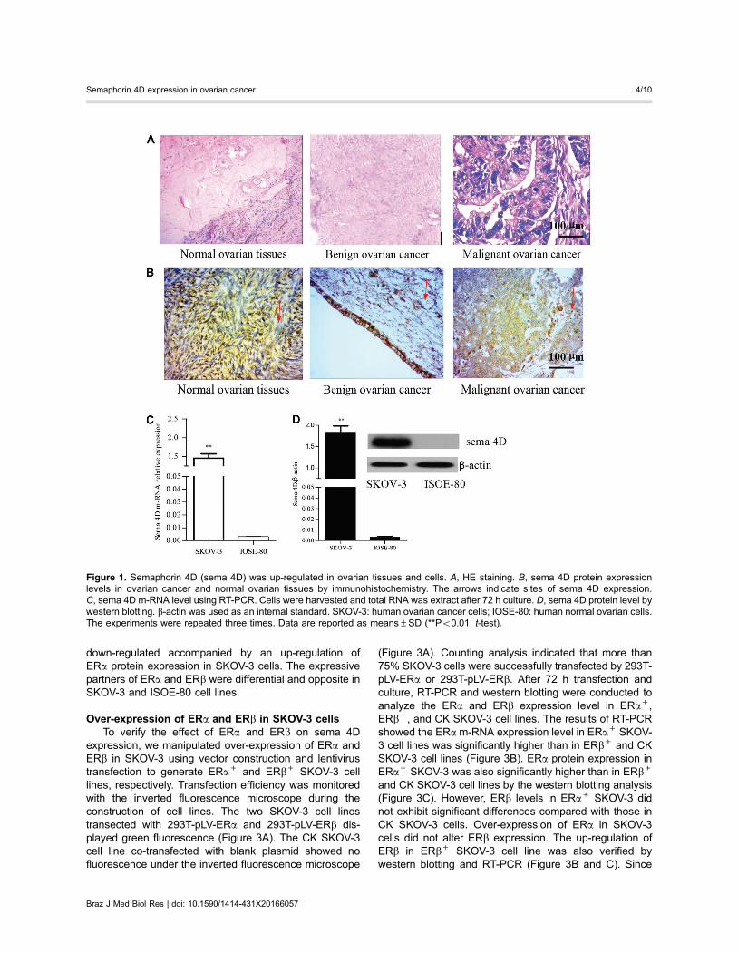
Sema 4D was highly expressed in ovarian cancer cellsand tissues. To confirm the role of sema 4D in ovariancancer, a total of 180 ovary samples including 60 benignovarian cancer, malignant ovarian cancer and normalovarian tissues each, were collected for the analysis ofsema 4D protein expression by immunohistochemistry.These ovary samples were pathologically confirmed byHE staining (Figure 1A). The results showed that expres-sion of sema 4D protein in benign ovarian cancer tissuesand malignant ovarian cancer tissues was 58.3% (35/60)and 90.2% (55/60), respectively. In contrast, the expres-sive proportion (33.3%, 20/60) of sema 4D proteins innormal ovarian tissues was significantly lower than thosein benign and malignant ovarian cancer tissues. Malignantissues classified as early pathological stage had a lowerproportion of sema 4D protein expression than thoseclassified as late stage (Figure 1B and Table 1), althoughvalues were not statistically significant. Meanwhile, sema4D m-RNA and expression levels in ovarian cancerSKOV-3 cells were both found to be significantly higherthan those in ISOE-80 cells (Figure 1C and D). Thoseobservations uniformly confirmed the fact that sema 4Dwas up-regulated in ovarian cancer cells and tissues.
ERa and ERb were differentially expressed in SKOV-3and ISOE-80 cells
As shown in Figure 2A and B, ISOE-80 cells have ahigher ERb expression than ERa both in nuclear acid andprotein levels. The approximate ratio of ERa/ERb wasrespectively 1/2.3 and 1/1.7 in nuclear acid and proteinlevels in ISOE-80 cells. Whereas the relative ERa m-RNAexpression in SKOV-3 cells was significantly enhancedcompared with those in ISOE-80. In contrast, a reducedERb m-RNA level in SKOV-3 cells was detected, resultingin the increased ratio of ERa:ERb (3.3/1) in m-RNA levels.Correspondingly, ERb protein expression was found to be
Braz J Med Biol Res | doi: 10.1590/1414-431X20166057
Semaphorin 4D expression in ovarian cancer 3/10
down-regulated accompanied by an up-regulation ofERa protein expression in SKOV-3 cells. The expressivepartners of ERa and ERb were differential and opposite inSKOV-3 and ISOE-80 cell lines.
Over-expression of ERa and ERb in SKOV-3 cellsTo verify the effect of ERa and ERb on sema 4D
expression, we manipulated over-expression of ERa andERb in SKOV-3 using vector construction and lentivirustransfection to generate ERa+ and ERb+ SKOV-3 celllines, respectively. Transfection efficiency was monitoredwith the inverted fluorescence microscope during theconstruction of cell lines. The two SKOV-3 cell linestransected with 293T-pLV-ERa and 293T-pLV-ERb dis-played green fluorescence (Figure 3A). The CK SKOV-3cell line co-transfected with blank plasmid showed nofluorescence under the inverted fluorescence microscope
(Figure 3A). Counting analysis indicated that more than75% SKOV-3 cells were successfully transfected by 293T-pLV-ERa or 293T-pLV-ERb. After 72 h transfection andculture, RT-PCR and western blotting were conducted toanalyze the ERa and ERb expression level in ERa+,ERb+, and CK SKOV-3 cell lines. The results of RT-PCRshowed the ERa m-RNA expression level in ERa+ SKOV-3 cell lines was significantly higher than in ERb+ and CKSKOV-3 cell lines (Figure 3B). ERa protein expression inERa+ SKOV-3 was also significantly higher than in ERb+
and CK SKOV-3 cell lines by the western blotting analysis(Figure 3C). However, ERb levels in ERa+ SKOV-3 didnot exhibit significant differences compared with those inCK SKOV-3 cells. Over-expression of ERa in SKOV-3cells did not alter ERb expression. The up-regulation ofERb in ERb+ SKOV-3 cell line was also verified bywestern blotting and RT-PCR (Figure 3B and C). Since
Figure 1. Semaphorin 4D (sema 4D) was up-regulated in ovarian tissues and cells. A, HE staining. B, sema 4D protein expressionlevels in ovarian cancer and normal ovarian tissues by immunohistochemistry. The arrows indicate sites of sema 4D expression.C, sema 4D m-RNA level using RT-PCR. Cells were harvested and total RNA was extract after 72 h culture. D, sema 4D protein level bywestern blotting. b-actin was used as an internal standard. SKOV-3: human ovarian cancer cells; IOSE-80: human normal ovarian cells.The experiments were repeated three times. Data are reported as means±SD (**Po0.01, t-test).
Braz J Med Biol Res | doi: 10.1590/1414-431X20166057
Semaphorin 4D expression in ovarian cancer 4/10

ERa m-RNA and protein levels in ERb+ SKOV-3 cellswere not significantly different from those in CK SKOV-3cells, the ERb over-expression did not affect the expres-sion of ERa as well.
Over-expression of ERa and ERb converselyregulated sema 4D expression and cell proliferation
As members of ligand-dependent transcription factor,ERa and ERb exert transcriptional regulation on thetargets only in the presence of a co-factor. E2 hormone isthe most common and natural co-factor of ERa and ERb inthe ovary. To initiate ERa and ERb activity on transcrip-tional regulation, a 10
–6 M E2 solution was incubated withcells for 6 h, and then followed by analysis of sema 4Dexpression using western blotting and RT-PCR. In ERa+
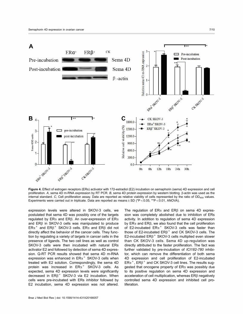
SKOV-3 cells, sema 4D m-RNA level was increased by44.2% after E2 incubation (Figure 4A). And sema 4D
protein levels were also enhanced by 38.8% in ERa+
SKOV-3 cells via E2 incubation (Figure 4B). However, theexpressive pattern of sema 4D in ERb+ SKOV-3 wascompletely different from those in ERa+ SKOV-3 cells.The sema 4D expressions in m-RNA and protein levelswere found to be reduced by 85.3 and 86.4 % in ERb+
SKOV-3 cells with E2 incubation, respectively (Figure 4Aand B). Although sema 4D mRNA levels were similarpre-incubation and post-incubation of E2 in CK SKOV-3cells, the protein expression of sema 4D was significantlyincreased with E2 incubation, which was possibly dueto the higher expression of ERa than those of ERb inCK SKOV-3 cells. The cell proliferation of ERa+ SKOV-3was significantly higher than those of ERb+ SKOV-3and CK SKOV-3 cells starting from 12 h of E2 incubation.The cell proliferation of ERb+ SKOV-3 cells was thelowest (Figure 4C).
Table 1. Semaphorin (sema) 4D protein expression levels in different ovariantissues.
Groups Total Sema `4D
(n) – + ++ +++
Normal ovarian tissues 60 40 18 2 0
Benign ovarian cancer 60 25* 18 14* 3Malignant ovarian cancer (I-II) 30 5* 4* 10* 11*Malignant ovarian cancer (III-IV) 30 0** 2** 12** 16**
Sema 4D protein expression was assayed using immunohistochemistry. Theresults of immunohistochemistry were evaluated according to the quantity ofpositive cells and density of cell staining in the visual field. Less than 5% positivecells in a visual field was scored 0; 5–25% scored 1; 26–50% scored 2; 51–75%scored 3; 76–100% scored 4. No or vague coloration in a visual field was scored 0;pale yellow scored 1; medium yellow scored 2; brown scored 3. (–) means 0-1scores; (+) means 2–3 scores; (++) means 4–5 scores; (+++) means 6–7scores. Ten visual fields were randomly selected in the single assay. Data arereported as numbers (*Po0.05, **Po0.01, ANOVA).
Figure 2. Estrogen receptor (ER)a and ERb were conversely expressed in SKOV-3 human ovarian cancer cells and ISOE-80 humannormal cancer cells. A, sema 4D protein expression by western blotting. B, Semaphorin 4D (sema 4D) m-RNA expression by RT PCR.b-actin was used as the internal standard. Data are reported as means±SD. (*Po0.05, **Po0.01, ANOVA).
Braz J Med Biol Res | doi: 10.1590/1414-431X20166057
Semaphorin 4D expression in ovarian cancer 5/10

ERa and ERb regulation on sema 4D expression andcell proliferation was disabled by inhibitor of estrogensignaling pathway
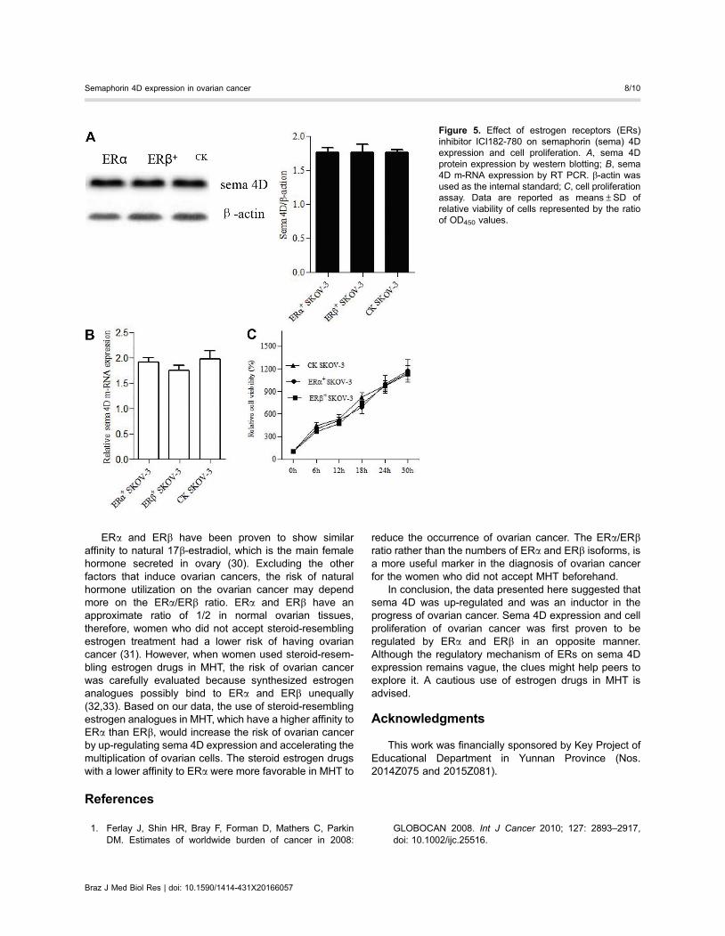
To confirm that E2 pre-incubation regulated sema 4Dexpression through ERa and ERb, an inhibitor of estrogensignal pathway, ICI182-780, which can inhibit both ERaand ERb activity, was pre-incubated with cells and thenfollowed by E2 treatment and detection of sema 4Dexpression. The western blotting results showed that thelevels of sema 4D proteins in both ERa+ and ERb+ SKOV-3cell lines were recovered to control levels (Figure 5A). Andthe m-RNA levels of sema 4D in ERa+ and ERb+ SKOV-3 cell lines were also parallel to those in CK SKOV-3cells (Figure 5B). The regulatory effect of ERa and ERbon the sema 4D expression was completely abolisheddue to inactivation of ERa and ERb by ICI182-780 inhib-itor and the cell proliferation of the three cell lines wasfound to be at the same level during the E2 incubation(Figure 5C).
Discussion
Sema 4D has been reported to be up-regulated inseveral cancers (20), advancing cancer progress. In thecurrent study, we analyzed sema 4D expression at the celland tissue levels. Sema 4D was found to be negativelyexpressed (40/60, –) or expressed in a relatively low rateof 33.3% (18/60, +; 2/60, ++) in normal ovarian tissue
and patients with ovarian benign tumors had a sema 4Dexpression rate of 58.3% (18/60, +; 14/60, ++; 3/60,+++). However, only 8.3% (5/60, –) of patients withovarian malignancy negatively expressed sema 4D at anearly pathological stage and patients at a late pathologicalstage showed higher sema 4D protein levels (2/30, +;12/30, ++; 16/30, +++) than those in an early stage(5/30, -; 4/30, +; 10/30, ++; 11/30, +++). Sema 4Dexpression was positively correlated with the progress ofovarian cancer. Likewise, we also found that sema 4D m-RNA and protein expression levels in ovarian cancerSKOV-3 cells were significantly higher than those in ISOE-80. A previous study by our group has reported that sema4D expression was gradually enhanced with the prog-ress of ovarian malignancy using immunohistochemistry.However, the association of sema 4D expression withthe ovarian cancer grade was not well assured due tothe small sample size used previously. Therefore, anexpanded sample size of ovarian malignancy was utilizedin the current study. Conclusively, the data further con-firmed that sema 4D played an inductive factor in ovariancancer as they function in other malignancies (27).
ERa expression levels were significantly higher thanERb in SKOV-3 cells. On the contrary, IESO-80 cellsshowed a significantly higher ERb levels than those ofERa in the present study. These observations confirmedthe fact that ERa is generally an oncogene and ERb is ananti-oncogene (28,29). Since sema 4D, ERa, and ERb
Figure 3. Over-expression of estrogen receptors (ERs) in SKOV-3 human ovarian cancer cells. The 293T pLV-ERa or 293T pLV-ERbvector co-transfected with SKOV-3 cells was used to generate ERa+ and ERb+ SKOV-3 cell lines, respectively. CK SKOV-3 cellsreferred to blank plasmid transfection. A, transfection efficiency validated by inverted fluorescence microscope. Green fluorescenceindicates that cells were successfully transfected. B, ERs m-RNA expression levels by RT-PCR. C, ERs protein expression levels bywestern blotting. b-actin was used as the internal standard. Data are reported as means±SD (*Po0.05, **Po0.01, ANOVA).
Braz J Med Biol Res | doi: 10.1590/1414-431X20166057
Semaphorin 4D expression in ovarian cancer 6/10
expression levels were altered in SKOV-3 cells, wepostulated that sema 4D was possibly one of the targetsregulated by ERa and ERb. An over-expression of ERaand ERb in SKOV-3 cells was manipulated to produceERa+ and ERb+ SKOV-3 cells. ERa and ERb did notdirectly affect the behavior of the cancer cells. They func-tion by regulating a variety of targets in cancer cells in thepresence of ligands. The two cell lines as well as controlSKOV-3 cells were then incubated with natural ERsactivator E2 and followed by detection of sema 4D expres-sion. Q-RT PCR results showed that sema 4D m-RNAexpression was enhanced in ERa+ SKOV-3 cells whentreated with E2 solution. Correspondingly, the sema 4Dprotein was increased in ERa+ SKOV-3 cells. Asexpected, sema 4D expression levels were significantlydecreased in ERb+ SKOV-3 via E2 incubation. Whencells were pre-incubated with ERs inhibitor followed byE2 incubation, sema 4D expression was not altered.
The regulation of ERa and ERb on sema 4D expres-sion was completely abolished due to inhibition of ERsactivity. In addition to regulation of sema 4D expressionby ERa and ERb, we also found that the cell proliferationof E2-incubated ERa+ SKOV-3 cells was faster thanthose of E2-incubated ERb+ and CK SKOV-3 cells. TheE2-incubated ERb+ SKOV-3 cells multiplied even slowerthan CK SKOV-3 cells. Sema 4D up-regulation wasdirectly attributed to the faster proliferation. The fact wasfurther validated by pre-incubation of ICI182-780 inhibi-tor, which can remove the differentiation of both sema4D expression and cell proliferation of E2-incubatedERa+, ERb+ and CK SKOV-3 cell lines. The results sug-gested that oncogene property of ERa was possibly dueto its positive regulation on sema 4D expression andacceleration of cell multiplication, whereas ERb negativelycontrolled sema 4D expression and inhibited cell pro-liferation.
Figure 4. Effect of estrogen receptors (ERs) activator with 17b-estradiol (E2) incubation on semaphorin (sema) 4D expression and cellproliferation. A, sema 4D m-RNA expression by RT PCR. B, sema 4D protein expression by western blotting. b-actin was used as theinternal standard. C, Cell proliferation assay. Data are reported as relative viability of cells represented by the ratio of OD450 values.Experiments were carried out in triplicate. Data are reported as means±SD (*Po0.05, **Po0.01, ANOVA).
Braz J Med Biol Res | doi: 10.1590/1414-431X20166057
Semaphorin 4D expression in ovarian cancer 7/10
ERa and ERb have been proven to show similaraffinity to natural 17b-estradiol, which is the main femalehormone secreted in ovary (30). Excluding the otherfactors that induce ovarian cancers, the risk of naturalhormone utilization on the ovarian cancer may dependmore on the ERa/ERb ratio. ERa and ERb have anapproximate ratio of 1/2 in normal ovarian tissues,therefore, women who did not accept steroid-resemblingestrogen treatment had a lower risk of having ovariancancer (31). However, when women used steroid-resem-bling estrogen drugs in MHT, the risk of ovarian cancerwas carefully evaluated because synthesized estrogenanalogues possibly bind to ERa and ERb unequally(32,33). Based on our data, the use of steroid-resemblingestrogen analogues in MHT, which have a higher affinity toERa than ERb, would increase the risk of ovarian cancerby up-regulating sema 4D expression and accelerating themultiplication of ovarian cells. The steroid estrogen drugswith a lower affinity to ERa were more favorable in MHT to
reduce the occurrence of ovarian cancer. The ERa/ERbratio rather than the numbers of ERa and ERb isoforms, isa more useful marker in the diagnosis of ovarian cancerfor the women who did not accept MHT beforehand.
In conclusion, the data presented here suggested thatsema 4D was up-regulated and was an inductor in theprogress of ovarian cancer. Sema 4D expression and cellproliferation of ovarian cancer was first proven to beregulated by ERa and ERb in an opposite manner.Although the regulatory mechanism of ERs on sema 4Dexpression remains vague, the clues might help peers toexplore it. A cautious use of estrogen drugs in MHT isadvised.
Acknowledgments
This work was financially sponsored by Key Project ofEducational Department in Yunnan Province (Nos.2014Z075 and 2015Z081).
References
1. Ferlay J, Shin HR, Bray F, Forman D, Mathers C, ParkinDM. Estimates of worldwide burden of cancer in 2008:
GLOBOCAN 2008. Int J Cancer 2010; 127: 2893–2917,doi: 10.1002/ijc.25516.
Figure 5. Effect of estrogen receptors (ERs)inhibitor ICI182-780 on semaphorin (sema) 4Dexpression and cell proliferation. A, sema 4Dprotein expression by western blotting; B, sema4D m-RNA expression by RT PCR. b-actin wasused as the internal standard; C, cell proliferationassay. Data are reported as means±SD ofrelative viability of cells represented by the ratioof OD450 values.
Braz J Med Biol Res | doi: 10.1590/1414-431X20166057
Semaphorin 4D expression in ovarian cancer 8/10
2. Jemal A, Siegel R, Ward E, Hao Y, Xu J, Murray T, et al.Cancer statistics CA. Cancer J Clin 2009; 59: 225–249,doi: 10.3322/caac.20006.
3. Deng H, Yin L, Zhang XT, Liu LJ, Wang ML, Wang ZY.ER-a variant ER-a 36 mediates anti-estrogen resistancein ER-positive breast cancer stem/progenitor cells. J SteroidBiochem Mol Biol 2014; 144: 417–426, doi: 10.1016/j.jsbmb.2014.08.017.
4. Suda T, Oba H, Takei H, Kurosumi M, Hayashi S,Yamaguchi Y. ER-activating ability of breast cancer stromalfibroblasts is regulated independently of alteration of TP53and PTEN tumor suppressor genes. Biochem Biophys ResCommun 2012; 428: 259–263, doi: 10.1016/j.bbrc.2012.10.035.
5. Rodvold JJ, Mahadevan NR, Zanetti M. Immune modulationby ER stress and inflammation in the tumor microenviron-ment. Cancer Lett 2015; 380: 227–236, doi: 10.1016/j.canlet.2015.09.009.
6. Santen RJ. Menopausal hormone therapy and breastcancer. J Steroid Biochem Mol Biol 2014; 142: 52–61,doi: 10.1016/j.jsbmb.2013.06.010.
7. Al-Safi ZA, Santoro N. Menopausal hormone therapy andmenopausal symptoms. Fertil Steril 2014; 101: 905–915,doi: 10.1016/j.fertnstert.2014.02.032.
8. Salehi F, Dunfield L, Phillips KP, Krewski D, VanderhydenBC. Risk factors for ovarian cancer: an overview withemphasis on hormonal factors. J Toxicol Environ Health B2008; 11: 301–321, doi: 10.1080/10937400701876095.
9. Riman T, Dickman PW, Nilsson S, Correia N, Nordlinder H,Magnusson CM, et al. Risk factors for invasive epithelialovarian cancer: results from a Swedish case-control study.Am J Epidemiol 2002; 156: 363–373, doi: 10.1093/aje/kwf048.
10. Purdie DM, Bain CJ, Siskind V, Russell P, Hacker NF, WardBG, et al. Hormone replacement therapy and risk ofepithelial ovarian cancer. Br J Cancer 1999; 81: 559–563,doi: 10.1038/sj.bjc.6690731.
11. Folsom AR, Anderson JP, Ross JA. Estrogen replacementtherapy and ovarian cancer. Epidemiology 2004; 15: 100–110, doi: 10.1097/01.ede.0000091606.31903.8e.
12. Spillman MA, Manning NG, Dye WW, Sartorius CA, PostMD, Harrell JC, et al. Tissue-specific pathways for estrogenregulation of ovarian cancer growth and metastasis. CancerRes 2010; 70: 8927–8936, doi: 10.1158/0008-5472.CAN-10-1238.
13. Armaiz-Pena GN, Mangala LS, Spannuth WA, Lin YG,Jennings NB, Nick AM, et al. Estrous cycle modulatesovarian carcinoma growth. Clin Cancer Res 2009; 15: 2971–2978, doi: 10.1158/1078-0432.CCR-08-2525.
14. Hall JM, Mcdonnell DP. Co-regulators in nuclear estrogenreceptor action: from concept to therapeutic targeting. MolInterv 2005; 5: 343–357, doi: 10.1124/mi.5.6.7.
15. Sotoca AM, Ratman D, van der Saag PT, Strom A,Gustafsson JA, Rietjens I, et al. Phytoestrogen-mediatedinhibition of proliferation of the human T47D breast cancercells depends on the ERalpha/ERbeta ratio. J SteroidBiochem Mol Biol 2008; 112: 171–178, doi: 10.1016/j.jsbmb.2008.10.002.
16. Stossi F, Barnett DH, Frasor J, Komm B, Lyttle CR,Katzenellenbogen BS. Transcriptional profiling of estrogen-regulated gene expression via estrogen receptor (ER) alpha
or ERbeta in human osteosarcoma cells: distinct and com-mon target genes for these receptors. Endocrinology 2004;145: 3473–3486, doi: 10.1210/en.2003-1682.
17. Guttilla IK, Adams BD, White BA. ERa, microRNAs, and theepithelial–mesenchymal transition in breast cancer. TrendsEndocrin Met 2012; 23: 73–82, doi: 10.1016/j.tem.2011.12.001.
18. Negishi M, Oinuma I, Katoh H. Plexins: axon guidance andsignal transduction. Cell Mol Life Sci 2005; 62: 1363–1371,doi: 10.1007/s00018-005-5018-2.
19. Roth L, Koncina E, Satkauskas S, Cremel G, Aunis D,Bagnard D. The many faces of semaphorins: from develop-ment to pathology. Cell Mol Life Sci 2009; 66: 649–666,doi: 10.1007/s00018-008-8518-z.
20. Rehman M, Tamagnone L. Semaphorins in cancer: biologi-cal mechanisms and therapeutic approaches. Semin CellDev Biol 2013; 24: 179–189, doi: 10.1016/j.semcdb.2012.10.005.
21. Basile JR, Castilho RM, Williams VP, Gutkind JS.Semaphorin 4D provides a link between axon guidanceprocesses and tumor-induced angiogenesis. Proc NatlAcad Sci USA 2006; 103: 9017–9022, doi: 10.1073/pnas.0508825103.
22. Worzfeld T, Ofermanns S. Semaphorins and plexins astherapeutic targets. Nat Rev Drug DiscoVR 2014; 13: 603–621, doi: 10.1038/nrd4337.
23. Qiang R, Wang F, Shi LY, Liu M, Chen S, Wan HY, et al.Plexin-B1 is a target of miR-214 in cervical cancer andpromotes the growth and invasion of HeLa cells. Int JBiochem Cell Biol 2011: 43; 632–641, doi: 10.1016/j.biocel.2011.01.002.
24. Sotoca AM, van den Berg H, Vervoort PT, van den Saag P,Strom A, Gustafsson JA, et al. Influence of cellular ERalpha/ERbeta ratio on the ERalpha agonist induced proliferationof human T47D breast cancer cells. Toxicol Sci 2008; 105:303–311, doi: 10.1093/toxsci/kfn141.
25. Bhata RA, Stauffera B, Unwallab RJ, Xu Z, Harrisa HA,Komm BS. Molecular determinants of ERa and ERbinvolved in selectivity of 16a-iodo-17b estradiol. J SteroidBiochem Mol Biol 2004; 88: 17–26, doi: 10.1016/j.jsbmb.2003.10.009.
26. Liu H, Yang Y, Xiao J, Yang S, Liu Y, Kang W, et al.Semaphorin 4D expression is associated with a poor clinicaloutcome in cervical cancer patients. Microvasc Res 2014;93: 1–8, doi: 10.1016/j.mvr.2014.02.007.
27. Liu Y, Zhou H, Ma L, Hou Y, Pan J, Sun C, et al. MiR-214suppressed ovarian cancer and negatively regulated sema-phorin 4D. Tumor Biol 2015; 37: 8239–8248, doi: 10.1007/s13277-015-4708-0.
28. Paruthiyil S, Parmar H, Kerekatte V, Cunha GR, FirestoneGL, Leitman DC. Estrogen receptor beta inhibits humanbreast cancer cell proliferation and tumor formation bycausing a G2cell cycle arrest. Cancer Res 2004; 64: 423–428, doi: 10.1158/0008-5472.CAN-03-2446.
29. Sotoca AM, van den Berg H, Vervoort PT, van den Saag P,Strom A, Gustafsson JA, et al. Influence of cellular ERalpha/ERbeta ratio on the ERalpha agonist induced proliferation ofhuman T47D breast cancer cells. Toxicol Sci 2008; 105:303–311, doi: 10.1093/toxsci/kfn141.
30. Bhata RA, Stauffera B, Unwallab RJ, Xu Z, Harrisa HA,Komm BS. Molecular determinants of ERa and ERb
Braz J Med Biol Res | doi: 10.1590/1414-431X20166057
Semaphorin 4D expression in ovarian cancer 9/10

involved in selectivity of 16a-iodo-17b estradiol. J SteroidBiochem Mol Biol 2004; 88: 17–26, doi: 10.1016/j.jsbmb.2003.10.009.
31. Paterni I, Granchi C, Katzenellenbogen JA, Minutolo F.Estrogen receptors alpha (ERa) and beta (ERb): Subtype-selective ligands and clinical potential. Steroids 2014; 90:13–29, doi: 10.1016/j.steroids.2014.06.012.
32. Jordan VC. Selective estrogen receptor modulation: apersonal perspective. Cancer Res 2001; 61: 5683–5687.
33. Anstead GM, Carlson KE, Katzenellenbogen JA. Theestradiol pharmacophore: ligand structure-estrogen receptorbinding affinity relationships and a model for the receptorbinding site. Steroids 1997; 62: 268–303, doi: 10.1016/S0039-128X(96)00242-5.
Braz J Med Biol Res | doi: 10.1590/1414-431X20166057
Semaphorin 4D expression in ovarian cancer 10/10


![4DFAB: A Large Scale 4D Database for Facial Expression ... · which hinders the use of 3D/4D FER system in real world scenarios. Henceforth, three databases, B3D(AC) [25], BP4D-Spontaneous](https://img.pdfslide.us/doc/110x75/6007bccd018c5771591ad670/4dfab-a-large-scale-4d-database-for-facial-expression-which-hinders-the-use.jpg)