Embed Size (px)
Citation preview

ARTICLE
Received 30 Jan 2016 | Accepted 14 Sep 2016 | Published 27 Oct 2016
Self-motion evokes precise spike timingin the primate vestibular systemMohsen Jamali1, Maurice J. Chacron1 & Kathleen E. Cullen1
The accurate representation of self-motion requires the efficient processing of sensory input
by the vestibular system. Conventional wisdom is that vestibular information is exclusively
transmitted through changes in firing rate, yet under this assumption vestibular neurons
display relatively poor detection and information transmission. Here, we carry out an analysis
of the system’s coding capabilities by recording neuronal responses to repeated presentations
of naturalistic stimuli. We find that afferents with greater intrinsic variability reliably
discriminate between different stimulus waveforms through differential patterns of precise
(B6 ms) spike timing, while those with minimal intrinsic variability do not. A simple
mathematical model provides an explanation for this result. Postsynaptic central neurons also
demonstrate precise spike timing, suggesting that higher brain areas also represent
self-motion using temporally precise firing. These findings demonstrate that two distinct
sensory channels represent vestibular information: one using rate coding and the other that
takes advantage of precise spike timing.
DOI: 10.1038/ncomms13229 OPEN
1 Department of Physiology McGill University, Montreal, Quebec, Canada H3G1Y6. Correspondence and requests for materials should be addressed toK.E.C. (email: [email protected]).
NATURE COMMUNICATIONS | 7:13229 | DOI: 10.1038/ncomms13229 | www.nature.com/naturecommunications 1

Understanding the set of transformations by which thebrain processes incoming sensory input to ensure accurateperception and behaviour remains a central problem
in neuroscience. The vestibular system provides informationabout our self-motion and spatial orientation relative to theworld that is required for ensuring gaze stability, balance andpostural control during everyday life. This essential sensorysystem comprises two classes of primary afferents that differin their morphology and response dynamics (reviewed in ref. 1).Historically, these two afferent classes have been termed regularand irregular because the distribution of resting dischargevariability is bimodal. The evolution of amniotes fromamphibian-like animals was accompanied by the appearance ofa new type of vestibular receptor cell that preferentially suppliesirregular afferents (reviewed in ref. 2). It has been hypothesizedthat the relatively late appearance of the type I hair cell inevolution demonstrates a neural adaptation to changes in naturalstimulus statistics following the transition from a water-basedto a land-based environment (reviewed in refs 1,3). However,this proposal is at odds with recent findings that despitedisplaying higher sensitivity, irregular afferents are actuallyworse at detecting and transmitting information aboutself-motion than their regular counterparts4,5. Accordingly, whythe vestibular system uses two distinct peripheral channels torepresent self-motion is currently an open question.
Conventional wisdom has been that the vestibular neuronsrepresent sensory input exclusively through firing rate (reviewedin ref. 3). Specifically, previous studies have characterized how thefiring rates of vestibular afferents and their target neurons in thevestibular nuclei encode self-motion information and determinemotion detection thresholds4,6–9. It is however possible that thelongstanding paradox regarding the functional role of two distinctchannels to represent self-motion mentioned above stems fromthe fact that other aspects of spiking activity have not beenconsidered. Theory suggests that temporal codes, in whichsensory input is instead represented by the precise timing ofaction potentials, more efficiently represent sensory stimuli thanrate codes10. In this context, rate coding has been commonlydefined as a neural code in which stimulus attributes areencoded by the number of spikes occurring during a timewindow whose length is determined by the stimulus timescale.In contrast, temporal coding has been defined as a neural codein which stimulus attributes are encoded by the precise timingof spikes within the same time window11–13. This then leadsto the question of whether the vestibular system takes advantageof precise spike timing to encode self-motion. Temporal precisionin action potential firing has been observed in many other sensorysystems14–20 and could, in theory, also exist in the vestibularsystem4. However, no study to date has directly tested whetherself-motion information is represented by precise spike timing.
Here we explicitly tested whether neurons in early vestibularpathways use precise spike timing to represent self-motion. Therecordings were made from vestibular afferents while monkeysexperienced repeated trials of naturalistic self-motion stimuli.We found that, while regular afferents primarily encodemotion stimuli through changes in firing rate, irregular afferentsinstead more reliably discriminated between different stimuluswaveforms through differential patterns of precise spike timing.A simple mathematical model reproduced our findings, andprovided an explanation of how the nature of the neural codeis determined by a balance between neuronal variability andsensitivity. Importantly, afferent target neurons in the centralvestibular nuclei also discriminated between self-motionstimuli through precise spike timing, suggesting that spike timingin higher brain areas ensures accurate self-motion perception andbehaviour.
ResultsPrecision of spike timing in the vestibular periphery. To studywhether the vestibular system uses precise spike timing torepresent self-motion information, single-unit recordings weremade from peripheral semicircular canal afferents (N¼ 22 regularand N¼ 35 irregular) and their central target neurons within thevestibular nuclei (N¼ 24). We applied naturalistic self-motionstimuli, and first quantified the information encoded throughchanges in firing rate. We then investigated whether spike timingrepresents self-motion information. Such an encoding strategy is,by definition nonlinear, and requires reliable and precisespike timing responses. Thus, if early vestibular pathways usespike timing to represent self-motion information, the followingthree conditions must be met: (1) neurons should respondnonlinearly to naturalistic self-motion stimuli, (2) neurons shoulddisplay low trial-to-trial variability in their responses to repeatedpresentations of the same stimulus, and (3) different self-motionstimuli should evoke distinctive and precise patterns ofaction potentials11. Accordingly, we tested whether all threeconditions were met by recording vestibular neural spikingresponses to repeated trials of naturalistic self-motion andthen establishing whether precise spike timing could be reliablyused to discriminate between different stimulus waveforms(see the ‘Methods’ section).
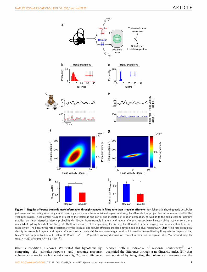
Information transmission via changes in firing rate. Vestibularafferents display a wide range of resting discharge variability andare typically classified as either irregular or regular (Fig. 1a–c).We applied single presentations of time-varying naturalisticstimuli and found that both irregular and regular afferentsresponded through changes in firing rate (Fig. 1d,e). Notably,firing rate modulations were markedly greater for irregularafferents due to their greater sensitivity (see SupplementaryFig. 1A for population averages), consistent with the results ofprevious studies that used artificial stimuli (that is, singlesinusoids and band-pass noise) to characterize these cells4,5. If theincreased variability of irregular afferents reduces theirinformation transmission through changes in firing rate (that is,rate coding), we hypothesized that they should encode lessinformation during natural stimulation. Indeed, using the directmethod21 and computing firing rate conditional probabilitydensity as a function of head velocity (see the ‘Methods’ section),we found that this was the case. The firing rate probability densityshowed a marked increase as a function of head velocity anddisplayed higher variance for irregular afferents as comparedwith their regular counterparts (compare Fig. 1f,g). Nevertheless,as shown for each population in Fig. 1h,i, regular afferentstransmitted significantly higher information than irregularafferents through changes in firing rate (Po0.01). Thus,consistent with previous results obtained using an indirectmethod4 (Supplementary Fig. 1B), regular afferents transmittedmore information about natural stimuli through changes in firingrate than irregular afferents. We further note that subdivision ofirregular afferents into two subgroups corresponding to putativemorphological origin22,23 did not alter the qualitative nature ofthis finding (Supplementary Fig. 2).
Irregular afferents display nonlinear responses. The responsesof typical irregular and regular afferents to repeated presentationsof naturalistic stimuli are shown in Fig. 2a,b, respectively.Irregular afferents appeared to exhibit more reliable spikingresponses across trials than their regular counterparts, leading usto hypothesize that irregular afferents use precise spike timing torepresent self-motion. If this is the case, then they should respondnonlinearly to repeated trials of naturalistic self-motion stimuli
ARTICLE NATURE COMMUNICATIONS | DOI: 10.1038/ncomms13229
2 NATURE COMMUNICATIONS | 7:13229 | DOI: 10.1038/ncomms13229 | www.nature.com/naturecommunications
(that is, condition 1 above). We tested this hypothesis bycomparing the stimulus–response and response–responsecoherence curves for each afferent class (Fig. 2c), as a difference
between both is indicative of response nonlinearity24. Wequantified the difference through a nonlinearity index (NI) thatwas obtained by integrating the coherence measures over the
b c
0 20 4010 30
Pro
babi
lity
0
0.2
ISI (ms)
0 20 4010 30
Pro
babi
lity
0
0.5
ISI (ms)
−50
200
0
050
d e
−500
50
Hea
d ve
loci
ty(d
eg s
–1)
Firi
ng r
ate
(spk
s–1
)
0
400
100 ms
f g
0
100 0.5
200
300
1
Firi
ng r
ate
(spk
s–1
)
Firi
ng r
ate
(spk
s–1
)
1.5
0
×10–4
0–50 50
0.5
1
0
×10–3
Head velocity (deg s–1) Head velocity (deg s–1)
Pro
babi
lity
dens
ity
Pro
babi
lity
dens
ity
0
100
200
300
0–50 50
h
MI fi
ring
rate
(bit)
1
1.5i
Nor
mal
ized
MI fi
ring
rate
0.2
0.1
0Regular
**
Irregular
0.5
0Regular Irregular
Irregular
RegularCentralneuron
Thalamus/cortexperception
Spinal cordto stabilize postureVestibular
nuclei
a
Irregular afferent Regular afferent
Figure 1 | Regular afferents transmit more information through changes in firing rate than irregular afferents. (a) Schematic showing early vestibular
pathways and recording sites. Single unit recordings were made from individual regular and irregular afferents that project to central neurons within the
vestibular nuclei. These central neurons project to the thalamus and cortex and mediate self-motion perception, as well as to the spinal cord for posture
stabilization. (b,c) Interspike interval probability distribution from example irregular and regular afferents, respectively. Insets: spiking activity from these
units. (d,e) Spiking (middle) and firing rate (bottom) response of example irregular and regular afferents to a time-varying head velocity stimulus (top),
respectively. The linear firing rate predictions for the irregular and regular afferents are also shown in red and blue, respectively. (f,g) Firing rate probability
density for example irregular and regular afferents, respectively. (h) Population-averaged mutual information transmitted by firing rate for regular (blue,
N¼ 22) and irregular (red, N¼ 35) afferents (P¼0.0028). (i) Population-averaged normalized mutual information for regular (blue, N¼ 22) and irregular
(red, N¼ 35) afferents (P¼ 1.6� 10� 5).
NATURE COMMUNICATIONS | DOI: 10.1038/ncomms13229 ARTICLE
NATURE COMMUNICATIONS | 7:13229 | DOI: 10.1038/ncomms13229 | www.nature.com/naturecommunications 3

stimulus frequency range and taking the ratio between the two(see the ‘Methods’ section for details). NI is null when theresponse-response and stimulus-response coherence curves are
equal, and approaches 100% with increasing levels ofnonlinearity. Indeed, consistent with our prediction, theexample irregular afferent showed strong response nonlinearity
−500
50
a b
−500
50
Hea
d ve
loci
ty(d
eg s
–1)
0
300
Firi
ng r
ate
(spk
s–1
)
0
300
100 ms
c
Stimulus Response
Linear Nonlinear
Frequency (Hz)0
1
SR
coh
eren
ce
Neuralsystem
√RR
coh
eren
ce
Frequency (Hz)0
1
0
1
Frequency (Hz)
Trial 2
Trial 3
Trial n
Trial 1
0
0.5
1
100 1010
0.5
1
Coh
eren
ce
100 101
SR coherence
SR coherence
d eIrregular afferent Regular afferent
1
Coh
eren
ce
1f
50 *
g
0
0.5
Frequency (Hz)
100 1010
0.5
Frequency (Hz)
100 101
0Non
linea
rity
inde
x (N
I)
Regula
r
Irreg
ular
√RR coherence
√RR coherence
Figure 2 | Irregular afferents display nonlinear responses to repeated stimulus presentations. (a,b) Spiking (middle) and firing rate (bottom) responses
of the same example irregular and regular afferents shown in Fig. 1 to repeated stimulus (top) presentations, respectively. (c) Schematic showing repeated
presentations of the same stimulus and different spiking responses to each trial. The stimulus–response (SR) coherence (blue) measures correlations between
stimulus and response (top). In contrast, the response–response (RR) coherence (red) measures correlations between responses to repeated stimulus
presentations (bottom). (d,e) SR (red and blue) and square-rooted RR (purple and cyan) coherence curves obtained for the same example irregular and
regular afferents shown in a and b, respectively. (f,g) Population-averaged SR (red and blue) and square-rooted RR (purple and cyan) coherence curves
obtained for irregular and regular afferents, respectively. Shaded bands illustrate s.e.m. Inset: population-averaged nonlinearity index for regular (blue, N¼ 22)
and irregular (red, N¼ 35) afferents (P¼6.3� 10� 6). ‘*’ indicates statistical significance at the P¼0.05 level using a Wilcoxon rank-sum.
ARTICLE NATURE COMMUNICATIONS | DOI: 10.1038/ncomms13229
4 NATURE COMMUNICATIONS | 7:13229 | DOI: 10.1038/ncomms13229 | www.nature.com/naturecommunications

(Fig. 2d, NI¼ 22%). In contrast, the example regular afferentshowed weak response nonlinearity (Fig. 2e, NI¼ 5.7%).Qualitatively similar results were obtained across our data set(Fig. 2f,g, respectively; irregular: 31.5±3.2%, regular: 9.6±2.9%;Po0.001), indicating that irregular afferents displayed stronglynonlinear responses to naturalistic self-motion stimuli.
Irregular afferents use spike timing to encode self-motion. Ifirregular afferents preferentially use spike timing to represent self-motion information as compared to regular afferents, then thereare two additional conditions that must be met: they shoulddisplay low trial-to-trial variability in their responses to repeatedpresentations of the same stimulus (condition 2) and differentself-motion stimuli should evoke distinctive and precisepatterns of action potentials (condition 3). To test whether theseconditions were met, we next compared the action potentialpatterns evoked by different stimuli. Specifically, we quantifiedthe ability of an ideal observer to discriminate between thesedifferent stimuli using the recorded spike trains.
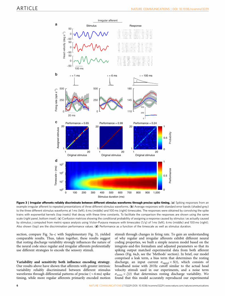
Figure 3a shows the spiking responses of the same exampleirregular afferent described above in Fig. 2 to three differenttime-varying stimuli with identical statistics (mean¼ 0 deg s� 1,standard deviation¼ 20 deg s� 1). Each stimulus was repeatedmultiple times and we found that, over short timescales (that is, 1and 6 ms; Fig. 3b, left and middle panels), the variabilitybetween responses to different stimuli was often substantiallylarger than that between responses to the same stimulus(indicated by the grey shading around the three responsewaveforms). In such cases, stimulus separation was possible andwas better at 6ms. In contrast, at longer (for example, 100 ms)timescales, there was significant overlap between responsesthereby limiting discriminability (Fig. 3b, right panel). To directlyquantify this observation, we used the Victor–Purpura distancemetric25 and compared the distance between a neuron’sresponses to a given stimulus and the distance betweenits responses to different stimuli. Then, to determine whether agiven response was correctly predicted as having been elicited bya given stimulus (see the ‘Methods’ section), the discriminationperformance was computed from the confusion matrix whoseelement ij gives the conditional probability that a responsegenerated by stimulus i is classified as being generated bystimulus j. Thus, if self-motion is represented by precise spiketiming in irregular afferents, then we should expect that theywould show maximum discrimination performance at timescalesmuch shorter than those contained in the stimulus.
The confusion matrices obtained from the example irregularafferent are illustrated in Fig. 3c for the same timescales shown inFig. 3b. Indeed, consistent with our prediction, discriminationperformance was maximal for timescales shorter than thosecontained in the stimulus (Fig. 3c, left and middle panels) andconsiderably less for longer timescales (Fig. 3c, right panel).Moreover, systematically varying both the timescale and thestimulus duration further revealed that peak discriminationperformance was consistently achieved for a timescale of 6 ms(Fig. 3d). Thus, given that this timescale is much shorter thanthose contained in the stimulus, we conclude that the precisespike timing patterns of this irregular afferent can indeed be usedto reliably discriminate between different stimuli.
We next performed the same analysis on the example regularafferent (Fig. 4a). In contrast to the irregular afferent shownabove in Fig. 3, the regular afferent’s responses to different stimuliconsistently overlapped for both short (1 ms) and long (100 ms)timescales (Fig. 4b, left and right panels) making stimulusseparation difficult. This neuron’s responses could be betterdiscriminated at an intermediate timescale (26 ms; Fig. 4b, middle
panel) as quantified by performance (Fig. 4c). Indeed, whentimescale and stimulus duration were systematically varied, weconsistently obtained maximum performance at an intermediatetimescale of B30 ms (Fig. 4d); a timescale comparable tothose contained in stimulus and therefore consistent with ratecoding. Furthermore, comparisons between this neuron andexample irregular afferent revealed that its performance at shorttimescales (that is, lower than those contained in the stimulus)was far poorer, and that its maximum performance wasactually much lower (compare maximum values in the middlepanels of Figs 4c and 3c). In summary, our analysis revealedthat unlike the example irregular afferent shown in Fig. 3, ourexample regular afferent did not represent self-motion throughprecise spike timing.
Figure 5 illustrates the comparison of the population-averagedresults for irregular and regular afferents. Discriminationperformance values were consistently greater for irregular thanregular afferents at all timescales ranging between 1 and 100 ms(Fig. 5a). Notably, irregular afferents displayed maximumperformance at lower timescales (B6 ms versus B30 ms; Fig. 5a,compare red and blue traces) as compared with regular afferents.Accordingly, irregular afferents displayed higher (B160 Hz)temporal precision (defined as the inverse of the timescale forwhich performance is maximal) than regular afferents (B30 Hz).Importantly, the peak performance of irregular afferents wassubstantially higher than that of their regular counterparts andfurthermore occurred at B6 ms—a timescale over which thestimulus does not vary significantly.
To better emphasize the implications of this result, Fig. 5breplots the performances of regular (blue trace) and irregular(red trace) afferents as a function of frequency (that is, the inverseof timescale) with the stimulus power spectrum (grey area)superimposed. At lower frequencies (for example, 1 Hz, leftmostgreen arrow) for which there is significant stimulus power, theperformance of regular afferents was greater than that of irregularafferents. This is consistent with our finding above thatsignificantly more information is transmitted by the firing rate ofregular versus irregular afferents (Fig. 1h,i). Indeed, the perfor-mance of regular afferents approaches its maximum value forfrequencies that are contained in the stimulus (that is, for whichthere is significant stimulus power; B20 Hz, middle green arrow).However, this is not the case for irregular afferents. Instead theirperformance is maximal at a much higher frequency (B100 Hz)where the stimulus power is negligible. Taken together, the resultsin Fig. 5b show that, whereas the performance of irregular afferentsincreases substantially (B50%) for frequencies greater than thosecontained in the stimulus (that is, 4B20 Hz and up to B100 Hz)or conversely for smaller and smaller timescales (that is, down toB10 ms), this was not the case for regular afferents. Instead, therewas only a negligible increase in their performance.
Thus, these results suggest that irregular afferents transmitsubstantially more information through precise spike timing thantheir regular counterparts. It is important to note that the higherspike timing precision of irregular afferents as compared withtheir regular counterparts was not due to differences in firing rate.This is because the mean firing rates of regular and irregularafferents in our data set during stimulation were not significantlydifferent from one another (regular: 105±6 spk s� 1; irregular:96±6 spk s� 1, P¼ 0.31, tstat¼ 1.03, df¼ 55).
We further found strong positive correlations between spiketiming precision (that is, the frequency at which performance ismaximal) and baseline variability (Fig. 5c, R¼ 0.8, Po0.001)as well as response nonlinearity (Fig. 5d, R¼ 0.7, Po0.001).As expected, a comparable analysis of our data using the vanRossum metric26, which has also been commonly used toquantify the distance between spike trains (see the ‘Methods’
NATURE COMMUNICATIONS | DOI: 10.1038/ncomms13229 ARTICLE
NATURE COMMUNICATIONS | 7:13229 | DOI: 10.1038/ncomms13229 | www.nature.com/naturecommunications 5
section, compare Fig. 5a–c with Supplementary Fig. 3), yieldedcomparable results. Thus, taken together, these results suggestthat resting discharge variability strongly influences the nature ofthe neural code since regular and irregular afferents preferentiallyuse different strategies to encode the sensory stimuli.
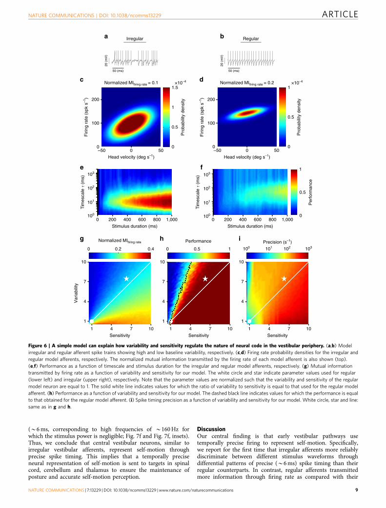
Variability and sensitivity both influence encoding strategy.Our results above have shown that afferents with greater intrinsicvariability reliably discriminated between different stimuluswaveforms through differential patterns of precise (B6 ms) spiketiming, while more regular afferents primarily encoded motion
stimuli through changes in firing rate. To gain an understandingof why regular and irregular afferents exhibit different neuralcoding properties, we built a simple neuron model based on theintegrate-and-fire formalism and adjusted parameters so that itsspiking output matched experimental data from both afferentclasses (Fig. 6a,b, see the ‘Methods’ section). In brief, our modelcomprised a leak term, a bias term that determines the restingdischarge, an input current ssignal� S(t), which consists ofbroadband noise with 20 Hz cutoff similar to the actual headvelocity stimuli used in our experiments, and a noise termsnoise� x(t) that determines resting discharge variability. Wefound that this model accurately reproduced our experimental
a
b
c
d
Irregular afferent
201
1 1 1
20
201
20
201
20
0
0.5
1
0 100 200 300 400 500 600 700 800 900 1,000
101
102
103
Stimulus duration (ms)
Tim
esca
le �
(ms)
0
0.5
1
Pro
babi
lity
Per
form
ance
Ass
igne
d st
imul
us
Original stimulus Original stimulus Original stimulus
100
Performance = 0.65 Performance = 0.99 Performance = 0.24
� = 6 ms
Firi
ng r
ate
(spk
s–1
)
0
500
20 ms
250
500
250
�
� = 1 ms � = 100 ms
100 ms−50
0
50−50
0
50−50
0
50
Hea
d ve
loci
ty (
deg
s–1)
Stimulus Response
110
145
180
0
0
500
250
Figure 3 | Irregular afferents reliably discriminate between different stimulus waveforms through precise spike timing. (a) Spiking responses from an
example irregular afferent to repeated presentations of three different stimulus waveforms. (b) Average responses with standard error bands (shaded grey)
to the three different stimulus waveforms at 1 ms (left), 6 ms (middle) and 100 ms (right) timescales. The responses were obtained by convolving the spike
trains with exponential kernels (top insets) that decay with these time constants. To facilitate the comparison the responses are shown using the same
scale (right panel, bottom inset). (c) Confusion matrices showing the conditional probability of assigning a response caused by stimulus i as actually caused
by stimulus j computed from metric-space analysis using Victor–Purpura measure with timescales (1/q) of 1 ms (left), 6 ms (middle) and 100 ms (right).
Also shown (top) are the discrimination performance values. (d) Performance as a function of the timescale as well as stimulus duration.
ARTICLE NATURE COMMUNICATIONS | DOI: 10.1038/ncomms13229
6 NATURE COMMUNICATIONS | 7:13229 | DOI: 10.1038/ncomms13229 | www.nature.com/naturecommunications

results. Although the model regular afferent transmitted moreinformation through changes in firing rate as compared with themodel irregular afferent (Fig. 6c,d), maximum discriminationperformance as computed from the confusion matrix wasconsistently achieved at timescales of B6 ms and B50 ms for theirregular and regular model afferents, respectively. Thus,consistent with our experimental observations, the modelirregular afferent more accurately represented time varyingstimuli via precise spike timing than the model regular afferent(Fig. 6e,f).
Why does resting discharge variability influence encodingstrategies used by the vestibular periphery? Using this model,it becomes possible to systematically and independently varysensitivity (ssignal) and variability (snoise). Our results show thathigher levels of variability lead to decreased information
transmission through firing rate, while increases in neuronalresponse sensitivity lead to increased information transmissionthrough firing rate (Fig. 6g). Furthermore, our model predictedthat the information transmitted will be approximately constantwhen sensitivity and variability were co-varied such that their ratio(that is, the input signal-to-noise ratio (SNR)) is constant (Fig. 6g,solid white line). Indeed, this finding is consistent withtheory because mutual information is determined by the inputSNR10. As such, our modelling results provide additional strongevidence in agreement with our previous observations, and suggestthat the irregular afferents transmit less information through firingrate because they displayed a greater amount of variability relativeto sensitivity (compare star with circle in Fig. 6g).
We next quantified the effects of sensitivity and variability ondiscrimination performance by precise spike timing. Results
Firi
ng r
ate
(spk
s–1
)
0
300
150
a
b
c
� = 26 ms � = 100 ms
1
20
1
20
1
20
0
0.5
1
Pro
babi
lity
20 ms
0
0.5
1
0 100 200 300 400 500 600 700 800 900 1,000
101
102
103
Tim
esca
le �
(ms)
100
� = 1 ms
�
Ass
igne
d st
imu l
us
Stimulus duration (ms)
d
Regular afferent
Stimulus Response
100 ms−50
0
50−50
0
50−50
0
50
Hea
d ve
loci
ty (
deg
s–1)
Per
form
ance
Performance = 0.15 Performance = 0.58 Performance = 0.38
110
160
135
125
135
145
0
300
150
0
150
300
201Original stimulus
201Original stimulus
201Original stimulus
Figure 4 | Regular afferents display low performance in discriminating between different stimulus waveforms through precise spike timing.
(a) Spiking responses from an example regular afferent to repeated presentations of three different stimulus waveforms. (b) Average responses with
standard error bands (shaded grey) to the three different stimulus waveforms at 1 ms (left), 26 ms (middle) and 100 ms (right) timescales. The responses
were obtained by convolving the spike trains with exponential kernels (top insets) that decay with these time constants. To facilitate the comparison, the
responses are shown using the same scale (right and middle panels, bottom insets). (c) Confusion matrices showing the conditional probability of assigning
a response caused by stimulus i as actually caused by stimulus j computed from metric-space analysis using Victor–Purpura measure with timescales (1/q)
of 1 ms (left), 26 ms (middle) and 100 ms (right). Also shown (top) are the discrimination performance values. (d) Performance as a function of the
timescale as well as stimulus duration.
NATURE COMMUNICATIONS | DOI: 10.1038/ncomms13229 ARTICLE
NATURE COMMUNICATIONS | 7:13229 | DOI: 10.1038/ncomms13229 | www.nature.com/naturecommunications 7

qualitatively different than those found for informationtransmission by firing rate were obtained. Specifically, we foundthat: (i) increasing variability and sensitivity led to decreasesand increases in discrimination performance, respectively;(ii) increasing both variability and sensitivity at the same rateactually increased performance (Fig. 6h, solid white line); and(iii) constant discrimination performance was achieved onlywhen variability increased at a rate that was roughly twice that ofsensitivity (Fig. 6h, dashed black line). Notably, the timescale forwhich maximum performance was achieved decreased to valuesmuch lower than those contained in the stimulus, which in turnresulted in high precision values (compare Fig. 6h and Fig. 6i).This indicates that co-varying sensitivity and variability whilekeeping the input SNR constant causes a transition in encodingstrategy in which coding by precise spike timing becomes moreimportant (Fig. 6i, white line). Thus, our model shows that bothvariability and sensitivity can strongly influence the informationcarried by both firing rate and spike timing, consistent with ourexperimental results shown above in Fig. 5c,d.
Taken together, our modelling results suggest that irregularafferents represent self-motion through precise spike timing moreaccurately than their regular counterparts because their increasedvariability is actually accompanied by a nearly proportionalincrease in sensitivity. The strong co-variation between variabilityand sensitivity previously demonstrated experimentally investibular afferents1,6,27 is consistent with our modelling resultsand thus provides support for this proposal.
Precise spike timing in central vestibular neuron responses.Thus far, we have addressed whether the peripheral vestibular
system uses precise spike timing to represent self-motioninformation. We found that afferents with greater intrinsicvariability reliably discriminated between different stimuluswaveforms through differential patterns of precise spike timing.A priori, if higher brain areas use temporal precision to representself-motion, then we speculate that postsynaptic central neuronsin the vestibular nuclei receiving direct synaptic input fromafferents (Fig. 7a) should also demonstrate precise spiketiming. Figure 7b illustrates the responses of an example centralvestibular neuron to time-varying naturalistic stimuli. Theneuron encoded the stimulus through changes in firing rate(Fig. 7b), consistent with previous results5,28. However, as was thecase for irregular afferents, the information transmitted throughfiring rate was relatively low because this neuron’s firing rateconditional probability displayed high variance, (compare Fig. 7cand Fig. 1f with Fig. 1g). It is important to note, however, thatfurther analysis of the precision of this neuron’s spiking inresponse to repeated stimulus presentations revealed strikinglysimilar results to those obtained above for our analysis ofirregular afferents. Specifically, when discrimination performancewas quantified from the confusion matrix, we found thatmaximum performance was consistently achieved at B6 ms(Fig. 7d)—a timescale that matches that of irregular afferents(compare with Fig. 3d).
Our analysis of the central neuron population furtherestablished that they actually transmitted less information thaneither regular or irregular afferents through changes in firing rate(Po0.001, Wilcoxon rank-sum test with Bonferroni correction;Fig. 7e). However, the discrimination performance from spiketiming was comparable to that of irregular afferents (Fig. 7f).Indeed, peak performance was achieved for low timescales
Nonlinearity index (NI)
Regular
Irregular
b
100
CV*
100
200
300 *
0Regular irregular
Pre
cisi
on (
s–1)
Pre
cisi
on (
s–1)
Pre
cisi
on (
s–1)
d
a
0
0.5
1
Nor
mal
ized
pow
er
Frequency (Hz)
0
0.2
0.4
0.6
0.8
1
Per
form
ance
100 101 102 103 100
100
101
101
10–1 10–1
102
101 102
102
103
103
101
102
103
0
0.2
Timescale (ms)
Per
form
ance
0.4
RegularIrregularChance0.6
0.8
1
c
Figure 5 | Irregular afferents display higher discrimination performance and greater precision than regular afferents. (a) Population-averaged
discrimination performance for regular (blue, N¼ 22) and irregular (red, N¼ 35) afferents as a function of timescale. The shaded red and blue bands show
the standard error. (b) Population-averaged discrimination performance for regular (blue, N¼ 22) and irregular (red, N¼ 35) afferents as a function of
frequency. The shaded red and blue bands show the standard error. The shaded grey area is the normalized power spectra of the stimulus as a function of
frequency. The three arrows highlight the performances at 1, 20 and 100 Hz. (c) Spike timing precision as a function of baseline variability as quantified by
CV* (see the ‘Methods’ section) for regular (blue) and irregular (red) afferents. Inset: population-averaged spike timing precision for regular (blue, N¼ 22)
and irregular (red, N¼ 35) afferents (P¼4.5� 10� 7). ‘*’ indicates statistical significance at the P¼0.05 level using a Wilcoxon rank-sum test. (d) Spike
timing precision as a function of nonlinearity for regular (blue) and irregular (red) afferents.
ARTICLE NATURE COMMUNICATIONS | DOI: 10.1038/ncomms13229
8 NATURE COMMUNICATIONS | 7:13229 | DOI: 10.1038/ncomms13229 | www.nature.com/naturecommunications
(B6 ms, corresponding to high frequencies of B160 Hz forwhich the stimulus power is negligible; Fig. 7f and Fig. 7f, insets).Thus, we conclude that central vestibular neurons, similar toirregular vestibular afferents, represent self-motion throughprecise spike timing. This implies that a temporally preciseneural representation of self-motion is sent to targets in spinalcord, cerebellum and thalamus to ensure the maintenance ofposture and accurate self-motion perception.
DiscussionOur central finding is that early vestibular pathways usetemporally precise firing to represent self-motion. Specifically,we report for the first time that irregular afferents more reliablydiscriminate between different stimulus waveforms throughdifferential patterns of precise (B6 ms) spike timing than theirregular counterparts. In contrast, regular afferents transmittedmore information through firing rate as compared with their
b
0
0.5
1
2000 400 600 800 1,000
101
102
103
Stimulus duration (ms)
2000 400 600 800 1,000
Stimulus duration (ms)
Tim
esca
le �
(ms)
Tim
esca
le �
(ms)
a
100
101
102
103
100
c d
50 (ms)
20 (
mV
)
20 (
mV
)
50 (ms)
Precision (s–1)
7
1
10
4
Sensitivity
4 711
g
1
7
0.50
107
1
10
4
Sensitivity
4
Var
iabi
lity
0.4
7
0.20
107
1
10
4
Sensitivity
41 10
0.5
1
0
Pro
babi
lity
dens
ity
0
Firi
ng r
ate
(spk
s–1
)
0–50 50
Head velocity (deg s–1)
0
1000.5
200
1
Firi
ng r
ate
(spk
s–1
)1.5
0
×10–4 ×10–4
0–50 50
Pro
babi
lity
dens
ity
Head velocity (deg s–1)
Normalized MIfiring rate = 0.1
Normalized MIfiring rate
Normalized MIfiring rate = 0.2
h i
e f
Irregular Regular
Per
form
ance
Performance
101 102 103100
100
200
Figure 6 | A simple model can explain how variability and sensitivity regulate the nature of neural code in the vestibular periphery. (a,b) Model
irregular and regular afferent spike trains showing high and low baseline variability, respectively. (c,d) Firing rate probability densities for the irregular and
regular model afferents, respectively. The normalized mutual information transmitted by the firing rate of each model afferent is also shown (top).
(e,f) Performance as a function of timescale and stimulus duration for the irregular and regular model afferents, respectively. (g) Mutual information
transmitted by firing rate as a function of variability and sensitivity for our model. The white circle and star indicate parameter values used for regular
(lower left) and irregular (upper right), respectively. Note that the parameter values are normalized such that the variability and sensitivity of the regular
model neuron are equal to 1. The solid white line indicates values for which the ratio of variability to sensitivity is equal to that used for the regular model
afferent. (h) Performance as a function of variability and sensitivity for our model. The dashed black line indicates values for which the performance is equal
to that obtained for the regular model afferent. (i) Spike timing precision as a function of variability and sensitivity for our model. White circle, star and line:
same as in g and h.
NATURE COMMUNICATIONS | DOI: 10.1038/ncomms13229 ARTICLE
NATURE COMMUNICATIONS | 7:13229 | DOI: 10.1038/ncomms13229 | www.nature.com/naturecommunications 9

Irregular
Regular
Thalamus/cortexperception
Spinal cordto stabilize postureVestibular
nuclei
Centralneuron
b
a
fe
c
0
250
100 ms
−500
50
0
1000.5
200
1
Firi
ng r
ate
(spk
s–1
)
1.5
0
×10–4
0–60 60
Pro
babi
lity
dens
ity
Head velocity (deg s–1)
0
0.5
1
2000 400 600 800 1,000
101
102
103
100
Tim
esca
le �
(ms)
Spike train length (ms)
d
1
0
Regula
r
Irreg
ular
0.5
VO
Normalized MIfiring rate=0.1
Nor
mal
ized
MI fi
ring
rate
0.2
**
*
**
**
0
0.1
Hea
d ve
loci
ty(d
eg s
–1)
Firi
ng r
ate
(spk
s–1
)
Per
form
ance
Per
form
ance
200
300
Pre
cisi
on (
s–1)
100
0
Regula
r
Irreg
ular
VO
Regula
r
Irreg
ular
VO
100 1010
0.2
0.4
0.6
0.8
1
Per
form
ance
102
RegularIrregularVO
103
Chance
Frequency (Hz)
Figure 7 | Central vestibular neurons reliably discriminated between different stimulus waveforms through precise spike timing. (a) Schematic
showing early vestibular pathways. Single unit recordings were made from central neurons within the vestibular nuclei that receive direct input from
afferents. (b) Spiking (middle) and firing rate (bottom) responses from an example central neuron to repeated stimulus (top) presentations. The linear
prediction is shown in green. (c) Firing rate probability density for this same neuron with information transmitted by firing rate indicated on top.
(d) Performance as a function of timescale and stimulus duration for this same neuron. (e) Population-averaged mutual information transmitted by firing
rate for regular (blue, N¼ 22), irregular (red, N¼ 35) afferents, and for central vestibular neurons (green, N¼ 24; regular–irregular: P¼ 1.6� 10� 5;
regular–central: P¼ 2.4� 10� 6; irregular–central: P¼ 7.5� 10�4). (f) Population-averaged discrimination performance by spike timing for regular
(blue, N¼ 22), irregular (red, N¼ 35) afferents and for central vestibular neurons (green, N¼ 24). Inset (top): population-averaged performance for regular
(blue), irregular (red) afferents and for central vestibular neuron (green) as a function of frequency (top). The shaded bands show the standard error. Inset
(bottom): population-averaged spike precision for regular (blue, N¼ 22), irregular (red, N¼ 35) afferents and for central vestibular neuron (green, N¼ 24;
regular–irregular: P¼4.5� 10� 7; regular–central: P¼ 5.5� 10�6; irregular–central: P¼0.53). ‘*’ indicates statistical significance at the P¼0.05 level using
a Wilcoxon rank-sum test with Bonferroni correction.
ARTICLE NATURE COMMUNICATIONS | DOI: 10.1038/ncomms13229
10 NATURE COMMUNICATIONS | 7:13229 | DOI: 10.1038/ncomms13229 | www.nature.com/naturecommunications

irregular counterparts. A simple mathematical model accuratelyreproduced our experimental data and further explained howvariability influences encoding strategies. Furthermore, we foundthat postsynaptic central vestibular neurons also reliablydiscriminate between different stimulus waveforms throughdifferential patterns of spike timing, and that the temporalprecision of this coding was comparable to that observed forirregular afferents. Taken together, our results indicate that earlyvestibular pathways use both firing rate and precision of spiketiming to represent self-motion. This constitutes a majorparadigm shift for the field as previous studies have insteadmostly focused on neural responses at relatively (450 ms) longtimescales (reviewed in ref. 3). Moreover, we provide new insightinto how the balance between neural variability (a widespreadphenomenon across brain structures) and sensitivity candetermine encoding strategies.
Conventional wisdom had been that early vestibular pathwaysuse a rate code to encode self-motion information (reviewed inref. 3). Indeed, this view is supported by numerous studiesshowing that both afferents and central neurons accuratelyencode the detailed time course of head rotations through linearlyrelated changes in firing rate over a wide range of frequenciesreviewed in refs 1,5,29. Moreover, prior investigationshad established that both peripheral30 and central28 vestibularneurons can respond nonlinearly to single repetitions ofnaturalistic stimuli1,4. However, while this property is requiredfor temporal coding, it is not sufficient. A major contribution ofour study is that it shows that early vestibular pathways useprecise spike timing to represent self-motion. Although ourresults show that self-motion information is present in the precisespike timing of central vestibular neurons, this information mustultimately be decoded to be behaviourally relevant. Onepossibility is that precise spike timing information is discardedat higher stages of vestibular processing, as observed in thesomatosensory system31,32 (also see refs 17,18,20). However, thisis unlikely because neurons in higher vestibular areas are actuallymuch less sensitive than the organism33–35, suggestingthat substantial pooling of neuronal activity must occur to driveperception5,6,33. We further speculate that, at the population level,both irregular afferents and central vestibular neurons will displayprecisely timed synchronized firing that carries information aboutnaturalistic stimuli, resulting in neuronal thresholds thatapproach perceptual values. Future studies should focus on howhigher vestibular areas (that is, thalamus, cortex) decode preciseand potentially synchronized spike timing information.
It is important to note that, under natural conditions,vestibular stimulation results from both active and passiveself-motion. Although all the stimuli in the present study werepassively applied, previous studies have established that afferentsrespond similarly to both classes of stimuli27,36,37. Thus, theencoding of self-motion by precise spike timing found inthe vestibular periphery is predicted to also be present duringactive self-motion. However, the central neurons that were thefocus of the current study display markedly attenuatedresponses to active self-motion (reviewed in ref. 3) owing tointegration of vestibular and extra-vestibular signals (for example,proprioception and motor). Further studies are needed touncover whether precise spike timing is also used to representactive self-motion in central vestibular pathways.
Our present results provide the first direct demonstration thatprecise spike timing of both irregular afferents and centralvestibular neurons can be used to reliably discriminate betweendifferent stimulus waveforms. We note that a previous study4
postulated a seemingly contradictory proposal, namely thatregular but not irregular afferents use temporal coding torepresent self-motion. This prior study, however, did not record
or quantify afferents responses to multiple presentations of thesame stimulus. Instead, temporal coding was only indirectlyinferred on the basis of the addition of artificial jitter to the spiketrain response to a single stimulus presentation. In contrast,our present findings directly quantified the precision of spikingto repeated stimulus presentations and directly showed thatdifferent spike patterns elicited by different stimuli can be usedfor discrimination.
By quantifying the precision of the spiking of both irregularafferents and central vestibular neurons to repeated stimuluspresentations, we further found that neurons at both stages ofprocessing represent time-varying stimuli through precise spiketiming, thereby providing evidence for coding via precise spiketiming in early vestibular pathways. In contrast and consistentwith previous studies, regular afferents encode self-motion stimulithrough changes in firing rate. Taken together, these resultsindicate that the vestibular nuclei neurons receive two parallelstreams of sensory input coded through firing rate and spiketiming. Prior studies tracing the projections of the physiologicallyidentified central VO neurons characterized in this report havedemonstrated terminations in the spinal cord, consistent with arole in the vestibulospinal reflexes that control posture38–40. Inaddition, VO neurons project to the cerebellum andthalamus41,42, and are thus thought to play a key role inrelaying self-motion information to higher-order areas thatcontribute to spatial perception and voluntary behaviour. Thus,the major contribution of our study is that it provides the firstevidence that the precise spike timing observed in the first twostages of vestibular processing facilitates the discrimination ofdifferent self-motion stimuli thus contributing to the control andaccuracy of these essential functions.
We posit that parallel streams of afferent sensory input codedpreferentially through firing rate and spike timing found at thevestibular periphery are not only preserved but are further refinedcentrally. Interestingly, previous studies have proposed that centralVO neurons primarily receive input from irregular afferents1,3.Our results showing that both irregular afferents and central VOneurons display similar spike-timing precision is consistent withthis proposal. The second primary class of neurons found in thevestibular nuclei, termed position-vestibular-pause (PVP), has amarkedly different projection pattern than VO neurons.Specifically, while PVP neurons also receive inputs fromvestibular afferents, in contrast to VO neurons, they project tothe extraocular motoneurons that control the eye muscles.Accordingly, PVP neurons mediate the vestibulo-ocular reflex(VOR), which stabilizes gaze by moving the eye in the oppositedirection to ongoing head motion. Previous studies have shownthat the VOR precisely follows and compensates for head motionover a temporal frequency range 425 Hz (ref. 43), implying thatdetailed information about the stimulus’ timecourse is preserved inthe VOR pathway. Since such information regarding the detailedpatterning of vestibular input is most reliably transmitted throughthe firing rates of regular afferents4, we speculate that PVP neuronspreferentially decode and transmit information originating fromregular afferents to extraocular motoneurons primarily throughchanges in firing rate. Further studies investigating the trial-to-trialvariability in the spiking responses of PVP neurons across repeatedpresentations of self-motion stimuli will be required to address thisquestion.
Our simple mathematical model provides an explanation of themechanism underlying the influence of variability on encodingstrategies in early vestibular pathways. Specifically, we foundthat co-varying sensitivity and variability to keep the inputsignal-noise relation constant triggered a transition from ratecoding to temporal coding (as commonly defined by Theunissenand Miller11). By increasing variability and sensitivity, we found a
NATURE COMMUNICATIONS | DOI: 10.1038/ncomms13229 ARTICLE
NATURE COMMUNICATIONS | 7:13229 | DOI: 10.1038/ncomms13229 | www.nature.com/naturecommunications 11

shift from rate coding to temporal coding as quantifiedby a decrease in discrimination performance through firingrate and an increase in discrimination performance throughprecise spiking, respectively. Notably, temporal coding on atimescale comparable to that found here for irregularvestibular afferents has been observed across other sensorypathways (visual15,16,44–46, olfactory47,48, somatosensory19,20 andauditory49–51). However, in contrast to our current findings, theseprior studies did not establish the influence of sensitivity andvariability on the nature of neural code. This raises the interestingquestion of whether the mechanism underlying the parallelcoding that we observed in early vestibular pathways mightsimilarly mediate the analogous coding by firing rate and spiketiming that has been reported in the somatosensory19,20 andauditory50,52 systems.
We speculate that the mechanism uncovered in the presentstudy reveals a general feature of neural coding in which atrade-off between sensitivity and variability determine thenature of the neural code. For instance, strong similaritiesbetween the vestibular and auditory periphery providesupport for common mechanisms. Irregular afferents are morelikely than regular afferents to have low voltage-activatedpotassium currents. Notably these currents, which are criticalfor determining the characteristics of the membrane recoverytime following an action potential53–55, are thought to contributeto the increased sensitivity and variability of irregular afferents2.Likewise, low voltage-activated potassium currents havebeen shown to play a key role in regulating precise spike timingin early auditory pathways56–60. Thus, we hypothesize thatsimilar heterogeneities in intrinsic properties of neurons in theauditory system as well as other sensory pathways are keydeterminants of the nature of the neural code.
MethodsAll the procedures were approved by the McGill University Animal CareCommittee and were in compliance with the guidelines of the Canadian Council onAnimal Care.
Surgical preparation. Two male (Macaca fascicularis) and two female (Macacamulatta) macaque monkeys were implanted with a head post for immobilizationand recording chambers, which were oriented stereotaxically towards the vestibularnerve and the vestibular nuclei, respectively. The surgical preparation was similarto that previously described61.
Data acquisition and experimental design. We made recordings from twoclasses of neurons: (1) vestibular afferents that innervate the horizontal semi-circular canals, and (2) a group of non-eye movement sensitive neurons in themedial vestibular nuclei, termed vestibular-only (VO) neurons using previouslydescribed methodology28. Horizontal semicircular canal afferents and VO neuronswithin the vestibular nuclei were identified as done previously28,37,62. Each neuronwas stimulated using a broadband noise angular velocity stimulus (20 Hz cutoff)that had a Gaussian distribution with zero mean and standard deviation ofB20 deg s� 1. At least four identical 20 s-long epochs of broadband noise stimuluswere concatenated to build a ‘frozen noise’ stimulus.
Analysis of neuronal discharges. Regularity of resting discharge was determinedby means of a normalized coefficient of variation (CV*, after Goldberg, Smith54) ofthe interspike intervals (ISIs) recorded during spontaneous activity. Afferents withCV*o0.1 were classified as regular whereas those with CV* Z0.1 were classified asirregular27,63. Irregular afferents were further subdivided into two groups based ontheir response gain at 2 Hz stimulation (high-gain and low-gain) corresponding toputative morphological origin as previously described22,23 for some analyses.
Neural firing rates fr(t) were generated by convolving the spike trains with aGaussian spike density function (standard deviation of 10 ms) as previouslydescribed64. Estimates of firing rates were computed using a least-squaresregression analysis between the stimulus and filtered spike trains that were alignedwith the stimulus waveform as described previously28. Note that the meanfiring rates during stimulation of regular and irregular afferents were notsignificantly different (regular: 105±6 spk s� 1; irregular: 96±6 spk s� 1; P¼ 0.31,tstat¼ 1.03, df¼ 55), consistent with previous characterizations of these neurons(see for example, ref. 27).
The response gain was computed from G(f)¼ |PSR(f)/PSS(f)| where PSR(f) is thecross-spectrum between the stimulus S(t) and spike train R(t), and PSS(f) is thepower spectrum of the stimulus S(t). Here R(t) is the binary sequencecorresponding to the spike train with bin width 1 ms. All spectral quantities (that is,power-spectra, cross-spectra) were estimated using multitaper estimationtechniques with eight Slepian functions65 as previously described4.
We note that the stimuli used in the present study do not elicit simple (that is,rectification, saturation) static nonlinearities in either regular or irregularafferents5,30. To detect the presence of other nonlinearities in the response, wequantified correlations between the neuronal response R(t) and the stimulusS(t) using the stimulus-response (SR) coherence CSR(f), as in ref. 24:
CSR fð Þ ¼ PSR fð Þj j2
PSS fð ÞPRR fð Þ ð1Þ
where PSR(f) is the cross-spectrum between S(t) and R(t), and PSS(f) and PRR(f) arethe power spectra of S(t) and R(t), respectively. The response–response (RR)coherence between sequences of action potentials was computed by:
CRR fð Þ ¼oPRi Rj fð Þ4i;j
�� ��2oPRi Ri fð Þ4ioPRj Rj fð Þ4j
ð2Þ
where PRiRj(f) is the cross-spectrum between binary sequence Ri(t) and Rj(t), andPRiRi(f) and PRjRj(f) are the power spectra of Ri(t) and Rj(t), respectively, andoy4 denotes the average. For k repetitions of the stimuli, the equation abovebecomes:
CRR fð Þ ¼2
kðk� 1ÞPk
i¼2
Pi� 1j¼1 PRi Rj fð Þ
��� ���2PRR fð Þ2
ð3Þ
Since in generalffiffiffiffiffiffiffiffiffiffiffiffiffiffiCRR fð Þ
p� CSR fð Þ, a linear model is optimal if the SR coherence
equals the square root of the RR coherence. A significant difference between thesetwo quantities indicates that a nonlinear model is necessary to explain therelationship between the stimulus S(t) and the response R(t) for a given frequencyf (ref. 24). Accordingly, we computed a nonlinearity index (NI) as previouslydescribed66:
NI ¼ 100� 1�R 20
0 CSR fð ÞdfR 200
ffiffiffiffiffiffiffiffiffiffiffiffiffiffiffiffiffiffiffiCRR fð Þdf
p !
ð4Þ
A perfectly linear response results in an NI of zero whereas with increasing non-linearity NI approaches 100%.
To determine the precision of spike timing in the activity of vestibular neurons,and to quantify the timescales at which these neurons operate to encode headvelocity, we used metric-space analysis of the spike train. First, we split each20 s-long epoch of broadband noise stimulus into 20 1 s-long segments (that is,20 different categories of head velocity stimuli). For each category, one spike trainwas randomly chosen as a template and the remaining spike trains were assigned toone of 20 categories of stimuli based on the spike distance measure (see below).This procedure was repeated 30 times by drawing different template choices andaverages were then computed to construct a confusion matrix (Figs 3c and 4c)whose element (i,j) gives the probability that a response was assigned as beinggenerated by stimulus j given that it was actually generated by stimulus i. Thediagonal elements of this matrix are the probabilities that a stimulus was correctlyassigned, whereas non-zero off-diagonal elements indicate misclassification. Foreach confusion matrix obtained from the metric-space analysis, we computed thediscrimination performance by averaging over the diagonal elements. Thediscrimination performance can thus vary between 0 (no discrimination) and 1(perfect discrimination). Note that the chance level for discrimination performancewas 0.05 (that is, 1/20) because we used 20 stimuli.
To determine the dissimilarity between two spike trains, we used two well-known measures of spike distance:
The Victor–Purpura metric (VPspike) is a cost-based metric that measuresdissimilarity between two spike trains based on the minimum cost of transforminga spike train into another spike train through a series of basic operations: insertionand deletion of a single spike are permitted for a cost of 1, and a spike can beshifted by an amount Dt for a cost of qDt25,67, where q (in units of s� 1) is aparameter that determines the relative sensitivity of the metric to spike count andspike timing68. When q¼ 0, spike trains are compared under the assumption of arate code, whereas for high values of q, they are compared under the assumption ofa temporal code25,67. The quantity 1/q is a measure of temporal precision in thismetric; by varying q (1r1/qr2,000 ms) and repeating the classification procedurementioned above, we investigated the impact of different timescales of the neuronalresponse on discrimination performance. We assessed the algorithm’s performanceby constructing the confusion matrix and computing the discriminationperformance as described above.
When using the van Rossum spike distance metric, each spike train wasconvolved with a decaying exponential kernel with time constant t:
f tð Þ¼XM
i¼1Hðt� tiÞe
�ðt� ti Þt ð5Þ
where ti is ith spike time, M is the total number of spikes and H(t) is the Heavisidestep function (H(x)¼ 0 if xo0 and H(x)¼ 1 if xZ0). The distance between two
ARTICLE NATURE COMMUNICATIONS | DOI: 10.1038/ncomms13229
12 NATURE COMMUNICATIONS | 7:13229 | DOI: 10.1038/ncomms13229 | www.nature.com/naturecommunications

spike trains Rj(t) and Rk(t) was then defined as the Euclidean distance between theircorresponding filtered traces, fRj
and fRk:
D2ðfRj ; fRk Þt ¼1t
Z10
½fRj � fRk �2dt ð6Þ
The parameter t is related to the quantity 1/q in the Victor–Purpura metric andgoverns the temporal precision of the metric. Again, we varied t between 1 and2,000 ms. When t is small, the metric acts as a ‘coincidence detector’ since evenminor differences in spike timing contribute to the distance, whereas at largertimescales, the difference in total spike count matters, thus the metric becomesmore of a ‘rate difference counter’26. Note that the qualitative nature of our resultsdoes not depend on the specific metric used; (compare Fig. 5 using Victor–Purpurawith Supplementary Fig. 3 using van Rossum).
We calculated mutual information between the stimulus and response using theinstantaneous firing rate (MIfiring rate). We computed MIfiring rate by first obtainingthe instantaneous firing rate fr(t) as described above and after adjusting for anytime shift between the stimulus and fr(t), we plotted the neuron’s time dependentfiring rate as a function of the shifted stimulus. Next, we used an angular velocitybin-width of 1 deg s� 1 and a firing rate bin-width of 1 spk s� 1, to construct a set ofstimulus S and response R. For each stimulus sAS and response rAR, wedetermined the conditional probability p(r|s) and then the joint probability p(s,r)by p(s)*p(r|s). Finally, the mutual information (MIfiring rate) between the stimulusset S and the response set R was computed as21:
MIfiring rate¼Xs2S
Xr2R
p s; rð Þlog2pðs; rÞ
p sð ÞpðrÞ
� �ð7Þ
To facilitate the comparison with the discrimination performance obtained usingmetric-space analysis, we normalized the MIfiring rate by the entropy of the response(that is, Hresp¼�
Pr2R
p rð Þlog2 p rð Þð Þ) such that:
Normalized MIfiringrate¼ MIfiringrate=Hresp ð8Þ
Note that the normalized MIfiring rate can vary between 0 and 1.We built a leaky integrate-and-fire neuron to model the activity of semicircular
canal afferents using equations as follows.
CmdVdt¼� gVðtÞþ Ibias þssignalS tð Þþ snoisex tð Þ
V tð Þ � y! V tþð Þ¼0ð9Þ
where Cm is the membrane capacitance (Cm¼ 1 nF), V(t) is the membranepotential, g is the membrane conductance for the leak current (g¼ 0.243 mS), Ibias isa bias current (to simulate the resting discharge of semicircular canal afferents), S(t)is the input current, which consisted of a broadband noise current (20 Hz cutoff)similar to the actual head velocity stimuli applied to stimulate the afferents, and x isa Gaussian white noise process with zero mean and standard deviations of snoise.To account for the known response dynamics of both regular and irregularsemicircular canal afferents, the stimulus S(t) used in the model was obtained byfiltering the original broadband noise current using the transfer functions ofregular and irregular units as done previously30. The parameters snoise and ssignal
determine the response variability and the strength of the signal, respectively.When V(t) is greater than or equal to the threshold y (that is, � 50 mV), V(t) isimmediately reset to 0 mV and a spike is said to have occurred at time t.Equation (9) was numerically integrated using an Euler–Maruyama algorithm witha time step of 0.025 ms. The spiking responses from the model were analysed in thesame way as the experimental data.
For the regular model neuron, parameter values were: Ibias¼ 4.14 nA,snoise¼ 0.28 nA, ssignal¼ 0.58 nA. For the irregular model neuron, parametervalues were: Ibias¼ 3.71 nA, snoise¼ 2.1 nA, ssignal¼ 2.9 nA. The parameter valueswere set such that responses of regular and irregular model neurons mimickedexperimental data. We also systematically varied both sensitivity (that is, ssignal)and variability (that is, snoise) in our model and computed information transmittedby firing rate and spike timing as described above for the experimental data. Notethat we used the van Rossum metric to minimize computation time as weextensively varied model parameters.
All the values are expressed as mean±s.e.m. Statistical significance was set atPo0.05, using Wilcoxon rank-sum tests unless otherwise indicated. To account formultiple comparisons, a Bonferroni correction was applied whenever applicable.
Data availability. All data supporting the findings of this study are availablewithin the article and the Supplementary Information file.
References1. Goldberg, J. M. Afferent diversity and the organization of central vestibular
pathways. Exp. Brain Res. 130, 277–297 (2000).2. Eatock, R. A. & Songer, J. E. Vestibular hair cells and afferents: two channels for
head motion signals. Annu. Rev. Neurosci. 34, 501–534 (2011).3. Cullen, K. E. The vestibular system: multimodal integration and encoding of
self-motion for motor control. Trends Neurosci. 35, 185–196 (2012).
4. Sadeghi, S. G., Chacron, M. J., Taylor, M. C. & Cullen, K. E. Neural variability,detection thresholds, and information transmission in the vestibular system.J. Neurosci. 27, 771–781 (2007).
5. Massot, C., Chacron, M. J. & Cullen, K. E. Information transmission anddetection thresholds in the vestibular nuclei: single neurons vs. populationencoding. J. Neurophysiol. 105, 1798–1814 (2011).
6. Jamali, M., Carriot, J., Chacron, M. J. & Cullen, K. E. Strong correlationsbetween sensitivity and variability give rise to constant discriminationthresholds across the otolith afferent population. J. Neurosci. 33, 11302–11313(2013).
7. Yu, X. J., Dickman, J. D., DeAngelis, G. C. & Angelaki, D. E. Neuronalthresholds and choice-related activity of otolith afferent fibers during headingperception. Proc. Natl Acad. Sci. USA 112, 6467–6472 (2015).
8. Yu, X. J., Thomassen, J. S., Dickman, J. D., Newlands, S. D. & Angelaki, D. E.Long-term deficits in motion detection thresholds and spike count variabilityafter unilateral vestibular lesion. J. Neurophysiol. 112, 870–889 (2014).
9. Jamali, M. et al. Neuronal detection thresholds during vestibular compensation:contributions of response variability and sensory substitution. J. Physiol. 592,1565–1580 (2014).
10. Borst, A. & Theunissen, F. E. Information theory and neural coding.Nat. Neurosci. 2, 947–957 (1999).
11. Theunissen, F. & Miller, J. P. Temporal encoding in the nervous system:a rigorous definition. J. Comput. Neurosci. 2, 149–162 (1995).
12. Dayan, P. & Abbott, L. F. Theoretical Neuroscience: Computational andMathematical Modeling of Neural Systems (MIT, 2001).
13. Aldworth, Z. N., Dimitrov, A. G., Cummins, G. I., Gedeon, T. & Miller, J. P.Temporal encoding in a nervous system. PLoS Comput. Biol. 7, e1002041(2011).
14. Bair, W. & Koch, C. Temporal precision of spike trains in extrastriate cortex ofthe behaving macaque monkey. Neural Comput. 8, 1185–1202 (1996).
15. Butts, D. A. et al. Temporal precision in the neural code and the timescales ofnatural vision. Nature 449, 92–95 (2007).
16. Uzzell, V. J. & Chichilnisky, E. J. Precision of spike trains in primate retinalganglion cells. J. Neurophysiol. 92, 780–789 (2004).
17. Mackevicius, E. L., Best, M. D., Saal, H. P. & Bensmaia, S. J. Millisecondprecision spike timing shapes tactile perception. J. Neurosci. 32, 15309–15317(2012).
18. Harvey, M. A., Saal, H. P., Dammann, 3rd J. F. & Bensmaia, S. J. Multiplexingstimulus information through rate and temporal codes in primatesomatosensory cortex. PLoS Biol. 11, e1001558 (2013).
19. Johansson, R. S. & Birznieks, I. First spikes in ensembles of human tactileafferents code complex spatial fingertip events. Nat. Neurosci. 7, 170–177(2004).
20. Zuo, Y. et al. Complementary contributions of spike timing and spike rate toperceptual decisions in rat S1 and S2 cortex. Curr. Biol. 25, 357–363 (2015).
21. Cover, T. M. & Thomas, J. A. Elements of Information Theory (Wiley, 1991).22. Lysakowski, A., Minor, L. B., Fernandez, C. & Goldberg, J. M. Physiological
identification of morphologically distinct afferent classes innervating the cristaeampullares of the squirrel monkey. J. Neurophysiol. 73, 1270–1281 (1995).
23. Ramachandran, R. & Lisberger, S. G. Transformation of vestibular signalsinto motor commands in the vestibuloocular reflex pathways of monkeys.J. Neurophysiol. 96, 1061–1074 (2006).
24. Roddey, J. C., Girish, B. & Miller, J. P. Assessing the performance of neuralencoding models in the presence of noise. J. Comput. Neurosci. 8, 95–112(2000).
25. Victor, J. D. & Purpura, K. P. Nature and precision of temporal coding in visualcortex: a metric-space analysis. J. Neurophysiol. 76, 1310–1326 (1996).
26. van Rossum, M. C. A novel spike distance. Neural Comput. 13, 751–763 (2001).27. Sadeghi, S. G., Minor, L. B. & Cullen, K. E. Response of vestibular-nerve
afferents to active and passive rotations under normal conditions and afterunilateral labyrinthectomy. J. Neurophysiol. 97, 1503–1514 (2007).
28. Massot, C., Schneider, A. D., Chacron, M. J. & Cullen, K. E. The vestibularsystem implements a linear-nonlinear transformation in order to encodeself-motion. PLoS Biol. 10, e1001365 (2012).
29. Cullen, K. E. & Roy, J. E. Signal processing in the vestibular system duringactive versus passive head movements. J. Neurophysiol. 91, 1919–1933 (2004).
30. Schneider, A. D., Jamali, M., Carriot, J., Chacron, M. J. & Cullen, K. E. Theincreased sensitivity of irregular peripheral canal and otolith vestibular afferentsoptimizes their encoding of natural stimuli. J. Neurosci. 35, 5522–5536 (2015).
31. Camarillo, L., Luna, R., Nacher, V. & Romo, R. Coding perceptualdiscrimination in the somatosensory thalamus. Proc. Natl Acad. Sci. USA 109,21093–21098 (2012).
32. Salinas, E., Hernandez, A., Zainos, A. & Romo, R. Periodicity and firing rate ascandidate neural codes for the frequency of vibrotactile stimuli. J. Neurosci. 20,5503–5515 (2000).
33. Liu, S., Yakusheva, T., Deangelis, G. C. & Angelaki, D. E. Directiondiscrimination thresholds of vestibular and cerebellar nuclei neurons.J. Neurosci. 30, 439–448 (2010).
NATURE COMMUNICATIONS | DOI: 10.1038/ncomms13229 ARTICLE
NATURE COMMUNICATIONS | 7:13229 | DOI: 10.1038/ncomms13229 | www.nature.com/naturecommunications 13

34. Liu, S., Gu, Y., DeAngelis, G. C. & Angelaki, D. E. Choice-related activity andcorrelated noise in subcortical vestibular neurons. Nat. Neurosci. 16, 89–97 (2013).
35. Gu, Y., Deangelis, G. C. & Angelaki, D. E. A functional link between areaMSTd and heading perception based on vestibular signals. Nat. Neurosci. 10,1038–1047 (2007).
36. Jamali, M., Sadeghi, S. G. & Cullen, K. E. Response of vestibular nerveafferents innervating utricle and saccule during passive and active translations.J. Neurophysiol. 101, 141–149 (2009).
37. Cullen, K. E. & Minor, L. B. Semicircular canal afferents similarly encode activeand passive head-on-body rotations: implications for the role of vestibularefference. J. Neurosci. 22, RC226 (2002).
38. Abzug, C., Maeda, M., Peterson, B. W. & Wilson, V. J. Cervical branching oflumbar vestibulospinal axons. J. Physiol. 243, 499–522 (1974).
39. Gdowski, G. T. & McCrea, R. A. Integration of vestibular and head movementsignals in the vestibular nuclei during whole-body rotation. J. Neurophysiol. 82,436–449 (1999).
40. Shinoda, Y., Ohgaki, T., Futami, T. & Sugiuchi, Y. Vestibular projections to thespinal cord: the morphology of single vestibulospinal axons. Prog. Brain Res.76, 17–27 (1988).
41. Marlinski, V. & McCrea, R. A. Self-motion signals in vestibular nuclei neuronsprojecting to the thalamus in the alert squirrel monkey. J. Neurophysiol. 101,1730–1741 (2009).
42. Meng, H., May, P. J., Dickman, J. D. & Angelaki, D. E. Vestibular signals inprimate thalamus: properties and origins. J. Neurosci. 27, 13590–13602 (2007).
43. Huterer, M. & Cullen, K. E. Vestibuloocular reflex dynamics during high-frequency and high-acceleration rotations of the head on body in rhesusmonkey. J. Neurophysiol. 88, 13–28 (2002).
44. Berry, M. J., Warland, D. K. & Meister, M. The structure and precision ofretinal spike trains. Proc. Natl Acad. Sci. USA 94, 5411–5416 (1997).
45. Meister, M., Lagnado, L. & Baylor, D.A. Concerted signaling by retinal ganglioncells. Science 270, 1207–1210 (1995).
46. Reich, D. S., Victor, J. D., Knight, B. W., Ozaki, T. & Kaplan, E. Responsevariability and timing precision of neuronal spike trains in vivo. J. Neurophysiol.77, 2836–2841 (1997).
47. Stopfer, M. & Laurent, G. Short-term memory in olfactory network dynamics.Nature 402, 664–668 (1999).
48. Shusterman, R., Smear, M. C., Koulakov, A. A. & Rinberg, D. Precise olfactoryresponses tile the sniff cycle. Nat. Neurosci. 14, 1039–1044 (2011).
49. Machens, C. K. et al. Single auditory neurons rapidly discriminate conspecificcommunication signals. Nat. Neurosci. 6, 341–342 (2003).
50. Chase, S. M. & Young, E. D. Spike-timing codes enhance the representationof multiple simultaneous sound-localization cues in the inferior colliculus.J. Neurosci. 26, 3889–3898 (2006).
51. Huetz, C., Philibert, B. & Edeline, J. M. A spike-timing code for discriminatingconspecific vocalizations in the thalamocortical system of anesthetized andawake guinea pigs. J. Neurosci. 29, 334–350 (2009).
52. Kayser, C., Logothetis, N. K. & Panzeri, S. Millisecond encoding precision ofauditory cortex neurons. Proc. Natl Acad. Sci. USA 107, 16976–16981 (2010).
53. Highstein, S. M. & Politoff, A. L. Relation of interspike baseline activity to thespontaneous discharges of primary afferents from the labyrinth of the toadfish,Opsanus tau. Brain Res. 150, 182–187 (1978).
54. Goldberg, J. M., Smith, C. E. & Fernandez, C. Relation between dischargeregularity and responses to externally applied galvanic currents in vestibularnerve afferents of the squirrel monkey. J. Neurophysiol. 51, 1236–1256 (1984).
55. Kalluri, R., Xue, J. & Eatock, R. A. Ion channels set spike timing regularity ofmammalian vestibular afferent neurons. J. Neurophysiol. 104, 2034–2051(2010).
56. Reyes, A. D., Rubel, E. W. & Spain, W. J. Membrane properties underlying thefiring of neurons in the avian cochlear nucleus. J. Neurosci. 14, 5352–5364(1994).
57. Kopp-Scheinpflug, C., Fuchs, K., Lippe, W. R., Tempel, B. L. & Rubsamen, R.Decreased temporal precision of auditory signaling in Kcna1-null mice: anelectrophysiological study in vivo. J. Neurosci. 23, 9199–9207 (2003).
58. Oertel, D., Bal, R., Gardner, S. M., Smith, P. H. & Joris, P. X. Detection ofsynchrony in the activity of auditory nerve fibers by octopus cells of themammalian cochlear nucleus. Proc. Natl Acad. Sci. USA 97, 11773–11779(2000).
59. Day, M. L., Doiron, B. & Rinzel, J. Subthreshold Kþ channel dynamics interactwith stimulus spectrum to influence temporal coding in an auditory brain stemmodel. J. Neurophysiol. 99, 534–544 (2008).
60. Kuznetsova, M. S., Higgs, M. H. & Spain, W. J. Adaptation of firing rateand spike-timing precision in the avian cochlear nucleus. J. Neurosci. 28,11906–11915 (2008).
61. Dale, A. & Cullen, K. E. The nucleus prepositus predominantly outputs eyemovement-related information during passive and active self-motion. J.Neurophysiol. 109, 1900–1911 (2013).
62. Roy, J. E. & Cullen, K. E. Dissociating self-generated from passively appliedhead motion: neural mechanisms in the vestibular nuclei. J. Neurosci. 24,2102–2111 (2004).
63. Goldberg, J. M., Desmadryl, G., Baird, R. A. & Fernandez, C. Thevestibular nerve of the chinchilla. IV. Discharge properties of utricular afferents.J. Neurophysiol. 63, 781–790 (1990).
64. Roy, J. E. & Cullen, K. E. Selective processing of vestibular reafference duringself-generated head motion. J. Neurosci. 21, 2131–2142 (2001).
65. Jarvis, M. R. & Mitra, P. P. Sampling properties of the spectrum and coherencyof sequences of action potentials. Neural Comput. 13, 717–749 (2001).
66. Chacron, M. J. Nonlinear information processing in a model sensory system.J. Neurophysiol. 95, 2933–2946 (2006).
67. Victor, J. D. & Purpura, K. P. Metric-space analysis of spike trains: theory,algorithms and application. Network Comput. Neural Syst. 8, 127–164 (1997).
68. Victor, J. D. Spike train metrics. Curr. Opin. Neurobiol. 15, 585–592 (2005).
AcknowledgementsWe thank Jerome Carriot and Corentin Massot for their contribution to data collection,Alexis Dale, Diana Mitchell, Jessica Brooks, Isabelle Mackrous, Guillaume Ducharme,Annie Kwan and Amy Wong for critically reading the manuscript and S. Nuara andW. Kucharski for excellent technical assistance. This study was supported by theCanadian Institutes for Health Research and the Canada research chairs.
Author contributionsAll the authors were involved in the experimental design. M.J. performed the researchand analysed the data. All the authors contributed towards writing the manuscript.
Additional informationSupplementary Information accompanies this paper at http://www.nature.com/naturecommunications
Competing financial interests: The authors declare no competing financial interests.
Reprints and permission information is available online at http://npg.nature.com/reprintsandpermissions/
How to cite this article: Jamali, M. et al. Self-motion evokes precise spike timing in theprimate vestibular system. Nat. Commun. 7, 13229 doi: 10.1038/ncomms13229 (2016).
This work is licensed under a Creative Commons Attribution 4.0International License. The images or other third party material in this
article are included in the article’s Creative Commons license, unless indicated otherwisein the credit line; if the material is not included under the Creative Commons license,users will need to obtain permission from the license holder to reproduce the material.To view a copy of this license, visit http://creativecommons.org/licenses/by/4.0/
r The Author(s) 2016
ARTICLE NATURE COMMUNICATIONS | DOI: 10.1038/ncomms13229
14 NATURE COMMUNICATIONS | 7:13229 | DOI: 10.1038/ncomms13229 | www.nature.com/naturecommunications