Embed Size (px)
Citation preview
REGULAR ARTICLES
Selective Pick-Up of Increased Iron by Deferoxamine-CoupledCellulose Abrogates the Iron-Driven Induction of Matrix-Degrading Metalloproteinase 1 and Lipid Peroxidation inHuman Dermal Fibroblasts In Vitro: A New Dressing Concept
Jutta Wenk,1 Angelika Foitzik,*1 Volker Achterberg,* Andrea Sabiwalsky, Joachim Dissemond,Christian Meewes, Andrea Reitz,* Peter Brenneisen, Meinhard Wlaschek, Wolfgang Meyer-Ingold,* andKarin Scharffetter-Kochanek*Beiersdorf AG, Hamburg, Germany; Department of Dermatology, University of Cologne, Germany
Using atomic absorption spectrum analysis, wefound iron levels in exudates from chronic woundsto be signi®cantly increased (3.71 6 1.56 mmol per gprotein) compared to wound ¯uids from acutewounds derived from blister ¯uids (1.15 6 0.62 mmolper g protein, p < 0.02), drainage ¯uids of acutewounds (0.87 6 0.34 mmol per g protein, p < 0.002),and pooled human plasma of 50 volunteers(0.42 mmol per g protein). Increased free iron and anincrease in reactive oxygen species released fromneutrophils represent pathogenic key steps that ± viathe Fenton reaction ± are thought to be responsiblefor the persistent in¯ammation, increased connectivetissue degradation, and lipid peroxidation contribut-ing to the prooxidant hostile microenvironment ofchronic venous leg ulcers. We herein designed aselective pick-up dressing for iron ions by covalentlybinding deferoxamine to cellulose. No leakageoccurred following gamma sterilization of thedressing and, more importantly, the deferoxamine-coupled cellulose dressing retained its iron complex-ing properties suf®cient to reduce iron levels foundin chronic venous ulcers to levels comparable tothose found in acute wounds. In order to study thefunctionality of the dressing, human dermal ®bro-
blasts were exposed to a Fenton reaction mimickingcombination of 220 mM Fe(III) citrate and 1 mMascorbate resulting in a 4-fold induction ofmatrix-degrading metalloproteinase 1 as determinedby a matrix-degrading metalloproteinase 1 speci®cenzyme-linked immunosorbent assay. This inductionwas completely suppressed by dissolved deferox-amine at a concentration of 220 mM or by an equi-molar amount of deferoxamine immobilized tocellulose. In addition, the Fe(III) citrate and ascor-bate driven Fenton reaction resulted in an 8-foldincrease in malondialdehyde, the major product oflipid peroxidation, as determined by high pressureliquid chromatography. This increase in malondi-aldehyde levels could be signi®cantly reduced in thepresence of the selective pick-up dressing coupledwith deferoxamine suggesting that the deferoxaminedressing, in fact, prevents the development of adamaging prooxidant microenvironment and alsoprotects from unfavorable consequences like matrix-degrading metalloproteinase 1 and lipid peroxideinduction. Key words: chronic wounds/dressing/Fentonreaction/Haber±Weiss reaction/iron/reactive oxygen species.J Invest Dermatol 116:833±839, 2001
Chronic venous leg ulcers represent the ®nal outcomeof lower extremity chronic venous insuf®ciency inmost cases. It is a debilitating recurrent complicationwith an estimated cost of 1 billion dollars annually inthe U.S.A. for its treatment. Moist wound healing
providing dressings in combination with compression therapy
represent the state-of-the-art ulcer treatment. Even though progresshas been achieved, current efforts de®ning the highly aggressivechronic wound microenvironment promise further improvementof this treatment modality.
Chronic leg ulcers fail to progress through the normal pattern ofwound repair involving in¯ammation, granulation tissue formation,and remodeling, but instead remain in a chronic in¯ammatory statewith little signs of healing. There is increasing evidence that thepersisting in®ltration of neutrophils and macrophages (Rosner et al,1995) in conjunction with elevated iron depositions in the ulcertissue (Ackerman et al, 1988) play a major role in the generation ofthe prooxidant hostile microenvironment in chronic venous legulcers. Venous hypertension results in enhanced erythrocyteextravasation into the interstitium through widened interendo-thelial pores (Wenner et al, 1980). Iron is released from hemoglobinof degraded erythrocytes, ferritin, hemosiderin (Thomas et al, 1985;
Manuscript received August 21, 2000; revised January 10, 2001;accepted for publication February 1, 2001.
Reprint requests to: Dr. Wolfgang Meyer-Ingold, Beiersdorf AG,Unnastrasse 48, D-20245 Hamburg, Germany. Email: [email protected]
Abbreviations: CDI, 1,1¢-carbonyldiimidazole; DFO, deferoxamine;LC/MS, liquid chromatography/mass spectrometry; MDA, malondialde-hyde; MMP-1, matrix-degrading metalloproteinase 1.
1These authors contributed equally.The authors declared no con¯ict of interest.
0022-202X/01/$15.00 ´ Copyright # 2001 by The Society for Investigative Dermatology, Inc.
833
Biemond et al, 1988), aconitase, and other iron±sulfur proteins(Fridovich, 1995) via the attack of reactive oxygen species (ROS)and proteolytic enzymes occurring in nonhealing wounds.Subsequently, iron in combination with hydrogen peroxidereleased by activated neutrophils generates the highly toxichydroxyl radical via the Fenton reaction (Halliwell andGutteridge, 1989) Fe2+ + H2O2 ® Fe3+ + OH´ + OH±. Theresulting ferric iron (Fe3+) can be reduced to ferrous iron (Fe2+) inthe Haber±Weiss reaction involving superoxide anions:Fe3+ + O2
±´ ® Fe 2+ + O2, thus perpetuating the Fenton reactionand tissue damage. The hydroxyl radical initiates local lipidperoxidation of polyunsaturated fatty acids in cellular membranesresulting in an in¯ammatory response with continuous recruitmentof neutrophils (Gutteridge et al, 1979; Weiss, 1989; Cheatle, 1991;Schaich, 1992; Falanga and Eaglstein, 1993).
In addition to direct damage of structural proteins of theextracellular matrix (Monboisse and Borel, 1992), the hydroxylradical and other ROS enhance the synthesis and activity of matrix-degrading metalloproteinases (Wysocki et al, 1993; Weckroth et al,1996; Saarialho-Kere, 1998) and serine proteases with concomitantinactivation of tissue inhibitors of matrix metalloproteinases (Mastand Schultz, 1996) and serine proteases (Grinnell et al, 1992; Raoet al, 1995; Grinnell and Zhu, 1996; Herrick et al, 1997) ®nallytilting the balance towards proteolysis. Apart from enhancedconnective tissue degradation, serine proteases have been identi®edto degrade key growth factors in tissue repair such as platelet-derived growth factor (Wlaschek et al, 1997; Yager et al, 1997) andothers.
The identi®cation of factors delaying wound healing has beencrucial for the development of a novel therapeutic concept that®nally led to a new dressing concept called the selective pick-upprinciple.2 According to this optimized concept, we focused on thestrategy to remove or eliminate the deleterious substances from thewound exudate by selectively acting biomolecules that are boundto the surface of traditional wound dressing materials instead ofapplying potentially bene®cial agents to the wounds (Meyer-Ingoldet al, 1998; Edwards et al, 1999). This selective sequestration islikely to result in a positive in¯uence on wound healing or even ina reversal of this process from chronicity to healing. Furthermore, amore general bene®t of selective pick-up dressings includes theirability to adjust the composition of wound ¯uids locally comparedto the systemic action of drugs.
In a ®rst attempt to disrupt the detrimental sequence of eventsleading to a prooxidant hostile microenvironment and impairedhealing ± and based on the herein reported ®nding of increased ironconcentrations in the exudate of chronic venous leg ulcers ± wehave designed and developed a ®rst prototype of a selective pick-updressing for iron ions based on the iron chelator deferoxamine(DFO) covalently immobilized to a cellulose dressing.
The in vitro ef®cacy and functionality of the iron pick-updressing was demonstrated by its iron-complexing properties, andits protective activity concerning the iron-driven upregulation ofmatrix-degrading metalloproteinase 1 (MMP-1) and lipid peroxid-ation in human dermal ®broblasts when subjected to differentin vitro settings simulating the hostile microenvironment of venousleg ulcers.
MATERIALS AND METHODS
Cell culture Dermal foreskin ®broblasts were established by outgrowthfrom biopsies of healthy human donors (Fleischmajer et al, 1981) at anage of 3±6 y and cultured in Dulbecco's modi®ed Eagle's mediumsupplemented with glutamine (2 mM), penicillin (400 U per ml),streptomycin (50 mg per ml), and 10% fetal bovine serum in ahumidi®ed atmosphere of 5% CO2 and 95% air at 37°C. Cells were usedat passages 5±15 corresponding to cumulative population doubling ratesof 10±32 (Bayreuther et al, 1992).
Generation of ROS and the Fenton reaction The Fenton reactionwas simulated by exposure of con¯uent ®broblasts to concentrations of220 mM Fe(III) citrate and 1 mM ascorbate in the presence or absence ofH2O2 in serum-free medium. Non-toxic concentrations of dissolvedDFO (220 mM), equimolar concentrations of DFO immobilized tocellulose, and cellulose dressings without DFO were used for theincubation. By means of the transwell chamber system (Costar,Bodenheim, Germany) we avoided mechanical stress induced by directcontact between cells and the dressing. Cytotoxicity was analyzed usingthe MTT [3-(4,5-dimethylthiazol-2yl)-2,5-diphenyltetrazolium bromide]assay (Green et al, 1984). The viability of cells was more than 80% ofnontreated controls at the used concentrations of Fe(III) citrate,ascorbate, dissolved DFO, and cellulose-coupled DFO. After a 4 hexposure of ®broblasts to different treatment modalities, which wasperformed in medium with or without serum, ®broblasts were washedwith phosphate-buffered saline and either fresh medium without serumwas added to determine the MMP-1 production after 24 h using aMMP-1 speci®c enzyme-linked immunosorbent assay (ELISA), or®broblasts were directly lyzed in water for the high pressure liquidchromatography (HPLC) based measurement of malondialdehyde(MDA), the major product of lipid peroxidation.
ELISA An MMP-1 ``sandwich'' ELISA assay was performed accordingto the manufacturer's protocol (Amersham Pharmacia Biotech,Braunschweig, Germany) using precoated 96-well immunoplates, rabbitantihuman MMP-1 antibodies, and an antirabbit horseradish peroxidaseconjugate. 3,3¢,5,5¢-Tetramethylbenzidine was used as peroxidasesubstrate. Optical densities were determined at 450 nm using a microtiterplate reader LP 400 (Sano® Diagnostics Pasteur, Freiburg, Germany).MMP-1 concentrations in the samples were determined against standardcurves using Graph Pad Software (San Diego, CA).
Measurement of thiobarbituric-acid-reactive substances Thio-barbituric-acid-reactive substances were measured as described previously(Yu et al, 1986) with minor modi®cations. Brie¯y, to 0.5 ml of celllysate, 20 ml 5% butylated hydroxytoluene, 0.35 ml 20% trichloroaceticacid, and 0.5 ml 1.4% thiobarbituric acid were added. The mixtureswere incubated at 95°C for 15 min. Following centrifugation, thesamples were analyzed using HPLC on a 4 3 250 mm LiChrospher 100RP-18 column with ¯uorescence detection. The mobile phase consistedof 55% potassium phosphate (50 mM, pH 5.5) and 45% methanol.Standard solutions of MDA (0.1±1 mM) were used for calibration. MDAconcentrations were expressed per mg protein. Protein was determinedusing the Bradford assay (Biorad, Munich, Germany).
Collection of wound exudates and determination of total iron andprotein content Wound ¯uids were collected from chronic venousleg ulcers of nine patients after coverage of the wounds with atransparent polyurethane semiocclusive ®lm dressing (Cuti®lm,Beiersdorf, Hamburg, Germany). After 30 min wound exudate wasaspirated through the ®lm dressing using a 30 G needle mounted on a2 ml syringe. Acute wound exudate was obtained by drainage from fourpatients who had undergone mastectomy and one patient after excisionof a myosarcoma, and from four patients by punctation of blisters frombullous pemphigoid. In addition, pooled human plasma derived from 50donors was analyzed. The ¯uids were centrifuged at 10,000 3 g, andsupernatants were stored frozen at below ±20°C before analysis. Onlyclear yellow exudates were analyzed for total iron levels using atomicabsorption spectroscopy after microwave pressure disintegration withnitric acid. This method requires at least 200 ml of ¯uid with a detectionlimit of 1 ppm. The total protein content of each ¯uid was determinedusing established procedures (Bradford, 1976).
Covalent coupling of DFO to cellulose dressing DFO wascovalently immobilized to a cellulosic support by using the 1,1¢-carbonyldiimidazole (CDI) method (Hearn, 1987) yielding a stableurethane bond. Brie¯y, the procedure comprises two steps: (i) activationof the hydroxyl groups of cellulose support material (cotton gauze,viscose fabric, or cellulose ®lm) was carried out with CDI in anhydrousacetone for 2 h according to an earlier published method (Feldhoff,1992); (ii) reaction of the CDI-activated cellulose with DFO mesylate(Sigma, Deisenhofen, Germany) was performed as earlier described(Feldhoff, 1992) except that 25 mM sodium bicarbonate solution,pH 7.7, was used for 4 h. The extensively washed DFO±cellulose wasair-dried at room temperature and sterilized by gamma irradiation at30 kGy. Depending on the cellulose matrix used, between 20 and100 mmol DFO could be bound to 1 g of cellulose fabric. For theproduction of placebo±cellulose all steps were carried out identicallywithout adding CDI and DFO.
2Meyer-Ingold W, Eichner W, Ettner N, Schink M: The compositionof chronic wound ¯uid can be modulated by a dressing. Wound Rep Reg 7,A 281, 1999 (abstr.)
834 WENK ET AL THE JOURNAL OF INVESTIGATIVE DERMATOLOGY
Leakage studies on gamma-irradiated immobilized DFO Thestability of gamma-sterilized DFO±cellulose was examined by extracting100 mg cellulose with 5 ml of either a methanol±water mixture (50:50vol/vol) or physiologic solution (0.9% NaCl, 1 mM MgCl2, 1 mMCaCl2) for 16 h at room temperature. The resulting extracts wereanalyzed directly, in the case of methanol, or after solvent exchange bysolid phase extraction, in the case of physiologic extracts, by means ofliquid chromatography/mass spectrometry (LC/MS) with a detectionlimit of 1 mg per ml for methanol extracts and 10 mg per ml forphysiologic extracts.
In a parallel set of experiments, dry pulverized DFO was gammairradiated (30 kGy) and subsequently analyzed for changes in its masspro®le by means of LC/MS.
A third approach was used for the detection of volatile breakdownproducts of DFO± and placebo±cellulose before and after gammasterilization applying headspace gas chromatography/mass spectrometrywith a detection limit of 1±50 ppm. Three hundred and ®fty milligramsof cellulose were equilibrated in gas-tight 20 ml vials at 80°C for 1 h.Thereafter analysis of 1 ml of the resulting headspace atmosphere wasperformed.
Determination of the amount of DFO coupled to cellulose (ironpick-up assay) and measurement of the kinetics of iron binding Inorder to determine the amount of functional DFO coupled to cellulosean indirect method was established. Accordingly, 15 mg of cellulose(placebo or verum) were incubated in 30 ml of a 150 mM FeSO4
solution (pH 5.6) for 19 h at 25°C by mixing the content of tubesintensely on an overhead rotator (Heidolph REAX 2, Schwabach,Germany). The concentration of iron (Fe2+ and Fe3+) in thesupernatants before and after addition of cellulose samples wasdetermined by means of the ready-to-use ferrozine-based assay MPR3(Roche Diagnostics, Mannheim, Germany). This assay does not detectDFO-bound iron. The amount of DFO coupled to cellulose wascalculated on the basis of the difference in iron concentration (D ironconcentration, mM) as DFO chelates iron stoichiometrically. Theresulting equation was: amount of immobilized DFO (mmol per g) =iron binding capacity (mmol per g) = D iron concentration (mM) 3 2 mlper g sample. The results were corrected by subtracting the amount ofunspeci®cally bound iron determined with placebo±cellulose. The resultswere expressed in mmol per cm2 by multiplying the iron bindingcapacity with the area weight (g per cm2).
The measurements of iron binding kinetics of gamma-sterilized DFO±cellulose were performed with slight modi®cations using an FeSO4
solution at a concentration of about 50 mM and adding 40 ml ironsolution per 15 mg cellulose sample (ratio DFO:Fe = 1.5:2.0). Sampleswere mixed intensely by overhead rotation at room temperature andiron concentrations were determined in the supernatants at de®ned timeintervals.
In order to evaluate potential protein effects on the iron bindingcharacteristics of DFO±cellulose a third setting was used: 50 mg(= 1.6 cm2) of gamma-sterilized cellulose dressing (placebo or verum)were added to 1.33 ml of a 49.6 mM FeSO4 solution supplemented with30 mg per ml bovine serum albumin (BSA) (albumin fraction V, Merck,Darmstadt, Germany). After a 24 h period of intense mixing at 25°C,the iron concentration in the supernatants was determined as describedabove.
RESULTS
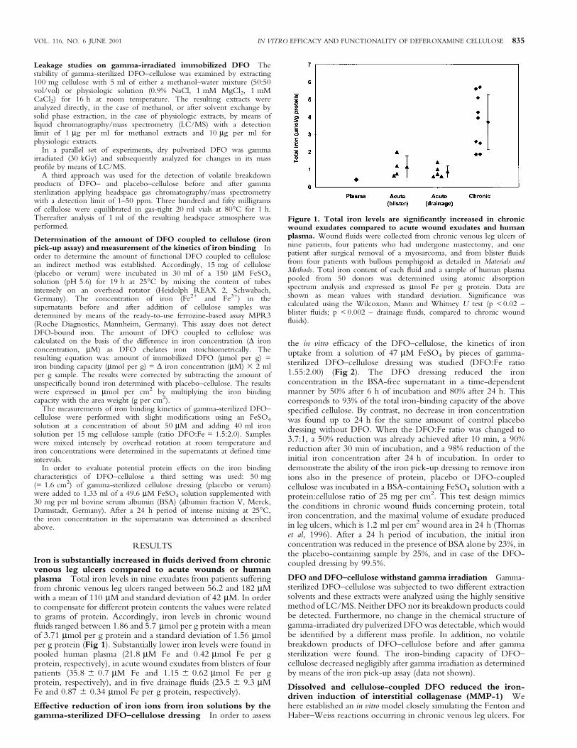
Iron is substantially increased in ¯uids derived from chronicvenous leg ulcers compared to acute wounds or humanplasma Total iron levels in nine exudates from patients sufferingfrom chronic venous leg ulcers ranged between 56.2 and 182 mMwith a mean of 110 mM and standard deviation of 42 mM. In orderto compensate for different protein contents the values were relatedto grams of protein. Accordingly, iron levels in chronic wound¯uids ranged between 1.86 and 5.7 mmol per g protein with a meanof 3.71 mmol per g protein and a standard deviation of 1.56 mmolper g protein (Fig 1). Substantially lower iron levels were found inpooled human plasma (21.8 mM Fe and 0.42 mmol Fe per gprotein, respectively), in acute wound exudates from blisters of fourpatients (35.8 6 0.7 mM Fe and 1.15 6 0.62 mmol Fe per gprotein, respectively), and in ®ve drainage ¯uids (23.5 6 9.3 mMFe and 0.87 6 0.34 mmol Fe per g protein, respectively).
Effective reduction of iron ions from iron solutions by thegamma-sterilized DFO±cellulose dressing In order to assess
the in vitro ef®cacy of the DFO±cellulose, the kinetics of ironuptake from a solution of 47 mM FeSO4 by pieces of gamma-sterilized DFO±cellulose dressing was studied (DFO:Fe ratio1.55:2.00) (Fig 2). The DFO dressing reduced the ironconcentration in the BSA-free supernatant in a time-dependentmanner by 50% after 6 h of incubation and 80% after 24 h. Thiscorresponds to 93% of the total iron-binding capacity of the abovespeci®ed cellulose. By contrast, no decrease in iron concentrationwas found up to 24 h for the same amount of control placebodressing without DFO. When the DFO:Fe ratio was changed to3.7:1, a 50% reduction was already achieved after 10 min, a 90%reduction after 30 min of incubation, and a 98% reduction of theinitial iron concentration after 24 h of incubation. In order todemonstrate the ability of the iron pick-up dressing to remove ironions also in the presence of protein, placebo or DFO-coupledcellulose was incubated in a BSA-containing FeSO4 solution with aprotein:cellulose ratio of 25 mg per cm2. This test design mimicsthe conditions in chronic wound ¯uids concerning protein, totaliron concentration, and the maximal volume of exudate producedin leg ulcers, which is 1.2 ml per cm2 wound area in 24 h (Thomaset al, 1996). After a 24 h period of incubation, the initial ironconcentration was reduced in the presence of BSA alone by 23%, inthe placebo-containing sample by 25%, and in case of the DFO-coupled dressing by 99.5%.
DFO and DFO±cellulose withstand gamma irradiation Gamma-sterilized DFO±cellulose was subjected to two different extractionsolvents and these extracts were analyzed using the highly sensitivemethod of LC/MS. Neither DFO nor its breakdown products couldbe detected. Furthermore, no change in the chemical structure ofgamma-irradiated dry pulverized DFO was detectable, which wouldbe identi®ed by a different mass pro®le. In addition, no volatilebreakdown products of DFO±cellulose before and after gammasterilization were found. The iron-binding capacity of DFO±cellulose decreased negligibly after gamma irradiation as determinedby means of the iron pick-up assay (data not shown).
Dissolved and cellulose-coupled DFO reduced the iron-driven induction of interstitial collagenase (MMP-1) Wehere established an in vitro model closely simulating the Fenton andHaber±Weiss reactions occurring in chronic venous leg ulcers. For
Figure 1. Total iron levels are signi®cantly increased in chronicwound exudates compared to acute wound exudates and humanplasma. Wound ¯uids were collected from chronic venous leg ulcers ofnine patients, four patients who had undergone mastectomy, and onepatient after surgical removal of a myosarcoma, and from blister ¯uidsfrom four patients with bullous pemphigoid as detailed in Materials andMethods. Total iron content of each ¯uid and a sample of human plasmapooled from 50 donors was determined using atomic absorptionspectrum analysis and expressed as mmol Fe per g protein. Data areshown as mean values with standard deviation. Signi®cance wascalculated using the Wilcoxon, Mann and Whitney U test (p < 0.02 ±blister ¯uids; p < 0.002 ± drainage ¯uids, compared to chronic wound¯uids).
VOL. 116, NO. 6 JUNE 2001 IN VITRO EFFICACY AND FUNCTIONALITY OF DEFEROXAMINE CELLULOSE 835

this purpose human dermal ®broblasts were incubated with220 mM Fe(III) citrate and 1 mM ascorbate in the presence ofDFO (220 mM) dissolved in PBS, an equimolar amount of DFOcoupled to cellulose, and control cellulose without DFO as detailedin Materials and Methods. We have used Fe(III) citrateconcentrations in this in vitro model that relate to the upper rangeof Fe concentrations found in ¯uid from chronic wounds. Exposureof human dermal ®broblast monolayer cultures to Fe(III) citrate andascorbate resulted in a 3-fold induction of the MMP-1 proteinconcentration compared to the mock-treated control. Exogenousaddition of H2O2 did not further enhance this induction, mostprobably indicating that intracellular H2O2 levels are suf®cient todrive the Fenton reaction (data not shown). This induction ofMMP-1 could be suppressed by 75% in the presence of dissolvedDFO at a concentration of 220 mM (Fig 3A). Similar results wereobserved in the presence of 10%-serum-containing medium duringthe incubation with Fe(III) citrate (data not shown) indicating thatthe effect of Fe(III) citrate on MMP-1 induction is independent ofserum. DFO-coupled cellulose suppressed the MMP-1 inductionin human dermal ®broblasts by 80% of the control cellulose.Interestingly, addition of DFO±cellulose even at 2 h after initiatingthe Fenton reaction with Fe(III) citrate and ascorbate resulted in analmost complete inhibition of the MMP-1 induction indicating thatiron-driven effects are at least in part responsible for the observedeffects (Fig 3B).
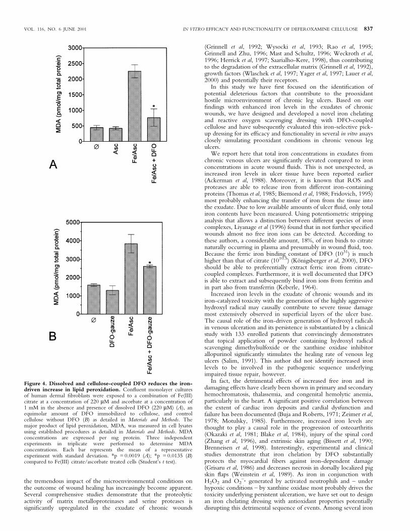
Dissolved and cellulose-coupled DFO reduced the iron-driven increase in lipid peroxidation Using the in vitrosetting with Fe(III) citrate and ascorbate to simulate conditions ofchronic venous leg ulcers, we have studied the effect of dissolvedand cellulose-coupled DFO on lipid peroxidation. The Fe(III)citrate, ascorbate driven Fenton reaction resulted in a 5-foldincrease in MDA, the major product of lipid peroxidation, asdetermined by HPLC based methods. This increase in MDA levelscould be signi®cantly reduced in the presence of dissolved DFO(Fig 4A) or the pick-up dressing coupled with DFO (Fig 4B)clearly indicating the functionality of dissolved and cellulose-coupled DFO.
DISCUSSION
Venous leg ulcers are common and cause considerable morbidity inthe adult population. As healing may be slow or may never be
achieved, ulcers create persistent and substantial demands onresources. A wide variety of dressings have been used, but to datethere is no dressing on the market that can claim to signi®cantlyenhance tissue repair. This may at least partly be due to the limitedknowledge on the microenvironment of chronic wounds. In fact,
Figure 3. Dissolved and cellulose-coupled DFO suppresses theiron-driven induction of interstitial collagenase (MMP-1).Con¯uent monolayer cultures of human dermal ®broblasts were exposedfor 4 h to a combination of Fe(III) citrate at a concentration of 220 mMand ascorbate at a concentration of 1 mM in the absence and presence ofdissolved DFO (220 mM) (A), an equimolar amount of DFOimmobilized to cellulose, or control cellulose without DFO (B) asdetailed in Materials and Methods. Thereafter, cells were washed and afteran incubation period of 24 h supernatants were collected and subjectedto an MMP-1 speci®c ELISA. Three independent experiments havebeen performed in triplicate. Each bar represents the mean of arepresentative experiment with standard deviation. *p = 0.0002 (A); *p= 0.0247, #p = 0.0026 (B) compared with cells exposed to Fe(III)citrate/ascorbate (Student's t test).
Figure 2. Effective reduction of iron ions in iron solution bygamma-sterilized DFO±cellulose. In order to determine the ironbinding capacity of active functional DFO-coupled cellulose andplacebo±cellulose, we have used the iron pick-up assay as detailed inMaterials and Methods. Brie¯y, 15 mg of cellulose sample (placebo orDFO verum) were added to 40 ml FeSO4 solution at an initialconcentration of 47 mM (ratio DFO:Fe = 1.5:2.0). The concentration ofiron was determined in the supernatants before and after addition ofcellulose samples as detailed in Materials and Methods. The results wereexpressed as mmol per l. Three independent experiments were performedin triplicate. Each point represents the mean value with standarddeviation.
836 WENK ET AL THE JOURNAL OF INVESTIGATIVE DERMATOLOGY
the tremendous impact of the microenvironmental conditions onthe outcome of wound healing has increasingly become apparent.Several comprehensive studies demonstrate that the proteolyticactivity of matrix metalloproteinases and serine proteases issigni®cantly upregulated in the exudate of chronic wounds
(Grinnell et al, 1992; Wysocki et al, 1993; Rao et al, 1995;Grinnell and Zhu, 1996; Mast and Schultz, 1996; Weckroth et al,1996; Herrick et al, 1997; Saarialho-Kere, 1998), thus contributingto the degradation of the extracellular matrix (Grinnell et al, 1992),growth factors (Wlaschek et al, 1997; Yager et al, 1997; Lauer et al,2000) and potentially their receptors.
In this study we have ®rst focused on the identi®cation ofpotential deleterious factors that contribute to the prooxidanthostile microenvironment of chronic leg ulcers. Based on our®ndings with enhanced iron levels in the exudates of chronicwounds, we have designed and developed a novel iron chelatingand reactive oxygen scavenging dressing with DFO-coupledcellulose and have subsequently evaluated this iron-selective pick-up dressing for its ef®cacy and functionality in several in vitro assaysclosely simulating prooxidant conditions in chronic venous legulcers.
We report here that total iron concentrations in exudates fromchronic venous ulcers are signi®cantly elevated compared to ironconcentrations in acute wound ¯uids. This is not unexpected, asincreased iron levels in ulcer tissue have been reported earlier(Ackerman et al, 1988). Moreover, it is known that ROS andproteases are able to release iron from different iron-containingproteins (Thomas et al, 1985; Biemond et al, 1988; Fridovich, 1995)most probably enhancing the transfer of iron from the tissue intothe exudate. Due to low available amounts of ulcer ¯uid, only totaliron contents have been measured. Using potentiometric strippinganalysis that allows a distinction between different species of ironcomplexes, Liyanage et al (1996) found that in not further speci®edwounds almost no free iron ions can be detected. According tothese authors, a considerable amount, 18%, of iron binds to citratenaturally occurring in plasma and presumably in wound ¯uid, too.Because the ferric iron binding constant of DFO (1031) is muchhigher than that of citrate (1010.5) (KoÈnigsberger et al, 2000), DFOshould be able to preferentially extract ferric iron from citrate-coupled complexes. Furthermore, it is well documented that DFOis able to extract and subsequently bind iron ions from ferritin andin part also from transferrin (Keberle, 1964).
Increased iron levels in the exudate of chronic wounds and itsiron-catalyzed toxicity with the generation of the highly aggressivehydroxyl radical may causally contribute to severe tissue damagemost extensively observed in super®cial layers of the ulcer base.The causal role of the iron-driven generation of hydroxyl radicalsin venous ulceration and its persistence is substantiated by a clinicalstudy with 133 enrolled patients that convincingly demonstratesthat topical application of powder containing hydroxyl radicalscavenging dimethylsulfoxide or the xanthine oxidase inhibitorallopurinol signi®cantly stimulates the healing rate of venous legulcers (Salim, 1991). This author did not identify increased ironlevels to be involved in the pathogenic sequence underlyingimpaired tissue repair, however.
In fact, the detrimental effects of increased free iron and itsdamaging effects have clearly been shown in primary and secondaryhemochromatosis, thalassemia, and congenital hemolytic anemia,particularly in the heart. A signi®cant positive correlation betweenthe extent of cardiac iron deposits and cardial dysfunction andfailure has been documented (Buja and Roberts, 1971; Zeimer et al,1978; Motulsky, 1985). Furthermore, increased iron levels arethought to play a causal role in the progression of osteoarthritis(Okazaki et al, 1981; Blake et al, 1984), injury of the spinal cord(Zhang et al, 1996), and extrinsic skin aging (Bissett et al, 1990;Brenneisen et al, 1998). Interestingly, experimental and clinicalstudies demonstrate that iron chelation by DFO substantiallyprotects the myocardial ®bers against iron-dependent damage(Grisaru et al, 1986) and decreases necrosis in dorsally localized pigskin ¯aps (Weinstein et al, 1989). As iron in conjunction withH2O2 and O2
±´ generated by activated neutrophils and ± underhypoxic conditions ± by xanthine oxidase most probably drives thetoxicity underlying persistent ulceration, we have set out to designan iron chelating dressing with antioxidant properties potentiallydisrupting this detrimental sequence of events. Among several iron
Figure 4. Dissolved and cellulose-coupled DFO reduces the iron-driven increase in lipid peroxidation. Con¯uent monolayer culturesof human dermal ®broblasts were exposed to a combination of Fe(III)citrate at a concentration of 220 mM and ascorbate at a concentration of1 mM in the absence and presence of dissolved DFO (220 mM) (A), anequimolar amount of DFO immobilized to cellulose, and controlcellulose without DFO (B) as detailed in Materials and Methods. Themajor product of lipid peroxidation, MDA, was measured in cell lysatesusing established procedures as detailed in Materials and Methods. MDAconcentrations are expressed per mg protein. Three independentexperiments in triplicate were performed to determine MDAconcentrations. Each bar represents the mean of a representativeexperiment with standard deviation. *p = 0.0019 (A); *p = 0.0135 (B)compared to Fe(III) citrate/ascorbate treated cells (Student's t test).
VOL. 116, NO. 6 JUNE 2001 IN VITRO EFFICACY AND FUNCTIONALITY OF DEFEROXAMINE CELLULOSE 837
chelators, DFO is the most speci®c and potent, with a stabilityconstant of 1031 (Keberle, 1964). Furthermore, DFO has beenextensively studied in a variety of experimental and clinical settingswith well-established ef®cacy and toxicity pro®les (BeDell andHulbert, 1999; Dollery, 1999).
We report here on the activation of functional groups of thecellulose dressing and successful covalent coupling of DFO to acellulose dressing. As a result, approximately 2 mmol DFO werecovalently bound per square centimeter of cellulose dressing,providing an iron complexing capacity suf®cient to cope with theiron levels found in chronic venous leg ulcers. Furthermore, itcould be shown that DFO±cellulose is able to bind iron out of aBSA-containing iron solution as an arti®cial chronic wound ¯uid.As stability after gamma sterilization represents an importantprerequisite for a dressing, leakage of gamma-sterilized DFO-coupled cellulose was analyzed. No leakage of DFO or itsbreakdown products from the dressing was detected. This is inline with data from the literature on the extraordinary stability ofurethane bonds (Bethell and Ayers, 1981).
As connective tissue breakdown and lipid peroxidation representpathogenic hallmarks preventing healing of chronic venous legulcers, we established a model that closely simulates the iron-drivenFenton reaction. Using this model system, we have screeneddissolved and cellulose-bound DFO for their protecting propertiesfrom Fe(III) citrate/ascorbate driven upregulation of MMP-1, themajor metalloproteinase in connective tissue breakdown, andenhanced lipid peroxidation. We found that exposure of ®broblaststo the Fenton reaction mimicking combination of 220 mM Fe(III)citrate and 1 mM ascorbate resulted in a 4-fold induction of MMP-1 levels. This induction was almost completely suppressed bydissolved DFO at a concentration of 220 mM or the equimolaramount of DFO covalently coupled to cellulose.
In addition, the Fe(III) citrate/ascorbate driven Fenton reactionresulted in a 5-fold increase in MDA, the major product of lipidperoxidation. The increase in MDA levels could be signi®cantlyreduced in the presence of dissolved DFO or DFO-coupledcellulose. Collectively, these data indicate that DFO in its dissolvedor bound form effectively prevents the development of aprooxidant microenvironnment and, furthermore, protects fromunfavorable consequences like MMP-1 and lipid peroxide induc-tion. We anticipate that the protective effect of DFO by the abovementioned mechanisms limits tissue injury and the perpetuatedrecruitment of activated neutrophils characteristic for chronicvenous leg ulcers. In fact, in a TPA (12-O-tetradecanoyl-phorbol-13-acetate) induced in¯ammation model, topical application ofDFO prior to TPA treatment could almost completely inhibit theneutrophil in¯ux into the interstitial tissue of mouse ears (Soybiret al, 1996).
Based on our promising results a double blind placebo controlledclinical study has been started to prove the principle and assess thetherapeutic value for patients suffering from nonhealing wounds.
We gratefully acknowledge the stimulating discussion with Cecilia Welling.
REFERENCES
Ackerman Z, Seidenbaum M, Loewenthal E, Rubinow A: Overload of iron in theskin of patients with varicose ulcers. Possible contributing role of ironaccumulation in progression of the disease. Arch Dermatol 124:1376±1378, 1988
Bayreuther K, Francz PI, Rodemann HP: Fibroblasts in normal and pathologicalterminal differentiation. Aging, Apoptosis Transformation Arch Geront Geriatr(Suppl.3):47±74, 1992
BeDell LS, Hulbert MK, eds: Physicians GenRx ± Mosby's Complete Drug Reference, 9thedn. St. Louis, MO: Mosby-Year Book, 1999
Bethell GS, Ayers JS: Investigation of the activation of various insolublepolysaccharides with 1,1¢-carbonyldiimidazole and of the properties of theactivated matrices. J Chromat 219:361±372, 1981
Biemond P, Swaak AJG, van Eijk HG, Koster JF: Superoxide dependent iron releasefrom ferritin in in¯ammatory diseases. Free Radic Biol Med 4:185±198, 1988
Bissett DL, Chatterjee R, Hannon DP: Photoprotection effect of superoxidescavenging antioxidants against ultraviolet radiation-induced chronic skin
damage in the hairless mouse. Photodermatol Photoimmunol Photomed 7:56±62,1990
Blake DR, Gallagher PH, Potter AR: The effect of synovial iron on the progressionof rheumatoid disease. Arthritis Rheum 27:495±501, 1984
Bradford MM: A rapid and sensitive method for the quantitation of microgramquantities of protein utilizing the principle of protein-dye binding. AnalBiochem 72:248±254, 1976
Brenneisen P, Wenk J, Klotz LO, et al: Central role of ferrous/ferric iron in theultraviolet B irradiation-mediated signaling pathway leading to increasedinterstitial collagenase (matrix-degrading metalloprotease (MMP-1) andstromelysin-1 (MMP-3) mRNA levels in cultured human dermal ®broblasts.J Biol Chem 273:5279±5287, 1998
Buja LM, Roberts WC: Iron in the heart: ethiology and clinical signi®cance. Am JMed 51:209±221, 1971
Cheatle T: Venous ulceration and free radicals. Br J Dermatol 124:508, 1991Dollery C: ed. Therapeutic Drugs CD-ROM Database Release 1.0. London: Churchill
Livingstone ± Harcourt Brace Ltd, 1999Edwards JV, Batiste SL, Gibbins EM, Goheen SC: Synthesis and activity of NH2- and
COOH-terminal elastase recognition sequences on cotton. J Peptide Res54:536±543, 1999
Falanga V, Eaglstein WH: The ``trap'' hypothesis of venous ulceration. Lancet341:1006±1008, 1993
Feldhoff PW: Comparison of coupling procedures for development of af®nitymembranes: opimization of the CDI method. Techniques in Protein ChemistryIII. London: Academic Press, 1992, pp 151±160
Fleischmajer R, Perlish JS, Krieg T, Timpl R: Variability in collagen and ®bronectinsynthesis by scleroderma ®broblasts in primary culture. J Invest Dermatol76:400±403, 1981
Fridovich I: Superoxide radical and superoxide dismutases. Annu Rev Biochem 64:97±112, 1995
Green LM, Reade JL, Ware CF: Rapid colorimetric assay for cell viability:application to the quantitation of cytotoxic and growth inhibitorylymphokines. J Immunol Methods 70:257±268, 1984
Grinnell F, Zhu M: Fibronectin degradation in chronic wounds depends on therelative levels of elastase, a1-proteinase inhibitor, and a2-macroglobulin.J Invest Dermatol 106:335±341, 1996
Grinnell F, Ho CH, Wysocki A: Degradation of ®bronectin and vitronectin inchronic wound ¯uid: analysis by cell blotting, immunoblotting, and celladhesion assays. J Invest Dermatol 98:410±416, 1992
Grisaru D, Goldfarb AW, Gotsman MS: Deferoxamine improves left ventricularfunction in beta-thalassemia. Arch Intern Med 146:2344±2349, 1986
Gutteridge JMC, Richmond R, Halliwell B: Inhibition of the iron-catalysedformation of hydroxyl radicals from superoxide and of lipid peroxidation bydesferrioxamine. Biochem J 184:469±472, 1979
Halliwell B, Gutteridge JMC: Free Radicals in Biology and Medicine, 2nd edn. Oxford:Clarendon Press, 1989
Hearn MTW: 1,1¢-Carbonyldiimidazole-mediated immobilization of enzymes andaf®nity ligands. Meth Enzymol 135:102±117, 1987
Herrick S, Ashcroft G, Ireland G, Horan M, McCollum C, Ferguson M: Up-regulation of elastase in acute wounds of healthy aged humans and chronicvenous leg ulcers are associated with matrix degradation. Lab Invest 77:281±288, 1997
Keberle H: The biochemistry of desferrioxamine and its relation to iron metabolism.Ann N Y Acad Sci 119:758±768, 1964
KoÈnigsberger LC, KoÈnigsberger E, May PM, Hefter GT: Complexation of iron(III)and iron(II) by citrate. Implications for iron speciation in blood plasma. J InorgBiochem 78:175±184, 2000
Lauer G, Sollberg S, Cole M, et al: Expression and proteolysis of vascular endothelialgrowth factor is increased in chronic wounds. J Invest Dermatol 115:12±18, 2000
Liyanage JA, Taylor DM, Williams DR: Chemical speciation in wound ¯uid leadingto enhanced bioavailability for healing. Chem Speciation Bioavailability 8:45±52,1996
Mast BA, Schultz GS: Interactions of cytokines, growth factors, and proteases in acuteand chronic wounds. Wound Rep Reg 4:411±420, 1996
Meyer-Ingold W, Eichner W, Ettner N, Schink M: Wound coverings for removal ofinterfering factors from wound ¯uid. Patent Application EP 0 945 144 A2, 1998
Monboisse JC, Borel JP: Oxidative damage to collagen. In Emerit I, Chance B: eds.Free Radicals and Aging. Experimentia (Suppl 62). Basel: BirkhaÈuser Verlag, 1992,pp 323±327
Motulsky AG: Hemochromatosis (iron storage disease). In Wyngaarden JB, SmithLH, eds. Cecil's Textbook of Medicine, 17th edn. Philadelphia: W.B. Saunders,1985, pp 1160±1163
Okazaki I, Brinckerhoff CE, Sinclair JF: Iron increases collagenase production byrabbit synovial ®broblasts. J Lab Clin Med 97:396±402, 1981
Rao CN, Ladin DA, Liu YY, Chilukuri K, Hou ZZ, Woodley DT: a1-Antitrypsinis degraded and non-functional in chronic wounds but intact and functional inacute wounds: the inhibitor protects ®bronectin from degradation by chronicwounds ¯uid enzymes. J Invest Dermatol 105:572±578, 1995
Rosner K, Ross C, Karlsmark T, Petersen AA, Gottrup F, Vejlsgaard GL:Immunohistochemical characterization of the cutaneous cellular in®ltrate indifferent areas of chronic leg ulcers. APMIS 103:293±299, 1995
Saarialho-Kere UK: Patterns of matrix metalloproteinase and TIMP expression inchronic ulcers. Arch Dermatol Res 290:S47±S54, 1998
Salim AS: The role of oxygen-derived free radicals in the management of venous(varicose) ulceration: a new approach. World J Surg 15:264±269, 1991
Schaich KM: Metals and lipid oxidation. Contemporary issues. Lipids 27:209±218,1992
838 WENK ET AL THE JOURNAL OF INVESTIGATIVE DERMATOLOGY

Soybir GR, Koyuneu H, Koksoy F: Protective effect of desferrioxamine against TPAcaused in¯ammation in CD-1 mouse skin. Surg Oncol 5:253±258, 1996
Thomas CE, Morehouse LA, Aust SD: Ferritin and superoxide-dependent lipidperoxidation. J Mol Biol 260:3275±3280, 1985
Thomas S, Fear M, Humphreys J, Disley L, Waring MJ: The effect of dressings onthe production of exudate from venous leg ulcers. Wounds 8:145±150, 1996
Weckroth M, Vaheri A, Lauharanta J, Sorsa T, Konttinen YT: Matrixmetalloproteinases, gelatinase and collagenase, in chronic leg ulcers. J InvestDermatol 106:1119±1124, 1996
Weinstein GS, Maves MD, McCormack ML: Deferoxamine decreases necrosis indorsally based pig skin ¯aps. Otolaryngol Head Neck Surg 101:559±561,1989
Weiss SJ: Tissue destruction by neutrophils. N Engl J Med 320:365±376, 1989Wenner A, Leu HJ, Spycher M, Brunner U: Ultrastructural changes of capillaries in
chronic venous insuf®ciency. Expl Cell Biol 48:1±14, 1980Wlaschek M, Peus D, Achterberg V, Meyer-Ingold W, Scharffetter-Kochanek K:
Protease inhibitors protect growth factor activity in chronic wounds. Br JDermatol 137:646±663, 1997
Wysocki AB, Staiano-Coico L, Grinnell F: Wound ¯uid from chronic leg ulcerscontains elevated levels of metalloproteinases MMP-2 and MMP-9. J InvestDermatol 101:64±68, 1993
Yager DR, Chen SM, Ward SI, Olutoye OO, Diegelmann RF, Cohen IK: Ability ofchronic wound ¯uids to degrade peptide growth factors is associated withincreased levels of elastase activity and diminished levels of proteinaseinhibitors. Wound Rep Reg 5:23±32, 1997
Yu LW, Latriano L, Duncan S, Hartwick RA, Witz G: High-performance liquidchromatography analysis of the thiobarbituric acid adducts of malonaldehydeand trans,trans-muconaldehyde. Anal Biochem 156:326±333, 1986
Zeimer R, Belkin M, Leitersdorff E: A non-invasive method for the evaluation oftissue iron deposition in beta thalassemia major. J Lab Clin Med 91:24±31, 1978
Zhang JR, Scherch HM, Hall ED: Direct measurement of lipid hydroperoxides iniron-dependent spinal neuronal injury. J Neurochem 66:355±361, 1996
VOL. 116, NO. 6 JUNE 2001 IN VITRO EFFICACY AND FUNCTIONALITY OF DEFEROXAMINE CELLULOSE 839