Embed Size (px)
Citation preview
HYPOCHROMI C ANEMIA & IRON METABOLISM
OBJECTIVE
• Iron metabolism
• Iron distribution & transport
• Dietary iron
• Iron absorption
• Iron requirements
• Disorders of iron metabolism
• Hypochromic anemia
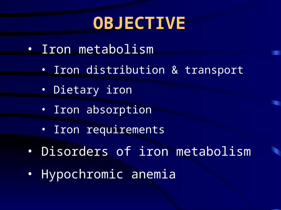
• To accept & donate electron (Fe2+ Fe3+)
• component of cytochromes, oxygen-binding
molecules
• cell growth,proliferation, differentiation
• damage tissues
H2O2 OH
Fe2+ Fe3+
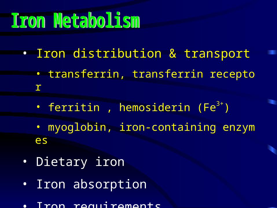
• Iron distribution & transport
• transferrin, transferrin receptor
• ferritin , hemosiderin (Fe3+)
• myoglobin, iron-containing enzymes
• Dietary iron
• Iron absorption
• Iron requirements
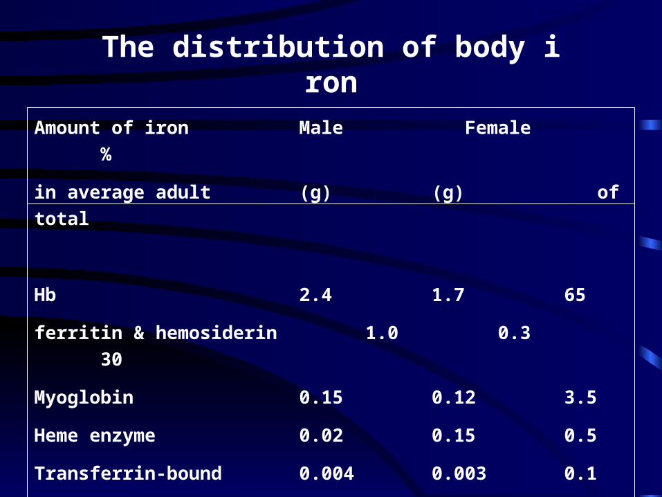
Amount of iron Male Female %
in average adult (g) (g) of total
Hb 2.4 1.7 65
ferritin & hemosiderin 1.0 0.3 30
Myoglobin 0.15 0.12 3.5
Heme enzyme 0.02 0.15 0.5
Transferrin-bound 0.004 0.003 0.1
iron
The distribution of body iron
• Iron distribution & transport
• Dietary iron
• ferric hydroxides
• ferric-protein complexs
• heme-protein complexes
• Iron absorption
• Iron requirements
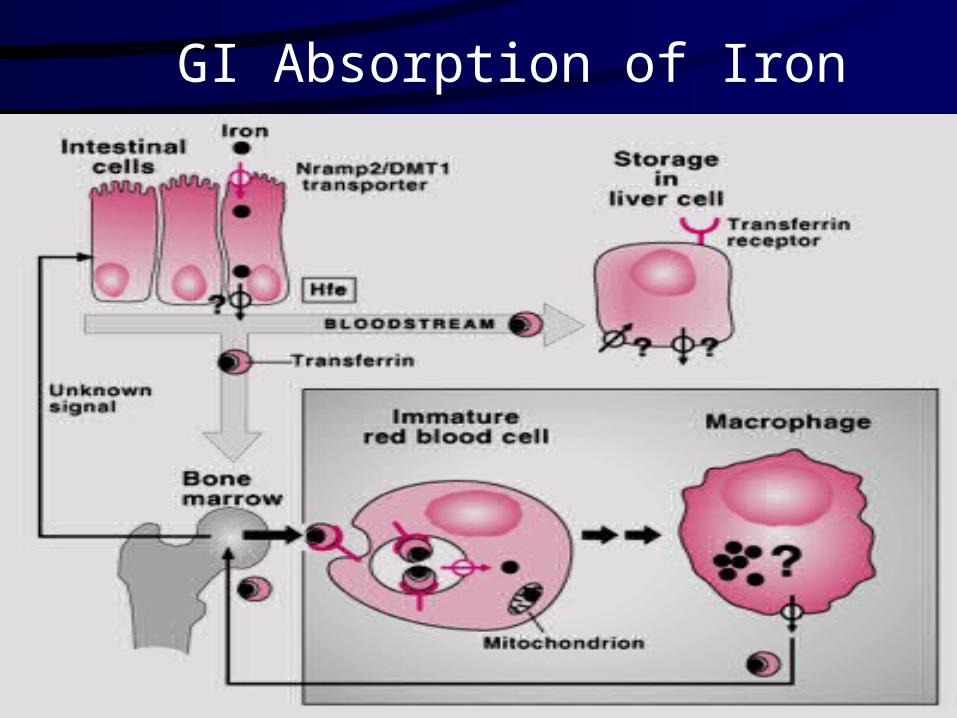
GI Absorption of Iron

INTRACELLULAR IRON TRANSPORT
• Iron distribution & transport
• Dietary iron
• Iron absorption
• Iron requirements
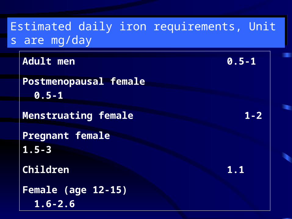
Adult men 0.5-1
Postmenopausal female 0.5-1
Menstruating female 1-2
Pregnant female 1.5-3
Children 1.1
Female (age 12-15) 1.6-2.6
Estimated daily iron requirements, Units are mg/dayEstimated daily iron requirements, Units are mg/day
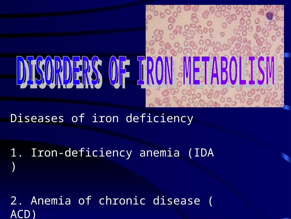
Diseases of iron deficiency
1. Iron-deficiency anemia (IDA)
2. Anemia of chronic disease (ACD)
Diseases of iron overload
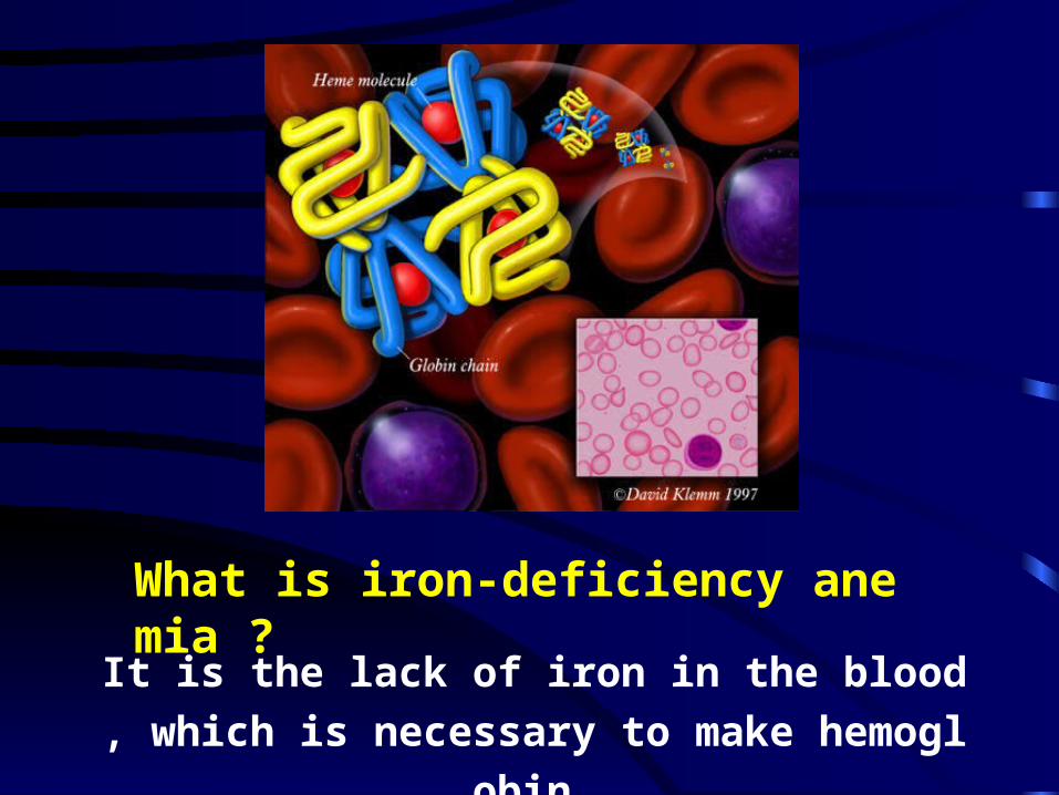
What is iron-deficiency anemia ?
It is the lack of iron in the blood, which is ne
cessary to make hemoglobin.
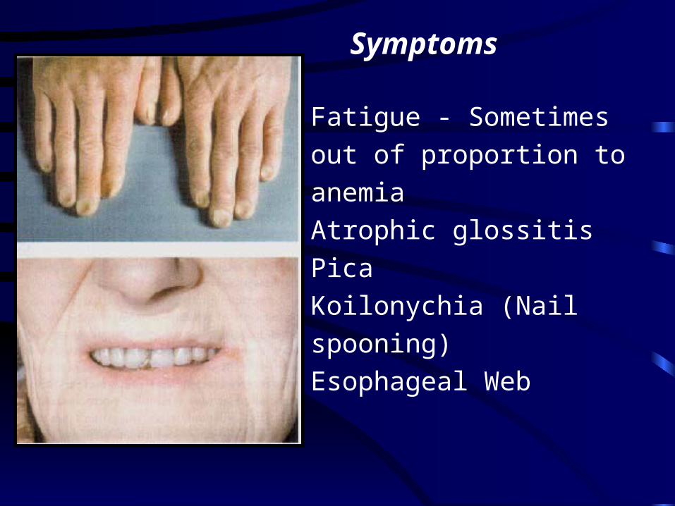
Symptoms
Fatigue - Sometimes out of
proportion to anemia
Atrophic glossitis
Pica
Koilonychia (Nail spooning)
Esophageal Web
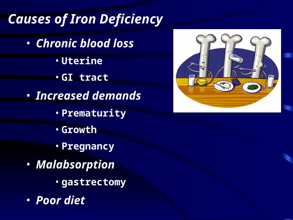
Causes of Iron Deficiency
• Chronic blood loss
• Uterine
• GI tract
• Increased demands
• Prematurity
• Growth
• Pregnancy
• Malabsorption
• gastrectomy
• Poor diet
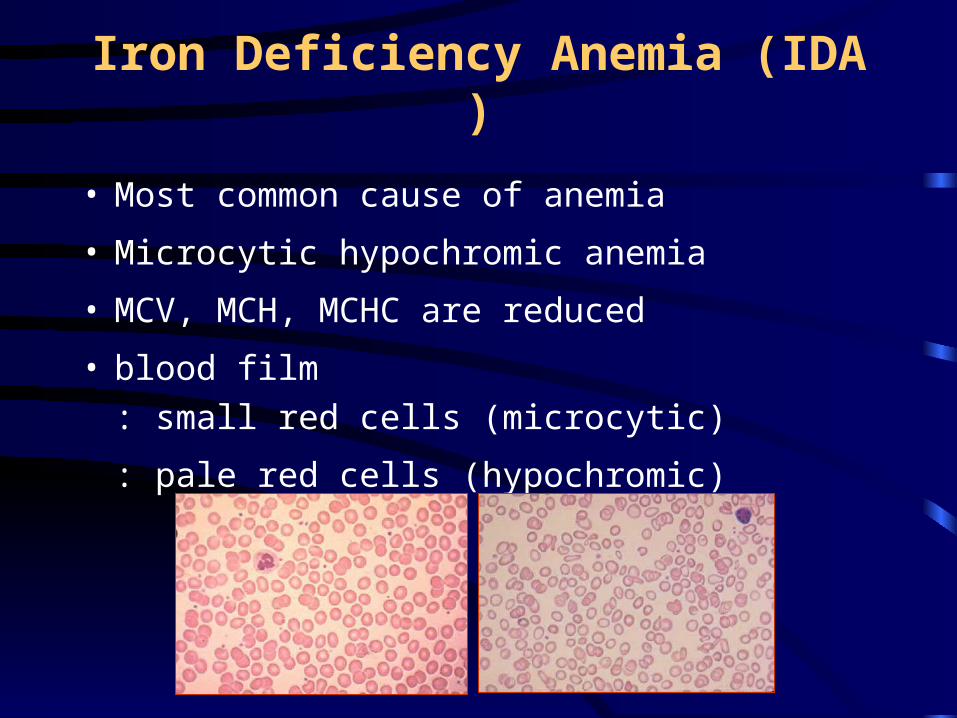
Iron Deficiency Anemia (IDA)
• Most common cause of anemia
• Microcytic hypochromic anemia
• MCV, MCH, MCHC are reduced
• blood film : small red cells (microcytic)
: pale red cells (hypochromic)
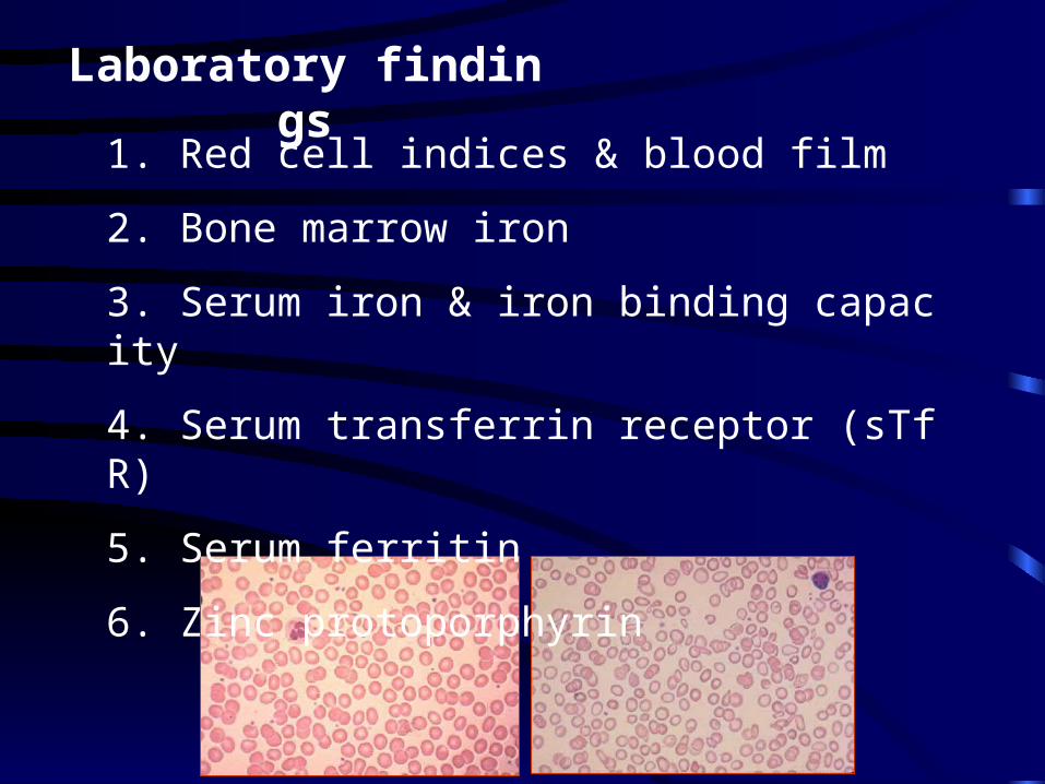
Laboratory findings
1. Red cell indices & blood film
2. Bone marrow iron
3. Serum iron & iron binding capacity
4. Serum transferrin receptor (sTfR)
5. Serum ferritin
6. Zinc protoporphyrin
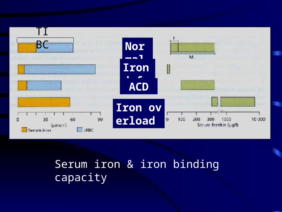
Normal Irondef.ACD
Iron overload
TIBC
Serum iron & iron binding capacity
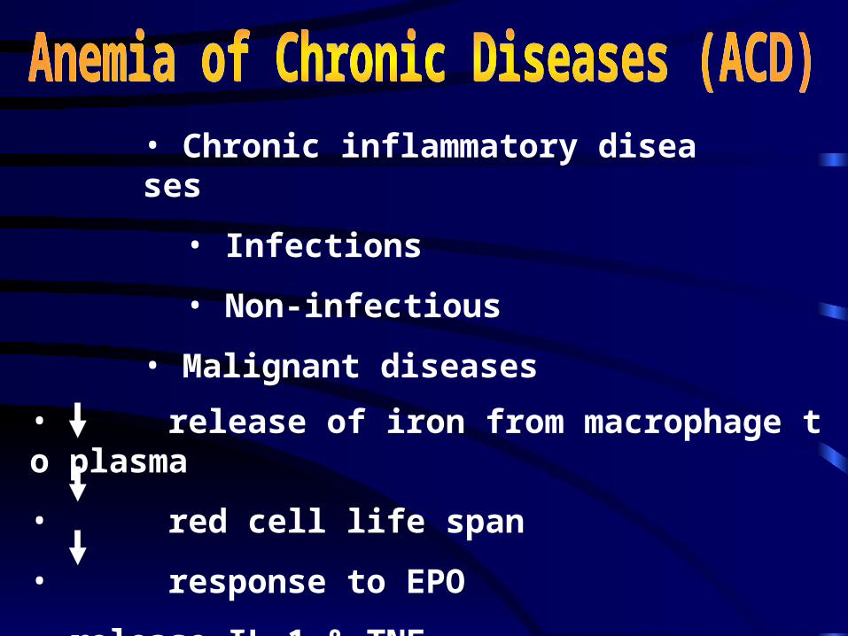
• Chronic inflammatory diseases
• Infections
• Non-infectious
• Malignant diseases
• release of iron from macrophage to plasma
• red cell life span
• response to EPO
• release IL-1 & TNF
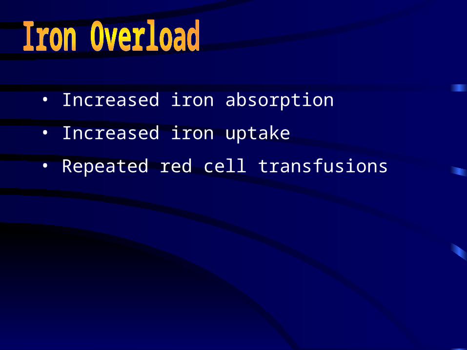
• Increased iron absorption
• Increased iron uptake
• Repeated red cell transfusions
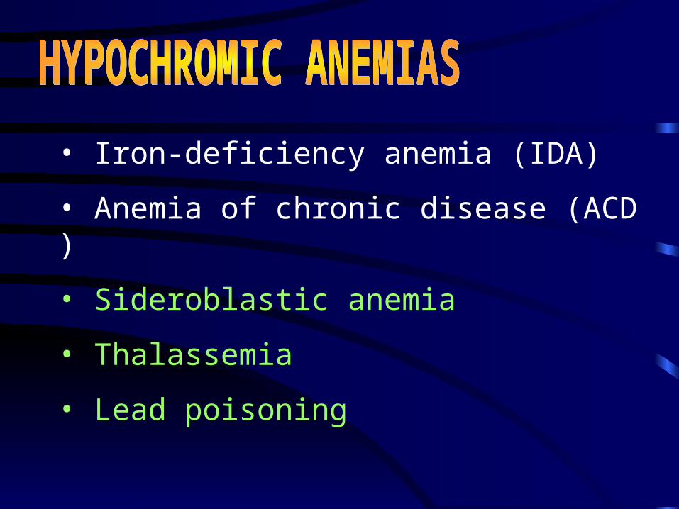
• Iron-deficiency anemia (IDA)
• Anemia of chronic disease (ACD)
• Sideroblastic anemia
• Thalassemia
• Lead poisoning
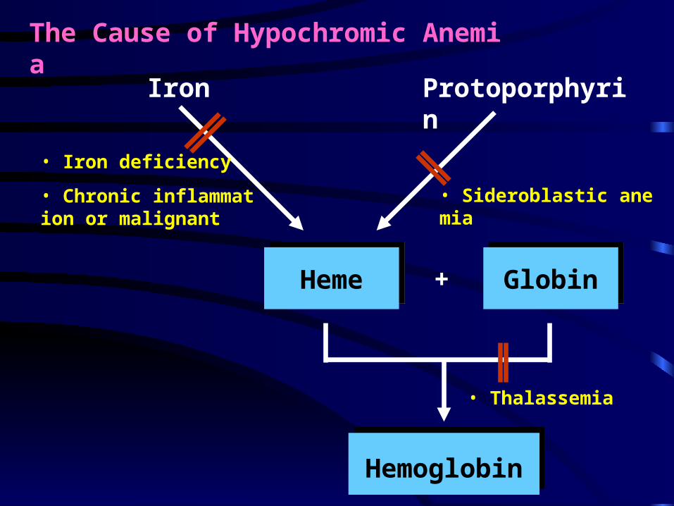
Iron Protoporphyrin
HemeHeme GlobinGlobin+
HemoglobinHemoglobin
• Iron deficiency
• Chronic inflammation or malignant
• Thalassemia
• Sideroblastic anemia
The Cause of Hypochromic Anemia
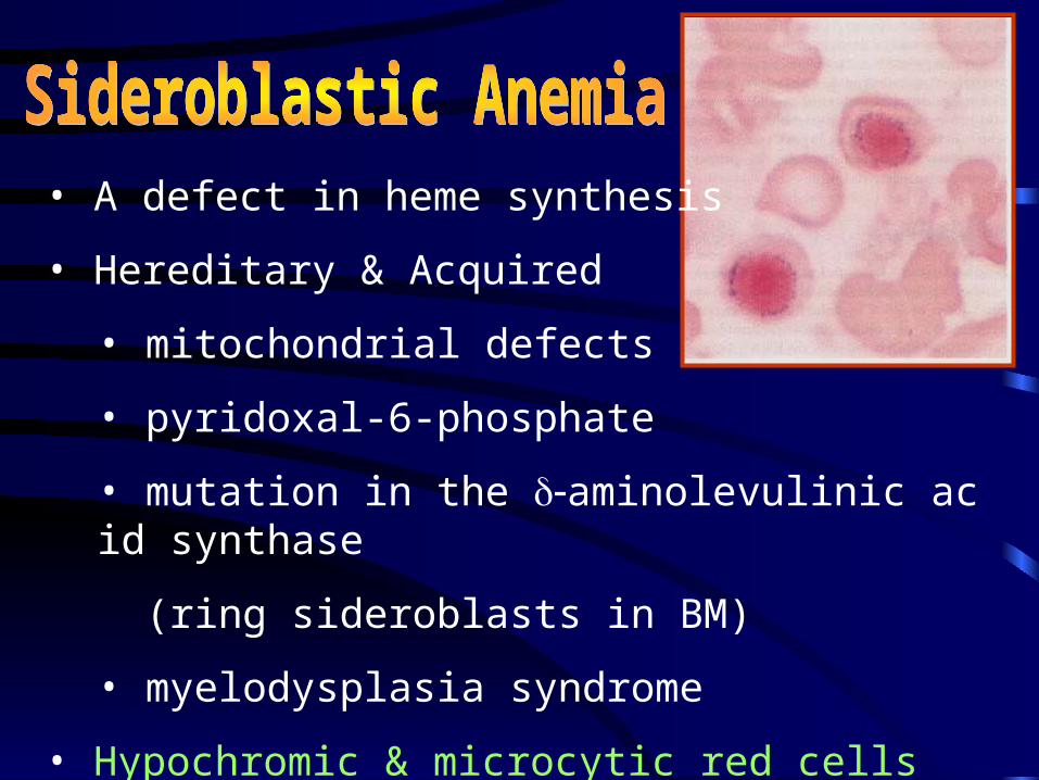
• A defect in heme synthesis
• Hereditary & Acquired
• mitochondrial defects
• pyridoxal-6-phosphate
• mutation in the aminolevulinic acid synthase
(ring sideroblasts in BM)
• myelodysplasia syndrome
• Hypochromic & microcytic red cells
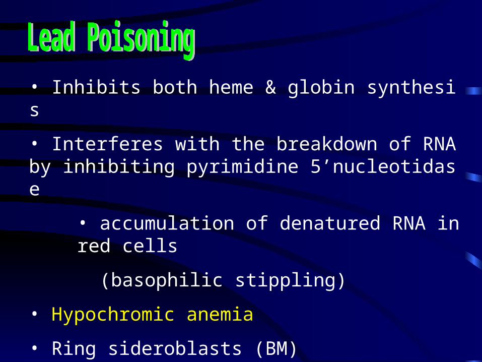
• Inhibits both heme & globin synthesis
• Interferes with the breakdown of RNA by inhibiting pyrimidine 5’nucleotidase
• accumulation of denatured RNA in red cells
(basophilic stippling)
• Hypochromic anemia
• Ring sideroblasts (BM)
• Free erythrocyte protoporphyrin is raised
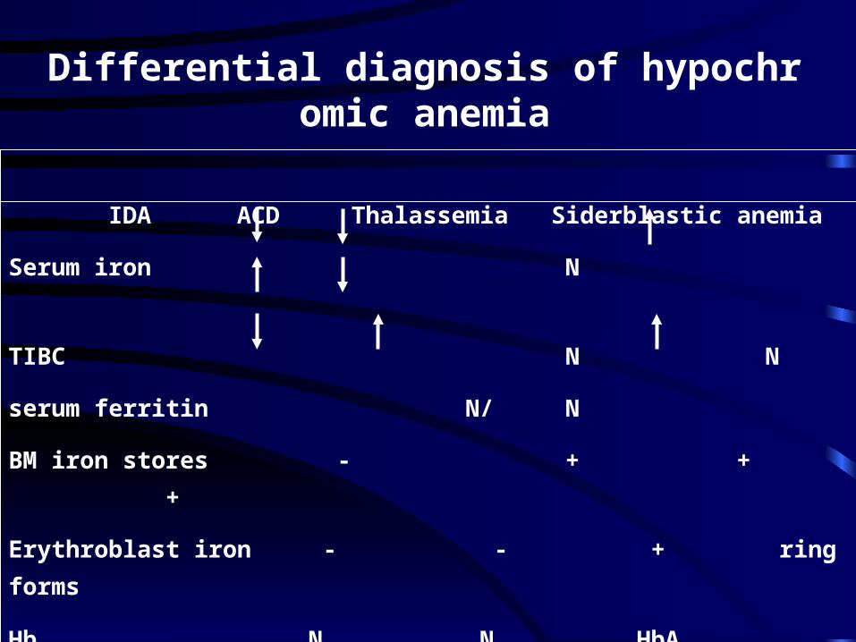
Differential diagnosis of hypochromic anemia
IDA ACD Thalassemia Siderblastic anemia
Serum iron N
TIBC N N
serum ferritin N/ N
BM iron stores - + + +
Erythroblast iron - - + ring forms
Hb N N HbA2 N
electrophoresis
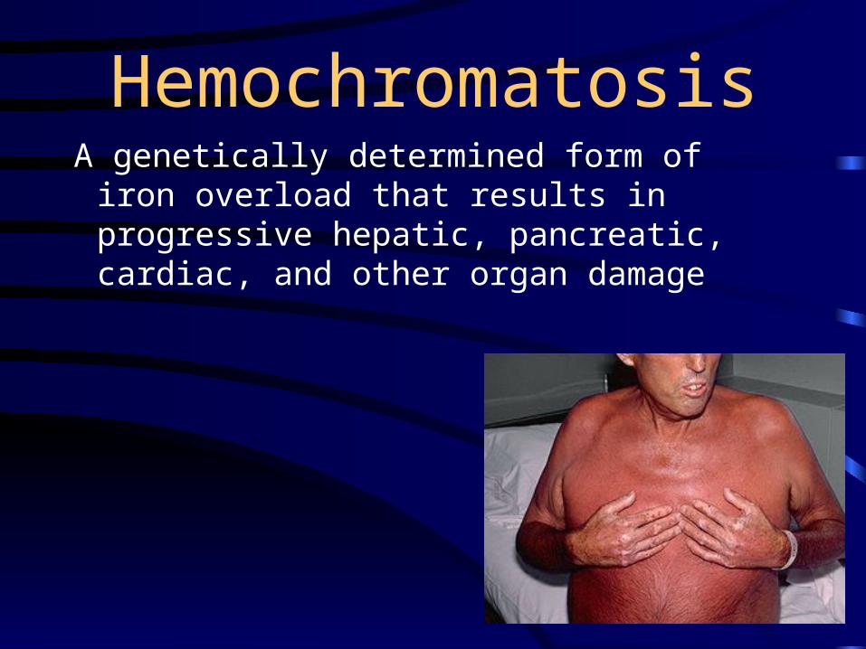
HemochromatosisA genetically determined form of iron
overload that results in progressive hepatic, pancreatic, cardiac, and other organ damage
Hemochromatosis• It is one of the most common genetic
disorders in the U.S.• Present in heterozygous (one gene) form
in 12% of nonblacks and 30% of blacks• Present in homozygous form (2 gene) in
1 in 200 nonblacks and 1 in 100 blacks• Homozygotes will die of iron overload
unless they give blood frequently• Homozygotes absorb three times more
iron from food than other people• Even heterozygotes may be at risk for
iron overload, increasing risk of heart disease
Hemochromatosis: Risk Factors
• Higher risk in people of northern European descent
• Men tend to manifest symptoms earlier because they have no way to dispose of excess iron (menstruation, pregnancy, lactation)
• Men may develop symptoms in their 30s but may not be diagnosed until their 50s
• Women often develop symptoms after menopause
Hemochromatosis: Symptoms
• Joint pain• Fatigue• Lack of energy• Abdominal pain• Loss of sex drive• Heart problems• Abnormal pigmentation of the
skin, making it look gray or bronze
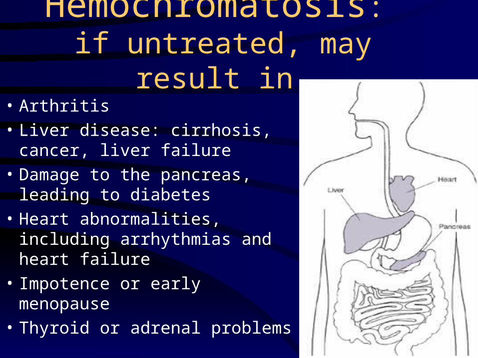
Hemochromatosis: if untreated, may result in
• Arthritis• Liver disease: cirrhosis,
cancer, liver failure• Damage to the pancreas,
leading to diabetes• Heart abnormalities,
including arrhythmias and heart failure
• Impotence or early menopause
• Thyroid or adrenal problems
Hemochromatosis: Diagnosis and
Treatment• Testing: serum ferritin and transferrin saturation can reveal excess stores of iron; followed by HFE (genetic) test and possible liver biopsy
• Treatment: regular phlebotomy to remove excess iron
• Avoidance of iron supplements and sources of iron in the diet, especially heme iron
• Awareness of iron cooking vessels