Embed Size (px)
Citation preview

Selected Pages from CHAPTER 8 of
Forensic Pathology, Second Edition
Vincent J. M. DiMaio;Dominick J DiMaio
ISBN: 0-8493-0072-XPub Date: 06/28/2001
Only the pages from this chapter that offer information relative to investigation of "Restraint Asphyxia" cases were scanned for inclusion within this PDF: pages 229-244 and 273-275; as well as all pages identifying the references cited within this chapter (275-277).
The black and white photos on these pages were not redacted from the scanned pages. However, several unnumbered pages were found within this chapter, containing Color Figure photos. Those pages were not scanned into the PDF: Color Figures 2.6, 3.13, 4.9, 4.6, 5.7, 6.8, 6.11(a,b,c), 7.15, 8.11, 8.24, 9.11, 9.19, 13.6, 13.7, 16.1, and 19.2.
When scanning these pages to RTF files, the yellow highlighting was ignored! Thus, this PDF does not contain the yellow highlight found within the jpg page scans found on http://www.charlydmiller.com/LIB/forensicpathasphyxia.html
For the purpose of clarification, I occasionally inserted text on some RTF pages, prior to creating this PDF. ALL inserted text appears between brackets ([ ]) in dark blue font color.
Asphyxia
8
Asphyxial deaths are caused by the failure of cells to receive or utilize oxygen. The deprivation of oxygen can be partial (hypoxia) or total (anoxia). The classical signs of asphyxia are visceral congestion, petechiae, cyanosis, and fluidity of blood. These are nonspecific, however, and can occur in deaths from other causes. Visceral congestion is due to obstructed venous return and cap-illovenous congestion. The latter is a result of the susceptibility of these vessels to hypoxia, with resultant dilatation of the vessels and stasis of blood.
Petechiae are pinpoint hemorrhages produced by rupture of small ves-sels, predominantly small venules. Rupture appears to be mechanical in etiology and is caused by sudden over distention and rupture of the vessels following abrupt increases in intravascular pressure.1'2 These are most com-mon in the visceral pleura and epicardium. In asphyxial deaths from stran-gulation, petechiae are classically seen in the conjunctivae and sclerae. Petechiae, as nonspecific markers,1'2 may be seen in the conjunctivae and sclerae in association with many different conditions, not all fatal, and not just in asphyxial deaths. They are routinely seen in the reflected scalp in all types of death and are of no diagnostic significance in this area. Petechiae of the epiglottis are also of no significance. Gordon and Mansfield documented development of epicardial petechiae after death.2
Petechiae can develop after death in dependent areas of the body e.g., an arm hanging over the side of a bed. Here, gravity causes increased intravascular congestion and pressure with resultant mechanical rupture of small vessels. If the petechiae become larger or confluent, they are called ecchymoses.
Cyanosis is, of course, nonspecific and caused by an increase in the amount of reduced hemoglobin. It does not become observable until at least 5 g of reduced hemoglobin is present. Postmortem fluidity of blood is not characteristic of asphyxia or any cause of death, but rather the result of a high rate of fibrinolysis that occurs in rapid deaths, possibly by high agonal levels of catecholamines.3
Asphyxial deaths can be loosely grouped into three categories:
1. Suffocation 2. Strangulation 3. Chemical asphyxia
229
230 Forensic Pathology
These deaths might be accidental, suicidal or homicidal in manner. Com-pared with other causes of homicide, homicides via asphyxia are relatively uncommon in the U.S. They predominantly involve strangulation — manual and ligature strangulation. In the last ten years, murders ascribed to stran-gulation have averaged 286 a year, with a range of 366 to 211. There seems to have been a gradual decrease in the number of such cases over the years. Murders caused by "asphyxiation" (no further description but excluding strangulation) have averaged 107 a year, with this number being fairly con-stant over the ten-year period.4
Suffocation
In deaths from suffocation, there is failure of oxygen to reach the blood. There are six general forms of suffocation:
1. Entrapment/environmental suffocation 2. Smothering 3. Choking 4. Mechanical asphyxia 5. Mechanical asphyxia combined with smothering 6. Suffocating gases
Entrapment / Environmental Suffocation
In suffocation by entrapment or environmental hazard, asphyxia is caused by inadequate oxygen in the environment. These deaths are almost exclusively accidental in nature. In entrapment, individuals find themselves trapped in an air-tight or relatively air-tight enclosure. Initially, there is sufficient oxygen to breathe. However, as respiration continues, they exhaust the oxygen and asphyxiate. The best example of this is a child trapped in a discarded refrig-erator. Fortunately, this specific form of death by entrapment is becoming rare, as modern refrigerators do not have a latch system of locking and can be pushed open from the interior. Suicide and homicide by entrapment are rare, but do occur.
In environmental suffocation, an individual inadvertently enters an area where there is gross deficiency of oxygen. This deficiency is not due to displacement of the oxygen by suffocating gases, which will be discussed in another section, but rather that the oxygen has been depleted by some mech-anism. Thus, the authors reported two deaths caused by lack of oxygen in an underground chamber.5 The normal percentage by volume of oxygen in the atmosphere is 20.946%. In this particular case, the percentage by volume was 9.6%. This lethal atmosphere was caused by fungus-like organisms and
Asphyxia 231
low forms of plant life present on the vault walls and in the sediment on the floor. The metabolic processes of the fungi and plant life resulted in depletion of oxygen by these organisms, with production of carbon dioxide. Thus, carbon dioxide, which is normally 0.033% in air, in this case, was 7.0%. The increased quantity of carbon dioxide, however, was insufficient in itself to have caused death by displacement of oxygen. It was the absolute lack of oxygen that caused death. At oxygen concentrations of 10 to 15%, there is impairment in judgment and coordination. Loss of consciousness occurs at 8 to 10%; death at 8% and less. At oxygen concentrations of 4 to 6%, there is loss of consciousness in 40 sec and death within a few min.
In deaths due to entrapment or environmental suffocation, the cause of death cannot be determined by autopsy alone, because there are no specific findings. All that one finds is nonspecific acute visceral congestion. It is only by an analysis of the circumstances leading up to and surrounding death, and the exclusion of other causes, that one can make a determination as to the cause of death.
Smothering
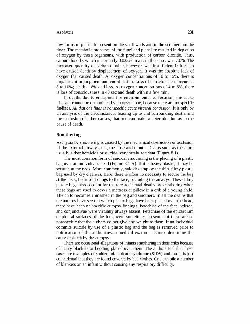
Asphyxia by smothering is caused by the mechanical obstruction or occlusion of the external airways, i.e., the nose and mouth. Deaths such as these are usually either homicide or suicide, very rarely accident (Figure 8.1).
The most common form of suicidal smothering is the placing of a plastic bag over an individual's head (Figure 8.1 A). If it is heavy plastic, it may be secured at the neck. More commonly, suicides employ the thin, filmy plastic bag used by dry cleaners. Here, there is often no necessity to secure the bag at the neck, because it clings to the face, occluding the airways. These filmy plastic bags also account for the rare accidental deaths by smothering when these bags are used to cover a mattress or pillow in a crib of a young child. The child becomes enmeshed in the bag and smothers. In all the deaths that the authors have seen in which plastic bags have been placed over the head, there have been no specific autopsy findings. Petechiae of the face, sclerae, and conjunctivae were virtually always absent. Petechiae of the epicardium or pleural surfaces of the lung were sometimes present, but these are so nonspecific that the authors do not give any weight to them. If an individual commits suicide by use of a plastic bag and the bag is removed prior to notification of the authorities, a medical examiner cannot determine the cause of death by the autopsy.
There are occasional allegations of infants smothering in their cribs because of heavy blankets or bedding placed over them. The authors feel that these cases are examples of sudden infant death syndrome (SIDS) and that it is just coincidental that they are found covered by bed clothes. One can pile a number of blankets on an infant without causing any respiratory difficulty.
232 Forensic Pathology
Figure 8.1 (A) Suicide of elderly female who secured plastic bag over head with tie around neck. (B) Accidental smothering in mentally retarded, 43-year-old male who wrapped face in duct tape. Deceased had performed this act before.
Occasionally, an alcoholic is found face down on a pillow, dead. Cir-cumferential oral and nasal pallor is noted and death is attributed to smothering. This pallor, however, can be caused post mortem by passive pressure of the dependent head on the pillow. Thus, the diagnosis cannot be made on this "evidence." The authors have grave reservations about this diagnosis. An individual would have to be in an alcoholic coma to smother this way. Alcoholic coma, however, puts them in grave danger of death anyway and this is more likely the cause of death, rather than the alleged suffocation.
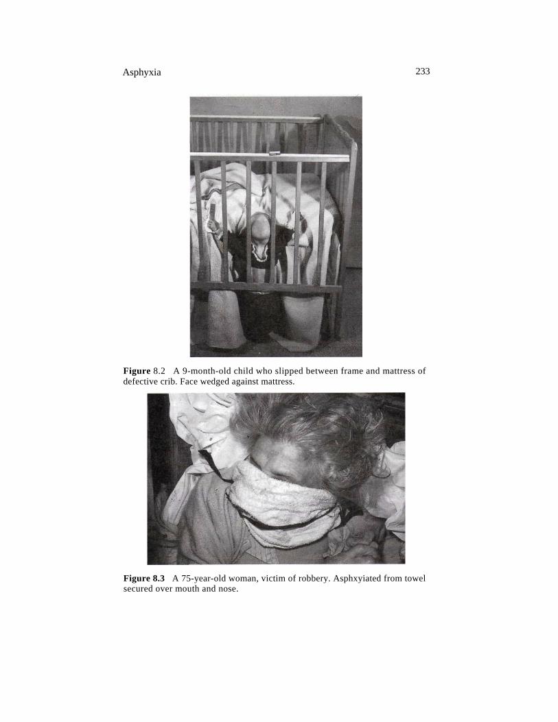
Accidental smotherings can occur with defective cribs. Here, an infant is trapped either between a too-small mattress and the frame of the crib, or between a defective crib and mattress, with the face wedged against the mattress (Figure 8.2). The child is unable to move and smothers.
Gags obstructing the nose and mouth can cause death by smothering. Such deaths, though unintentional, are still homicides if the victims die during the commission of a crime. Typically, a gag is placed around the face obstructing the mouth and nose (Figure 8.3). Victims are usually elderly individuals who are either unable to struggle sufficiently to move the gag or who are unusually susceptible to the anoxia by virtue of natural disease. Mucus and fluids may accumulate in the nasal cavities and airways, contrib-uting to asphyxia. In the elderly, there may be congestion of the face with
Asphyxia 233
Figure 8.2 A 9-month-old child who slipped between frame and mattress of defective crib. Face wedged against mattress.
Figure 8.3 A 75-year-old woman, victim of robbery. Asphxyiated from towel secured over mouth and nose.
234 Forensic Pathology
scattered fine petechiae of the sclerae, cpnjunctivae, and skin of the face. This has not been the case in young individuals in whom petechiae are usually absent. It is the discovery of the gag obstructing the airways that makes the diagnosis, not alleged signs of asphyxia.
In homicide by smothering, the implements used are usually pillows, bedding, and the hands. Infants may be placed in plastic bags. The victims tend to be very young, very old, debilitated, or incapacitated by restraints, disease or drugs. It is extremely difficult to smother adults in full control of their faculties.
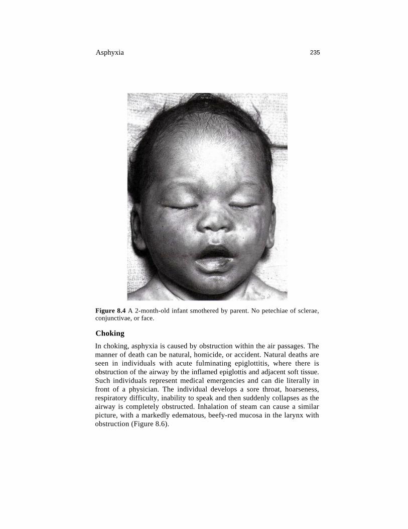
When a pillow is used, it is placed over the face and pushed down. This causes obstruction of the nose and mouth, asphyxia, and death. There are usually no marks on the face. The face is not congested and there are no petechiae of the sclerae or conjunctivae (Figure 8.4). Abrasion injuries of the face will occur only if the victim puts up a vigorous resistance. In a review of 15 smothering deaths involving children below the age of 2 years, of the 13 who could be evaluated for the presence of petechiae, only one had findings. This child had a single petechia of the conjunctiva and a single area of scleral hemorrhage. Because of the circumstances of this case, there was the possibility that the child might also have been choked. Pushing the face into the bedding will accomplish the same end as using a pillow.
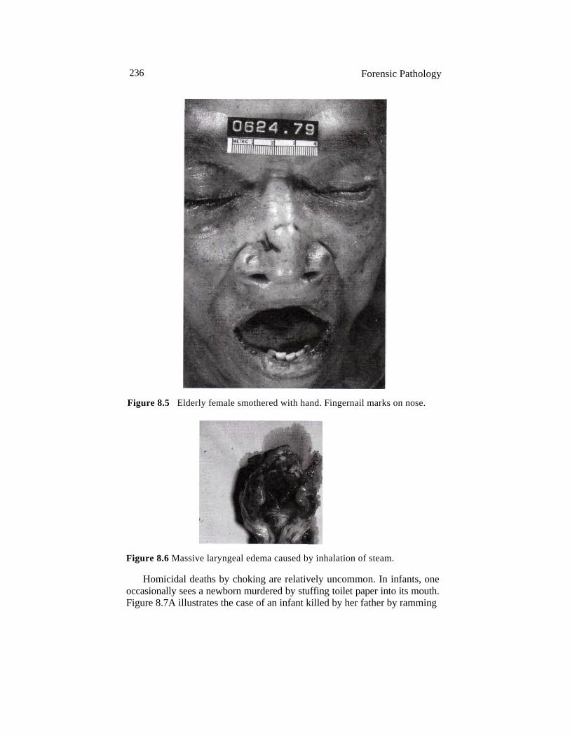
Smothering can also be accomplished using the hands. The nose is pinched off with one hand, while the other hand is used to push the jaw closed. In small children, one hand can accomplish both these tasks. In infants and adults unable to put up any effective resistance, an autopsy will fail to disclose any injury due to this process. In adults, even those who can muster only a minimal struggle, there may be abrasions on the nose or chin from the fin-gernails, and contusions of the lips from pressure of the palm (Figure 8.5).
The sequence of physiological events in smothering is:
• Bradycardia (decrease in heart rate) • Decrease in respiration to agonal gasps with eventual cessation of
respiration • Slowing and finally flattening of the electroencephalogram (EEC)
The heart will continue to beat even after flattening of the EEC. In infants, bradycardia has been observed to start 30 sec after the initiation of smoth-ering, and flattening of the EEC at 90 sec.6 If, after cessation of respiration, the pillow or hand is removed from the face, respiration will not usually restart spontaneously. The individual must be resuscitated. Violent struggles with increased utilization of oxygen can speed up this sequence of events, just as natural disease could make the individual more susceptible to the effects of hypoxia.
Asphyxia 235
Figure 8.4 A 2-month-old infant smothered by parent. No petechiae of sclerae, conjunctivae, or face.
Choking
In choking, asphyxia is caused by obstruction within the air passages. The manner of death can be natural, homicide, or accident. Natural deaths are seen in individuals with acute fulminating epiglottitis, where there is obstruction of the airway by the inflamed epiglottis and adjacent soft tissue. Such individuals represent medical emergencies and can die literally in front of a physician. The individual develops a sore throat, hoarseness, respiratory difficulty, inability to speak and then suddenly collapses as the airway is completely obstructed. Inhalation of steam can cause a similar picture, with a markedly edematous, beefy-red mucosa in the larynx with obstruction (Figure 8.6).
236 Forensic Pathology
Figure 8.5 Elderly female smothered with hand. Fingernail marks on nose.
Figure 8.6 Massive laryngeal edema caused by inhalation of steam.
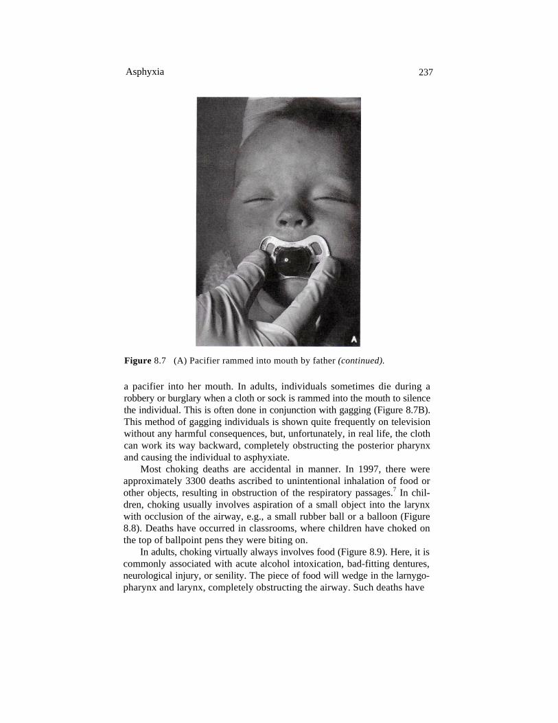
Homicidal deaths by choking are relatively uncommon. In infants, one occasionally sees a newborn murdered by stuffing toilet paper into its mouth. Figure 8.7A illustrates the case of an infant killed by her father by ramming
Asphyxia 237
Figure 8.7 (A) Pacifier rammed into mouth by father (continued).
a pacifier into her mouth. In adults, individuals sometimes die during a robbery or burglary when a cloth or sock is rammed into the mouth to silence the individual. This is often done in conjunction with gagging (Figure 8.7B). This method of gagging individuals is shown quite frequently on television without any harmful consequences, but, unfortunately, in real life, the cloth can work its way backward, completely obstructing the posterior pharynx and causing the individual to asphyxiate.
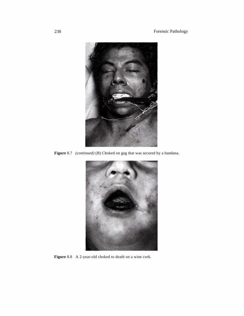
Most choking deaths are accidental in manner. In 1997, there were approximately 3300 deaths ascribed to unintentional inhalation of food or other objects, resulting in obstruction of the respiratory passages.7 In chil-dren, choking usually involves aspiration of a small object into the larynx with occlusion of the airway, e.g., a small rubber ball or a balloon (Figure 8.8). Deaths have occurred in classrooms, where children have choked on the top of ballpoint pens they were biting on.
In adults, choking virtually always involves food (Figure 8.9). Here, it is commonly associated with acute alcohol intoxication, bad-fitting dentures, neurological injury, or senility. The piece of food will wedge in the larnygo-pharynx and larynx, completely obstructing the airway. Such deaths have
238 Forensic Pathology
Figure 8.7 (continued) (B) Choked on gag that was secured by a bandana.
Figure 8.8 A 2-year-old choked to death on a wine cork.
Asphyxia 239
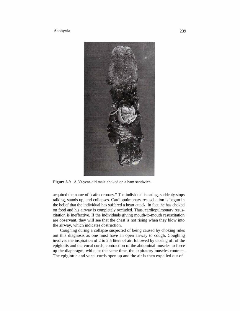
Figure 8.9 A 39-year-old male choked on a ham sandwich.
acquired the name of "cafe coronary." The individual is eating, suddenly stops talking, stands up, and collapses. Cardiopulmonary resuscitation is begun in the belief that the individual has suffered a heart attack. In fact, he has choked on food and his airway is completely occluded. Thus, cardiopulmonary resus-citation is ineffective. If the individuals giving mouth-to-mouth resuscitation are observant, they will see that the chest is not rising when they blow into the airway, which indicates obstruction.
Coughing during a collapse suspected of being caused by choking rules out this diagnosis as one must have an open airway to cough. Coughing involves the inspiration of 2 to 2.5 liters of air, followed by closing off of the epiglottis and the vocal cords, contraction of the abdominal muscles to force up the diaphragm, while, at the same time, the expiratory muscles contract. The epiglottis and vocal cords open up and the air is then expelled out of
240 Forensic Pathology
the lungs, under pressure, usually carrying with it any foreign material present in the bronchi and trachea.8 The expelled air can reach velocities of 75 to 100 miles per hour.
Occasionally, choking deaths occur when an individual falls into finely ground material, such as cornmeal or sawdust. There is involuntary inhala-tion and the airway is completely occluded by this material.
The finding of small amounts of food material in the airway at autopsy does not indicate that the individual choked to death. Approximately 20-25% of all individuals aspirate food agonally, independent of the cause of death.9
One can attribute a death to aspiration only if the airway from the larynx down is completely occluded by food. Death caused by massive aspiration of food is rarely seen in a medical examiner's office. It is most common in comatose patients who have impaired functioning of the central nervous system.
The diagnosis of choking death is made at autopsy when the airway is found occluded. If the individual had an occluded airway and the object or food was removed during resuscitation, the only way to make the diagnosis would be by history. There are no specific autopsy findings indicative of choking except for occlusion of the airway.
Some medical personnel will ascribe a death to choking even though the airway was never completely occluded. They suggest that laryngospasm is the cause of death. There is, however, no objective evidence that this can occur. If laryngospasm did occur, one would expect relaxation of the larynx as the victim became agonal. This, in turn, would lead to opening up of the airway and recovery. Others hypothesize that a fatal "vagal reaction" or "reflex cardiac death," mediated through the parasympathetic nervous system, occurred through hypersensitivity of the larynx to aspirated food. Again, there is just no objective proof that this entity exists.
Mechanical Asphyxia
In mechanical asphyxia, pressure on the outside of the body prevents respi-ration. Mechanical asphyxia is almost always accidental in manner. [When not associated with Restraint actively being applied.] It can be subdivided into three types:
1. Traumatic asphyxia (a term often used interchangeably with mechan-ical asphyxia)
2. Positional asphyxia 3. Riot-crush or "human pile" deaths
Traumatic Asphyxia Traumatic asphyxia occurs when a heavy weight presses down on an individ-ual's chest or upper abdomen, making respiration impossible. One common form of
Asphyxia 241
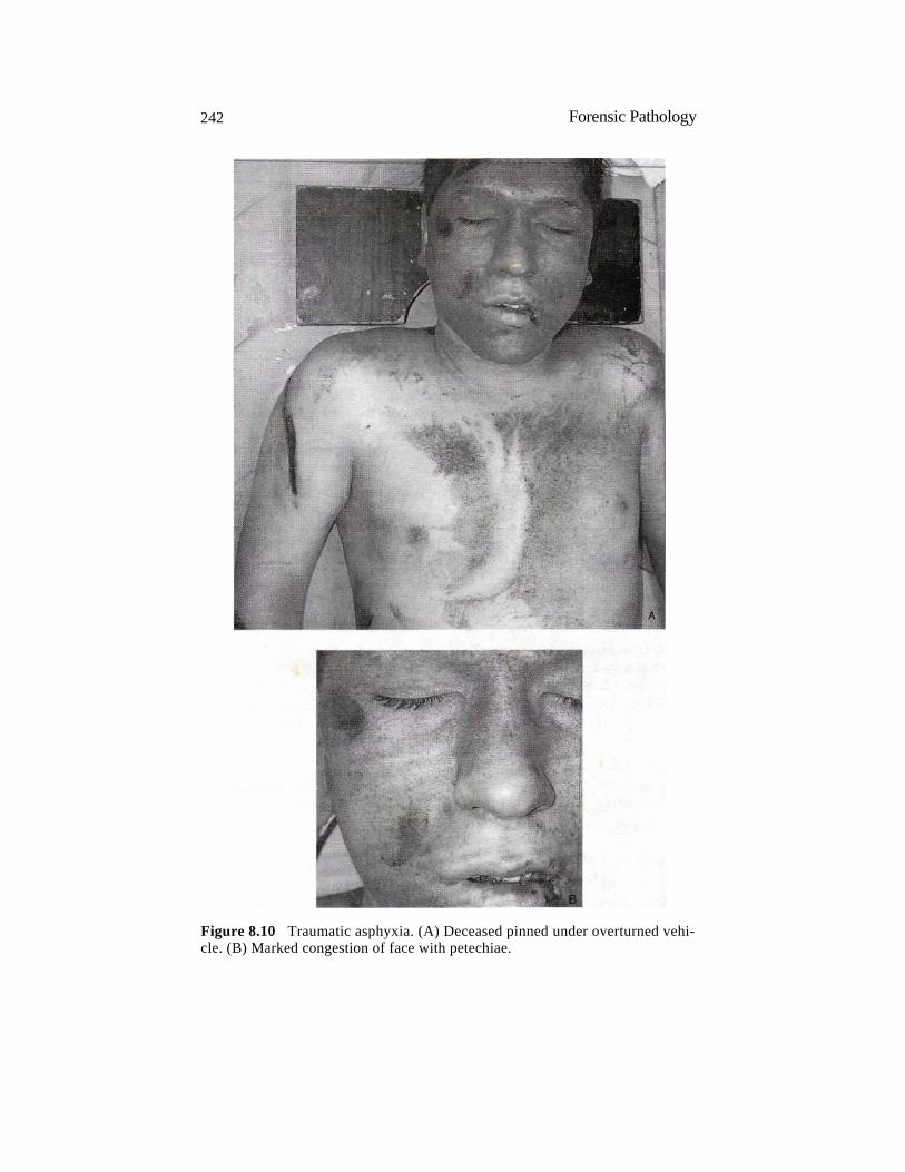
traumatic asphyxia is individuals under a car, repairing it, when the jack slips and the vehicle falls on top of them (Figure 8.10). At autopsy, there is con-gestion of the head, neck, and upper trunk with numerous petechiae in these areas, the sclerae, the conjunctivae and the periorbital skin. Retinal hemor-rhages may also be present. Internally, there is often no evidence of trauma in spite of the heavy weight on the chest. Individuals who survive an episode of traumatic asphyxia usually make an uneventful recovery, though occasion-ally there is some permanent visual impairment due to retinal hemorrhage. One individual who survived described a severe crushing pain and suffusion of his face followed by immediate unconsciousness.10 Rarely, traumatic asphyxia is homicidal. Thus, in one instance, an individual was knocked to the ground and a refrigerator and stereo were piled on top of him. An occasionally encountered form of accidental traumatic asphyxia involves individuals buried in cave-ins with their heads above the ground.
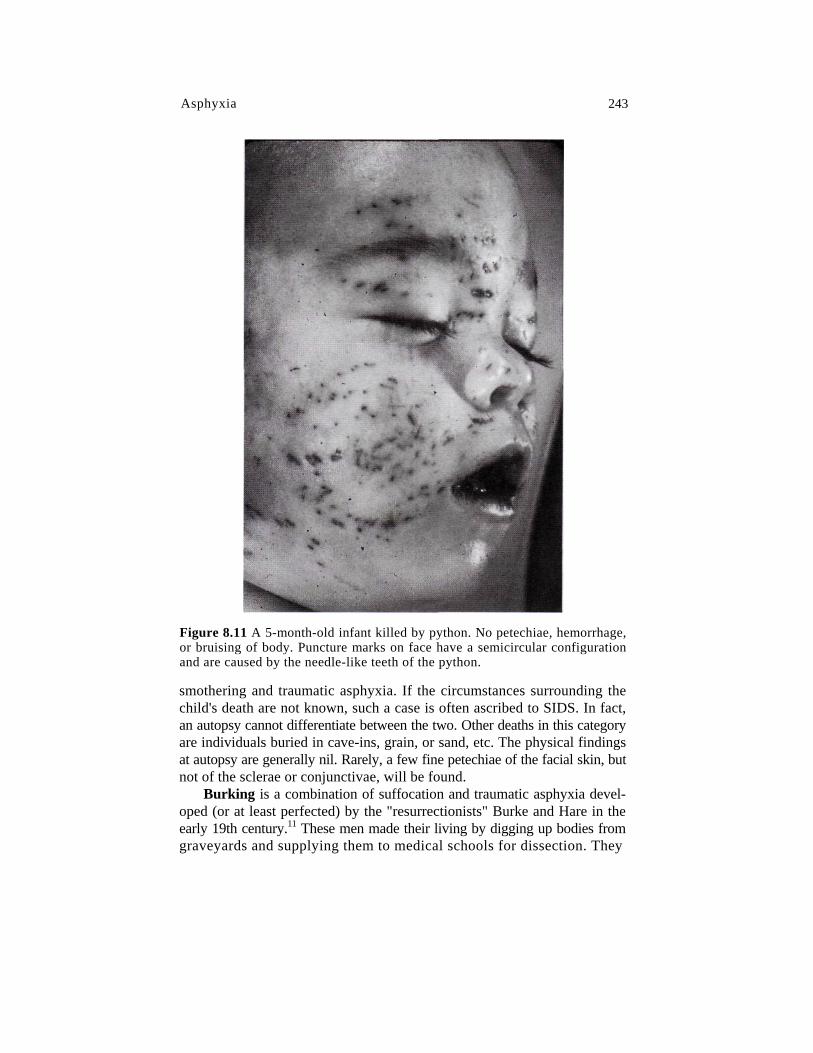
The most unusual case of traumatic asphyxia seen by the authors was that of a 5-month-old infant killed by a python. The snake wrapped itself around the baby, tightening its coils whenever the child exhaled. At autopsy, the only marks on the child were teeth marks on the face where the snake had tried to swallow the child whole (his head was too big for the snake's mouth) (Figure 8.11). There were no petechiae, hemorrhage, or bruising.
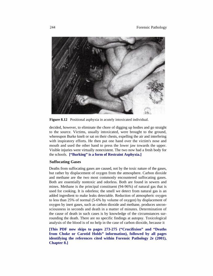
Positional asphyxia Positional asphyxia [when not associated with Restraint being actively applied] is virtually always an accident and is associated with alcohol or drug intoxication. In this entity, individuals become trapped in restricted spaces, where, because of the position of their bodies, they cannot move out of that area or position. This results in restriction of their ability to breathe, followed by death (Figure 8.12). There is usually marked congestion, cyanosis, and petechiae. Positional asphyxia might occur if individuals fall down a well and are wedged between the walls. Every time they exhale, they slip farther and farther down the well, preventing inhalation.
Riot-crush Riot-crush, as the name implies, occurs in riots, when the chest is compressed by stampeding people piling on top of each other. Respiratory movements are, thus, prohibited by this human pile.
Traumatic Asphyxia Combined with Smothering Traumatic asphyxia combined with smothering is a combination of both these entities. It can be accidental or homicidal. An accidental form is overlay, where an infant is placed in bed for the night with either an adult or a larger child. Subsequently, the infant is found dead. During the night, the other individual rolled onto the infant, killing it by a combination of
242 Forensic Pathology
Figure 8.10 Traumatic asphyxia. (A) Deceased pinned under overturned vehi-cle. (B) Marked congestion of face with petechiae.
Asphyxia 243
Figure 8.11 A 5-month-old infant killed by python. No petechiae, hemorrhage, or bruising of body. Puncture marks on face have a semicircular configuration and are caused by the needle-like teeth of the python.
smothering and traumatic asphyxia. If the circumstances surrounding the child's death are not known, such a case is often ascribed to SIDS. In fact, an autopsy cannot differentiate between the two. Other deaths in this category are individuals buried in cave-ins, grain, or sand, etc. The physical findings at autopsy are generally nil. Rarely, a few fine petechiae of the facial skin, but not of the sclerae or conjunctivae, will be found.
Burking is a combination of suffocation and traumatic asphyxia devel-oped (or at least perfected) by the "resurrectionists" Burke and Hare in the early 19th century.11 These men made their living by digging up bodies from graveyards and supplying them to medical schools for dissection. They
244 Forensic Pathology
Figure 8.12 Positional asphyxia in acutely intoxicated individual.
decided, however, to eliminate the chore of digging up bodies and go straight to the source. Victims, usually intoxicated, were brought to the ground, whereupon Burke knelt or sat on their chests, expelling the air and interfering with inspiratory efforts. He then put one hand over the victim's nose and mouth and used the other hand to press the lower jaw towards the upper. Visible injuries were virtually nonexistent. The two now had a fresh body for the schools. [“Burking” is a form of Restraint Asphyxia.]
Suffocating Gases
Deaths from suffocating gases are caused, not by the toxic nature of the gases, but rather by displacement of oxygen from the atmosphere. Carbon dioxide and methane are the two most commonly encountered suffocating gases. Both are essentially nontoxic and odorless. Both are found in sewers and mines. Methane is the principal constituent (94-96%) of natural gas that is used for cooking. It is odorless; the smell we detect from natural gas is an added ingredient to make leaks detectable. Reduction of atmospheric oxygen to less than 25% of normal (5-6% by volume of oxygen) by displacement of oxygen by inert gases, such as carbon dioxide and methane, produces uncon-sciousness in seconds and death in a matter of minutes. Determination of the cause of death in such cases is by knowledge of the circumstances sur-rounding the death. There are no specific findings at autopsy. Toxicological analysis of the blood is of no help in the case of carbon dioxide, because it
[This PDF now skips to pages 273-275 (“Crucifixion” and “Deaths from Choke or Carotid Holds” information), followed by all pages identifying the references cited within Forensic Pathology 2e (2001), Chapter 8.]
Asphyxia 273
with a simple noose around the neck, there may be elaborate binding with multiple turns of the rope around the body, or the hands bound either in front of or behind the body. Analysis of the binding will reveal that the individual was capable of binding himself. He is not completely suspended; his feet are on the ground. Thus, he can relieve the pressure of the noose just by standing a little straighter. In some instances, rather than a noose, a ligature or some other device capable of applying pressure to the neck is used. There is always some self-rescue device so that the individual can relieve pressure before losing consciousness. Unfortunately, because of equipment failure, a flaw in design or construction of the device, or loss of control by the individual, accidental deaths occur.
Crucifixion [a form of “Restraint Asphyxia”!] One unusual, historical, form of asphyxia was crucifixion.37 The victim was typically nailed to a cross with nails driven through the wrists into the crossbar and through the feet into the upright post. Death was caused by shock, both hypovolemic and secondary to the pain of nailing, plus dehy-dration and asphyxia. The weight of the body on the outstretched arms would interfere with exhalation by maintaining the intercostal muscles in an inha-lation state. Exhalation would then become primarily diaphragmatic. Over a prolonged time, this would lead to impaired respiration and asphyxia.
Death Caused by Upside-Down Suspension If an individual is suspended upside down for a long enough period of time death can result. The mechanisms of death might be either acute cardiac or respiratory failure or a combination of both. The length of time it takes for death to occur depends on the health of the individual. It could range from a few hours to a day, perhaps somewhat longer.
Deaths from Choke or Carotid Holds [Both of which can cause Restraint Asphyxia due to different mechanisms.] Neck holds are used by law enforcement agencies to subdue violent individ-uals. Rarely, one will encounter a death alleged to have occurred due to application of either a choke hold or a carotid sleeper hold.38'41 These terms are often used interchangeably, but, in fact, refer to two different holds whose purpose is to produce transient cerebral ischemia and unconsciousness. Nei-ther involves use of a mechanical implement. [This statement is false.] Rather, the arm and forearm are used to compress the neck, producing cerebral ischemia and unconsciousness. [A physical form of restraint.] Occasionally, a baton, large metal flashlight, or some other device [thus, constituting use of a “mechanical implement” for restraint], will be used to compress the neck. The authors have seen a number of deaths involving use of such instruments. In such cases, there is usually extensive hemorrhage in the neck and fractures of the hyoid or larynx.
274 Forensic Pathology
With choke (bar arm) holds, the forearm is placed straight across the front of the neck. The free hand grips the wrist, pulling it back, collapsing the airway and displacing the tongue rearward, which occludes the hypophar-ynx. Incapacitation is caused by collapse of the airway and the carotid arteries with resultant decrease in the supply of oxygen to the brain. Compression of the carotid arteries is the prime mechanism for loss of consciousness. If too much force is used, there could be fracture of the larynx or hyoid. In two cases reported by Reay and Eisele and in a case seen by the authors, there were unilateral fractures of the greater cornu of the thyroid cartilage.38 Both of Reay and Eisele's cases had fractures on the left side of the neck, the right forearm was across the neck and the left hand was used to pull it backward. Thus, pressure was eccentrically transferred to the neck, predominantly to the left side. In the case seen by the authors, the left forearm was across the neck and the fractures were on the right side of the neck. The authors' case also had a fracture of the hyoid bone on the same side. Following loss of consciousness, the chokehold is released and the victim should regain con-sciousness within 30 sec. There should be no permanent sequelae. Obviously, if the choke hold is maintained for too long, death will ensue, and one now has a case of manual strangulation.
In the carotid sleeper hold, symmetrical force is applied by the forearm and upper arm to the front of the neck such that there is compression of only the carotid arteries and jugular veins and not the trachea. The arm is placed about the neck with the antecubital fossa or crook of the arm centered at the midline of the neck. The free hand grips the wrist of the other arm and pulls it backward, creating a pincher effect. This produces transient cerebral ischemia. The carotid sleeper hold impedes blood flow in the carotid arteries by pressure exerted on both sides of the neck by the pincher effect of the arm and forearm. If properly applied, the compression of the carotid arteries will cause loss of consciousness in approximately 10-15 sec. On relaxation of the hold, cerebral blood flow will be restored and consciousness will return in approximately 10-20 sec, without any serious side effects. [This statement is entirely theoretical. There is no guarantee that consciousness will return after interrupting carotid blood flow and asphyxiating the brain into unconsciousness!] Experiments by Reay and Holloway demonstrated that, during application of the carotid sleeper hold, blood flow is decreased an average of 85% to the head.39 The range in five subjects was 82 to 96%. The time to minimum blood flow averaged 6 sec (range 3.2 to 7.2 sec).
In theory, the carotid sleeper hold will cause rapid unconsciousness without injury to the individual. Unfortunately, in violently struggling indi-viduals, a carotid sleeper hold can easily and unintentionally be converted into a choke hold, as the individual twists and turns to break the hold.
Maintenance of the pressure in a carotid sleeper hold, after loss of con-sciousness, becomes manual strangulation and, if continued long enough, will cause death. One would not expect trauma to the structures of the neck
Asphyxia 275
in such an instance. The compression of the carotid arteries, with resultant decreased cerebral blood flow, can theoretically precipitate a stroke in an individual with atherosclerotic disease of the carotid or cerebral vasculature. The pressure can cause dislodgment of atherosclerotic material with a stroke caused by an embolus. Blood flow to the brain is from both the carotid and the vertebral arteries. If the vertebral arteries have impaired blood flow due to atherosclerosis, then occlusion of the carotid arteries can threaten an already compromised circulation, resulting in thrombosis or stroke [or death].
Both choke and carotid sleeper holds are safe if properly used, though the latter is the safer of the two. In weighing how much force is acceptable in a situation, one must realize that any action involving force always has the potential of producing severe injury and death.
References
1. Ely SF and Hirsch CS, Asphyxial deaths and petechiae: a review. / Forens Sci 2000;45(6):1274-1277.
2. Gordon I and Mansfield RA, Subpleural, subpericardial and subendocardial hemorrhages. / Forens Med 1955; 2:31-50.
3. Gilg T et al., Investigations on postmortem coagulation and fibrinolytic reac tions in blood. Beitr Gericht Med 1986; 44:399-405.
4. U.S. Department of lustice, Federal Bureau of Investigation, Crime in the United States: 1999. Superintendent of Documents, Washington D.C., 2000.
5. DiMaio DJ and DiMaio VJM, Two deaths caused by a lack of oxygen in a water vault. / Forens Sci 1974; 19:398.40 1.
6. Rossen CL et al., Two siblings and recurrent cardiorespiratory arrest: Mun- chausen syndrome by proxy or child abuse? Pediatrics 1983; 71:715-720.
7. National Safety Council, Accident Facts:1998 Itasca, 111. 8. Guyton AC and Hall JE, Textbook of Medical Physiology 10th ed. W.B. Saun-
ders, Philadelphia 2000. 9. Knight BH, The significance of the postmortem discovery of gastric contents
in the air passages. / Forens Sci 1975; 6:229-234.
10. Feldman EA, Traumatic asphyxia: Report of three cases./ Trauma 1969; 9:345- 353.
11. Roughead W, Burke and Hare, ed 3. Edinburgh, Cited in Poison C, Gee DJ, Knight B: The Essentials of Forensic Medicine, New York, Pergamon Press, 1985.
12. Camps FE and Hunt AC, Pressure on the neck. ] Forens Med 1959; 6:116-135. 13. Brouardel P, Cited in Poison CJ, Gee DJ, Knight B, The Essentials of Forensic
Medicine. New York, Pergamon Press, 1985.
276 Forensic Pathology
14. Ikai M et al., Physiological studies on choking in judo, in Bulletin of the Association for the Scientific Studies on Judo, Part 1, Studies in General, 1958; pp 1-12.
15. Suzuki E, Medical studies on choking in judo, with special reference to elec- troencephalographic investigation, in Bulletin of the Association for the Scien tific Studies on Judo, 1958; pp 23-48.
16. Rossen R; Kabat H., and Anderson JP, Acute arrest of cerebral circulation in man. Arch Neurology and Psych 1943; 50:510-528.
17. Rao Vj and Weti CV, The forensic significance of conjunctival petechiae. Am JForens Med Pathol 1988; 9:32-34.
18. Harm T and Rajs J, Types of injuries and interrelated conditions of victims and assailants in attempted and homicide strangulations. Forens Sci Int 1981; 18:101-123.
19. Feigin G. Frequency of neck organ fractures in hanging. Am J Forensic Med. Path 1999; 20(2):128-130.
20. DiMaio VJM, Accidental hangings due to pacifiers. JAMA 1973; 226:790. 21. Rauchschwalbe R and Mann NC, Pediatric window-cord strangulations in
the United States, 1981-1995. JAMA. 1997; 277:1696-1698. 22. Petruk J et al., Fatal asphyxiations in children involving drawstrings on cloth
ing. Can. Med. Assoc. J. 1996; 155(10):1417-1419. 23. Casper JL, Handbook of the Practice of Forensic Medicine, vol 2, ed 3, GW
Balfour (trans). London, New Syndenham Society, 1982, pp 169-182. 24. Spence MW et al.,. Craniocervical injuries in judicial hangings: an anthro-
pologic analysis of six cases. Am J Forens Med Pathol 1999; 20 (4):309-322. 25. Hartshorne NJ, Reay DT, Judicial Hanging (letter), Am J Forens Med Pathol
1995; 16 (1):87. 26. DiMaio VJM, Homicidal Asphyxia, Am J Forens Med Pathol, 2000; 21 (1): 1 -4. 27. Habal M, Meguid MM, and Murray JE, The long scarf syndrome — A
potentially fatal and preventable hazard. JAMA 1972; 221:1269,1270. 28. Weiss S, Baker JP, The carotid sinus reflex in health and disease. Medicine
1933; 12:297-354. 29. Thomas JE, Hyperactive carotid sinus reflex and carotid sinus syncope. Mayo
ClinProc 1969; 44:127-139. 30. Simpson K and Knight B, Forensic Medicine, ed 9. Baltimore, Edward Arnold,
1985. 31. Paparo GP and Siegel H, On the significance of posterior crico-arytenoid
muscle hemorrhage. Forens Sci 1976; 7:61-65. 32. Raven KP, Reay DT, and Harruff RC, Artifactual injuries of the larynx pro
duced by resuscitative intubation. Am J Forens Med Pathol, 1999; 20(l):31-36. 33. Baselt RC, Disposition of Toxic Drugs and Chemicals in Man, 5th ed, Chem
ical Toxicology Institute, Foster City, 2000.
Asphyxia 277
34. Chandra H et al, Chronic cyanide exposure — A biochemical and industrial hygiene study. J Anal Toxicol 1980; 4:161-165.
35. Uva JL, Review: autoerotic asphyxiation in the United States, / Forens Sci, 1995; 40(4):574-581.
36. Byard RW, Hucker SJ, and Hazelwood RR, Fatal and near-fatal autoerotic asphyxial episodes in women. Am ] Forens Med Pathol, 1993; 14(l):70-73.
37. Edwards WD, Gabel WJ, and Hosmer FE, On the physical death of Jesus Christ. JAMA 1986; 255:1455-1463.
38. Reay DT and Eisele JW, Death from law enforcement neck holds. Am J Forens Med Pathol, 1982; 3:253-258.
39. Reay DT and Holloway GA, Changes in carotid blood flow produced by neck compression. Am J Forens Med Pathol, 1982; 3:199-202.
40. Koiwai EK, Deaths allegedly caused by use of "choke holds" (ShimeWazal). J Forens Sci 1987; 32:419-432.
41. Kornblum RN, Medical analysis of police choke holds and general neck trauma: I, II. Trauma, February 1986; 27(51:7-60; June 1986;28(l):13-64.