Embed Size (px)
Citation preview
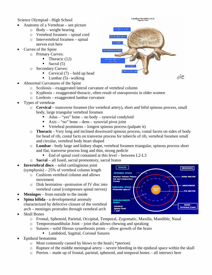
Science Olympiad - High School • Anatomy of a Vertebrae – see picture
o Body – weight bearing o Vertebral foramen – spinal cord o Intervertebral foramen – spinal
nerves exit here • Curves of the Spine
o Primary Curves: Thoracic (12) Sacral (5)
o Secondary Curves: Cervical (7) – hold up head Lumbar (5)– walking
• Abnormal Curvatures of the Spine o Scoliosis – exaggerated lateral curvature of vertebral column o Kyphosis – exaggerated thoracic, often result of osteoporosis in older women o Lordosis – exaggerated lumbar curvature
• Types of vertebrae o Cervical – transverse foramen (for vertebral artery), short and bifid spinous process, small
body, large triangular vertebral foramen Atlas – “yes” bone – no body – synovial condyloid Axis – “no” bone – dens – synovial pivot joint Vertebral prominens – longest spinous process (palpate it)
o Thoracic - Very long and inclined downward spinous process, costal facets on sides of body for head of rib, costal facts on tranverse process for tubercle of rib, vertebral foramen small and circular, vertebral body heart shaped
o Lumbar - body large and kidney shape, vertebral foramen triangular, spinous process short and flat, tranverse process long and thin, strong pedicle
End of spinal cord contained at this level – between L2-L3 o Sacral – all fused, sacral promontory, sacral hiatus
• Invertebral discs – solid cartilaginous joint (symphysis) – 25% of vertebral column length
o Cushions vertebral column and allows movement
o Disk herniation –protrusion of IV disc into vertebral canal (compresses spinal nerves)
• Meninges – from outside to the inside • Spina bifida - a developmental anomaly
characterized by defective closure of the vertebral arch – meninges protrudes through vertebral arch
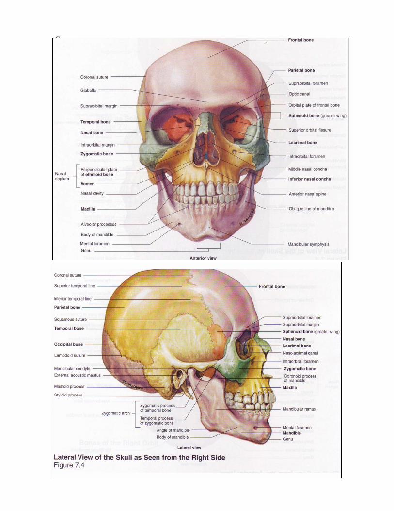
• Skull Bones o Frontal, Sphenoid, Parietal, Occipital, Temporal, Zygomatic, Maxilla, Mandible, Nasal o Temporomandibular Joint – joint that allows chewing and speaking o Sutures – solid fibrous synarthrosis joints – allow growth of the brain
Lambdoid, Sagittal, Coronal Sutures • Epidural hematoma
o Most commonly caused by blows to the head ( *pterion) o Rupture of the middle meningeal artery – severe bleeding in the epidural space within the skull o Pterion – made up of frontal, parietal, sphenoid, and temporal bones – all intersect here
o