Embed Size (px)
Citation preview

Assessment Manager: SGB Page 1 of 21
Schedule of Accreditation issued by
United Kingdom Accreditation Service
2 Pine Trees, Chertsey Lane, Staines-upon-Thames, TW18 3HR, UK
8130
Accredited to ISO 15189:2012
North Bristol NHS Trust
Issue No: 001 Issue date: 24 September 2018
Cellular Pathology
Southmead Hospital
Westbury on Trym
Bristol
BS10 5NB
Contact: Mr Andrew Heryet
Tel: +44 (0) 1173 232706
Fax: +44 (0) 1173 238476
E-Mail: [email protected]
Website: www.nbt.nhs.uk/severn-pathology
Testing performed by the Organization at the locations specified below
Locations covered by the organisation and their relevant activities
Laboratory locations:
Location details Activity Location code
Address Cellular Pathology Southmead Hospital Westbury on Trym Bristol BS10 5NB
Local contact Mr Andrew Heryet Tel: +44 (0) 1173 232706 Fax: +44 (0) 1173 238476 E-Mail: [email protected]
Histopathology Diagnostic Cytopathology Gynaecological Cytopathology Rapid frozen section for intra-operative diagnosis FNA material/slide preparation and cellularity assessment
A

8130
Accredited to
ISO 15189:2012
Schedule of Accreditation issued by
United Kingdom Accreditation Service 2 P ine Trees , Cher t sey Lane, S ta i nes -upon-Thames , TW 18 3HR, UK
North Bristol NHS Trust
Issue No: 001 Issue date: 24 September 2018
Testing performed by the Organisation at the locations specified
Assessment Manager: SGB Page 2 of 21
Site activities performed away from the locations listed above:
Location details Activity Location code
Address Brunel Building Southmead Hospital Westbury on Trym Bristol BS10 5NB
Local contact Mr Andrew Heryet Tel: +44 (0) 1173 232706 Fax: +44 (0) 1173 238476 E-Mail: [email protected]
One Stop Nucleic Acid assays OSNA B
Address Cellular Pathology Bristol Royal Infirmary Marlborough Street Bristol BS2 8HW
Local contact Mr Andrew Heryet Tel: +44 (0) 1173 232706 Fax: +44 (0) 1173 238476 E-Mail: [email protected]
Dissection and preparation and reporting of biopsies Diagnostic cytology, screening and reporting of urgent Head and Neck biopsies Rapid frozen section for intra-operative diagnosis
C
Address Mortuary Southmead Hospital Westbury on Trym Bristol BS10 5NB
Local contact Mr Andrew Heryet Tel: +44 (0) 1173 232706 Fax: +44 (0) 1173 238476 E-Mail: [email protected]
Storage of bodies
Address Perinatal Mortuary St Michaels Hospital Southwell Street Bristol BS2 8EG
Local contact Mr Andrew Heryet Tel: +44 (0) 1173 232706 Fax: +44 (0) 1173 238476 E-Mail: [email protected]
Storage of bodies

8130
Accredited to
ISO 15189:2012
Schedule of Accreditation issued by
United Kingdom Accreditation Service 2 P ine Trees , Cher t sey Lane, S ta i nes -upon-Thames , TW 18 3HR, UK
North Bristol NHS Trust
Issue No: 001 Issue date: 24 September 2018
Testing performed by the Organisation at the locations specified
Assessment Manager: SGB Page 3 of 21
DETAIL OF ACCREDITATION
Materials/Products tested
Type of test/Properties
measured/Range of measurement
Standard specifications/
Equipment/Techniques used
Location
Code
HUMAN TISSUES AND FLUIDS
Histopathology
Macroscopic and microscopic examination using documented in-house procedures in conjunction with manufacturer’s instructions (where relevant)
A
Formalin fixed tissues Preparation and examination
to identify or exclude morphological and cytological abnormalities
Macroanalysis and dissection Leica IPC Cassette writer and in-house procedures: CP-HiCU-SOP-017 CP-HiCU-SOP-018
A, C
CP-HiCU-SOP-4 Bone Marrow trephine
CP-HiCU-SOP-5 Thorasic
CP-HiCU-SOP-6 Cystectomy and Cytoprostatectomy
CP-HiCU-SOP-7 Testes
CP-HiCU-SOP-8 Kidney
CP-HiCU-SOP-9 Adrenal
CP-HiCU-SOP-13 GI
CP-HiCU-SOP-14 Skin
CP-HiCU-SOP-16 Bone and soft tissue
CP-HiCU-SOP-31 Prostate
CP-HiCU-SOP-32 Penile
CP-HiCU-SOP-33 Parathyroid
CP-HiCU-SOP-37 Placenta
CP-HiCU-SOP-51 Gynae
CP-HiCU-SOP-53 Thyroid
CP-HiCU-SOP-54 Parathyroid
CP-HiCU-SOP-55 Head and Neck
CP-HiCU-SOP-77 Breast
Tissue processing Automated method using Leica Peloris tissue processors CP-HiML-SOP-18
A

8130
Accredited to
ISO 15189:2012
Schedule of Accreditation issued by
United Kingdom Accreditation Service 2 P ine Trees , Cher t sey Lane, S ta i nes -upon-Thames , TW 18 3HR, UK
North Bristol NHS Trust
Issue No: 001 Issue date: 24 September 2018
Testing performed by the Organisation at the locations specified
Assessment Manager: SGB Page 4 of 21
Materials/Products tested
Type of test/Properties
measured/Range of measurement
Standard specifications/
Equipment/Techniques used
Location
Code
HUMAN TISSUES AND FLUIDS (cont’d)
Histopathology (cont’d) Preparation and examination to identify or exclude morphological and cytological abnormalities (cont’d)
Macroscopic and microscopic examination using documented in-house procedures in conjunction with manufacturer’s instructions (where relevant)
A
Embedding Manual method using Leica EG115H/E1150C, EG1160 and Histocare Arcadia embedding centres and in-house procedure: CP-HiML-SOP-19
Microtomy Automated method using Leica RM2135/2235/2245/2255 microtomes and in-house procedure:
CP-HiML-SOP-20 Formalin fixed, paraffin embedded (FFPE) tissue
Routine morphological staining
Routine Haematoxylin and Eosin staining and coverslipping using: Leica Autostainer CV5030 and XL ST5020/CV5030 stainer / cover slippers and in-house procedure: CP-HiML-SOP-21 CP-HiML-SOP-22
A
Fresh Tissue Frozen Tissue
Intra-operative diagnosis Rapid Frozen Section Leica CM1950 Leica CM1850 UV using and manual Haematoxylin and Eosin (H and E) stain with in-house procedure: CP-HiCU-SOP-19 CP-HiCU-SOP-12
A C
Identification of Hirschprungs
disease Manual Acetyl Cholinesterase (ACHE) stain with in-house procedure: CP-HiSD-SOP-37
A

8130
Accredited to
ISO 15189:2012
Schedule of Accreditation issued by
United Kingdom Accreditation Service 2 P ine Trees , Cher t sey Lane, S ta i nes -upon-Thames , TW 18 3HR, UK
North Bristol NHS Trust
Issue No: 001 Issue date: 24 September 2018
Testing performed by the Organisation at the locations specified
Assessment Manager: SGB Page 5 of 21
Materials/Products tested
Type of test/Properties
measured/Range of measurement
Standard specifications/
Equipment/Techniques used
Location
Code
HUMAN TISSUES AND FLUIDS (cont’d)
Histopathology (cont’d) Preparation and examination to identify or exclude morphological and cytological abnormalities (cont’d)
Macroscopic and microscopic examination using documented in-house procedures in conjunction with manufacturer’s instructions (where relevant)
A
Formalin fixed, paraffin embedded (FFPE) tissue
Detection of:
Special Staining Manual Techniques using in-house procedures and the following stains:
Acid and neutral mucins
Alcian blue-PAS CP-HiSD-SOP-ABPASD
Amyloid Congo red
CP-HiSD-SOP-CR
Copper deposits Dimethylaminobenzylidine
Rhodanine DMABR CP-HiSD-SOP-DMABR
Haematopoietic cells Giemsa
CP-HiSD-SOP-Giemsa
H. pylori Giemsa modified
CP-HiSD-SOP-ModifiedGiemsa
Reticulin fibres Gordon and Sweets reticulin stain
CP-HiSD-SOP-Retic
Gram positive and negative
bacteria Gram CP-HiSD-SOP-Gram
Basement membrane, Fungi Grocott methenamine silver
CP-HiSD-SOP-Grocott
General morphology nuclei,
cytoplasm & connective tissue Haematoxylin and eosin manual CP-HiSD-SOP-Manual
General morphology nuclei,
cytoplasm & connective tissue Haematoxylin and eosin rapid CP-HiSD-SOP-Rapid
Connective tissue collagen Haematoxylin Van Gieson HVG
CP-HiSD-SOP-HVG

8130
Accredited to
ISO 15189:2012
Schedule of Accreditation issued by
United Kingdom Accreditation Service 2 P ine Trees , Cher t sey Lane, S ta i nes -upon-Thames , TW 18 3HR, UK
North Bristol NHS Trust
Issue No: 001 Issue date: 24 September 2018
Testing performed by the Organisation at the locations specified
Assessment Manager: SGB Page 6 of 21
Materials/Products tested
Type of test/Properties
measured/Range of measurement
Standard specifications/
Equipment/Techniques used
Location
Code
HUMAN TISSUES AND FLUIDS (cont’d)
Histopathology (cont’d) Preparation and examination to identify or exclude morphological and cytological abnormalities (cont’d)
Macroscopic and microscopic examination using documented in-house procedures in conjunction with manufacturer’s instructions (where relevant)
A
FFPE Tissue (cont’d) Detection of: (cont’d) Special Staining Manual (cont’d)
Fibrin & connective tissue Martius scarlet blue MSB CP-HiSD-SOP-MSB
Melanin Masson Fontana
CP-HiSD-SOP-MassonFontana
Connective tissue Masson Trichrome
CP-HiSD-SOP-MBT
Removal of melanin pigment Melanin Bleach
CP-HiSD-SOP-MelaninBleach
Elastic fibres & connective
tissue Miller’s elastic Van Gieson EVG CP-HiSD-SOP-EVG
Neutral mucins Oil red O
CP-HiSD-SOP-OilRedO
Hepatitis B surface antigen
and copper associated protein Orcein (Shikata) CP-HiSD-SOP-Orcein
Polysaccharides including
glycogen Periodic acid Schiff PAS CP-HiSD-SOP-PAS
Basement membrane
Periodic acid silver PAAg CP-HiSD-SOP-PAAG
Haemosiderin Perl’s Prussian blue
CP-HiSD-SOP-Perls
Lipofuscin, melanin and
argentaffin granules Schmorls CP-SHIS-SOP-004 (no24)
Amyloid Sirius red
CP-SHIS-SOP-004 (no25)
Mast cells Toluidine blue
CP-SHIS-SOP-004 (no26)

8130
Accredited to
ISO 15189:2012
Schedule of Accreditation issued by
United Kingdom Accreditation Service 2 P ine Trees , Cher t sey Lane, S ta i nes -upon-Thames , TW 18 3HR, UK
North Bristol NHS Trust
Issue No: 001 Issue date: 24 September 2018
Testing performed by the Organisation at the locations specified
Assessment Manager: SGB Page 7 of 21
Materials/Products tested
Type of test/Properties
measured/Range of measurement
Standard specifications/
Equipment/Techniques used
Location
Code
HUMAN TISSUES AND FLUIDS (cont’d)
Histopathology (cont’d) Preparation and examination to identify or exclude morphological and cytological abnormalities (cont’d)
Macroscopic and microscopic examination using documented in-house procedures in conjunction with manufacturer’s instructions (where relevant)
A
FFPE Tissue (cont’d) Detection of: (cont’d) Special Staining Manual (cont’d): Fungi Tome for fungi
CP-SHIS-SOP-004 (no27)
Calcium salts Von Kossa
CP-SHIS-SOP-004 (no 28)
Acid fast bacilli e.g.
Mycobacterium leprae
Wade Fite CP-SHIS-SOP-004 (no29)
Acid fast bacilli e.g.
Mycobacterium tuberculosis Ziehl-Neelsen ZN CP-HiSD-SOP-ZN
FFPE Tissue Detection of the following:
Immunohistochemistry Automated methods using Bond III IHC instruments using in-house procedures: CP-HiSD-SOP-ICC CP-HiSD-SOP-36 Incorporating the following antibodies:
High MW Cytokeratin 34BE12 Pan-cytokeratin CK1-8, 10,
14-16 and 19 AE1/3
Adenovirus Adenovirus Carcinoma marker liver,
kidney, colon & prostate AMACR P504s
Carcinoma marker prostate AMACR/34BE12 Amyloid Amyloid A component Germ Cell Tumours Alpha Feto Protein (AFP)

8130
Accredited to
ISO 15189:2012
Schedule of Accreditation issued by
United Kingdom Accreditation Service 2 P ine Trees , Cher t sey Lane, S ta i nes -upon-Thames , TW 18 3HR, UK
North Bristol NHS Trust
Issue No: 001 Issue date: 24 September 2018
Testing performed by the Organisation at the locations specified
Assessment Manager: SGB Page 8 of 21
Materials/Products tested
Type of test/Properties
measured/Range of measurement
Standard specifications/
Equipment/Techniques used
Location
Code
HUMAN TISSUES AND FLUIDS (cont’d)
Histopathology (cont’d) Preparation and examination to identify or exclude morphological and cytological abnormalities (cont’d)
Macroscopic and microscopic examination using documented in-house procedures in conjunction with manufacturer’s instructions (where relevant)
A
FFPE Tissue (cont’d) Detection of the following:
(cont’d)
Immunohistochemistry Automated methods using Bond III IHC instruments using in-house procedures: CP-HiSD-SOP-ICC CP-HiSD-SOP-36 Incorporating the following antibodies:
Human p80 protein ALK-1 (Lymphoma) Tumour Suppressor Gene BAF47 T-cells, B-cells in the mantle
zone bcl-2
Follicular centre B-cells bcl-6 B cells, plasma cells follicular
lymphomas and lymphomas BOB -1
Marker in renal
glomerulopathy C1q (renal)
Marker in renal
glomerulopathy C3c (renal)
Marker in renal
glomerulopathy C4d (renal)
Ovarian epithelial
malignancies, seminal vesicle carcinoma, anaplastic lymphoma
Ca 125
Medullary Thyroid Tumours Calcitonin Mesothelial cell lining Calretinin

8130
Accredited to
ISO 15189:2012
Schedule of Accreditation issued by
United Kingdom Accreditation Service 2 P ine Trees , Cher t sey Lane, S ta i nes -upon-Thames , TW 18 3HR, UK
North Bristol NHS Trust
Issue No: 001 Issue date: 24 September 2018
Testing performed by the Organisation at the locations specified
Assessment Manager: SGB Page 9 of 21
Materials/Products tested
Type of test/Properties
measured/Range of measurement
Standard specifications/
Equipment/Techniques used
Location
Code
HUMAN TISSUES AND FLUIDS (cont’d)
Histopathology (cont’d) Preparation and examination to identify or exclude morphological and cytological abnormalities (cont’d)
Macroscopic and microscopic examination using documented in-house procedures in conjunction with manufacturer’s instructions (where relevant)
A
FFPE Tissue (cont’d) Detection of the following:
(cont’d) Immunohistochemistry Automated methods (cont’d) Bond III IHC instruments and in-house procedures: CP-HiSD-SOP-ICC CP-HiSD-SOP-36
Epithelial cells, low molecular
weight CAM5.2
T-cell marker CD2 T-cell marker CD3 T-helper cells CD4 T-cells, B-cells in the mantle
zone CD5
Cytotoxic T-cells CD8 Mature B cells, follicular
dendritic cells, CALLA CD10
Dendritic cells CD11c Immature B lymphocytes CD15 B-cell marker CD20 Mature B-cells and FDC CD21 FDC and EBV transformed
blasts CD23
Inflammatory and malignant conditions
CD25
RS cells CD30

8130
Accredited to
ISO 15189:2012
Schedule of Accreditation issued by
United Kingdom Accreditation Service 2 P ine Trees , Cher t sey Lane, S ta i nes -upon-Thames , TW 18 3HR, UK
North Bristol NHS Trust
Issue No: 001 Issue date: 24 September 2018
Testing performed by the Organisation at the locations specified
Assessment Manager: SGB Page 10 of 21
Materials/Products tested
Type of test/Properties
measured/Range of measurement
Standard specifications/
Equipment/Techniques used
Location
Code
HUMAN TISSUES AND FLUIDS (cont’d)
Histopathology (cont’d) Preparation and examination to identify or exclude morphological and cytological abnormalities (cont’d)
Macroscopic and microscopic examination using documented in-house procedures in conjunction with manufacturer’s instructions (where relevant)
A
FFPE Tissue (cont’d) Detection of the following:
(cont’d) Immunohistochemistry Automated methods (cont’d) Bond III IHC instruments and in-house procedures: CP-HiSD-SOP-ICC CP-HiSD-SOP-36
Endothelial Cell Marker CD31 Endothelial marker CD34 T-cell and myeloid cells CD43 (MT1) Leukocyte common antigen CD45 (LCA) Neutral cell adhesion molecule CD56 Platelet glycoprotein IIIa CD61 Macrophages CD68 (KP1) Monocytes & Macrophages CD68 (PG-M1) Mast cells and GIST CD117 Plasma cells CD138 B-cell marker CD79a Ewings sarcoma, T cell
Lymphoma CD99
Intestinal epithelial cells,
colorectal + other adenocarcinomas
CDX2
Carcinoembryonic antigen CEA
Neuroendocrine Chromogranin A
8130
Accredited to
ISO 15189:2012
Schedule of Accreditation issued by
United Kingdom Accreditation Service 2 P ine Trees , Cher t sey Lane, S ta i nes -upon-Thames , TW 18 3HR, UK
North Bristol NHS Trust
Issue No: 001 Issue date: 24 September 2018
Testing performed by the Organisation at the locations specified
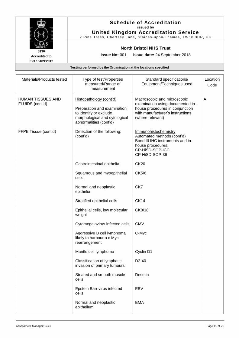
Assessment Manager: SGB Page 11 of 21
Materials/Products tested
Type of test/Properties
measured/Range of measurement
Standard specifications/
Equipment/Techniques used
Location
Code
HUMAN TISSUES AND FLUIDS (cont’d)
Histopathology (cont’d) Preparation and examination to identify or exclude morphological and cytological abnormalities (cont’d)
Macroscopic and microscopic examination using documented in-house procedures in conjunction with manufacturer’s instructions (where relevant)
A
FFPE Tissue (cont’d) Detection of the following:
(cont’d) Immunohistochemistry Automated methods (cont’d) Bond III IHC instruments and in-house procedures: CP-HiSD-SOP-ICC CP-HiSD-SOP-36
Gastrointestinal epithelia CK20 Squamous and myoepithelial
cells CK5/6
Normal and neoplastic
epithelia CK7
Stratified epithelial cells CK14 Epithelial cells, low molecular
weight CK8/18
Cytomegalovirus infected cells CMV Aggressive B cell lymphoma
likely to harbour a c Myc rearrangement
C-Myc
Mantle cell lymphoma Cyclin D1 Classification of lymphatic
invasion of primary tumours D2-40
Striated and smooth muscle
cells Desmin
Epstein Barr virus infected
cells EBV
Normal and neoplastic
epithelium EMA

8130
Accredited to
ISO 15189:2012
Schedule of Accreditation issued by
United Kingdom Accreditation Service 2 P ine Trees , Cher t sey Lane, S ta i nes -upon-Thames , TW 18 3HR, UK
North Bristol NHS Trust
Issue No: 001 Issue date: 24 September 2018
Testing performed by the Organisation at the locations specified
Assessment Manager: SGB Page 12 of 21
Materials/Products tested
Type of test/Properties
measured/Range of measurement
Standard specifications/
Equipment/Techniques used
Location
Code
HUMAN TISSUES AND FLUIDS (cont’d)
Histopathology (cont’d) Preparation and examination to identify or exclude morphological and cytological abnormalities (cont’d)
Macroscopic and microscopic examination using documented in-house procedures in conjunction with manufacturer’s instructions (where relevant)
A
FFPE Tissue (cont’d) Detection of the following:
(cont’d) Immunohistochemistry Automated methods (cont’d) Bond III IHC instruments and in-house procedures: CP-HiSD-SOP-ICC CP-HiSD-SOP-36
Lymphatic endothelium Factor VIII
Fibrohistiocytic Tumour Factor XIIIa Erythroid cells,
erythroleukemia Glycophorin A
Hepatocellular
carcinomas(HCC), yolk sac tumours
Glypican-3
Cytotoxic T cells granules Granzyme B Smooth muscle h-Caldesmon Trophoblasts HCG GVHD Skin HLA-DP, DQ, DR Melanosomes HMB 45 Helicobacter pylori H. pylori Alpha chains IgA (renal) Gamma chains IgG (renal) Mu chains IgM (renal) Immunoglobulin G IgG IgG4 related disease IgG4

8130
Accredited to
ISO 15189:2012
Schedule of Accreditation issued by
United Kingdom Accreditation Service 2 P ine Trees , Cher t sey Lane, S ta i nes -upon-Thames , TW 18 3HR, UK
North Bristol NHS Trust
Issue No: 001 Issue date: 24 September 2018
Testing performed by the Organisation at the locations specified
Assessment Manager: SGB Page 13 of 21
Materials/Products tested
Type of test/Properties
measured/Range of measurement
Standard specifications/
Equipment/Techniques used
Location
Code
HUMAN TISSUES AND FLUIDS (cont’d)
Histopathology (cont’d) Preparation and examination to identify or exclude morphological and cytological abnormalities (cont’d)
Macroscopic and microscopic examination using documented in-house procedures in conjunction with manufacturer’s instructions (where relevant)
A
FFPE Tissue (cont’d) Detection of the following:
(cont’d) Immunohistochemistry Automated methods (cont’d) Bond III IHC instruments and in-house procedures: CP-HiSD-SOP-ICC CP-HiSD-SOP-36
Sex chord stromal tumours Inhibin Islets of Langerhans tumours Insulin Cell proliferation marker Ki 67 Breast cancer Mammoglobin Melanocytes MELAN-A Melanocytes (triple antibody
cocktail pan melanoma screen)
Melanoma cocktail
Broad range cytokeratin MNF116 B-cells MUM-1 Myeloid cells Myeloperoxidase Classification of
rhabdomyosarcoma Myo-D1
Classification of
rhabdomyosarcoma and Wilms tumours
Myogenin
Skeletal and cardiac muscle Myoglobin Neuroblastoma cells Neuroblastoma 84A Classification of tumours with
neuronal differentiation Neurofilament protein (NFP)

8130
Accredited to
ISO 15189:2012
Schedule of Accreditation issued by
United Kingdom Accreditation Service 2 P ine Trees , Cher t sey Lane, S ta i nes -upon-Thames , TW 18 3HR, UK
North Bristol NHS Trust
Issue No: 001 Issue date: 24 September 2018
Testing performed by the Organisation at the locations specified
Assessment Manager: SGB Page 14 of 21
Materials/Products tested
Type of test/Properties
measured/Range of measurement
Standard specifications/
Equipment/Techniques used
Location
Code
HUMAN TISSUES AND FLUIDS (cont’d)
Histopathology (cont’d) Preparation and examination to identify or exclude morphological and cytological abnormalities (cont’d)
Macroscopic and microscopic examination using documented in-house procedures in conjunction with manufacturer’s instructions (where relevant)
A
FFPE Tissue (cont’d) Detection of the following:
(cont’d) Immunohistochemistry Automated methods (cont’d) Bond III IHC instruments and in-house procedures: CP-HiSD-SOP-ICC CP-HiSD-SOP-36
Neuroendocrine marker Neuro specific Endolase (NSE) Seminoma OCT-2 Germ Tumour cells OCT3/4 Tumour suppressor gene P53 mRNA/ p16 HPV biomarkers P16 mRNA/P16 HPV biomarkers P16 Basal cell marker P63 ALL Pax 5 Ovarian serous carcinoma,
clear cell renal cell carcinoma, papillary thyroid carcinoma
PAX8
Follicular helper T cells PD-1 Placental alkaline
phosphatase PLAP
Prostatic intraepithelial
neoplasia PIN cocktail
Placental alkaline
phosphatase PLAP
Prostatic specific antigen PSA

8130
Accredited to
ISO 15189:2012
Schedule of Accreditation issued by
United Kingdom Accreditation Service 2 P ine Trees , Cher t sey Lane, S ta i nes -upon-Thames , TW 18 3HR, UK
North Bristol NHS Trust
Issue No: 001 Issue date: 24 September 2018
Testing performed by the Organisation at the locations specified
Assessment Manager: SGB Page 15 of 21
Materials/Products tested
Type of test/Properties
measured/Range of measurement
Standard specifications/
Equipment/Techniques used
Location
Code
HUMAN TISSUES AND FLUIDS (cont’d)
Histopathology (cont’d) Preparation and examination to identify or exclude morphological and cytological abnormalities (cont’d)
Macroscopic and microscopic examination using documented in-house procedures in conjunction with manufacturer’s instructions (where relevant)
A
FFPE Tissue (cont’d) Detection of the following:
(cont’d) Immunohistochemistry Automated methods (cont’d) Bond III IHC instruments and in-house procedures: CP-HiSD-SOP-ICC CP-HiSD-SOP-36
Melanoma, neuroendocrine
cells S100
Smooth muscle cells Smooth muscle actin SV40 infected cells SV40 Neuroendocrine marker Synaptophysin Early B-Cell, thymocyte
marker TdT
Lumen of thyroid follicles Thyroglobulin Cytotoxic T cell granule.
Anaplastic large cell lymphoma
TIA-1
Levels of Trypsin in pancreas TRYPSIN Thyroid transcription factor-1 TTF-1 Mesenchymal marker Vimentin Wilms Tumour gene product Wilms Tumour

8130
Accredited to
ISO 15189:2012
Schedule of Accreditation issued by
United Kingdom Accreditation Service 2 P ine Trees , Cher t sey Lane, S ta i nes -upon-Thames , TW 18 3HR, UK
North Bristol NHS Trust
Issue No: 001 Issue date: 24 September 2018
Testing performed by the Organisation at the locations specified
Assessment Manager: SGB Page 16 of 21
Materials/Products tested
Type of test/Properties
measured/Range of measurement
Standard specifications/
Equipment/Techniques used
Location
Code
HUMAN TISSUES AND FLUIDS (cont’d)
Histopathology (cont’d) Preparation and examination to identify or exclude morphological and cytological abnormalities (cont’d)
Macroscopic and microscopic examination using documented in-house procedures in conjunction with manufacturer’s instructions (where relevant)
A
FFPE Tissue (cont’d) Detection of the following:
(cont’d) Immunohistochemistry Automated method using Roche Ventana Benchmark and in-house procedures: Incorporating the following antibodies:
Cancer marker Beta Catenin Monocytes and macrophages CD1
Estrogen Receptor ER Malignant mesothelioma,
colorectal cancer Glut-1
Hepatocellular neoplasms Glutamine synthetase Breast carcinoma Her2 Human Herpes virus 8
(Kaposi, AIDS) HHV8
Hepatocytes Hepatocyte Specific Antigen (HSA) Mismatch Repair Protein
Expression MLH1
Mismatch Repair Protein Expression
MSH1
Mismatch Repair Protein Expression
MSH6
Mismatch Repair Protein Expression
PMS1

8130
Accredited to
ISO 15189:2012
Schedule of Accreditation issued by
United Kingdom Accreditation Service 2 P ine Trees , Cher t sey Lane, S ta i nes -upon-Thames , TW 18 3HR, UK
North Bristol NHS Trust
Issue No: 001 Issue date: 24 September 2018
Testing performed by the Organisation at the locations specified
Assessment Manager: SGB Page 17 of 21
Materials/Products tested
Type of test/Properties
measured/Range of measurement
Standard specifications/
Equipment/Techniques used
Location
Code
HUMAN TISSUES AND FLUIDS (cont’d)
Histopathology (cont’d) Preparation and examination to identify or exclude morphological and cytological abnormalities (cont’d)
Macroscopic and microscopic examination using documented in-house procedures in conjunction with manufacturer’s instructions (where relevant)
A
FFPE Tissue (cont’d) Detection of the following:
(cont’d) Immunohistochemistry Automated method using Roche Ventana Benchmark and in-house procedures: Incorporating the following antibodies:
Progesterone Receptor PR Smooth muscle cells Smooth muscle myosin Mantle Cell lymphoma
transcription factor SOX11
FFPE tissue on glass slides
Preparation and examination to identify or exclude morphological and cytological abnormalities
In-Situ Hybridisation Bond III IHC instruments CP-HiSD-SOP-36 CP-HiSD-SOP-BOND
Detection and identification of:
Latent EBV infection EBER-ISH
Kappa light chains, light
chain restriction or monoclonality
Kappa-ISH
Lambda light chains, light
chain restriction or monoclonality
Lambda-ISH
Herpes simplex virus infected
cells HSV type I
Herpes simplex virus infected
cells HSV type II

8130
Accredited to
ISO 15189:2012
Schedule of Accreditation issued by
United Kingdom Accreditation Service 2 P ine Trees , Cher t sey Lane, S ta i nes -upon-Thames , TW 18 3HR, UK
North Bristol NHS Trust
Issue No: 001 Issue date: 24 September 2018
Testing performed by the Organisation at the locations specified
Assessment Manager: SGB Page 18 of 21
Materials/Products tested
Type of test/Properties
measured/Range of measurement
Standard specifications/
Equipment/Techniques used
Location
Code
HUMAN TISSUES AND FLUIDS (cont’d)
Histopathology (cont’d) Preparation and examination to identify or exclude morphological and cytological abnormalities (cont’d)
Macroscopic and microscopic examination using documented in-house procedures in conjunction with manufacturer’s instructions (where relevant)
Slides prepared in-house from sample types above
Morphological assessment and interpretation/diagnosis
Microscopy (qualitative analysis) In-house procedures:
A, C
CP-QMS-SOP-23 Report content
CP-QMS-SOP-25 Double reporting using: Leica DM1000 Leica DM2000 Leica DM2500 Leica DM3000 Leica DM4000 Leica DMLB Leica DMLB2 Leica DMLS Leica DMRB Leica DMR Laborlux S Nikon E600 Nikon Eclipse 400 Nikon Eclipse 50i Nikon Eclipse 80i Nikon Eclipse Ci-E Nikon Eclipse E400 Nikon Eclipse E600 Olympus BH2 Olympus BX40 Olympus BX40CY Olympus BX40CYF2 Olympus BX41 Olympus BX45 Olympus BX45TF Olympus CX40RF200
Intraoperative breast surgery sentinel lymph node samples
Detection of Cytokeratin 19 (CK19) mRNA
One stop nucleic acid method (OSNA) in-house method CP-NHIS-SOP-266 and CP-NHIS-SOP209-216 and: Sysmex RD-1
B

8130
Accredited to
ISO 15189:2012
Schedule of Accreditation issued by
United Kingdom Accreditation Service 2 P ine Trees , Cher t sey Lane, S ta i nes -upon-Thames , TW 18 3HR, UK
North Bristol NHS Trust
Issue No: 001 Issue date: 24 September 2018
Testing performed by the Organisation at the locations specified
Assessment Manager: SGB Page 19 of 21
Materials/Products tested
Type of test/Properties
measured/Range of measurement
Standard specifications/
Equipment/Techniques used
Location
Code
HUMAN TISSUES AND FLUIDS (cont’d)
Histopathology (cont’d) Preparation and examination to identify or exclude morphological and cytological abnormalities (cont’d)
Macroscopic and microscopic examination using documented in-house procedures in conjunction with manufacturer’s instructions (where relevant)
A
Skin biopsies Detection and identification of
antigens and antibodies: Using Leica CM1950 Microtome and Leica microscope with UV attachment and filters to enable viewing of fluorescein isothiocyanate (FITC) with in-house procedures: CP-HiSD-SOP-6 CP-HiSD-SOP-27
IgA
FITC conjugated anti Human IgA
IgG
FITC conjugated anti Human IgG
IgM
FITC conjugated anti Human IgM
C3 Complement
FITC conjugated anti Human C3
C1q Complement
FITC conjugated anti Human C1q

8130
Accredited to
ISO 15189:2012
Schedule of Accreditation issued by
United Kingdom Accreditation Service 2 P ine Trees , Cher t sey Lane, S ta i nes -upon-Thames , TW 18 3HR, UK
North Bristol NHS Trust
Issue No: 001 Issue date: 24 September 2018
Testing performed by the Organisation at the locations specified
Assessment Manager: SGB Page 20 of 21
Materials/Products tested
Type of test/Properties
measured/Range of measurement
Standard specifications/
Equipment/Techniques used
Location
Code
HUMAN TISSUES AND FLUIDS (cont’d)
Gynaecological Cytology A
Cervical Cytology samples (Gynae)Cervical/Vaginal cells
Preparation and screening to identify or exclude morphological and cytological abnormalities (cont’d)
Liquid based Cytology processing Automated method using T5000 processor and in-house procedure: CP-CyCer-SOP-41 T2000 processor CP-CyCer-SOP-4 Leica ST5020/CV5030 stainer and coverslipper CP-CyCer-SOP-42 CP-CyCer-SOP-43
Papanicolaou staining CP-CyCER-SOP58
Slides prepared in-house from sample types listed above
Morphological assessment and interpretation/diagnosis
Microscopy qualitative analysis In-house procedures: CP-CyCER-SOP-76 using microscopes listed above
Cervical Cytology samples Vaginal/Cervical cells
Detection of high-risk HPV DNA
Hologic Tomcat aliquotter and Hologic Panther Nucleic Acid Amplification Test (NAAT) using APTIMA HPV Assay and in-house procedure: CP-CyMol-SOP-14

8130
Accredited to
ISO 15189:2012
Schedule of Accreditation issued by
United Kingdom Accreditation Service 2 P ine Trees , Cher t sey Lane, S ta i nes -upon-Thames , TW 18 3HR, UK
North Bristol NHS Trust
Issue No: 001 Issue date: 24 September 2018
Testing performed by the Organisation at the locations specified
Assessment Manager: SGB Page 21 of 21
Materials/Products tested
Type of test/Properties
measured/Range of measurement
Standard specifications/
Equipment/Techniques used
Location
Code
HUMAN TISSUES AND FLUIDS (cont’d)
Diagnostic Cytology A
Fluids Brushings Imprints, Fine-needle aspirations
Preparation and screening to identify or exclude morphological and cytological abnormalities (cont’d)
Preparation/Centrifugation Manual method using:
CSF Urine
Hettich Rotina 46 centrifuge and in-house procedure: CP-CyNG-SOP-4
Thermo Shandon Cytospin4
cytocentrifuge and in-house procedure: CP-CyNG-SOP-36
Slides prepared from sample types listed above
Papanicolaou staining
Automated method using Leica ST5020/CV5030 Stainer and Coverslipper And in-house procedures: CP-CyNG-SOP-36 CP-CyNG-SOP-42
Slides prepared from sample types listed above
Romanowsky Stains: Giemsa and May Grunwald Giemsa CP-CyNG-SOP-7
Synovial fluid
Identification of crystals: Monosodium urate Calcium pyrophosphate Basic Calcium Phosphate Cholesterol
Polarising microscopy manual method using: Leica DMLB microscope with compensated polarised filter and in-house procedure: CP-CyNG-SOP-57
Stained slides prepared in-house from sample types above
Morphological assessment and Interpretation/diagnosis
Microscopy (qualitative analysis) in house procedure CP-CyNG-SOP-57 and microscopes listed above
END