Embed Size (px)
Citation preview
Biological NanomachinesBiological Nanomachines
Yann R ChemlaDept. of Physics, University of Illinois at Urbana‐Champaign
Saturday Physics for Everyone, Sept. 14, 2013
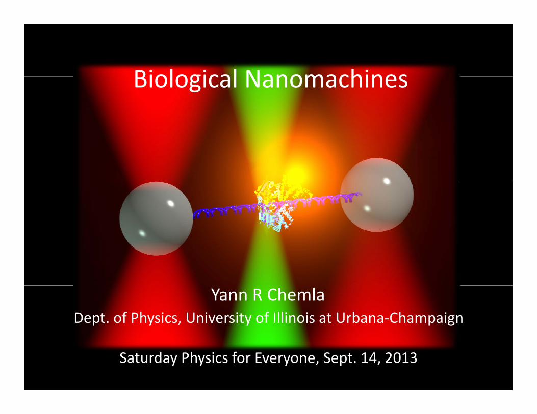
Biophysics at Illinois
WHAT IS BIOPHYSICS?Part I:
WHAT IS BIOPHYSICS?
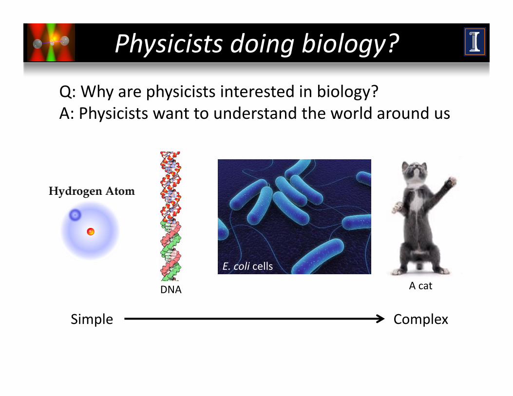
Physicists doing biology?
Q: Why are physicists interested in biology?A: Physicists want to understand the world around us
E. coli cells
Simple Complex
A catDNA
Simple Complex
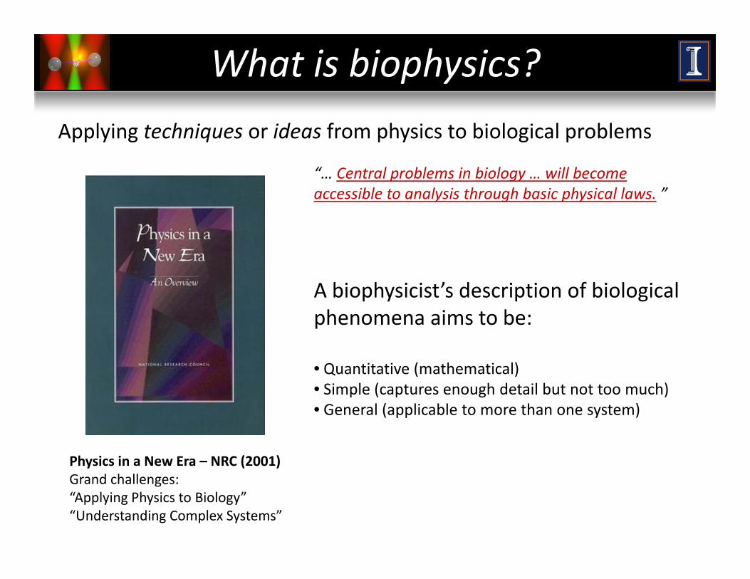
What is biophysics?
Applying techniques or ideas from physics to biological problems
“… Central problems in biology … will become p gyaccessible to analysis through basic physical laws. ”
A biophysicist’s description of biological phenomena aims to be:p
• Quantitative (mathematical)• Simple (captures enough detail but not too much)
Physics in a New Era – NRC (2001)
• General (applicable to more than one system)
Grand challenges: “Applying Physics to Biology”“Understanding Complex Systems”

MOLECULAR NANOMACHINESPart II:
MOLECULAR NANOMACHINES
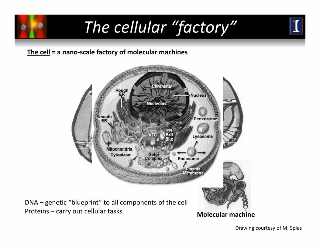
The cellular “factory”The cell = a nano‐scale factory of molecular machines
DNA genetic “blueprint” to all components of the cell
Molecular machine
Drawing courtesy of M. Spies
DNA – genetic “blueprint” to all components of the cellProteins – carry out cellular tasks
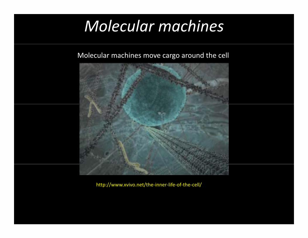
Molecular machines
Molecular machines move cargo around the cell
http://www.xvivo.net/the‐inner‐life‐of‐the‐cell/
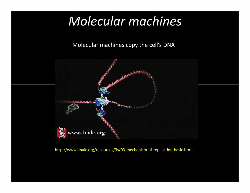
Molecular machines
Molecular machines copy the cell’s DNA
http://www.dnalc.org/resources/3s/03‐mechanism‐of‐replication‐basic.html
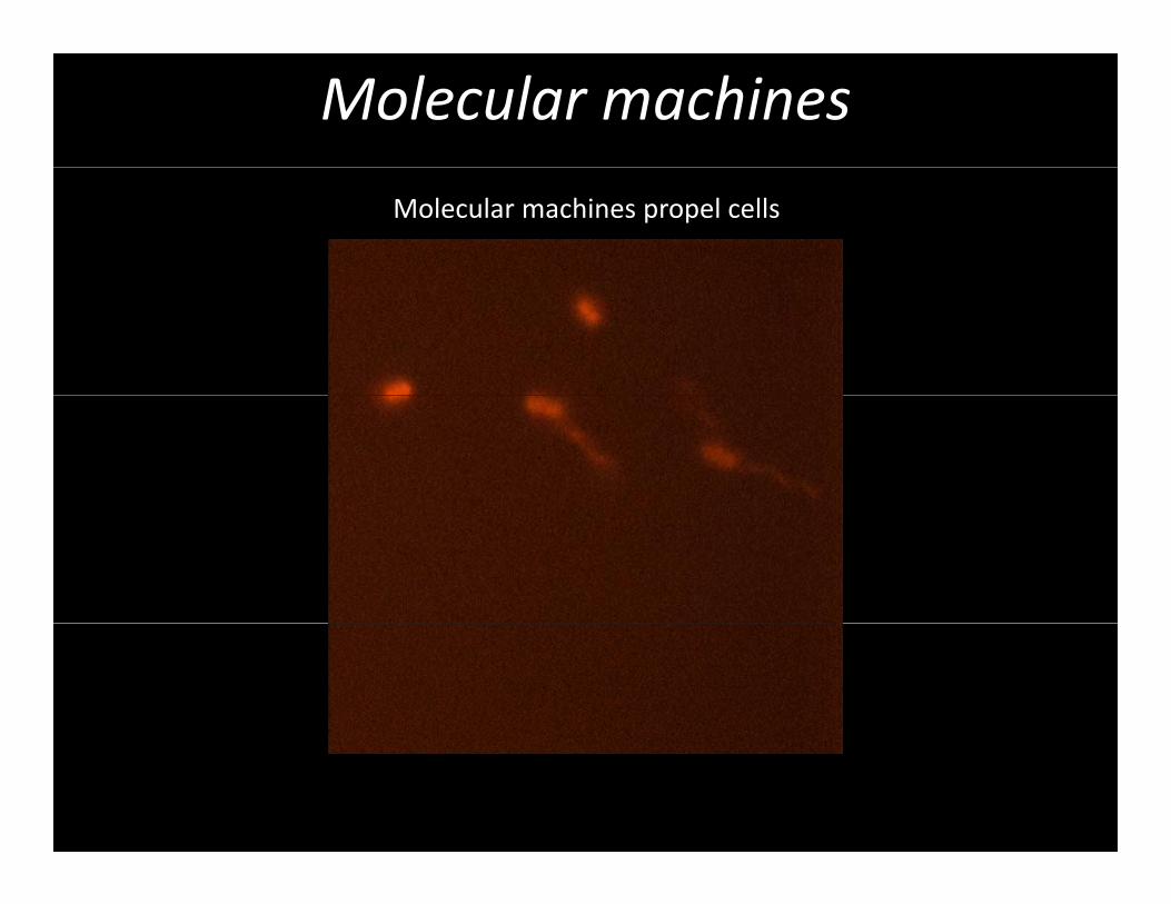
Molecular machines
Molecular machines propel cells
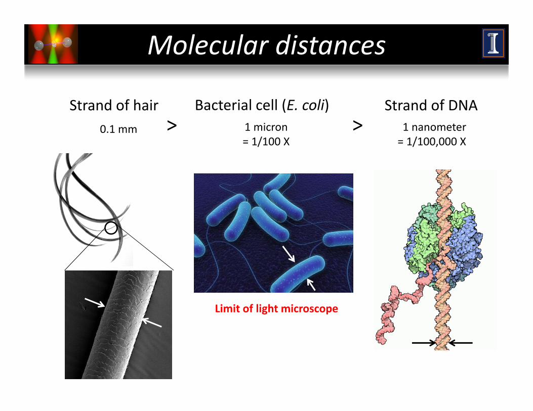
Molecular distances
0 1 mm
Strand of hair Bacterial cell (E. coli)1 micron
Strand of DNA1 nanometer> >0.1 mm 1 micron
= 1/100 X1 nanometer= 1/100,000 X
> >
Limit of light microscope
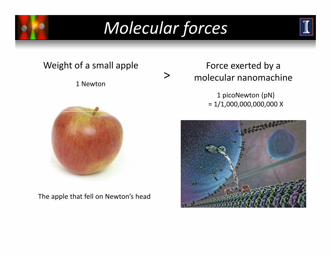
Molecular forces
Force exerted by a molecular nanomachine
Weight of a small apple>
1 Newtonmolecular nanomachine
1 picoNewton (pN)= 1/1,000,000,000,000 X
The apple that fell on Newton’s head
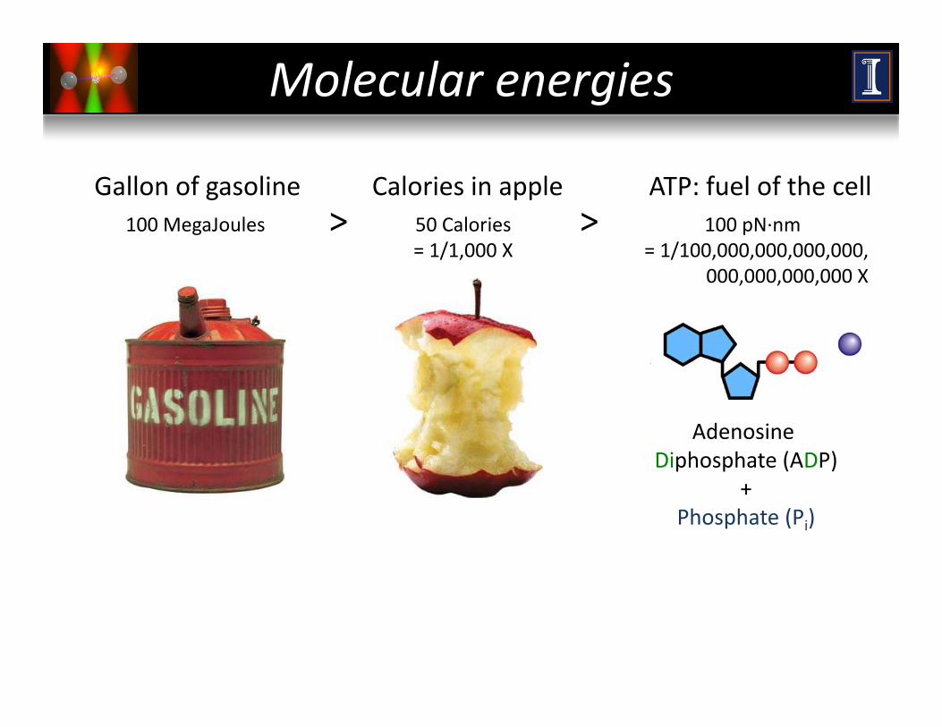
Molecular energies
100 MegaJoules
Gallon of gasoline Calories in apple50 Calories
ATP: fuel of the cell> > 100 pN∙nm100 MegaJoules 50 Calories
= 1/1,000 X> > 100 pN∙nm
= 1/100,000,000,000,000,000,000,000,000 X
Adenosine Triphosphate (ATP)
Adenosine Diphosphate (ADP)
+Phosphate (Pi)
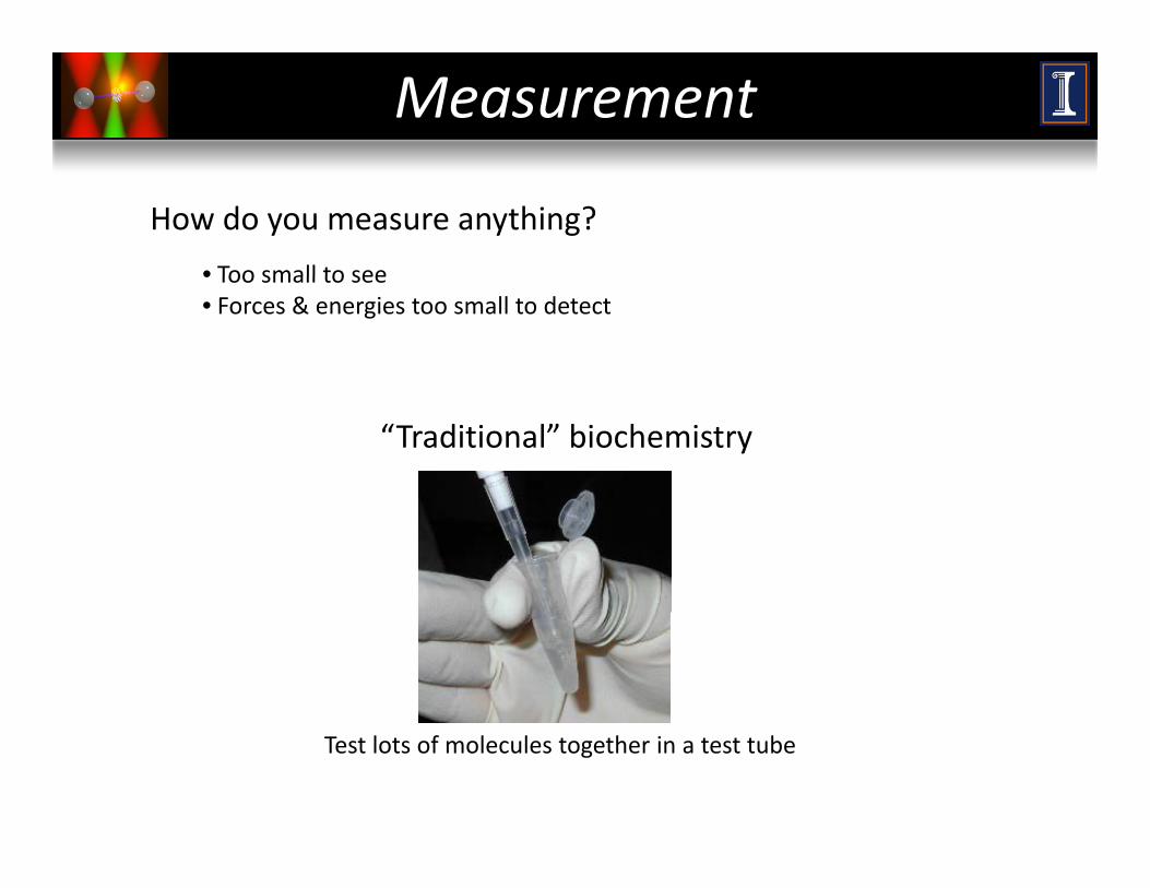
Measurement
How do you measure anything?
• Too small to see• Too small to see• Forces & energies too small to detect
“Traditional” biochemistry
Test lots of molecules together in a test tube
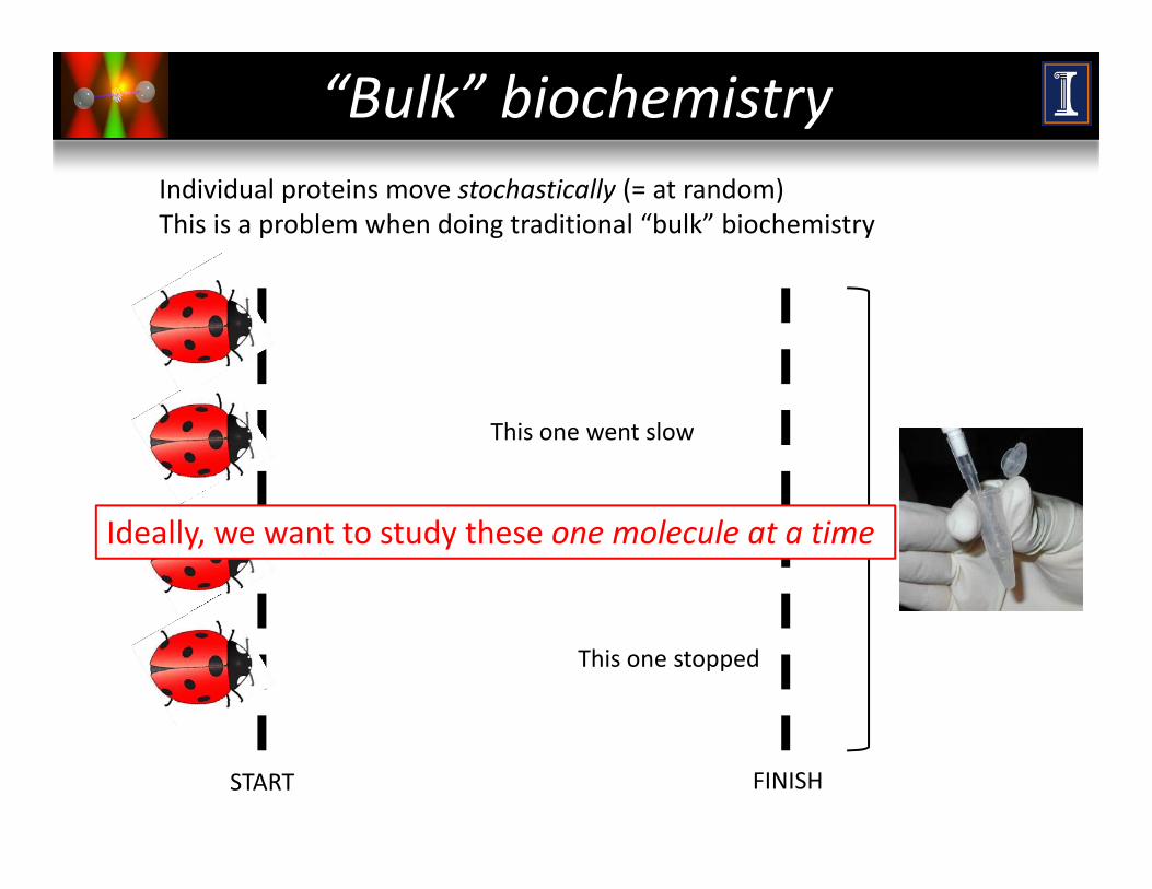
“Bulk” biochemistryIndividual proteins move stochastically (= at random)This is a problem when doing traditional “bulk” biochemistry
This one went slow
This one pausedIdeally, we want to study these one molecule at a time
This one stopped
START FINISH
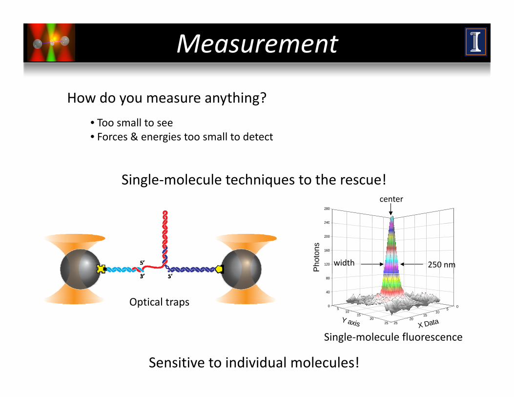
Measurement
How do you measure anything?
• Too small to see• Too small to see• Forces & energies too small to detect
Single‐molecule techniques to the rescue!
240
280
center
120
160
200
240
Pho
tons
width 250 nm
0
40
80
05
1015
2025
510
1520
25 X DataY axis
Optical traps
Sensitive to individual molecules!
2525 X Dataaxis
Single‐molecule fluorescence

SINGLE‐MOLECULE TECHNIQUESPart II:
SINGLE MOLECULE TECHNIQUES
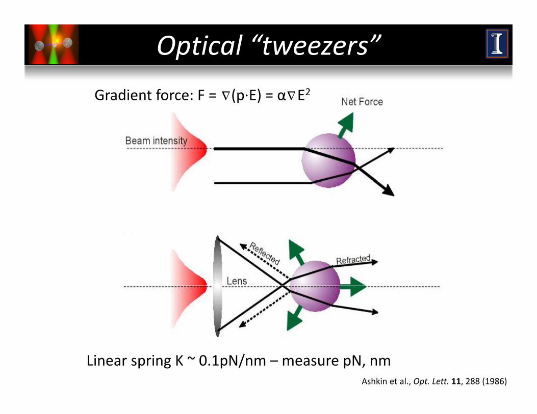
Optical “tweezers”
Gradient force: F = (p∙E) = α E2∆ ∆
Linear spring K ~ 0.1pN/nm – measure pN, nmAshkin et al., Opt. Lett. 11, 288 (1986)
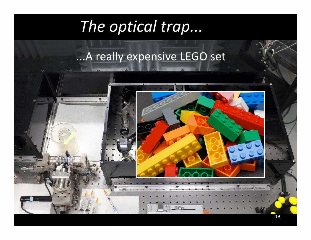
The optical trap...
...A really expensive LEGO set
19
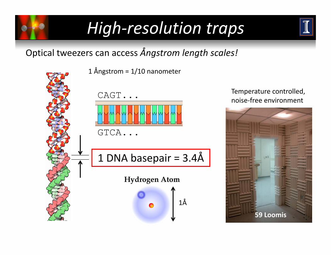
High‐resolution trapsOptical tweezers can access Ångstrom length scales!
1 Ångstrom = 1/10 nanometer
Temperature controlled, noise‐free environment
CAGT...
GTCA...
1 DNA basepair = 3.4Å
1Å
59 Loomis
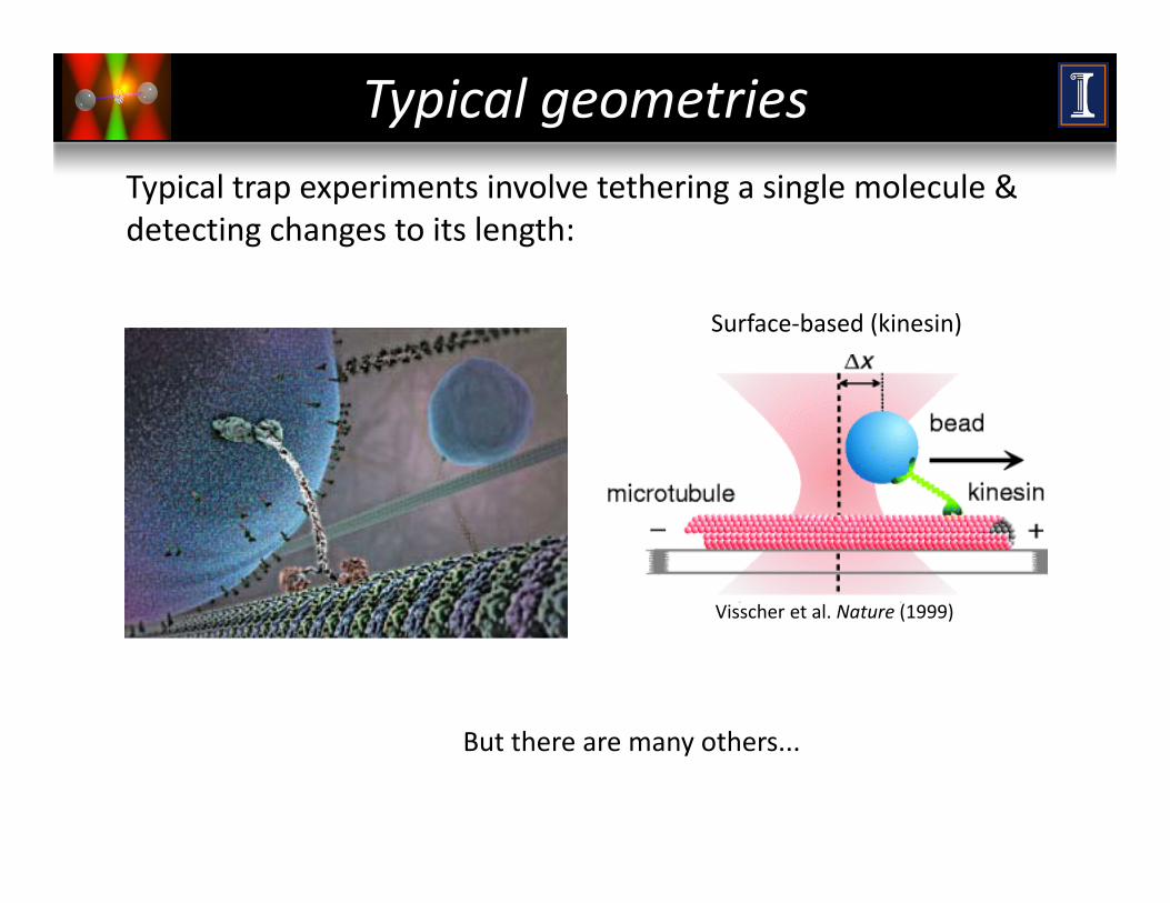
Typical geometriesTypical trap experiments involve tethering a single molecule & detecting changes to its length:
Surface‐based (kinesin)
Visscher et al. Nature (1999)
B t th thBut there are many others...
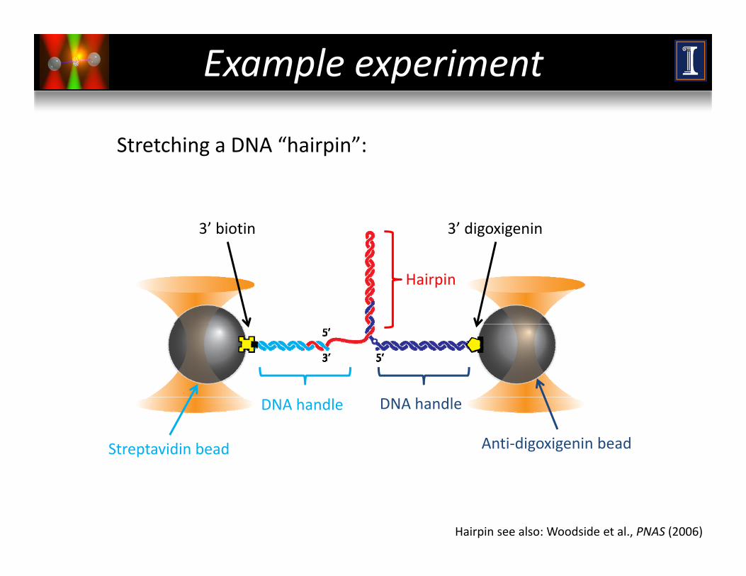
Example experiment
Stretching a DNA “hairpin”:
3’ biotin 3’ digoxigenin
Hairpin
Streptavidin bead Anti‐digoxigenin bead
DNA handle DNA handle
Hairpin see also: Woodside et al., PNAS (2006)
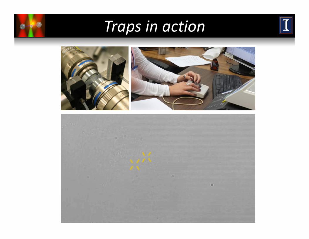
Traps in action
Gone fishing
24
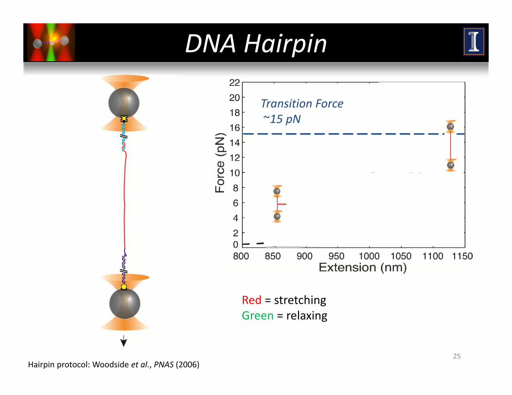
DNA Hairpin
Transition Force~15 pN15 pN
Red = stretchingGreen = relaxing
Hairpin protocol: Woodside et al., PNAS (2006)25

DNA MOTORSPart III:
DNA MOTORS
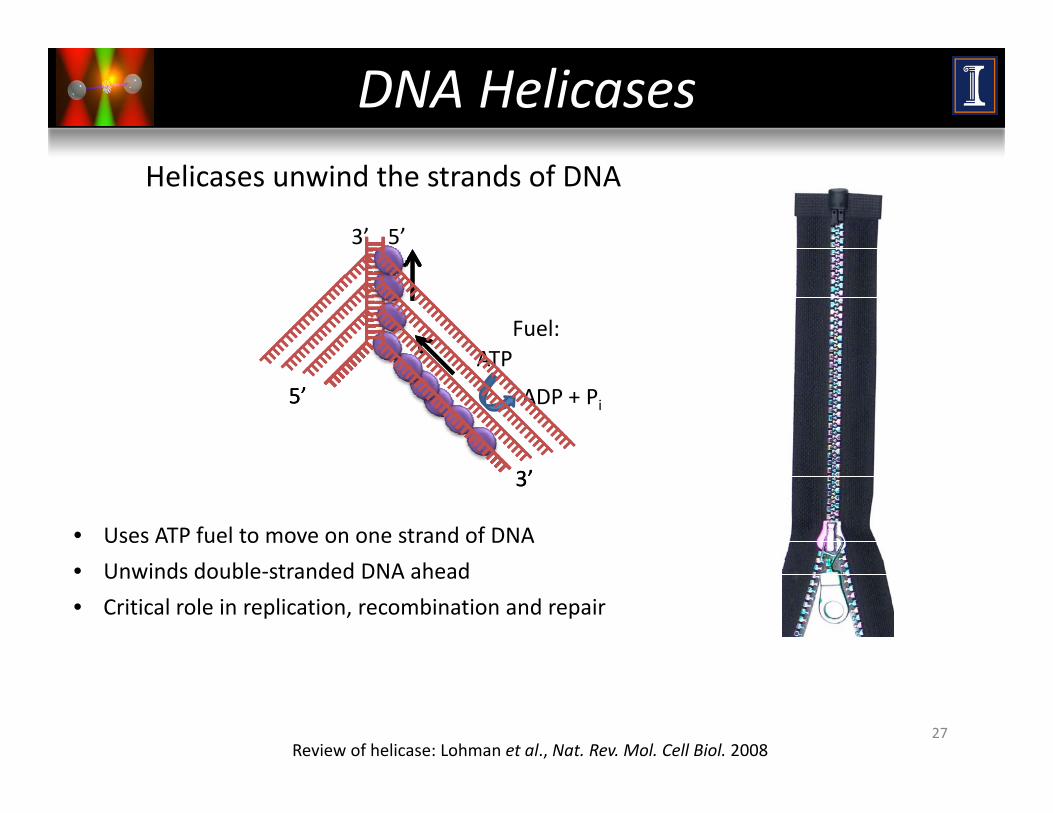
DNA Helicases
5’3’
Helicases unwind the strands of DNA
ATPFuel:
5’
ATP
ADP + Pi5’
• Uses ATP fuel to move on one strand of DNA
3’3’
• Unwinds double‐stranded DNA ahead
• Critical role in replication, recombination and repair
27Review of helicase: Lohman et al., Nat. Rev. Mol. Cell Biol. 2008
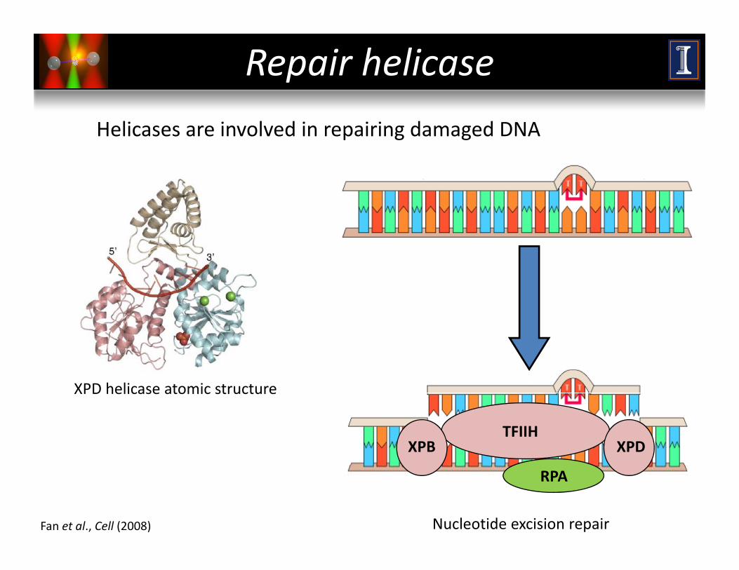
Repair helicase
Helicases are involved in repairing damaged DNA
XPD helicase atomic structure
TFIIHXPB XPD
XPD helicase atomic structure
RPA
Fan et al., Cell (2008) Nucleotide excision repair
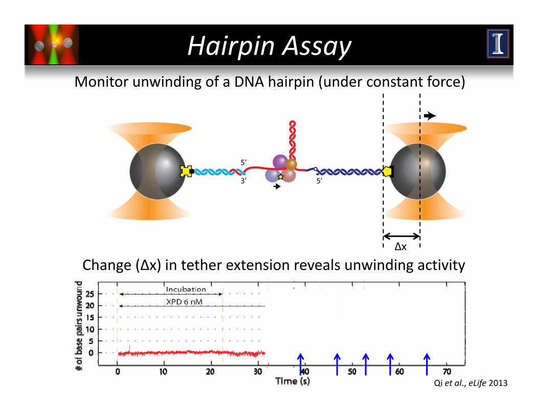
M i i di f DNA h i i ( d f )
Hairpin AssayMonitor unwinding of a DNA hairpin (under constant force)
Change (Δx) in tether extension reveals unwinding activity Δx
Qi et al., eLife 2013
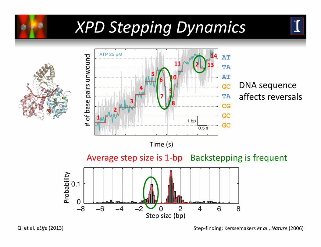
XPD Stepping Dynamics
5 10
11 12 1314 AT
TAAT
23
47
8
9
106 ATGCTACG
DNA sequence affects reversals
12 CG
GCGC
Time (s)
Average step size is 1‐bp Backstepping is frequent
Step‐finding: Kerssemakers et al., Nature (2006)
Step size (bp)
Qi et al. eLife (2013)
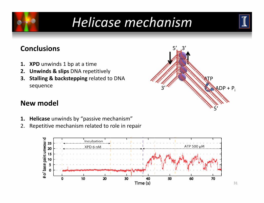
Helicase mechanism
3’5’Conclusions
1 XPD unwinds 1 bp at a time
3’
ATP
ADP + P
1. XPD unwinds 1 bp at a time2. Unwinds & slips DNA repetitively3. Stalling & backstepping related to DNA
sequence
5’
3 ADP + Pi
New model
1. Helicase unwinds by “passive mechanism”2. Repetitive mechanism related to role in repair
31

Fishing... in the dark
32
“Wouldn’t it be nice to see what’s on the fishing line?”
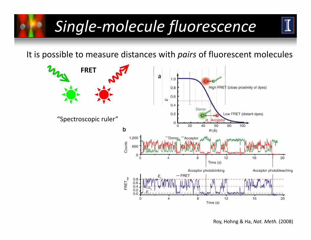
Single‐molecule fluorescence
It is possible to see light from a single molecule!It is possible to measure distances with pairs of fluorescent molecules
240
280
FRET
120
160
200
240
Phot
ons
0
40
80
05
1015
2025
510
1520
25 X DataY axis
“Spectroscopic ruler”
Courtesy of Paul Selvin
X Dis
Roy, Hohng & Ha, Nat. Meth. (2008)
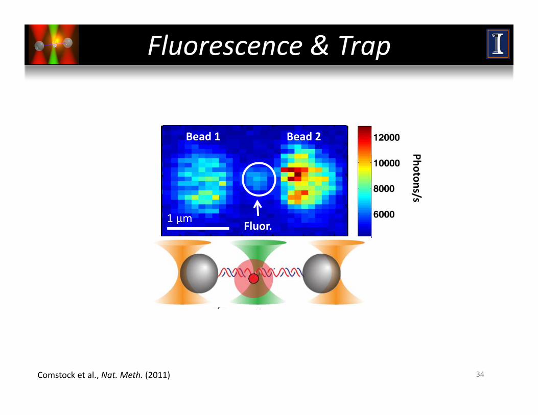
Fluorescence & Trap
PhotoBead 1 Bead 2
ons/s
1 μmFluor.
34Comstock et al., Nat. Meth. (2011)
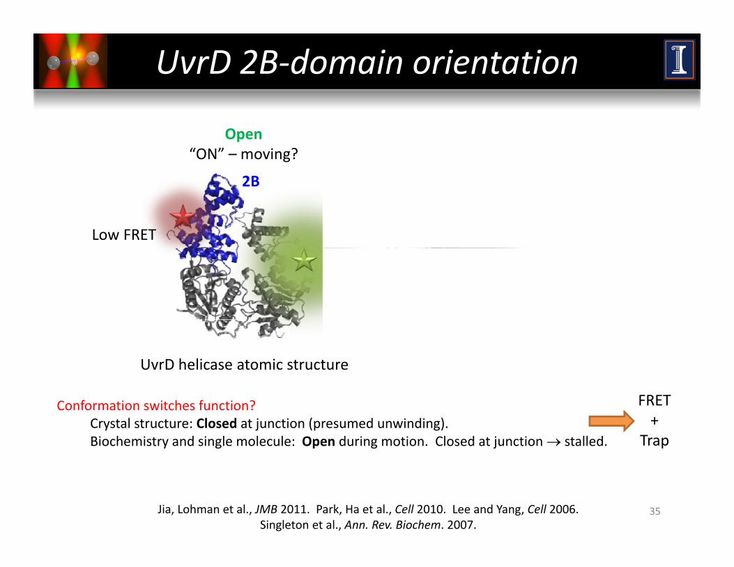
UvrD 2B‐domain orientation
Closed“Off” – stalled?
Open“ON” – moving?
~160°2B 2B160
rotation
High FRETLow FRET
2B
UvrD helicase atomic structure
Conformation switches function?Crystal structure: Closed at junction (presumed unwinding).Biochemistry and single molecule: Open during motion. Closed at junction → stalled.
FRET+
Trap
35Jia, Lohman et al., JMB 2011. Park, Ha et al., Cell 2010. Lee and Yang, Cell 2006. Singleton et al., Ann. Rev. Biochem. 2007.
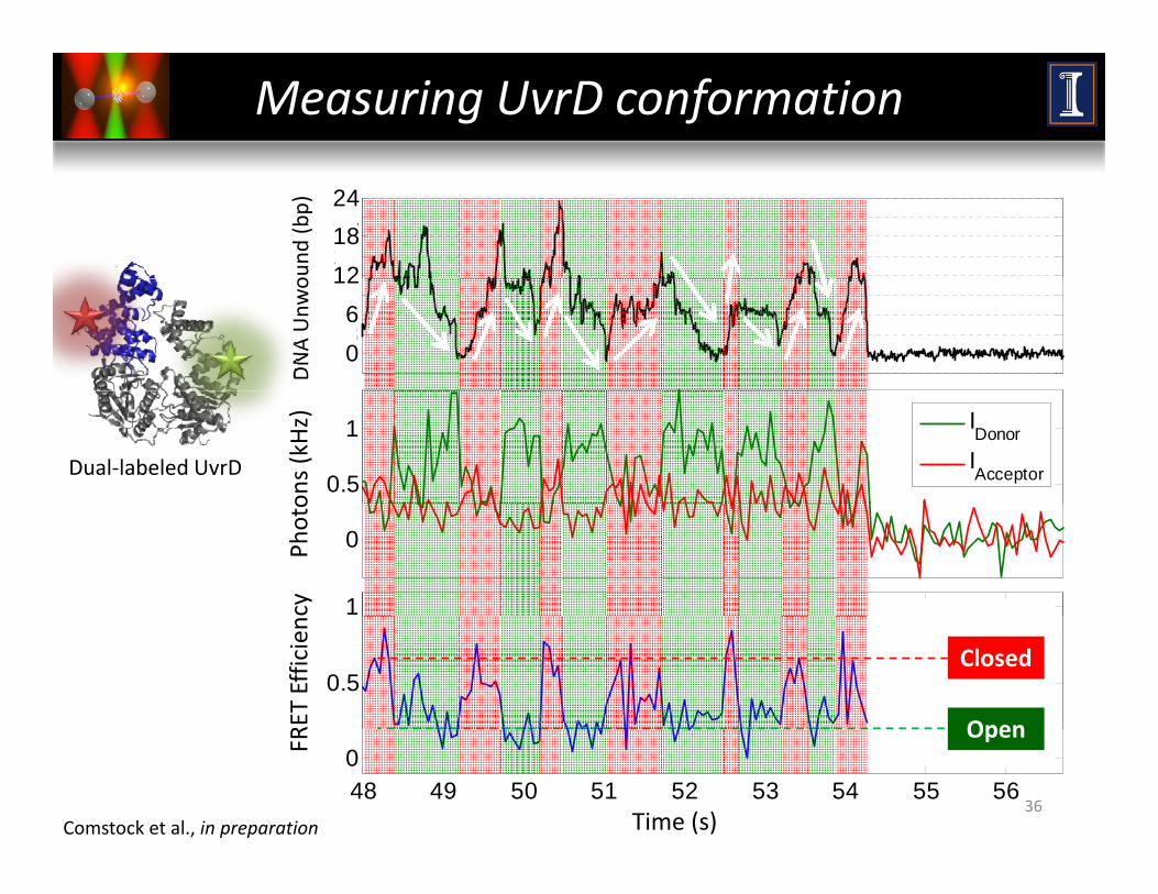
Measuring UvrD conformation
1215182124
ound
(bp)
-30369
12
DNA Unw
o
0.5
1 IDonorIAcceptor
ons (kHz)
Dual‐labeled UvrD
0
1
Photo
cy
0.5
RET Efficien
Open
Closed
48 49 50 51 52 53 54 55 560
Time (s)
FR
36
Open
Comstock et al., in preparation
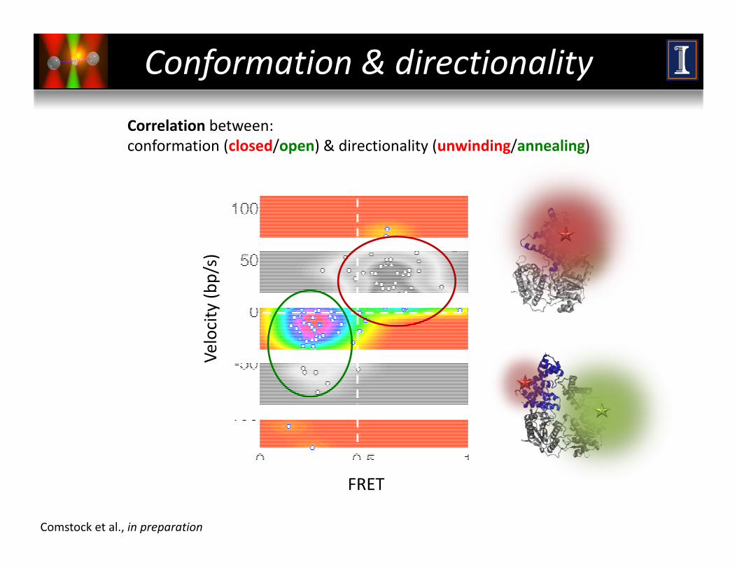
Conformation & directionality
Correlation between:conformation (closed/open) & directionality (unwinding/annealing)
ity (b
p/s)
Veloci
FRET
Comstock et al., in preparation
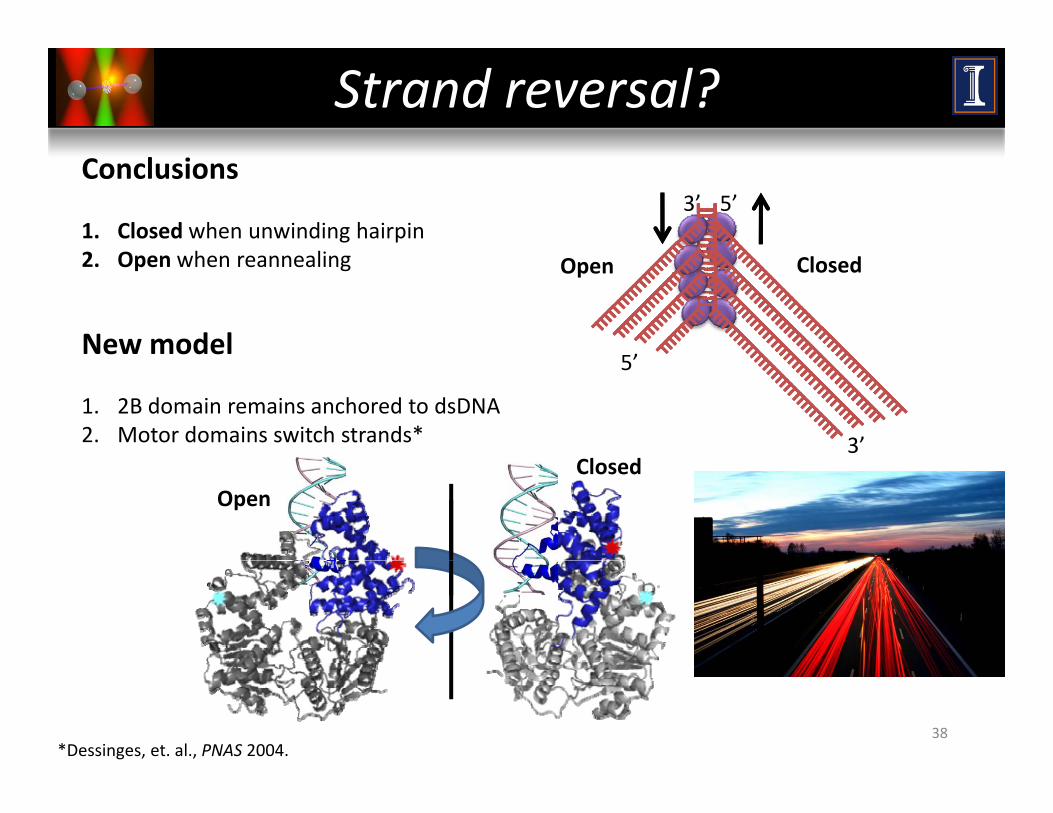
Strand reversal?Conclusions
1. Closed when unwinding hairpin2 O h li
5’3’
2. Open when reannealing
New model
ClosedOpen
5’
1. 2B domain remains anchored to dsDNA2. Motor domains switch strands*
5’
3’l dClosed
Open
38*Dessinges, et. al., PNAS 2004.

OUTLOOKPart IV:
OUTLOOK
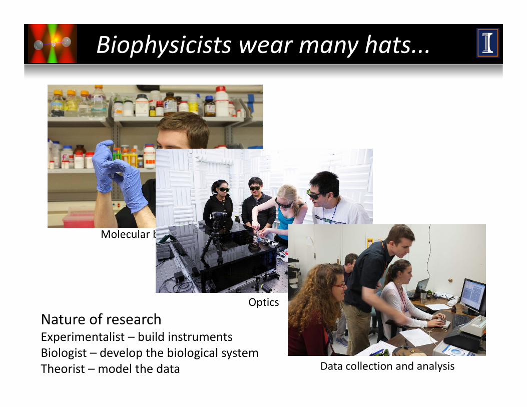
Biophysicists wear many hats...
Molecular biology
Optics
Nature of researchl b ld
Data collection and analysis
Experimentalist – build instrumentsBiologist – develop the biological systemTheorist – model the data
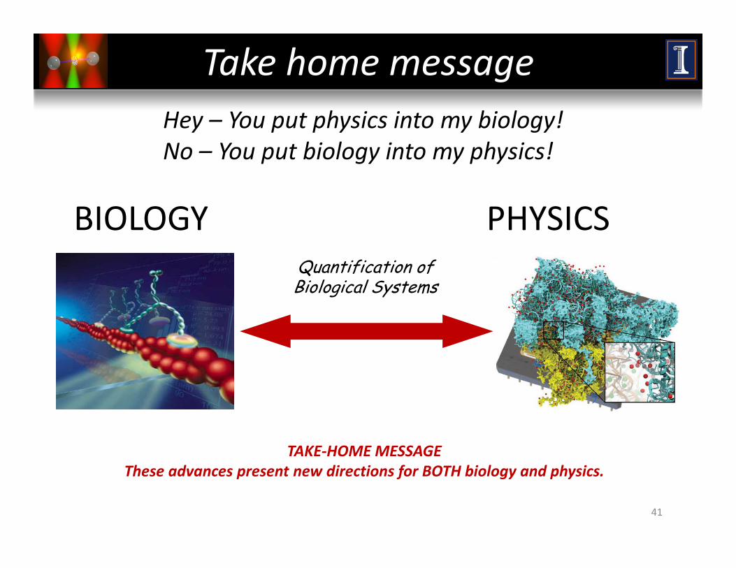
Take home messageHey – You put physics into my biology!No – You put biology into my physics!
BIOLOGY PHYSICSQuantification of Biological Systems
TAKE‐HOME MESSAGE
41
These advances present new directions for BOTH biology and physics.

AcknowledgementsAcknowledgements
XPD XPD HelicaseHelicase steppingsteppingZhi Qi†Zhi Qi†XPD XPD HelicaseHelicase steppingsteppingZhi Qi†Zhi Qi†Zhi Qi†Zhi Qi†Maria Spies & Robert PughMaria Spies & Robert Pugh(Univ. of Iowa)(Univ. of Iowa)
Zhi Qi†Zhi Qi†Maria Spies & Robert PughMaria Spies & Robert Pugh(Univ. of Iowa)(Univ. of Iowa)
UvrDUvrD trap & fluorescencetrap & fluorescence::Matt Comstock*Matt Comstock*UvrDUvrD trap & fluorescencetrap & fluorescence::Matt Comstock*Matt Comstock*Kevin WhitleyKevin WhitleyTaekjip Ha (Univ. of Illinois)Taekjip Ha (Univ. of Illinois)Tim Lohman &Tim Lohman & HaifengHaifeng JiaJia
Kevin WhitleyKevin WhitleyTaekjip Ha (Univ. of Illinois)Taekjip Ha (Univ. of Illinois)Tim Lohman &Tim Lohman & HaifengHaifeng JiaJia Now at:Now at:Tim Lohman & Tim Lohman & HaifengHaifeng JiaJia(Washington Univ.)(Washington Univ.)Tim Lohman & Tim Lohman & HaifengHaifeng JiaJia(Washington Univ.)(Washington Univ.) † † Columbia Univ.Columbia Univ.
* Michigan State Univ.* Michigan State Univ.
Funding:Funding:


![Artificial nanomachines based on interlocked molecules · Artificial nanomachines based on interlocked molecules S1781 microscopies [16], capable of visualizing or manipulating](https://img.pdfslide.us/doc/110x75/5ea3814d675d087a8517e008/artiicial-nanomachines-based-on-interlocked-molecules-artiicial-nanomachines.jpg)


![GENES, MEMES, LANGUAGE, AND NANOMACHINES: A … · 2020-06-22 · Twibell Book Proof (Do Not Delete) 4/10/20 12:44 PM 2019] Genes, Memes, Language, and Nanomachines 67 critical theorist](https://img.pdfslide.us/doc/110x75/5f5d1e4023261209686fdf16/genes-memes-language-and-nanomachines-a-2020-06-22-twibell-book-proof-do.jpg)