Embed Size (px)
Citation preview

Safety and Efficacy
Cosmetic Services
Anti-aging
Oxidative Stress and Anti-oxidant capacity
Skin is repetitively exposed to environmental oxidative stress such
as ultraviolet (UV) radiation, pollutants, and chemical oxidants
leading to premature skin aging.
Inside the cutaneous tissue, cellular metabolism and mitochondrial
respiration can also result in production of oxidative species and
reactive oxygen species (ROS).
Skin possesses various endogenous anti-oxidant systems, such as
small molecules or enzymes, but oxidative stress can overwhelm
these protective systems.
This unbalance will accelerate aging by damaging DNA, proteins,
lipids, and other cellular constituents. ROS and other free radicals
also affect the regulation of gene transcription.
Test for radical scavenger activity
This assay is based on the decrease of absorbance of the free
radical DPPH (1,1-diphenyl-2-picrylhydrazyl). DPPH is a stable free
radical which has an unpaired valence electron at one atom of
nitrogen bridge. Scavenging of DDPH radical is the basis of the
popular DDPH antioxidant assay.
Sample concentration: 10 study concentrations.
Time points: Kinetic
Replicates: 3/condition
Positive inhibitors: Ascorbic acid, Butylated hydroxytoluene (BHT)
or Trolox.
End point: Kinetic spectrophotometric measurement
of the DDPH absorbance (517nm) decrease.
Anti-aging
Oxidative stress and Anti-oxidant action
Measurement of intracellular reactive oxygen species
(ROS)
ROS formation is induced by H2O2/ tert-Butyl Hydroperoxid
(TBHP)/ UV light, in different cell types: human primary cultures
(Epidermal Progenitor Keratinocytes, Epidermal Keratinocytes,
Dermal Fibroblasts, HUVEC, Dendritic cells, Hepatocytes), skin cell
lines (HaCat, human microvascular endothelial cells, 3T3) and
current cell lines.
Sample concentration: 3 study concentrations.
Time points: To be selected by customer.
Replicates: 3/condition
Positive inhibitors: Trolox, Quercetin or NAC.
End point: Measurement of fluorescence on treated and non-
treated cells, using the carboxy-H2-DCFDA (Oxidative stress
indicator).
Test for lipid peroxidation (Fenton reaction) and
malondialdehyde (MDA) measurement
Sample concentration: 3 study concentrations.
Time points: To be selected by customer
Replicates: 3/condition.
Positive inhibitors: Deferoxamine, Trolox, NAC, BHT
Inducer: H2O2 alone, H202 + iron
(to induce the Fenton reaction).
End point: nmols MDA/mg protein.
This assay is based on the fluorimetric
measurement of MDA, the major product of lipid
peroxidation of polyunsaturated fatty acids.
Measurement of this aldehyde in cell supernatants
and lysates provides a good index of lipid
peroxidation.
Anti-aging
Oxidative stress and Anti-oxidant action
Test for Lipid peroxide content under UV induction or
other stimuli
This assay allows measuring the ratio-fluorescence microscopy of
lipid oxidation in living cells using C11-BODIPY581:591. This
fluorescent probe is readily incorporated into cellular membranes
and is about twice as sensitive to oxidation as arachidonic acid;
thereby it is relatively insensitive to nitric oxide and superoxide.
Sample concentration: 3 study concentrations.
Time points: To be selected by customer.
Replicates: 3/condition.
Positive inhibitors: Butylated hydroxytoluene (BHT) or Trolox.
Inducer: UVA/B (using a Sun simulator)
End point: Lipid peroxide using C11-fluor probe – Fluorescence
measurement.
Test for protection of glutathione depleted levels
This assay allows to assess the protective effect of active
compound or anti-oxidants on the recovery of glutathione levels
depleted by either tert-butyl hydroperoxide (TBHP) or by L-
buthionine-S,R-sulfoximine (BSO).
Sample concentration: 3 study concentrations.
Time points: To be selected by customer
Replicates: 3/condition
Positive inhibitors: NAC or Butylated hydroxytoluene (BHT).
Inducer: TBHP or BSO.
End point: nmols glutathione/mg protein.
Anti-aging
Aging and Photo-aging
Skin is repetitively exposed to environmental oxidative stress such
as ultraviolet (UV) radiation, pollutants, and chemical oxidants
leading to premature skin aging. Ultraviolet (UV) light produces
reactive oxygen species (ROS) in skin, which accelerate aging by
damaging DNA, proteins, lipids, and other cellular constituents.
UV is a major factor known to cause premature aging by
producing higher levels of matrix metalloproteinases (MMP),
enzymes which take part to skin remodelling, wound healing and
other skin physiological functions.
Among these MMPs, MMP-9 is a type IV collagen hydrolase
implicated in different processes which lead to skin extracellular
matrix degradation and its collapse. In addition, MMP-1 or
collagenase may contribute to loss of interstitial collagen and
extracellular matrix (ECM) alteration.
These MMPs are induced directly by reactive oxygen species (ROS)
or indirectly by inflammatory cytokines produced such as
interleukin-1.In addition, intrinsic or chronological Skin aging is
clinically associated with increased fragility, loss of elasticity and
transparent quality of skin.
Decrease in proliferative capacity of skin cells, increased
expression of matrix metalloproteinase (MMP) and reduced
collagen and elastin synthesis are the responsible phenomenon of
these effects. To evaluate the effectiveness of a cosmetics product
in improving all this aging effects on skin, the following in vitro
tests are presented.
Anti-aging
Aging and Photo-aging
Tests for DNA damage after UV irradiation
Evaluation of the DNA-photoprotective effect on UV-irradiated
human skin cells.
Pre-treated human skin cells are UV-irradiated for DNA damage
induction (DNA strand breaks, modifications and oxidative
damage). The Single Cell Gel Electrophoresis assay (Comet assay)
is used for the assessment of the extent of DNA-damage.
Cell types and models: Epidermal Keratinocytes (progenitor,
neonatal and/or adult), Human Dermal Fibroblasts or human
reconstructed skin models.
Sample concentration: 3 study concentrations.
Time points: To be selected by customer.
Replicates: 3/condition
Positive controls: Octyl methoxycinnamate (OMC), 4-tert-butyl-4'-
methoxydibenzoylmethane (BM-DBM) or sun cream. Inducer:
UVA/B (Sun simulator system).
End point: Percentage of DNA tail as an indicator of DNA strand
break index (DNA-damage).
Anti-aging
Aging and Photo-aging
Tests for DNA repair kinetics after UV irradiation
To the aim of evaluating the repair system promotion potential of
a compound or product, skin cells are UV-irradiated in order to
induce DNA-damage. Thereupon cells are exposed to the test item
and the DNA repair process is analysed over time by means of
Comet assay.
Cell types and models: Epidermal Keratinocytes (progenitor,
neonatal and/or adult), Human Dermal Fibroblasts or human
reconstructed skin models.
Sample concentration: 3 study concentrations.
Time points: Kinetic evaluation post-irradiation.
Replicates: 3/condition
Inducer: UVA/B (Sun simulator system).
End point: Percentage of DNA tail as an indicator of DNA strand
break index (DNA-damage).
Cellular stress, cycle or aging genes and associated
protein expression
A wide range of cellular stress, cycle and aging cell markers are
analyzed on skin cells: SIRT-1, AP-1, P16, COX, MMPs, MNSOD,...
by RT-PCR, Multiplexing analysis, western Blot or ELISA.
Immunolocalization and microscopically image analysis is also
available.
Cell types and models: Epidermal Keratinocytes (progenitor,
neonatal and/or adult), Human Dermal Fibroblasts or human
reconstructed skin models.
Sample concentration: 3 study concentrations.
Time points: To be selected by customer.
Replicates: 3/condition
End point: level of protein expression.
Anti-aging
Aging and Photo-aging
Cells proliferation inductor effect
The stimulation of the skin cells proliferation by a product can be
evaluated by means of several well known methodologies: 3H-
thymidine incorporation (DNA Radiolabeling based method) or 5-
bromo-2'-deoxyuridine (BrdU) incorporation test.
3H-thymidine or BrdU (Thymidine analog) are incorporated into
the newly synthesized DNA of replicating cells (during the S phase
of the cell cycle) during DNA replication. Then, de novo
synthetized DNA is quantified by processing radioactive culture
samples and measurement in a liquid scintillation counter, or by
ELISA if the BrdU method is used.
Sample concentration: 3 study concentrations.
Time points: To be selected by customer.
Replicates: 3/condition
Positive Control: TGF- 1, VEGF and others.
End point:
- Disintegrations per minute (DPM) - radiation values directly
proportional to the amount of synthesized DNA.
- Absorbance values (450nm) after ELISA processing of treated
cell monolayer.
Cell types and models: Epidermal Keratinocytes
(progenitor, neonatal and/or adult), Human Dermal
Fibroblasts, melanocytes, endothelial cells, lymphocytic
cells or human reconstructed skin models.
Anti-aging
Aging and Photo-aging
Moisturising effect and Glicosaminoglycans (GAG) and
Hyaluronic Acid synthesis “de novo”
Measurement of de novo synthesis of Glycosaminoglycans
[90-95% Hyaluronic Acid (HA)] on skin cells exposed to the
product by means of 3H-Glucosamine incorporation.
After treatment with the product, cells are incubated with 3H-
Glucosamine (radioactive material) in culture medium. The newly
Glycosaminoglycans (HA) produced molecules, radioactively
labeled, are isolated and quantified in a liquid scintillation counter.
The amount of GAG/HA produced by product-exposed cells are
compared to non-treated cells.
Cell types and models: Epidermal Keratinocytes (progenitor,
neonatal and/or adult), Human Dermal Fibroblasts, chondrocytes,
or human reconstructed skin models.
Sample concentration: 3 study concentrations.
Time points: To be selected by customer.
Replicates: 3/conditionPositive
Control: TGF- 1.
End point: Disintegrations per minute (DPM) - radiation values
directly proportional to the amount of synthesized GAG and HA.
Anti-aging
Aging and Photo-aging
Synthesis and expression of Extracellular Matrix (ECM)
proteins
Evaluation of the improvement in ECM proteins production in order
to increase skin firmness, elasticity and palliate aging effects
associated to the test item.
ECM proteins to be detected: Fibrillar collagen types (Collagen
type I, type III, type V), Network-forming collagens (Collagen type
IV and type VII), Fibril-associated collagens (Type XII and type
XVI), elastin, fibronectin and others. Inmunocytoquemistry or
inmunohistoqumistry will be performed for ECM protein analysis
using specific human primary antibodies. Immunolocalization and
microscopically image analysis is also available.
Cell types and models: Epidermal Keratinocytes (progenitor,
neonatal and/or adult), Human Dermal Fibroblasts or human
reconstructed skin models.
Sample concentration: 3 study concentrations.
Time points: To be selected by customer.
Replicates: 3/condition
Positive Control: TGF- 1.
End point: Absorbance values after Inmunocytoquemistry
and ECM protein production index.
Anti-aging
Aging and Photo-aging
ECM protection by MMP synthesis modulation or MMP
inhibition
Evaluation of the MMP synthesis modulation ability of a product on
induced skin cells. MMP production and release is evaluated on
cultured skin cell pre-treated with the test item.
Different members of the MMP family are quantified in stimulated
cell cultures (supernatants) by ELISA or Multiplexing technique.
Specific MMP activity is also possible to be evaluated by ELISA or
Zymography.
Cell types and models: Human Dermal Fibroblasts and
Chondrocytes.
MMP to be analysed: MMP-1, MMP-2, MMP-3, MMP-9, MMP-13 and
others.
Sample concentration: 3 study concentrations.
Time points: To be selected by customer.
Replicates: 3/condition
Inducer: UVA/B (Sun simulator), H2O2 or IL-1 .
Positive Control: Dexamethasona or TGF- 1.
End point: MMP production (ng MMP/mg protein) or MMP Activity
(band intensity and area analysis).
Anti-inflammatory and calming
Exposure of the skin to several external stimuli, stress and chronic
UV irradiation, induces a variety of biological effects including
inflammation, reactive oxygen species (ROS), photo-aging
(wrinkle formation and skin thickening), and, in some cases,
cancer development.
In addition, during inflammation and UV radiation higher levels of
matrix metalloproteases (MMPs) and inflammatory mediators,
such as cytokines and chemokine (Interleukins 1 (IL-6), 6 (IL-6),
8 (IL-8), RANTES, TGF- , etc.) or immunomodulatory cytokines
(mainly interleukins 10 and 12), and several growth factors (IGF,
VEGF, TGF- , etc.) are produced by skin and surrounding cells. In
parallel, other key inflammatory mediators such prostaglandins,
mainly PGE2, are also produced in irradiated and inflamed skin.
Evaluation of Induced-Inflammatory mediators release
measurement on different cell types
Evaluation of inflammatory mediators release on stimulated
human cells/models: Release of IL-1 , IL-1 , IL-6, IL-8, IL-10,
IL-12, E-Selectin, G-CSF, ICAM-1, TNF- , IFN- , INF- , RANTES,
and other inflammatory mediators measurement (Flow Cytometric
multiplexing analysis).
Cell types and models: Human Epidermal Keratinocytes
(progenitor, neonatal, adult), Dermal Fibroblasts, Dendritic Cells,
Human Follicle Dermal Papilla Cells, Endotehial Cells, monocytic
cells, lymphocytic cells and others. Human reconstructed
epidermis, Full thickness skin model and skin biopsies.
Sample concentration: 3 study concentrations.
Time points: To be selected by customer Replicates: 3/condition
Positive inhibitors: Dexamethasone and others.
Inducer: UVA/B (sun simulator system), PMA, LPS or other stimuli
(IL-1 or TNF )
End point: ng inflammatory mediator/mg protein.
Inflammatory responses
Anti-inflammatory and calming
Arachidonic acid mediators
Evaluation of PGE2 release on stimulated human cells/models.
Cell types and models: Human Epidermal Keratinocytes
(progenitor, neonatal and/or adult), Human Dermal Fibroblasts,
Dendritic Cells, Endotehial Cells and others. Human reconstructed
epidermis, Full thickness skin model and skin biopsies.
Sample concentration: 3 study concentrations.
Time points: To be selected by customer
Replicates: 3/condition
Positive inhibitors: Indometacin and NS398.
Inducer: PMA or other stimuli like IL-1 or TNF .
End point: ng PGE2/mg protein.
Induced COX-2 and PG: Expression and Inhibition
Evaluation of the COX-2 and PG expression/production on
stimulated human cells/models through quantitative RT-PCR,
Western blot or ELISA.
Cell types and models: Human Epidermal Keratinocytes
(progenitor, neonatal and/or adult), Human Dermal Fibroblasts,
Dendritic Cells, Endotehial Cells and others. Human reconstructed
epidermis, Full thickness skin model and skin biopsies.
Sample concentration: 3 study concentrations.
Time points: To be selected by customer
Replicates: 3/condition
Positive inhibitors: NS398.
Inducer: IL-1 or TNF .
End Point: ng PG/mg Protein
Inflammatory responses
Skin repair
Wound healing
Skin is permanently subjected to several stresses and outer
aggressions that can damage its physical integrity. Improvement
of cell response to an injury will lead to accelerated wound-
healing.
Wound healing is a complex process in which a variety of cellular
and matrix components act in concert to re-establish the integrity
of injured tissue. The complexity of this process may be simplified
into the main biological phenomenons in the healing response.
Migration and proliferation of fibroblasts in dermis and of
epidermic keratinocytes are the crucial processes in the one of the
wound repair phases, the regenerative phase.
Sample concentration: 3 study concentrations.
Time points: To be selected by customer.
Replicates: 3/condition
Positive Control: TGF- 1, VEGF and others.
End point: Absorbance values (450nm) after ELISA processing of
treated cell monolayer.
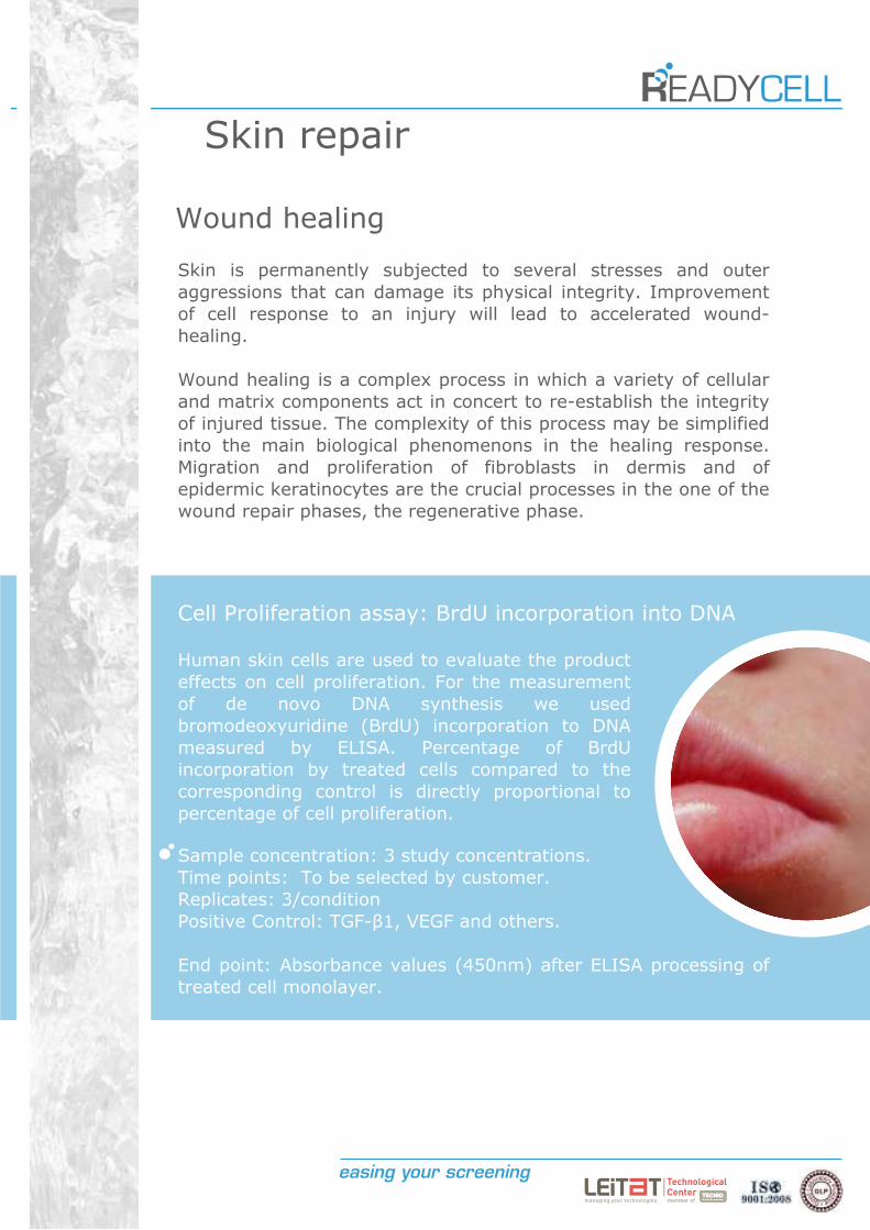
Human skin cells are used to evaluate the product
effects on cell proliferation. For the measurement
of de novo DNA synthesis we used
bromodeoxyuridine (BrdU) incorporation to DNA
measured by ELISA. Percentage of BrdU
incorporation by treated cells compared to the
corresponding control is directly proportional to
percentage of cell proliferation.
Cell Proliferation assay: BrdU incorporation into DNA
Skin repair
Wound healing
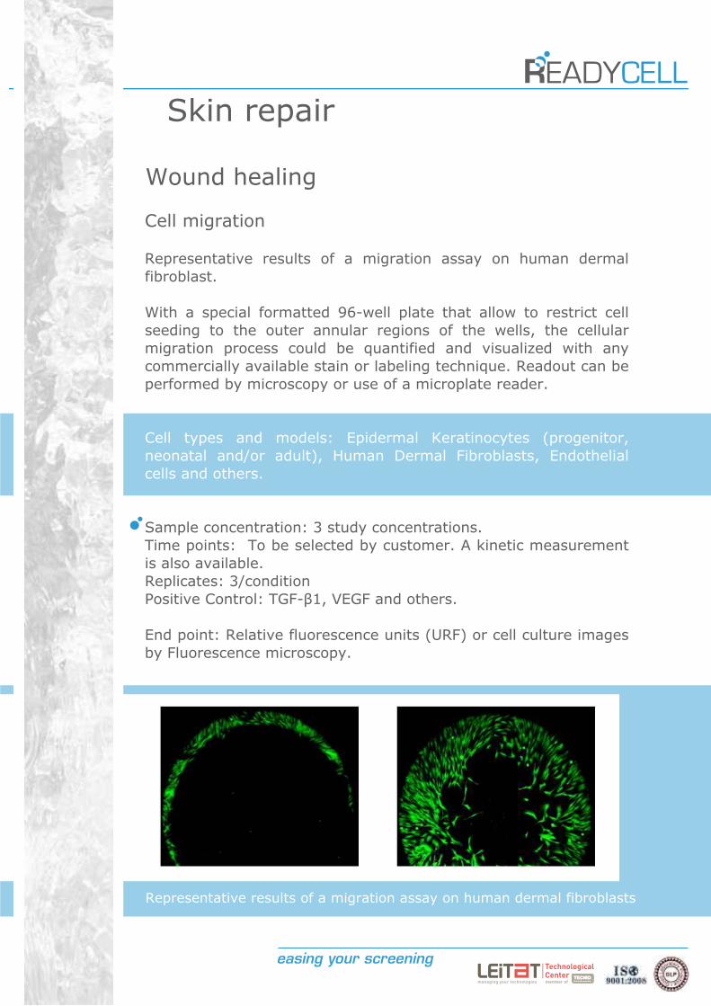
Cell migration
Representative results of a migration assay on human dermal
fibroblast.
With a special formatted 96-well plate that allow to restrict cell
seeding to the outer annular regions of the wells, the cellular
migration process could be quantified and visualized with any
commercially available stain or labeling technique. Readout can be
performed by microscopy or use of a microplate reader.
Cell types and models: Epidermal Keratinocytes (progenitor,
neonatal and/or adult), Human Dermal Fibroblasts, Endothelial
cells and others.
Sample concentration: 3 study concentrations.
Time points: To be selected by customer. A kinetic measurement
is also available.
Replicates: 3/condition
Positive Control: TGF- 1, VEGF and others.
End point: Relative fluorescence units (URF) or cell culture images
by Fluorescence microscopy.
Representative results of a migration assay on human dermal fibroblasts
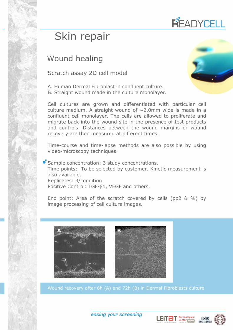
Wound recovery after 6h (A) and 72h (B) in Dermal Fibroblasts culture
Skin repair
Wound healing
Scratch assay 2D cell model
A. Human Dermal Fibroblast in confluent culture.
B. Straight wound made in the culture monolayer.
Cell cultures are grown and differentiated with particular cell
culture medium. A straight wound of ~2.0mm wide is made in a
confluent cell monolayer. The cells are allowed to proliferate and
migrate back into the wound site in the presence of test products
and controls. Distances between the wound margins or wound
recovery are then measured at different times.
Time-course and time-lapse methods are also possible by using
video-microscopy techniques.
Sample concentration: 3 study concentrations.
Time points: To be selected by customer. Kinetic measurement is
also available.
Replicates: 3/condition
Positive Control: TGF- 1, VEGF and others.
End point: Area of the scratch covered by cells (pp2 & %) by
image processing of cell culture images.
A B
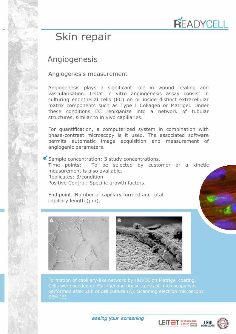
Formation of capillary-like network by HUVEC on Matrigel coating.
Cells were seeded on Matrigel and phase-contrast microscopy was
performed after 20h of cell culture (A). Scanning electron microscopy
SEM (B).
Skin repair
Angiogenesis
Angiogenesis measurement
Angiogenesis plays a significant role in wound healing and
vascularisation. Leitat in vitro angiogenesis assay consist in
culturing endothelial cells (EC) on or inside distinct extracellular
matrix components such as Type I Collagen or Matrigel. Under
these conditions EC reorganize into a network of tubular
structures, similar to in vivo capillaries.
For quantification, a computerized system in combination with
phase-contrast microscopy is it used. The associated software
permits automatic image acquisition and measurement of
angiogenic parameters.
Sample concentration: 3 study concentrations.
Time points: To be selected by customer or a kinetic
measurement is also available.
Replicates: 3/condition
Positive Control: Specific growth factors.
End point: Number of capillary formed and total
capillary length (µm).
A B
Smoothing
Anti Wrinkle effect
Wrinkles are an obvious, visible signs and a very important
indicator of ageing. Cosmetic products and actives focus on
ameliorating wrinkles require subjective and objective
measurements about wrinkles characteristics and presence, before
and after treatment, for an accurate assessment of efficacy.
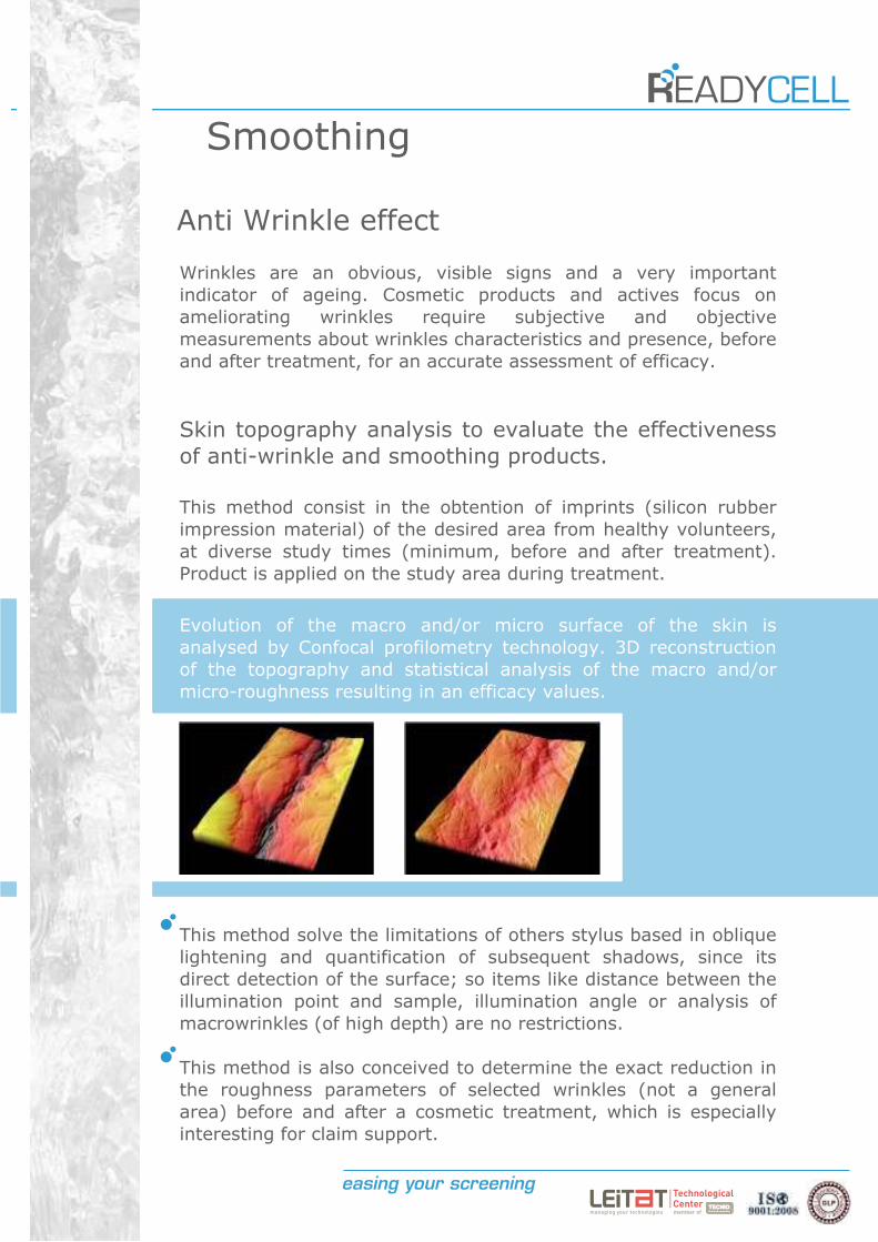
Skin topography analysis to evaluate the effectiveness
of anti-wrinkle and smoothing products.
This method consist in the obtention of imprints (silicon rubber
impression material) of the desired area from healthy volunteers,
at diverse study times (minimum, before and after treatment).
Product is applied on the study area during treatment.a
Evolution of the macro and/or micro surface of the skin is
analysed by Confocal profilometry technology. 3D reconstruction
of the topography and statistical analysis of the macro and/or
micro-roughness resulting in an efficacy values.
This method solve the limitations of others stylus based in oblique
lightening and quantification of subsequent shadows, since its
direct detection of the surface; so items like distance between the
illumination point and sample, illumination angle or analysis of
macrowrinkles (of high depth) are no restrictions.
This method is also conceived to determine the exact reduction in
the roughness parameters of selected wrinkles (not a general
area) before and after a cosmetic treatment, which is especially
interesting for claim support.
Toxicity and Safety
Acute systemic toxicity
OECD GD 129
BALB/c 3T3 Neutral Red Uptake Assay (3T3 NRU assay)Cytotoxicity test (MTT test) in mouse 3T3 fibroblast cells.
Normal Human Keratinocyte Neutral Red Uptake Assay (NHK
NRU assay)Cytotoxicity test in Normal Human Keratinocytes.
Acute systemic toxicity assessment through different assays,
on several species, in different cell types and systems.NRU, LDH release, MTT, WST-1, Resazurin test, ect. in human or
animal primary cells or cell lines (kidney, liver, pancreas, intestinal,
skin, etc.), among others, on demand.
ECVAM report on Acute systemic toxicity (2002) and
INVITTOX Protocol nº 51
LLC-RK1 Cell Test for NephrotoxicityCytotoxicity, Barrier integrity (Transepithelial resistance,TEER)
and paracellular permeability in LLC-PK1 (kidney proximal tubule
cell line).
ECVAM report on Acute systemic toxicity (2002) and
INVITTOX Protocol nº 86
MDCK test for acute toxicityCytotoxicity, Barrier integrity (Transepithelial resistance,TEER) and
paracellular permeability in MDCK (dog kidney epithelial cell line).
ECVAM report on Acute systemic toxicity (2002) and
INVITTOX Protocol nº 24
HepG2 Cell Test for HepatotoxicityCytotoxicity, Protein content and Cell growth. Morphology and
Cytoskeletal alterations, followed by Ph modifications in HepG2 liver
cell line (hepatoma).
Toxicity and Safety
Acute systemic toxicity
INVITTOX Protocol nº 41
Chondrocyte functional toxicity testAlteration analysis on Proteoglycans production by chondrocytes
(Alcian Blue test) in Rabbit articular chondrocytes.
ECVAM report on Acute systemic toxicity (2002)
Haematotoxicity testAdenosine triphosphate (ATP) content, energy production and
metabolism y HL-60 human acute promyelocytic leukemia cell line.
INVITTOX Protocol nº 101
Haematotoxicity test for acute neutropenia
Colony Forming Unit-Granulocyte/Macrophage (CFU-GM) Assay in
Human Cord Blood Mono Nuclear Cells (Hu-CBMNC) or Murine bone
marrow Mono Nuclear Cells (MNC)

Toxicity and Safety
Acute oral toxicity
OECD guideline nº 425
Up and Down procedureAnimal survival rate, LD50, periodically clinical observations, body
Weight and food/water consumption alterations, pathological analysis.
The assay could be performed in different rodent species (rat
preferred).
OECD guideline nº 407
Repeat Dose 28-dayDaily clinical observations (health conditions, morbidity and mortality),
Functional test (sensory reactivity test, motor activity, ect.). Body
weight and food/water consumption alterations, Haematology,
biochemical analysis, gross necropsy and Histopathology. This assay is
performed in different rodent species.
OECD guideline nº 420
Fixed Dose ProcedureAnimal survival rate, periodically clinical observations, body Weight and
food/water consumption alterations, pathological analysis. This assay
could be performed in different rodent species (rat preferred).
OECD guideline nº 423
Acute Toxic Class MethodAnimal survival rate, periodically clinical observations, body Weight and
food/water consumption alterations, performed in different rodent
species (rat preferred).
Toxicity and Safety
Acute dermal toxicity
OECD GD 129
Basal cytoxicity test on skin cells
NRU, LDH release, MTT, WST-1, Resazurin test, ect. performed in
Human skin primary cells or cell lines (Human epidermal progenitor
cells, keratinocytes, dermal fibroblasts, melanocytes, HACAT, etc.),
among others on demand.
OECD guideline nº 402 (in vivo)
Acute dermal toxicityPeriodically clinical observations and pathological analysis in rat, rabbit
or guinea pig.
OECD guideline nº 410 (in vivo)
Repeated Dose Dermal Toxicity: 21/28-day StudyDaily clinical observations (health general conditions and toxicity
signs), haematology, biochemical analysis, gross necropsy and
histopathology in rat, rabbit or guinea pig.
OECD guideline nº 411 (in vivo)
Subchronic Dermal Toxicity: 90-day StudyDaily clinical observations (health general conditions and toxicity
signs), haematology, ophthalmological examination, biochemical
analysis, gross necropsy and histopathology in rat, rabbit or guinea
pig.
Toxicity and Safety
Skin corrosion
OECD guideline nº 430
In Vitro Skin Corrosion: Transcutaneous Electrical Resistance
Test Method (TEER)TEER measurement and Sulforhodamine B dye permeation analysis in
rat skin discs.
OECD guideline nº 431
In Vitro Skin Corrosion: Reconstructed Human Epidermis
(RHE) Test MethodCell Viability Measurements (MTT test) in Reconstructed Human
Epidermis (RHE).
Optional: Histological analysis.
Skin irritation
OECD guideline nº439
Reconstructed Human Epidermis (RHE) Test MethodCell Viability Measurements (MTT test) in Reconstructed Human
Epidermis (RHE).
Optional: Cytokine and inflammatory mediators release quantification
and histological analysis.
OECD guideline nº404 (in vivo)
Acute Dermal Irritation/CorrosionClinical observations and grading of the skin reaction (internal score) in
albino rabbit.
Toxicity and Safety
Ocular corrosives and severe irritants
identification
OECD guideline nº437
Bovine Corneal Opacity and Permeability Test Method for
Identifying Chemicals Inducing Serious Eye Damage and
Chemicals Not Requiring Classification for Eye Irritation or
Serious Eye DamageOpacity (light transmission through the cornea) quantification using an
Opacitometer and permeability of sodium fluorescein dye. Assay
performed in Bovine Eye (Following selection criteria detailed on the
OECD guideline).
OECD guideline nº438
Isolated Chicken Eye Test Method for Identifying Chemicals
Inducing Serious Eye Damage and Chemicals Not Requiring
Classification for Eye Irritation or Serious Eye DamageCorneal opacity, swelling, fluorescein retention, and morphological
effects performed in Chicken Eye (Following selection criteria detailed
on the OECD guideline).
Optional: Photographs acquisition.
OECD guideline nº460
Fluorescein Leakage Test Method for identifying Ocular
Corrosives and Severe IrritantsFluorescein permeability as an indicator of barrier function in MDCK
dog kidney epithelial cell line.
Toxicity and Safety
Eye irritation
INVITTOX protocol nº96
Hen’s Egg Test on the Chorio-allantoic Membrane (HET-CAM)Macroscopical observation of coagulation, haemorrhage and lysis of
blood vessels in the Chorio-allantoic Membrane in Hen's egg at day 10
after fertilisation.
Protocol Reference Pending (Under prevalidation phase by
ECVAM under a multicentric study)
Reconstructed Human Corneal Epithelium (RHCE) Test MethodCell Viability Measurements (MTT test) in Reconstructed Human
Corneal Epithelium (RHCE)
Optional: Cytokine and inflammatory mediators release quantification
and histological analysis.
Toxicity and Safety
Skin sensitization
OECD guideline nº442A (in vivo)
Local Lymph Node Assay (LLNA): DA
Proliferation of lymphocytes in the lymph nodes of the animals,
through the ATP content measurement by bioluminescence technique
(luciferase enzyme). Model: Mouse (CBA/J)
OECD guideline nº442B (in vivo)
Local Lymph Node Assay (LLNA): BrdU-ELISAProliferation of lymphocytes in the lymph nodes of the animals,
through the BrdU incorporation test. Model: Model: Mouse (CBA/J).
OECD guideline nº406 (in vivo)
Skin sensitizationClinical observations and grading of the skin reaction (internal score):
Erythema, swelling, etc. in Guinea pig.
Skin absorption
OECD Guideline nº 428
Skin absorption in vitro methodPermeation and skin absorption of the test item through skin by
chemical analytic techniques (UPLC/HPLC-UV, UPLC/HPLC-MS, HPLC-
MS/MS, HPLC-QTOF, etc) in Human and pig skin biopsies.
Toxicity and Safety
Phototoxicity
OECD Guideline nº 101
UV-VIS ABSORPTIONUV-VIS absorption spectrum analysis of the test item by
Spectrophotometric analysis.
OECD Guideline nº 432
In vitro 3T3 NRU phototoxicity testPhoto-Irritation-Factor (PIF) or Mean Photo-effect (MEF) calculation.
Classification of the product: No phototoxic, probable phototoxic or
phototoxic. Model: 3T3 murine fibroblast cell line.
Mutagenesis
OECD Guideline nº 471
Bacterial Reverse Mutation TestRevertant colonies cuantification (+/- S9). Data statistical analysis.
Strain Models: S. typhimurium (TA1535, TA1537, TA97, TA97a, TA98,
TA102 or TA100), E. coli (WP2 uvrA or WP2 uvrA (pKM101)).
OECD Guideline nº 476
In vitro Mammalian Cell Gene Mutation TestCytotoxicity and viability determination, colony quantification and
mutant frequencies calculation (+/- S9). Cellular models: L5178Y,
CHO, AS52, V79 or TK6 cells.
Toxicity and Safety
Genotoxicity
OECD Guideline nº 487
In vitro Micronucleous Test (Mnvit)Micronucleous quantification by fluorescence microscopy analysis (+/-
Cyt. B) in Cultured primary human peripheral blood lymphocytes and
cell lines (HL-60, CHO, L5178Y, etc.).
ASTM-E2186: Standard Guide for Determining DNA Single-
Strand Damage in Eukaryotic Cells Using the Comet Assay
COMET ASSAY in vitroDNA damage rate (Percentage DNA tail) in Cultured primary human
peripheral blood lymphocytes, hepatocytes, kupffer cells and cell lines
(HL-60, CHO, L5178Y, etc.).

Contact
ReadyCell S.L.Barcelona Science Park
Baldiri Reixac 10
08028 Barcelona
SpainTel +34 93 403 70 77
Fax +34 93 789 19 06
www.readycell.com
Follow: