Embed Size (px)
Citation preview

8/8/2019 Rueda 2000
http://slidepdf.com/reader/full/rueda-2000 1/31
This document contains text automatically extracted from a PDF or image file. Formatting may havebeen lost and not all text may have been recognized.
To remove this note, right-click and select "Delete table".
8/8/2019 Rueda 2000
http://slidepdf.com/reader/full/rueda-2000 2/31
0026-895X/00/040814-07$3.00/0
M
OLECULAR
8/8/2019 Rueda 2000
http://slidepdf.com/reader/full/rueda-2000 3/31
P
HARMACOLOGY
Vol. 58, No. 4
Copyright © 2000 The American Society for Pharmacology and Experimental Therapeutics 12/852445
Mol Pharmacol 58:814–820, 2000 Printed in U.S.A.
The c-Jun CB
N-Terminal 1
Cannabinoid Kinase
Receptor Is Coupled to the Activation of DANIEL RUEDA, ISMAEL GALVE-ROPERH, AMADOR HARO, and MANUEL GUZM
AN
´
Department of Biochemistry and Molecular Biology I, School of Biology, Complutense University, Madrid, Spain
Received January 10, 2000; accepted June 19, 2000 This paper is available online at http://www.molpharm.org
ABSTRACT
Cannabinoids tor. This G-protein-coupled exert most of receptor their effects has been through shown the CBto be 1
recep-
func-
tionally coupled to inhibition of adenylyl cyclase, modulation of ion
814
blocked, whereas mastoparan mimicked, the evoked activation of JNK, supporting the involvement CB
1
of receptor-
a G
i
channels, and activation of extracellular signal-regulated kinase.
Using Chinese hamster ovary cells stably receptor cDNA, we show here that transfected 9-tetrahydrocannabinol
with the CB1
(THC), the major active component of marijuana, induces the
activation of c-Jun N-terminal kinase (JNK). Western blot analysis
showed that both JNK-1 and JNK-2 were stimulated by THC. The
effect of THC was also exerted by endogenous cannabinoids
(anandamide and 2-arachidonoylglycerol) and synthetic cannabi-
noids (CP-55,940, HU-210, and methanandamide), and was pre-
vented by the selective CB
1
-
protein, phosphoinositide 3 -kinase and Ras. THC-induced JNK
stimulation was prevented by tyrphostin AG1296, pointing to the
implication of platelet-derived growth factor receptor transactiva-tion, and was independent of ceramide generation. Experiments
performed with several types of neural cells that endogenously
express the CB
1
/G
o
receptor suggested that long-term JNK activa-
tion may be involved in THC-induced binoid receptor was also shown to be cell death. coupled to The the activation
8/8/2019 Rueda 2000
http://slidepdf.com/reader/full/rueda-2000 4/31
CB1
canna-
of
p38 mitogen-activated protein kinase. Data indicate that activa-
tion of JNK and p38 mitogen-activated protein kinase may be
responsible cannabinoid for receptor.
some of the cellular responses elicited by the CB
1
Cannabinoids, the active components of Cannabis sativa
(marijuana) and their endogenous counterparts, exert most
of their central and peripheral effects by binding to specific
G-protein-coupled receptors (Howlett, 1995; Felder and
Glass, 1998). To date, two different cannabinoid receptors
have been characterized and cloned from mammalian tis-
sues: CB
1
antagonist SR141716. Pertussis
toxin, wortmannin, and a Ras farnesyltransferase inhibitor peptide
discovery of a family of endogenous ligands of cannabinoidreceptors (Devane et al., 1992; Di Marzo et al., 1994; Martin
et al., 1999) and the potential therapeutic applications of
cannabinoids (Voth and Schwartz, 1997) have focused a lot of
attention on cannabinoids during the last years.
One of the most ubiquitous mechanisms of signal trans-
(Matsuda et al., 1990) and CB
2
(Munro et al.,
duction in response to environmental stimuli is the activa-
1993). The CB
1
receptor is distributed mainly in the central
tion of mitogen- and stress-activated protein kinase cascadesnervous system but is also present in peripheral nerve ter-
(Minden and Karin, 1998; Garrington and Johnson, 1999).
minals as well as in extraneural organs such as testis,
Members of the extracellular signal-regulated kinase (ERK)
uterus, spleen, and tonsils. By contrast, the expression of the
family are strongly activated by polypeptide growth factors
CB
2
receptor is almost exclusively restricted to cells and
whose receptors have tyrosine kinase activity and by tumor-
organs of the immune system (Matsuda et al., 1990; Munro et
promoting phorbol esters, whereas they are usually more
al., 1993; Felder and Glass, 1998). Several signaling path-weakly activated by stress stimuli and proinflammatory cy-
ways triggered by the activation of these receptors have
tokines. In contrast, members of the c-Jun N-terminal kinase
already been described. For example, both the CB
1
and the
(JNK) and the p38 mitogen-activated protein kinase (MAPK)
CB
8/8/2019 Rueda 2000
http://slidepdf.com/reader/full/rueda-2000 5/31
2
receptor signal inhibition of CB
1
receptor is coupled to modulation adenylyl cyclase and the
of Ca2 and K chan-
families are potently activated by stress signals but usually
more modestly by polypeptide growth factors and phorbolnels (Howlett, 1995; Felder and Glass, 1998). The recent
esters. In particular, the widely distributed JNKs may be-
come activated in response to UV- or X-irradiation, heat
This study was supported by grants from Comisio´n Interministerial de
Ciencia y Tecnologı´a (PM 98/0079) and Comunidad Auto´noma de Madrid
(CAM 08.5/0017/98).
shock, osmotic shock, proinflammatory cytokines, and cer-
tain mitogens (Ip and Davis, 1998; Minden and Karin, 1998).
In a manner parallel to the regulation of the related ERK, the
ABBREVIATIONS: ERK, extracellular signal-regulated kinase; CHO, Chinese hamster ovary; EGF, epidermal growth
factor; JNK, c-Jun N-terminal
kinase; MAPK, mitogen-activated protein kinase; PDGF, platelet-derived growth factor; PI3K, phosphoinositide 3 -
kinase; THC, 9-tetrahydro-cannabinol.
8/8/2019 Rueda 2000
http://slidepdf.com/reader/full/rueda-2000 6/31
JNK family members are activated after their phosphoryla-
tion on threonine and tyrosine residues by the dual-specific-
8/8/2019 Rueda 2000
http://slidepdf.com/reader/full/rueda-2000 7/31
ity upstream kinases MKK4 and MKK7. Once activated,
JNK phosphorylates the transactivating domain of transcrip-
tion factors, such as c-Jun, ATF2, and Elk-1, thereby increas-
ing their stability and transcriptional activity. This in turn
results in the control of the expression of genes that directly
contribute to the mammalian stress response through
changes in the cell cycle, DNA repair, or apoptosis (Ip andDavis, 1998; Minden and Karin, 1998; Garrington and John-
son, 1999).
In spite of the well established role of JNK in the regula-
tion of cell differentiation, proliferation, and death in the
central nervous system, the possible coupling of the CB
1
cannabinoid receptor to JNK has not been studied to date.
However, several observations indicate that this may be a
conceivable possibility: a) the CB
1
cannabinoid receptor is
coupled to ERK activation (Bouaboula et al., 1995a, b;
Sa´nchez et al., 1998b), b) cannabinoids may induce antipro-liferative effects through the CB
1
receptor (De Petrocellis et
al., 1998; Sa´nchez et al., 1998a; Chan et al., 1999; Galve-
Roperh et al., 2000), and c) by releasing G-protein -sub-
units, the CB
1
cannabinoid receptor should be able to acti-
vate small G-proteins such as Ras and Rac, which lie
upstream of the JNK cascade (Ip and Davis, 1998; Minden
and Karin, 1998). The present work was therefore under-
taken to test whether JNK is activated by the CB
1cannabi-
noid receptor.
Materials and MethodsReagents. The following materials were kindly donated: Chinese
hamster ovary (CHO) cells stably transfected with the rat CB
1
can-
nabinoid receptor cDNA by Dr. T. I. Bonner (National Institutes of
Health, Bethesda, MD) and Dr. Z. Vogel (The Weizmann Institute,
Rehovot, Israel); SR 141716 by Sanofi Recherche (Montpellier,
France); CP-55,940 by Dr. J. A. Ramos and Dr. J. J. Ferna´ndez-Ruiz
(Complutense University, Madrid, Spain); HU-210 by Prof. R.
Mechoulam (Hebrew University, Jerusalem, Israel); and the anti-
actin monoclonal antibody by Dr. P. M. P. van Bergen en Henegou-
wen (Utrecht University, The Netherlands). 9-Tetrahydrocannabi-
nol (THC), anandamide, and methanandamide were from Sigma (St.
Louis, MO). 2-Arachidonoylglycerol was from Cayman Chemical
(Ann Arbor, MI).
Cell Culture. Wild-type CHO cells were maintained in Dulbecco’s
modified Eagle’s medium supplemented with 8% fetal calf serum and
nonessential amino acids. CHO cells transfected with the CB
8/8/2019 Rueda 2000
http://slidepdf.com/reader/full/rueda-2000 8/31
1
recep-
tor cDNA were grown in the same medium supplemented with 0.5
mg/ml geneticin. Wild-type and transfected CHO cells are referred to
as CHO-wt and CHO-CB
1
cells, respectively. The rat glioma C6.9and C6.4 subclones (Galve-Roperh et al., 2000), the human astrocy-
toma U373 MG (Sa´nchez et al., 1998a), rat cortical primary astro-
cytes (Sa´nchez et al., 1998b), and rat cortical primary neurons
(Sa´nchez et al., 1998a) were cultured as described previously. Twen-
ty-four hours before the experiment, cells were transferred to their
respective serum-free media. Stock solutions of cannabinoids were
prepared in dimethyl sulfoxide. Control incubations had the corre-
sponding dimethyl sulfoxide content. No significant influence of di-
methyl sulfoxide was observed on any of the parameters determined
at the final concentration used (0.1%, v/v). Cell viability was deter-
mined by Trypan blue exclusion.
Assay of JNK and p38 MAPK Activity. Cells were exposed to
the different agents for the times indicated. Cells were subsequentlywashed with ice-cold phosphate-buffered saline (10 mM sodium
phosphate, 150 mM NaCl, pH 7.4) and scraped in lysis buffer con-
Cannabinoid Receptor 815
Activation of JNK by the CB
1
sisting of 50 mM Tris-HCl, pH 7.5, 0.1% (w/v) Triton X-100, 1 mM
EDTA, 1 mM EGTA, 50 mM NaF, 10 mM sodium -glycerophos-
phate, 5 mM sodium pyrophosphate, 1 mM sodium orthovanadate,
0.1% (v/v) 2-mercaptoethanol, 0.5 M microcystin-LR, 17.5 g/ml
phenylmethylsulfonyl fluoride, 5 g/ml leupeptin, 2 g/ml aprotinin,
20 g/ml soybean trypsin inhibitor, 5 g/ml benzamidine. Lysates
were centrifuged for 15 min at 13,000 g , and the activity of JNK and
p38 MAPK was monitored as the incorporation of [ -32P]ATP into
specific substrates (c-Jun 1–169 and MAPK activated protein ki-
nase-2 46–600) following dodecyl sulfate-polyacrylamide gel electro-
phoresis, autoradiography, and radioactive counting of the phosphor-
ylated substrate bands according to manufacturer’s instructions
(Upstate Biotechnology, Lake Placid, NY).
Western Blot Analysis of JNK Isoforms. Cells lysates were
obtained as described above for determination of JNK activity. Sam-
ples were subjected to SDS-polyacrylamide gel electrophoresis in10% gels, and proteins were transferred from SDS gels onto nitro-
cellulose membranes. The blots were then blocked with 5% fat-free
dried milk in 50 mM Tris-HCl, pH 7.8, 100 mM NaCl, 0.1% Tween 20
(TBST). They were subsequently incubated for 1 h at 4°C with a
monoclonal anti-phospho-Thr-183/Tyr-185-JNK antibody (1:1000 in
TBST supplemented with 1% fat-free dried milk) that recognizes
phosphorylated JNK-1 and JNK-2 isoforms (Santa Cruz Biotechnol-
ogy, Santa Cruz, CA). After the blots were washed thoroughly, they
8/8/2019 Rueda 2000
http://slidepdf.com/reader/full/rueda-2000 9/31
were incubated with anti-mouse peroxidase-conjugated secondary
antibody (1:10,000) for 1 h at 4°C and finally subjected to luminog-
raphy with an electrochemiluminescence detection kit. Loading con-
trols were carried out with an anti-actin antibody.
Ceramide and Sphingomyelin Levels. Cells were transferred
to chemically defined medium supplemented with 1 Ci of
L-[U-
14C]serine per well. After 48 h, reactions were started by the addition
of the cannabinoids and were terminated after different times by
aspiration of the medium and addition of 1 ml of methanol. Lipids
were extracted and saponified, and ceramide and sphingomyelin
were resolved by thin-layer chromatography in parallel with stan-
dards on silica-gel G60 plates with chloroform:methanol:water (100:
42:6, v/v/v) as developing system until the front reached two-thirds of
the plate. The solvent was then evaporated and plates were subse-
quently run with chloroform:methanol:acetic acid (94:1:5, v/v/v) until
the front reached the top of the plate (Bla´zquez et al., 1999).
Statistical Analysis. Results shown represent the means S.D.
of the number of experiments indicated in every case. Statisticalanalysis was performed by ANOVA. A post hoc analysis was made by
the Student-Neuman-Keuls test.
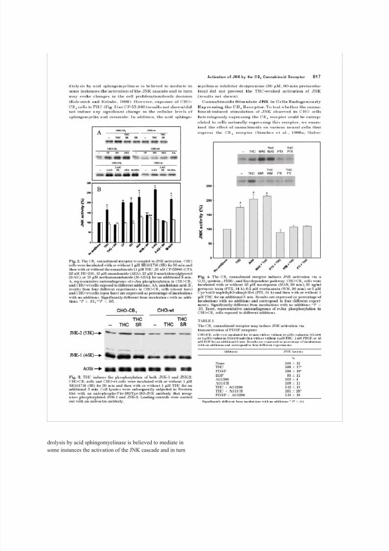
ResultsThe CB
1
Cannabinoid Receptor Is Coupled to JNK
Activation. CHO cells stably transfected with the CB
1
re-
ceptor cDNA constitute a well characterized model to study
the signal transduction pathways modulated by this recep-
tor. Here we used these cells to test the possible coupling of
the CB
1
receptor to JNK activation. CHO cells were treated
for different times with THC, the major active component of
marijuana. Cells were subsequently lysed, and JNK activity
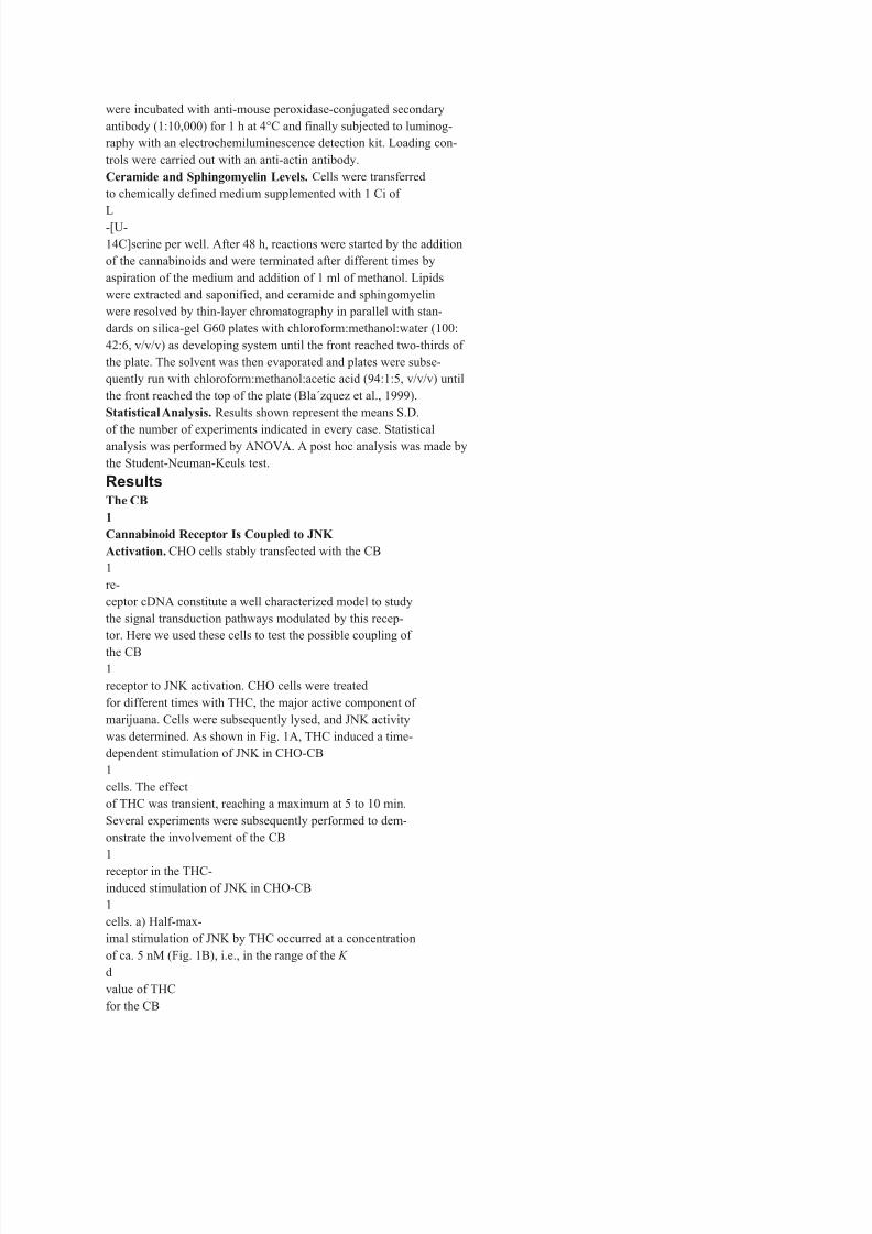
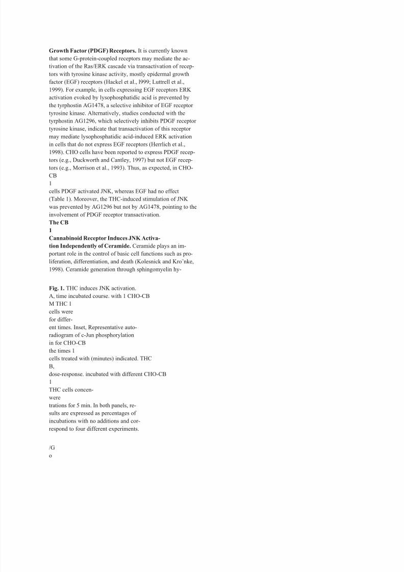
was determined. As shown in Fig. 1A, THC induced a time-
dependent stimulation of JNK in CHO-CB
1
cells. The effect
of THC was transient, reaching a maximum at 5 to 10 min.
Several experiments were subsequently performed to dem-
onstrate the involvement of the CB
1
receptor in the THC-
induced stimulation of JNK in CHO-CB
1
cells. a) Half-max-
imal stimulation of JNK by THC occurred at a concentration
of ca. 5 nM (Fig. 1B), i.e., in the range of the K
d
value of THC
for the CB

8/8/2019 Rueda 2000
http://slidepdf.com/reader/full/rueda-2000 10/31
1
receptor (Howlett, 1995). b) The synthetic can-
nabinoids CP-55,940 and HU-210 were able to stimulate
JNK to an extent similar to THC (Fig. 2). c) The stimulatory
8/8/2019 Rueda 2000
http://slidepdf.com/reader/full/rueda-2000 11/31
effect of THC on JNK was abolished by treatment of cells
with SR141716, a selective CB
8/8/2019 Rueda 2000
http://slidepdf.com/reader/full/rueda-2000 12/31
1
receptor antagonist, which
did not exert any effect per se on JNK activity (Fig. 2). d) The
stimulation of JNK induced by THC, CP-55,940, and HU-210
in CHO-CB
1
cells was not evident in CHO-wt cells (Fig. 2).The endogenous cannabinoids anandamide and 2-arachy-
donoylglycerol stimulated JNK to an extent higher than
other cannabinoid agonists (Fig. 2). These endocannabinoids
are known to be actively degraded by cellular fatty acid
amide hydrolase to arachidonic acid and ethanolamine or
glycerol, respectively (Martin et al., 1999). As shown in Fig.
2, in CHO-CB
1
cells the effect of anandamide and 2-arachi-
donoylglycerol on JNK was not completely blocked by
SR141716. In addition, in CHO-wt cells, anandamide and
2-arachidonoylglycerol induced a slight, although reproduc-
ible, activation of JNK. Methanandamide, a stable syntheticanalog of anandamide (Martin et al., 1999), also stimulated
JNK in CHO-CB
1
cells. Moreover, arachidonic acid stimu-
lated JNK in CHO-wt cells (151 6% stimulation by 15 M
arachidonic acid, n 3, P .01 versus incubations with no
additions; Fig. 2). These observations indicate that endocan-
nabinoids stimulate JNK activity mostly by a CB
1
receptor-
dependent mechanism, with a minor contribution of a CB
1
receptor-independent mechanism.The two major JNK isoforms (i.e., JNK-1 and JNK-2) may
become differently activated by extracellular stimuli (cf. Ip
and Davis, 1998; Minden and Karin, 1998). The contribution
of these two isoforms to THC-induced JNK activation was
assessed by Western blot with an antibody that recognizes
phosphorylated ( activated) JNK-1 and JNK-2. As shown in
Fig. 3, CHO cells expressed mostly the JNK-1 isoform. How-
ever, densitometric analysis of the luminograms showed that
THC induced a similar increase in the phosphorylation of
JNK-1 (33 10% over incubations with no additions, n 3)
and JNK-2 (30 6% over incubations with no additions, n
3) in CHO-CB
1cells. This effect was not evident in CHO-wt
cells and was prevented by SR141716 (Fig. 3).
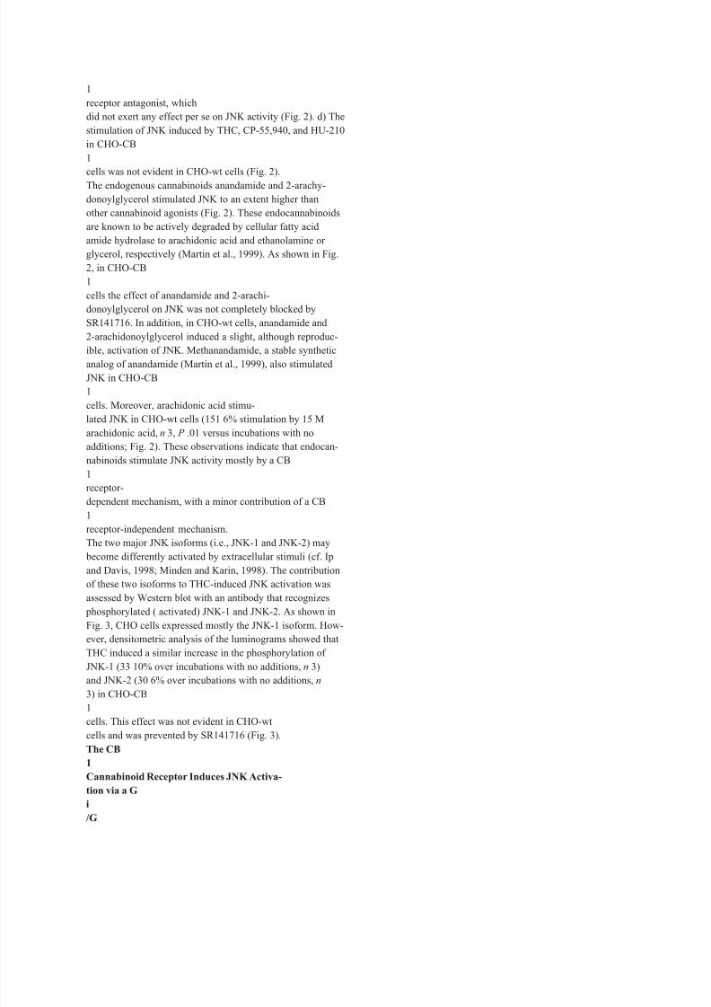
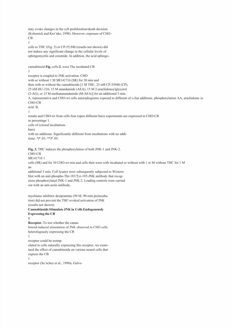
The CB
1
Cannabinoid Receptor Induces JNK Activa-
tion via a G
i
/G
8/8/2019 Rueda 2000
http://slidepdf.com/reader/full/rueda-2000 13/31
o
-Protein-, Phosphoinositide 3 -kinase
(PI3K)-, and Ras-Dependent Pathway. The CB
1
receptor
is coupled to G
i/G
o
-proteins (Howlett, 1995). To further in-
vestigate the signal transduction pathway responsible for
JNK activation, the possible involvement of a G
i
/G
o
-protein
was studied by examining the effect of pertussis toxin and
816 Rueda et al.
-protein
mastoparan. As shown in Fig. 4, blockade of G
i
dissociation with pertussis toxin abrogated the THC-induced
activation of JNK in CHO-CB
1
cells. In addition, induction of
G
i
/G
o
-protein dissociation with mastoparan induced a re-markable stimulation of JNK that was not additive to that
exerted by THC.
It has been shown that wortmannin, a PI3K inhibitor,
blocks the cannabinoid-induced stimulation of ERK
(Bouaboula et al., 1997) and glucose metabolism (Sa´nchez et
al., 1998b) in cells expressing the CB
1
receptor. Class I
B
PI3Ks, i.e., PI3K isoenzymes devoid of Src homology domains
and activated by G-protein -subunits, may mediate the
activation of small G-proteins such as Ras and therefore
stimulate the ERK cascade (Fruman et al., 1998). As shownin Fig. 4, the THC-induced activation of JNK in CHO-CB
1
cells was fully prevented by wortmannin and the Ras farne-
syltransferase inhibitor Cys-Val-2-naphthyl-3-alanyl-Met.
The CB
1
Cannabinoid Receptor May Induce JNK Ac-
tivation through Transactivation of Platelet-Derived
8/8/2019 Rueda 2000
http://slidepdf.com/reader/full/rueda-2000 14/31
Growth Factor (PDGF) Receptors. It is currently known
that some G-protein-coupled receptors may mediate the ac-
tivation of the Ras/ERK cascade via transactivation of recep-
tors with tyrosine kinase activity, mostly epidermal growth
factor (EGF) receptors (Hackel et al., l999; Luttrell et al.,
1999). For example, in cells expressing EGF receptors ERK
activation evoked by lysophosphatidic acid is prevented bythe tyrphostin AG1478, a selective inhibitor of EGF receptor
tyrosine kinase. Alternatively, studies conducted with the
tyrphostin AG1296, which selectively inhibits PDGF receptor
tyrosine kinase, indicate that transactivation of this receptor
may mediate lysophosphatidic acid-induced ERK activation
in cells that do not express EGF receptors (Herrlich et al.,
1998). CHO cells have been reported to express PDGF recep-
tors (e.g., Duckworth and Cantley, 1997) but not EGF recep-
tors (e.g., Morrison et al., 1993). Thus, as expected, in CHO-
CB
1
cells PDGF activated JNK, whereas EGF had no effect
(Table 1). Moreover, the THC-induced stimulation of JNK was prevented by AG1296 but not by AG1478, pointing to the
involvement of PDGF receptor transactivation.
The CB
1
Cannabinoid Receptor Induces JNK Activa-
tion Independently of Ceramide. Ceramide plays an im-
portant role in the control of basic cell functions such as pro-
liferation, differentiation, and death (Kolesnick and Kro¨nke,
1998). Ceramide generation through sphingomyelin hy-
Fig. 1. THC induces JNK activation.
A, time incubated course. with 1 CHO-CB
M THC 1
cells were
for differ-
ent times. Inset, Representative auto-
radiogram of c-Jun phosphorylation
in for CHO-CB
the times 1
cells treated with (minutes) indicated. THC
B,
dose-response. incubated with different CHO-CB
1
THC cells concen-
were
trations for 5 min. In both panels, re-
sults are expressed as percentages of
incubations with no additions and cor-
respond to four different experiments.
/G
o

8/8/2019 Rueda 2000
http://slidepdf.com/reader/full/rueda-2000 15/31
3 4 -1 0 1 2
200 180 160 140 120 100
8/8/2019 Rueda 2000
http://slidepdf.com/reader/full/rueda-2000 16/31
drolysis by acid sphingomyelinase is believed to mediate in
some instances the activation of the JNK cascade and in turn
8/8/2019 Rueda 2000
http://slidepdf.com/reader/full/rueda-2000 17/31
may evoke changes in the cell proliferation/death decision
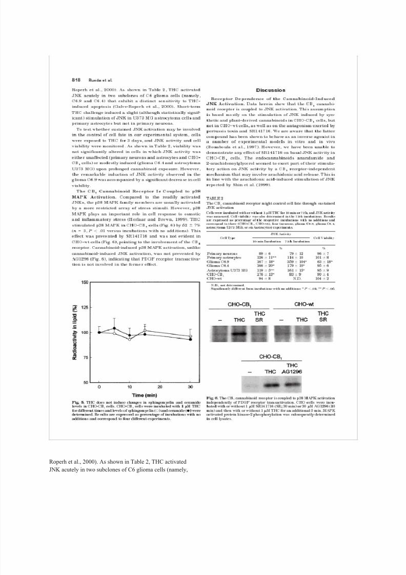
(Kolesnick and Kro¨nke, 1998). However, exposure of CHO-
CB
1
cells to THC (Fig. 5) or CP-55,940 (results not shown) did
not induce any significant change in the cellular levels of
sphingomyelin and ceramide. In addition, the acid sphingo-
cannabinoid Fig. cells 2. were The incubated CB
1
receptor is coupled to JNK activation. CHO
with or without 1 M SR141716 (SR) for 30 min and
then with or without the cannabinoids [1 M THC, 25 nM CP-55940 (CP),
25 nM HU-210, 15 M anandamide (AEA), 15 M 2-arachidonoylglycerol
(2-AG), or 15 M methananandamide (M-AEA)] for an additional 5 min.
A, representative and CHO-wt cells autoradiograms exposed to different of c-Jun additions. phosphorylation AA, arachidonic in
CHO-CB
acid. B,
1
results and CHO-wt from cells four (open different bars) experiments are expressed in CHO-CB
as percentage 1
cells of (closed incubations
bars)
with no additions. Significantly different from incubations with no addi-
tions: * P .01; ** P .05.
Fig. 3. THC induces the phosphorylation of both JNK-1 and JNK-2.
CHO-CB
SR141716 1
cells (SR) and for 30 CHO-wt min and cells then were with incubated or without with 1 or M without THC for 1 M
an
additional 5 min. Cell lysates were subsequently subjected to Western blot with an anti-phospho-Thr-183/Tyr-185-JNK antibody that recog-
nizes phosphorylated JNK-1 and JNK-2. Loading controls were carried
out with an anti-actin antibody.
myelinase inhibitor desipramine (50 M, 90-min preincuba-
tion) did not prevent the THC-evoked activation of JNK
(results not shown).
Cannabinoids Stimulate JNK in Cells Endogenously
Expressing the CB
1
Receptor. To test whether the canna-
binoid-induced stimulation of JNK observed in CHO cells
heterologously expressing the CB
1
receptor could be extrap-
olated to cells naturally expressing this receptor, we exam-
ined the effect of cannabinoids on various neural cells that
express the CB
1
receptor (Sa´nchez et al., 1998a; Galve-
8/8/2019 Rueda 2000
http://slidepdf.com/reader/full/rueda-2000 18/31
cannabinoid Fig. 4. G
incubated i
/G
o
-protein-, The CB
PI3K-, 1
with or receptor induces JNK activation via aand Ras-dependent without 15 M mastoparan pathway. (MAS, CHO-CB
20 min), 1
cells 50 ng/ml
were
pertussis toxin (PTX, 14 h), 0.2 M wortmannin (WM, 20 min), or 5 M
Cys-Val-2-naphthyl-3-alanyl-Met (FTI, 14 h) and then with or without 1
M THC for an additional 5 min. Results are expressed as percentage of
incubations with no additions and correspond to four different experi-
ments. Significantly different from incubations with no additions: * P
.01. Inset, representative autoradiograms of c-Jun phosphorylation in
CHO-CB
1
cells exposed to different additions.TABLE 1
The transactivation CB
1
cannabinoid of PDGF receptor receptors
may induce JNK activation via
CHO-CB
or 1 M tyrphostin 1
cells were AG1478 incubated and for then 10 min with with or without or without 1 30 M tyrphostin AG1296
M THC, 1 nM PDGF, or 15
nM EGF for an additional 5 min. Results are expressed as percentage of incubations
with no additions and correspond to four different experiments.
Additions JNK Activity
% None 100 12
THC 168 17*
PDGF 196 19*
EGF 93 11
AG1296 103 4
AG1478 109 11
THC AG1296 112 13
THC AG1478 181 26*
PDGF AG1296 110 19
Significantly different from incubations with no additions: * P .01.
Cannabinoid Receptor 817
Activation of JNK by the CB
1
250 200
MAS n
8/8/2019 Rueda 2000
http://slidepdf.com/reader/full/rueda-2000 19/31
Roperh et al., 2000). As shown in Table 2, THC activated
JNK acutely in two subclones of C6 glioma cells (namely,
8/8/2019 Rueda 2000
http://slidepdf.com/reader/full/rueda-2000 20/31
C6.9 and C6.4) that exhibit a distinct sensitivity to THC-
induced apoptosis (Galve-Roperh et al., 2000). Short-term
THC challenge induced a slight (although statistically signif-
icant) stimulation of JNK in U373 MG astrocytoma cells and
primary astrocytes but not in primary neurons.
To test whether sustained JNK activation may be involved
in the control of cell fate in our experimental system, cellswere exposed to THC for 3 days, and JNK activity and cell
viability were monitored. As shown in Table 2, viability was
not significantly altered in cells in which JNK activity was
either unaffected (primary neurons and astrocytes and CHO-
CB
1
cells) or modestly induced (glioma C6.4 and astrocytoma
U373 MG) upon prolonged cannabinoid exposure. However,
the remarkable induction of JNK activity observed in the
glioma C6.9 was accompanied by a significant decrease in cell
viability.
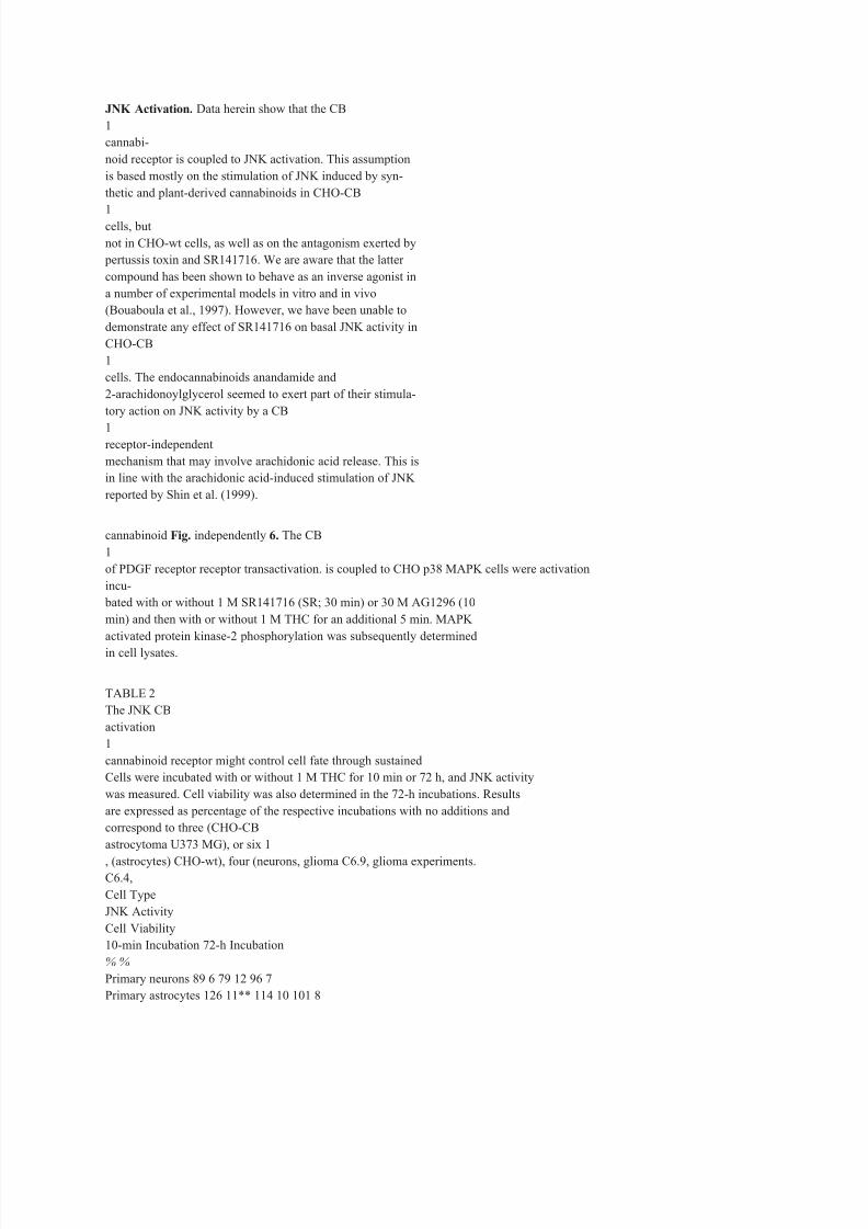
The CB
1Cannabinoid Receptor Is Coupled to p38
MAPK Activation. Compared to the readily activated
JNKs, the p38 MAPK family members are usually activated
by a more restricted array of stress stimuli. However, p38
MAPK plays an important role in cell response to osmotic
and inflammatory stress (Herlaar and Brown, 1999). THC
stimulated p38 MAPK in CHO-CB
1
cells (Fig. 6) by 52 7%
(n 3, P .01 versus incubations with no additions). This
effect was prevented by SR141716 and was not evident in
CHO-wt cells (Fig. 6), pointing to the involvement of the CB
1receptor. Cannabinoid-induced p38 MAPK activation, unlike
cannabinoid-induced JNK activation, was not prevented by
AG1296 (Fig. 6), indicating that PDGF receptor transactiva-
tion is not involved in the former effect.
Fig. 5. THC does not induce changes in sphingomyelin and ceramide
levels for different in CHO-CB
times 1
and cells. levels CHO-CB
of sphingomyelin 1
cells were incubated (E) and ceramide with 1 M THC
(F) were
determined. Results are expressed as percentage of incubations with no
additions and correspond to four different experiments.
818 Rueda et al.
DiscussionReceptor Dependence of the Cannabinoid-Induced
8/8/2019 Rueda 2000
http://slidepdf.com/reader/full/rueda-2000 21/31
JNK Activation. Data herein show that the CB
1
cannabi-
noid receptor is coupled to JNK activation. This assumption
is based mostly on the stimulation of JNK induced by syn-
thetic and plant-derived cannabinoids in CHO-CB
1cells, but
not in CHO-wt cells, as well as on the antagonism exerted by
pertussis toxin and SR141716. We are aware that the latter
compound has been shown to behave as an inverse agonist in
a number of experimental models in vitro and in vivo
(Bouaboula et al., 1997). However, we have been unable to
demonstrate any effect of SR141716 on basal JNK activity in
CHO-CB
1
cells. The endocannabinoids anandamide and
2-arachidonoylglycerol seemed to exert part of their stimula-
tory action on JNK activity by a CB
1receptor-independent
mechanism that may involve arachidonic acid release. This is
in line with the arachidonic acid-induced stimulation of JNK
reported by Shin et al. (1999).
cannabinoid Fig. independently 6. The CB
1
of PDGF receptor receptor transactivation. is coupled to CHO p38 MAPK cells were activation
incu-
bated with or without 1 M SR141716 (SR; 30 min) or 30 M AG1296 (10
min) and then with or without 1 M THC for an additional 5 min. MAPK
activated protein kinase-2 phosphorylation was subsequently determined
in cell lysates.
TABLE 2
The JNK CB
activation
1
cannabinoid receptor might control cell fate through sustained
Cells were incubated with or without 1 M THC for 10 min or 72 h, and JNK activity
was measured. Cell viability was also determined in the 72-h incubations. Results
are expressed as percentage of the respective incubations with no additions and
correspond to three (CHO-CB
astrocytoma U373 MG), or six 1
, (astrocytes) CHO-wt), four (neurons, glioma C6.9, glioma experiments.C6.4,
Cell Type
JNK Activity
Cell Viability
10-min Incubation 72-h Incubation
% %
Primary neurons 89 6 79 12 96 7
Primary astrocytes 126 11** 114 10 101 8
8/8/2019 Rueda 2000
http://slidepdf.com/reader/full/rueda-2000 22/31
Glioma C6.9 167 18* 359 104* 63 18*
Glioma C6.4 166 20* 179 10* 95 6
Astrocytoma U373 MG 119 5** 161 15* 95 9
CHO-CB
CHO-wt 1
176 13* 83 9 90 4
94 8 N.D. 104 2 N.D., not determined.
Significantly different from incubations with no additions: * P .01; ** P .05.
in
activity
Radio
50 I I I O 10 20 30
Time (min)
THC THC THC SR -- THC SR
8/8/2019 Rueda 2000
http://slidepdf.com/reader/full/rueda-2000 23/31
Mechanism of JNK Activation. Many studies have dem-
onstrated an involvement of the ERK and JNK cascades in
8/8/2019 Rueda 2000
http://slidepdf.com/reader/full/rueda-2000 24/31
the regulation of cell proliferation by G-protein-coupled re-
ceptors (Gutkind, 1998; Luttrell et al., 1999). Cannabinoid
receptors are coupled to G
i
/G
o
-proteins (Howlett, 1995). The-subunits released from heterotrimeric G
i
- and G
o
-proteins
are known to mediate the stimulation of small G-proteins
such as Ras involved in the activation of the ERK and JNK
cascades via class I
B
PI3Ks (Fruman et al., 1998; Minden and
Karin, 1998; Luttrell et al., 1999). More recently, the -sub-
units released from G
i- (Mochizuki et al., 1999) and G
o
-
proteins (Jordan et al., 1999) have been shown to mediate
ERK activation via a Ras- and PI3K-independent pathway. It
is well established that Ras is the major small G-protein
involved in the activation of the Raf-1/ERK cascade; however,
the JNK cascade may be also activated by Rac and in some
instances by Rho and Cdc42 (Minden and Karin, 1998; Yam-
auchi et al., 1999). Although conflicting results exist concern-
ing the relative importance of Ras and Rac in the activation
of the JNK cascade, most likely both proteins are activatory
components of the JNK cascade, Ras lying upstream of Rac(Scita et al., 1999). The CB
1
cannabinoid receptor seems to be
coupled to the activation of the ERK and JNK cascades
through a common upstream mechanism involving G
i
/G
o
-
protein -subunits, class I
B
PI3K, and Ras. Current re-
search is focused on the characterization of other elementslinking the CB
1
receptor to the JNK cascade, one of which
could be focal adhesion kinase. This kinase, which has been
shown to be phosphorylated by anandamide in primary neu-
rons, is capable of activating small G-proteins such as Ras
and may play an important role in the regulation of neuronal
activity, plasticity, and survival (Girault et al., 1999).
8/8/2019 Rueda 2000
http://slidepdf.com/reader/full/rueda-2000 25/31
Some G-protein-coupled receptors such as those for lyso-
phosphatidic acid, thrombin, and angiotensin II may mediate
the activation of the Ras/ERK cascade via transactivation of
receptors with tyrosine kinase activity (Hackel et al., 1999;
Luttrell et al., 1999). Our data indicate for the first time that
the CB
1cannabinoid receptor may stimulate a protein kinase
cascade such as the JNK cascade via transactivation of
PDGF receptors. Interestingly, cannabinoid-induced stimu-
lation of p38 MAPK was not prevented by AG1296, pointing
to the existence of divergent pathways for JNK and p38
MAPK activation. Transactivation of EGF and PDGF recep-
tors has been suggested to be the link between the release of
G
i
/G
o
-protein -subunits, leading to class I
BPI3K/Src acti-
vation, and the stimulation of Ras via Shc/Grb-2/Sos (Hackel
et al., 1999; Luttrell et al., 1999). Although cannabinoid
receptors do not share a very high sequential homology with
other receptors, they show a certain structural similarity
with members of the EDG family of G-protein-coupled recep-
tors (Yamaguchi et al., 1996). Ligands of these receptors
include bioactive long-chain fatty acid derivatives such as
lysophosphatidic acid and sphingosine 1-phosphate, whose
structure resembles that of endocannabinoids. Lysophospha-
tidic acid and sphingosine 1-phosphate play an important
role in the regulation of key cell functions such as prolifera-
tion, protection from apoptosis, differentiation, migration,and Ca2 mobilization. Of interest, both lysophosphatidic
acid receptors (Herrlich et al., 1998) and sphingosine 1-phos-
phate receptors (Pyne et al., 1999) are capable of stimulating
ERK via transactivation of PDGF receptors.
Cannabinoid Receptor 819
Activation of JNK by the CB
1
Physiological Considerations. Although the actual bio-logical functions of the endogenous cannabinoid system are
as yet unknown, it is believed that endogenous cannabinoids
might play a role in brain development and function. Thus,
the significance of the endogenous cannabinoid system is
supported by the high levels of cannabinoid receptors found
in brain; the specific mechanisms of endocannabinoid syn-
thesis, uptake, and degradation in neural cells; and the neu-
romodulatory properties of endogenous cannabinoids (Felder
8/8/2019 Rueda 2000
http://slidepdf.com/reader/full/rueda-2000 26/31
and Glass, 1998; Martin et al., 1999). One of the most in-
triguing and unexplored actions of cannabinoids is their abil-
ity to control cell growth. Thus, cannabinoids have been
shown to induce antiproliferative effects through the CB
1
receptor in a number of cultured cell systems (De Petrocellis
et al., 1998; Sa´nchez et al., 1998a; Chan et al., 1999; Galve-Roperh et al., 2000). Data in this report may help to explain
the signal transduction mechanisms involved in cell growth
control by cannabinoids. Thus, there might be a threshold
above which cannabinoid-induced long-term JNK activation
would lead to neural cell death. In the context of these find-
ings, one could speculate that by modulating the balance
among ERK, JNK, and p38 MAPK activities the CB
1
canna-
binoid receptor might regulate the fate of neural cells (re-
garding, e.g., proliferation, differentiation, and death) in re-
sponse to environmental stimuli (cf. Datta and Greenberg,
1998; Derkinderen et al., 1999). Moreover, we have recentlyshown that cannabinoids are able to modulate through the
CB
1
receptor the activity of the PI3K/protein kinase B path-
way, which serves as a pivotal antiapoptotic signal (Go´mez
del Pulgar et al., 2000). It is clear that further research is
required to understand the physiological role of cannabinoids
as modulators of cell fate.
Note Added in Proof. It has been recently shown that
anandamide stimulates ERK, JNK and p38 MAPK in the
ECV cell line derived from human umbilical vein endothelial
cells via CB
1receptor-dependent and independent mecha-
nisms [Liu J, Mirshahi F, Sanyal AJ, Khanolkar AD, Makri-
yannis A and Kunos G (2000) Functional CB1 cannabinoid
receptors in human vascular endothelial cells. Biochem J
346:835–840.].
Acknowledgments
We are indebted to Dr. C. Sa´nchez and T. Go´mez del Pulgar for
expert assistance in the determination of ceramide and sphingomy-
elin levels.
References
Bla´zquez C, Sa´nchez C, Daza A, Galve-Roperh I and Guzma´n M (1999) The stimu-
lation of ketogenesis by cannabinoids in astrocytes defines carnitine palmitoyl-
transferase I as a new ceramide-activated enzyme. J Neurochem 72:1759–1768.Bouaboula M, Bourrie´ B, Rinaldi-Carmona M, Shire D, Le Fur G and Casellas P
(1995a) Stimulation of cannabinoid receptor CB1 induces krox-24 expression in
human astrocytoma cells. J Biol Chem 270:13973–13980.
Bouaboula M, Poinot-Chazel C, Bourrie´ B, Canat X, Calandra B, Rinaldi-Carmona
M, Le Fur G and Casellas P (1995b) Activation of mitogen-activated protein
kinases by stimulation of the central cannabinoid receptor CB1. Biochem J 312:
637–641.
Bouaboula M, Perrachon S, Milligan L, Canat X, Rinaldi-Carmona M, Portier M,

8/8/2019 Rueda 2000
http://slidepdf.com/reader/full/rueda-2000 27/31
Barth F, Calandra B, Pecceu F, Lupker J, Maffrand JP, Le Fur G and Casellas P
(1997) A selective inverse agonist for central cannabinoid receptor inhibits mito-
gen-activated protein kinase activation stimulated by insulin or insulin-like
growth factor 1: Evidence for a new model of receptor/ligand interactions. J Biol
Chem 272:22330–22339.
Chan GCK, Hinds TR, Impey S and Storm DR (1999) Hippocampal neurotoxicity of
9-tetrahydrocannabinol. J Neurosci 18:5322–5332.Datta SR and Greenberg ME (1998) Molecular mechanisms of neuronal survival and
apoptosis. Horm Signal 1:257–306.
8/8/2019 Rueda 2000
http://slidepdf.com/reader/full/rueda-2000 28/31
8/8/2019 Rueda 2000
http://slidepdf.com/reader/full/rueda-2000 29/31

8/8/2019 Rueda 2000
http://slidepdf.com/reader/full/rueda-2000 30/31
820 Rueda et al.
Luttrell LM, Daaka Y and Lefkowitz RJ (1999) Regulation of tyrosine kinase cas-
cades by G-protein-coupled receptors. Curr Opin Cell Biol 11:177–183.
Martin BR, Mechoulam R and Razdan RK (1999) Discovery and characterization of
endogenous cannabinoids. Life Sci 65:573–595.
Matsuda LA, Lolait SJ, Brownstein M, Young A and Bonner TI (1990) Structure of a cannabinoid receptor and functional expression of the cloned cDNA. Nature
(Lond) 346:561–564.
Minden A and Karin M (1998) The JNK family of MAP kinases: Regulation and
function. Horm Signal 1:209–233.
Mochizuki N, Ohba Y, Kiyokawa E, Kurata T, Murakami T, Ozaki T, Kitabatake A,
Nagashima K and Matsuda M (1999) Activation of the ERK/MAPK pathway by an
isoform of rap1GAP associated with G
i
. Nature (Lond) 400:891–894.
Morrison P, Takishima K and Rosner MR (1993) Role of threonine residues in
regulation of the epidermal growth factor receptor by protein kinase C and mito-
gen-activated protein kinase. J Biol Chem 268:15536–15543.
Munro S, Thomas KL and Abu-Shaar M (1993) Molecular characterization of a
peripheral receptor for cannabinoids. Nature (Lond) 365:61–65.
Pyne S, Rahkit S, Conway AM, McKie A, Darroch P, Tate R and Pyne N (1999)
Extracellular actions of sphingosine 1-phosphate through endothelial differentia-
tion gene products in mammalian cells: Role in regulating proliferation and apo-
ptosis. Biochem Soc Trans 27:404–409.
Sa´nchez C, Galve-Roperh I, Canova C, Brachet P and Guzma´n M (1998a) 9-
Tetrahydrocannabinol induces apoptosis in C6 glioma cells. FEBS Lett 436:6–10.
Sa´nchez C, Galve-Roperh I, Rueda D and Guzma´n M (1998b) Involvement of sphin-
gomyelin hydrolysis and the mitogen-activated protein kinase cascade in the
9-tetrahydrocannabinol-induced stimulation of glucose metabolism in primary
astrocytes. Mol Pharmacol 54:834–843.
Scita G, Nordstrom J, Carbone R, Tenca P, Giardina G, Gutkind S, Bjarnegard M,
Betsholtz C and Di Fiore PP (1999) EPS8 and E3B1 transduce signals from Ras to
Rac. Nature (Lond) 401:290–293.
Shin EA, Kim KH, Han SI, Ha KS, Kim JH, Kang KI, Kim HD and Kang HS (1999)
Arachidonic acid induces the activation of the stress-activated protein kinase,
membrane ruffling and H
2
O
2
production via a small GTPase Rac1. FEBS Lett
452:355–359.
Voth E and Schwartz R (1997) Medicinal applications of delta-9-tetrahydrocannab-
inol and marijuana. Ann Intern Med 126:791–798.
Yamaguchi F, Tokuda M, Hatase O and Brenner S (1996) Molecular cloning of thenovel human G protein-coupled receptor (GPCR) gene mapped on chromosome 9.
Biochem Biophys Res Commun 227:608–614.
Yamauchi J, Kaziro Y and Itoh H (1999) Differential regulation of mitogen-activated
protein kinase kinase 4 (MKK4) and 7 (MKK7) by signaling from G protein
subunit in human embryonal kidney 293 cells. J Biol Chem 274:1957–1965.
Send reprint requests to: Dr. Manuel Guzma´n, Department of Biochemistry
and Molecular Biology I, School of Biology, Complutense University, 28040
Madrid, Spain. E-mail: [email protected]

8/8/2019 Rueda 2000
http://slidepdf.com/reader/full/rueda-2000 31/31