Embed Size (px)
Citation preview

I
R~rinted from:
I . fu'gery of the
Liver, Pancreas ~nd Biliary'fract
I
fAited by
JOHN S. NAJARIAN, MD.
rOHN P. DELANEY,.M.D. I .
I

Library of Congress Catalog Card No. 14-27862
ISBN ().88312-O25-6
Copyright 0' 1975
~~ ~ MIAMI. FIJ)RIDA.3m61
PrinWd in thl·U.SA

Judd Lecture
Portal Hepatotrophic Factors: A Century of Controversy
Thomas E. Stanl, M.D., Ph.D.
For this Judd Lecture, I would like to emphasize some of our work that has broad implications in physiology as well as in medicine and surgery. It is not a talk on transplantation. But what I will have to say should be of interest to liver transplanters and to those employing shunt procedures to decompress esophageal varices. It should also provide insight into some really important advances in the treatment of certain inborn errors of metabolism, as well as probably having some application to liver regeneration. This will be a review of one of the most persistently controversial problems in experimental surgery and hepatic physiology, namely, whether or not there are specific factors in the venous blood flowing up through the portal vein that are of any importance in the maintenance of liver structure and function.
Anatomical Notes
There may be some for whom a brief anatomical review is in order. The double blood supply of the liver consists of a hepatic artery which, of course, transmits completely mixed blood from the heart, and the portal vein which contains less completely mixed blood from the various splanchnic viscera. About 20% or 25% of the blood coming 'to the liver is brought by the hepatic artery and the other three-quarters to four-fifths comes in the portal blood supply. The double blood supply is retained down to the smallest structural unit of the liver, the lobule.
The capillary circulation of the liver and the means by which the two aforementioned separate sources of blood are brought to bear on the same
Thomas E. Starzl, M.D., Ph.D., Department of Surgery, University of Colorado Medical Center and the Denver Veterans Administration Hospital, Denver.
495

496 T.E.STARZL
capillary bed has been clarified in recent years. Both the arterial and portal venous branches perfuse the hepatic sinusoids lined with Kupffer cells, drain into the lobular central veins, and then into the larger hepatic veins and on to the heart. The portal blood supply is intimately related to the hepatocytes of the so-called limiting plate. These hepatocytes are rich in pentose nucleic acids, and they are the most active centers of regeneration. Variations in the relative quantity of blood from the portal vein and the hepatic artery can be imposed by vascular sphincters.
Historical Vignettes
From some of the anatomical features I have just described, most old-time biologists concluded that there was a special reason for the interposition of the liver between the splanchnic viscera and the heart. It is obvious that the special ingredients could include alimentary nutriments (mainly from the intestines) or hormones (from both the pancreas and bowel). A teleologist of the old days might also have concluded, and most did, that diversion of the portal venous blood around the liver should have been incompatible with life.
It was at this point that the famous paper of Nicholas Eck arrived on the scene and thoroughly upset the apple cart. Eck's article was published in the Military Medical Journal, Moscow, of 1877 [1]. It consisted of about one page. First, it described the technique of the operation by which the portal vein and vena cava could be anastomosed, thereby diverting the portal blood past the liver. Second, it described eight dog experiments. Seven of the animals died within one week, leaving a single chronic survivor.
This sole experimental subject lived for about 2~ months and ran away from the dog farm. Doctor Eck obviously envisioned the possible clinical use of his venous fistula, since he discussed its possible application for the treatment of ascites in humans. He said, "I consider the main reason to doubt that such an operation can be carried out on human beings has been removed because it was established that the blood of the portal vein, without any danger to the body, could be diverted directly into the general circulation and this by means of a perfectly safe operation," a not atypical surgical exaggeration since the mortality had been 88%. Doctor Eck was never heard from again in scientific circles, but the ripples from his innocent manuscript have continued to agitate surgical and physiologic waters for almost a century, involving many scientific great names in the process.

JUDD LECIURE 497
Eck's disanning conclusion of the safety of Eck fistula was challenged in 1893 by Hahn and his co-workers, including the famous Pavlov of St. Petersburg, Russia' [2J. Hahn and Pavlov described the syndrome in dogs which soon came to be called meat intoxication because it could be caused or made worse by eating meat. Animals affected by this disorder developed lethargy, ataxia, convulsions and death.
The essential technical feature of Eck's fistula was the coaptation of the portal vein and vena cava with two rows of interrupted sutures. Then, the tissue between the anterior and posterior rows of sutures was either ripped out or cut out by a pull-out suture inserted with a special instrument. The crude nature of the anastomotic technique made it difficult or impossible to conSistently ensure a wide open anastomosis as can be routinely achieved with vascular suture techniques today. The inconsistency of resuits by different workers with Eck fistula probably was, therefore, partly explained by variations in the size of the resulting hole created between the vessels.
The work of Pavlov and Hahn tilted the balance back slightly toward the concept that there were factors in the portal blood that were at least important, if not vital, for the support of liver structure and function, as did also the publications of Rous and Larimore [3] who were fascinated with the atrophy affecting the liver after the performance of Eck fistula. In a statement made in 1920, Rous and Larimore said: "If so, its completeness (that is, the atrophy) would indicate that the liver has no essential activity - none on which its maintenance depends - that is not intimately connected with substances derived from organs drained by the portal system."
The somewhat mystical concept of Rous and Larimore was neither supported nor denied by the works and writings of Dr. F. C. Mann of the Mayo Clinic, who in a summary of his work presented at the Mount Sinai Hospital, New York, in 1944, said, "We have made several investigations for the purpose of determining if a specific physiologic stimulus is responsible for hepatic restoration. None has been found to date. The results of all of our experiments appear to indicate that the restoration of the liver after partial removal is dependent upon the flow of portal blood through the organ. It appears that the restoration of hepatic tissue occurs primarily in order to maintain the portal pathways and the restoration of functioning hepatic tissue is secondary" [4] . In many ways this hypothesis of Mann has dominated all thinking about hepatic physiology until very recently. To put it more clearly, it seemed to state that the important factor in portal blood flow was not the qualitative characteristics of the blood but rather its quantity.

498
\,
~VJ
-\, \'~1 9.~l .. : . '.'. l'ib) ~ n.
\ "
T.E.STARZL
FIG. 1. Technique of portacaval transposition performed on a child in October 1963, in an effort to alleviate the symptoms of glycogen storage disease. Note that all the portal blood is diverted around the liver but that vena caval blood is used to replace this loss. (By permission of Surgery, 57 :687,1965.)
The flow theory seemed to derive crucial or even incontrovertible support from a paper which is truly a classic, authored by C. Gardner Child who retired last June as Chairman of the Department of Surgery at the University of Michigan. The paper was published in 1953 [5].
With this operation which, incidentally, is shown in Figure 1 as carried out in a human, the splanchnic venous blood is diverted by an end-to-end anastomosis to the vena cava flowing behind the liver, but the lost portal blood is replaced with an inflow to the hilar portal vein from the inferior vena cava. With the blood flow replacement, Child avoided in dogs most of the adverse effects of Eck fistula. The concept became completely accepted that the quality of portal venous inflow was not a prime determinant for ·good hepatic function, structure or the capacity for regeneration. Instead, it now became the fashionable dogma that the quantity of total blood flow was the main consideration.
The numerous publications of Dr. Bernie Fisher, today a Professor of Surgery at the University of Pittsburgh, were particularly persuasive in support of the flow hypothesis. In one of Fisher's publications from 1954, data appeared to show that the capacity of regeneration of a partially resected dog liver was, if anything, gre,ater than normal when splanchnic

JUDD LECfURE
It: ... > :J t; ~ I
E .. .... Z
§ ~ C!)
TCA SOLUABLE GLYCOGEN
PRE -t- POST TRANSPOSITION
roL_~W~
TCA INSOLUABLE GLYCOGEN
PRE -+- POST TRANSPOSITION
PR£ -+- POST TRANSPOSITION
499
FIG. 2. Mean changes in liver glycogen concentration in 17 dogs following portacaval transposition. Vertical lines represent ±1 standard error. (By permission of Surgery, 57:687,1965.
vencus blood was diverted and replaced by arterial blood [6] . For reasons still not understood, it is now my opinion that these observations, as well as other later ones by Fisher, were either incorrect or else misinterpreted by him.
I found it hard to believe the flow hypothesis and in 1958 I submitted a research application from Northwestern University (Chicago) for $28,000. The application proposed to determine if subtle changes were not brought about by portal diversion, specifically affecting carbohydrate and insulin metabolism. It was suggested that portal diversion procedures might help disease states of carbohydrate metabolism such as diabetes mellitus (and also glycogen storage disease). It alluded to an interplay between the liver and incoming blood-borne endocrine factors. And, as a bonus, it promised to deliver a new method of creating cirrhosis in dogs.
Actually, the grant contributed little to the subject of hepatotrophic factors except for the kinds of data shown in Figure 2. The dogs which provided this information [7] underwent Child's portacaval transposition and afterwards were perfectly healthy. Nevertheless, the total and labile liver glycogen within approximately a month fell to about half of its previous value. In many of these animals the total hepatic blood flow was measured and found to be essentially normal. Thus, there were easily demonstrable major changes in these livers effected by the loss of portal
500 T.E.STARZL
blood even though there was replacement with vena caval blood, a vital clue pointing to the existence of portal blood factors.
The Challenge to the Flow Hypothesis
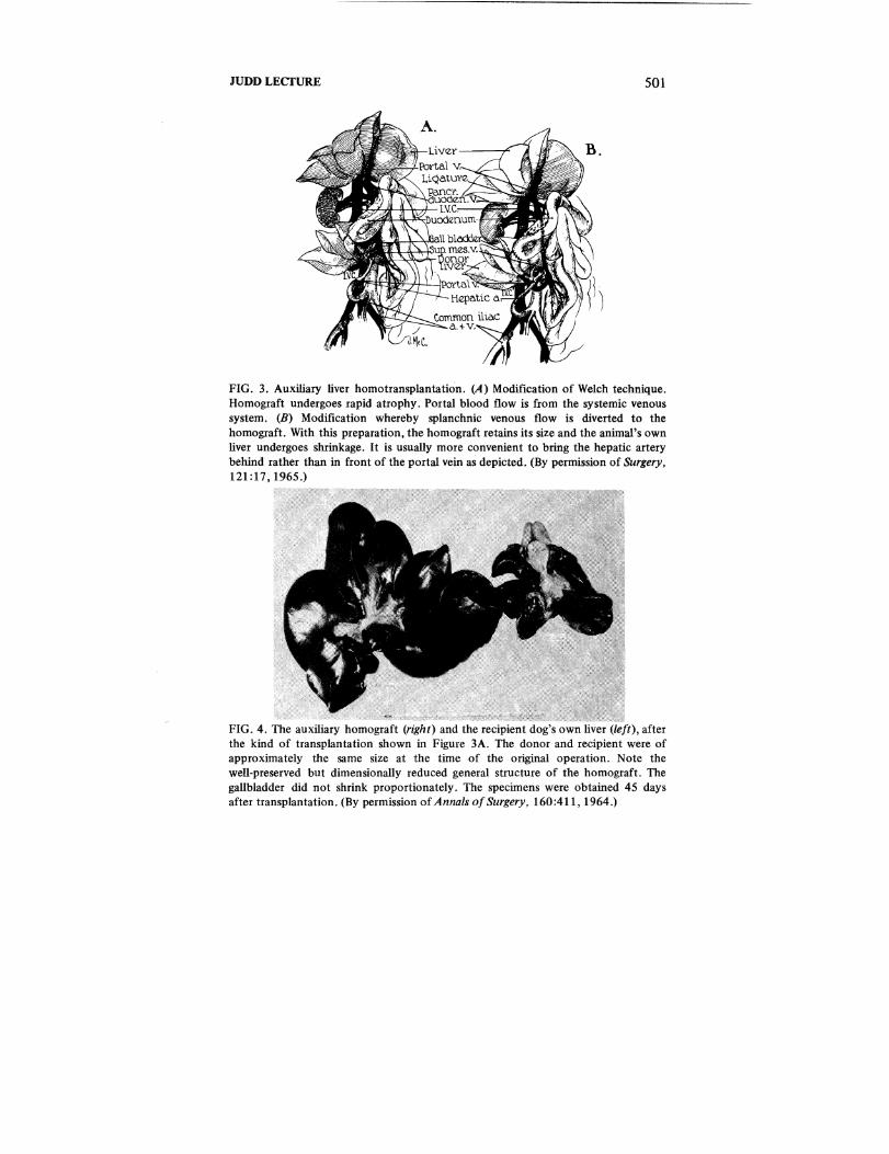
However, the most significant step in reopening the hepatotrophic issue was taken during laboratory efforts to evaluate auxiliary liver homotransplantation for the treatment of patients with non-neoplastic hepatic disease such as cirrhosis. The auxiliary operation was that originally described by C. Stuart Welch of Albany [8] . It involved the transplantation of an extra canine liver in the right paravertebral gutter or the pelvis of a nonrelated mongrel recipient. The hepatic arterial supply was derived from the iliac artery. Portal venous inflow was reconstituted by anastomosing the distal inferior vena cava to the homograft portal vein, providing a blood supply for this extra liver much the same as with Child's transposition (Fig. 3). Outflow was into the vena cava. In our laboratory in 1963, auxiliary transplantation in immunosuppressed canine recipients (using Imuran) was attempted for the first time and with very curious results [9]. It was soon found that these auxiliary homografts underwent rapid shrinkage which was usually evident within two weeks and which was very advanced at all times after one month (Fig. 4). The gross appearance and lobar proportions of the now diminutive homografts remained relatively unaltered except for size. The duct system was spared from the shrinkage. But within the parenchyma, there was massive loss of hepatocytes from focal or widespread necrosis, reticulum collapse and the consequent crowding together of intrahepatic portal tracts. The blood flow in these auxiliary transplants was shown by Daloze, one of our Canadian Fellows, to be actually greater than in the native liver [10] . Thus, the remarkable atrophy was not compatible with the blood flow hypothesis of Child and Fisher. Instead, it was speculated from the beginning "that competition of the homograft with the dog's own liver for nutritional or some other portal substrate may have been an unfavorable condition" [9].
We supported the hypothesis of competition between co-existing livers the following year in a paper by Marchioro in 1965 [11]. In these experiments canine homografts were placed in the right paravertebral gutter the same way as with the classical Welch procedure except that the portal vein of the homograft was connected to the superior mesenteric vein of the recipient (Fig. 3B). Splanchnic flow through the auxiliary liver was then promoted by ligating the portal vein at the hilum of the host liver.
JUDD LECTURE 501
FIG. 3. Auxiliary liver homotransplantation. (A) Modification of Welch technique. Homograft undergoes rapid atrophy. Portal blood flow is from the systemic venous system. (B) Modification whereby splanchnic venous flow is diverted to the homograft. With this preparation, the homograft retains its size and the animal's own liver undergoes shrinkage. It is usually more convenient to bring the hepatic artery behind rather than in front of the portal vein as depicted. (By permission of Surgery, 121: 17,1965.)
FIG. 4. The auxiliary homograft (right) and the recipient dog's own liver (left), after the kind of transplantation shown in Figure 3A. The donor and recipient were of approximately the same size at the time of the original operation. Note the well-preserved but dimensionally reduced general structure of the homograft. The gallbladder did not shrink proportionately. The specimens were obtained 45 days after transplantation. (By permission of Annals of Surgery, 160:411,1964.)

502 T.E.STARZL
These animals, which were also treated with azathioprine, now usually had atrophy of their own livers but not of the homografts. These donors and recipients had been of approximately the same size before operation but afterwards in most cases the dono! liver or homograft outweighed the native organ.
Histopathologically, the characteristic injury suffered by the native liver which was starved of splanchnic blood consisted of centrilobular atrophy or variable cell necrosis, and collapse of the reticulin-supporting framework of the liver.
I will not bore you with the details of confirmation of these fmdings except to say that important supporting studies in the auxiliary transplant model were made from our laboratories by Halgrimson [12] and Faris [13] and by Thomford [14] of the Mayo Clinic and Tretbar [15] of the Cleveland Clinic all by 1966. However, the transplant preparations which had made apparent the foregoing physiologic effects had two serious flaws which prevented the hepatotrophic concept from being accepted by many observers. First, the total flows delivered to the two co-existing livers were often different. Second, there was by definition an additional inherent inequality of the two organs, since the homograft was often under immunologic attack despite host immunosuppression, whereas the animal's own liver was not. Consequently, we undertook other experiments which were designed to circumvent one or both deficiencies.
FIG. s. Partial (split) portacaval transposition. Note that the entire vena caval flow is directed into either the left or right portal venous branch. (By permission of Surgery, Gynecology and Obstetrics, 137:179, 1973.)
-- -
JUDD LECI'URE 503
Partial Portacaval Transposition
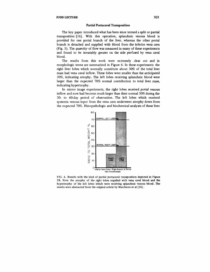
The key paper introduced what has been since termed a split or partial transposition [16]. With this operation, splanchnic venous blood is provided for one portal branch of the liver, whereas the other portal branch is detached and supplied with olood from the inferior vena cava (Fig. 5). The quantity of flow was measured in many of these experiments and found to be invariably greater on the side perfused by vena caval blood.
The results from this work were extremely clear cut and in morphologic terms are summarized in Figure 6. In these experiments the right liver lobes which normally constitute about 30% of the total liver mass had vena caval inflow. These lobes were smaller than the anticipated 30%, indicating atrophy. The left lobes receiving splanchnic blood were larger than the expected 70% normal contribution to total liver mass, indicating hypertrophy.
In mirror image experiments, the right lobes received portal venous inflow and now had become much larger than their normal 30% during the 30- to 60-day period of observation. The left lobes which received systemic venous input from the vena cava underwent atrophy down from the expected 70%. Histopathologic and biochemical analyses of these liver
~ o
fI <.9 W :s: .J
~
80
o 30·-Fc..:;..;;.;::..;..:;.;;;.;;,-== f-
o f- 20 o ~ 10 Ct:
O.L...-Inferior Vena Cova- Right Branch of Porto I
Vein Anastomosis
FIG. 6. Results with the kind of partial portacaval transposition depicted in Figure SB. Note the atrophy of the right lobes supplied with vena caval blood and the hypertrophy of the left lobes which were receiving splanchnic venous blood. The results were abstracted from the original article by Marchioro et al [161.

504 T. E.STARZL
fragments showed that the advantaged lobe complexes which were receiving splanchnic venous blood had hypertrophy of the individual hepatocytes, that these hepatocytes were also undergoing hyperplasia and that the hepatocytes were glycogen-rich.
Findings of Other Authors
These original papers describing split transposition were published in 1965 and 1967. Within a year or two, J. B. Price of Columbia [17] , Sun Lee and James Chandler of San Diego [18, 19] and ther associates performed analogous experiments exploiting the qualities of the double liver fragment model. Price used canine partial hepatic autografts and Lee and Chandler used isografts of inbred rat livers. All of these experiments showed hypertrophy in the hepatic tissue which was perfused ~ith
splanchnic blood and atrophy of the other hepatic fragments. In addition, with quantitative studies of DNA synthesis. Lee and Chandler provided further evidence that the hepatotrophic effects of splanchnic venous blood upon the liver included hyperplasia as well as hypertrophy [19]. By this time, it had become increasingly accepted that portal hepatotrophic factors were probably not just artifacts of transplantation and other experimental maneuvers, but were prime determinants of the initiation and control of liver hypertrophy and hyperplasia in many circumstances, presumably including regeneration.
However, claims of the hepatotrophic factor concept were not received by the surgical community with unmitigated joy. The most vigorous resistance came from Dr. Bernie Fisher of Pittsburgh who, in a widely quoted paper published in 1967, vigorously criticized the hepatotrophic hypothesis [20]. He presented data purporting to indicate that his previous position about the primacy of flow had been once more vindicated. The summary of this article began, "Studies of auxiliary liver transplantation have revived the concept that portal blood contains specific nutrients essential for the maintenance of the morphologic and functional integrity of hepatic tissue. Prior investigations from this and other laboratories provided no support to such a contention. It was concluded from these studies that the volume of flow via the portal system was the essential factor." Since the perpetrators of this controversy were identified as being from our Colorado group, we had all begun to feel more than a little nervous at our apparent adversary relationship to such a distinguished surgical scientist.
Consequently, it was with no small relief, to say nothing of amazement when scarcely three years later, Fisher published the first of two

JUDD LECI'URE 505
papers [21] in support of that very concept to which he had been previously implacably opposed. In rats, he had exploited the double liver fragment model originally developed with auxiliary liver transplantation and then carried out partial resections of one or the other of these co-existing livers. The critical observation was that the liver remnant receiving systemic blood did not regenerate well at all. In discussing these results, Fisher conceded that . . . "The conclusion from this study that there is a portal blood factor which is capable of stimulating hepatic parenchymal cell replication might seem to be in conflict with other findings previously reported from this laboratory."
I hope I will not be accused of small-mindedness in making the Fisher saga a subplot to the larger story of hepatotrophic substances which I am attempting to tell. I have mentioned Fisher only to indicate that sometimes the most outspoken critics of an idea became its most ardent advocates. When that occurred, thanks to the clever exploitation of the double liver fragment model, opposition to the hepatotrophic concept markedly diminished. Most students of hepatology began to concede the qualitative specialness of portal venous blood. But, now, I would like to look to three additional questions concerning the source, the nature and the mechanism of action of these special hepatotrophic substances.
Recent Experiments With Partial Portacaval Transposition
One approach was simply to do biochemical analyses of the regional liver tissues that were receiving different kinds of portal venous input, using again the split transposition model (Fig. 5). The differences in glycogen I mentioned earlier were highly quantifiable, as summarized in Table 1, from a batch of experiments in which the splanchnic venous blood went to the right lobes. * In addition to having higher glycogen concentration, the right or splanchnic-fed lobes had more glucokinase and lower concentrations of cyclic AMP and active phosphorylase. Of course, the glycogen, cyclic AMP and glucokinase findings all suggested that the right or hypertrophic lobes were being affected by endogenous insulin. It could be equally well suggested that the increased cyclic AMP and active phosphorylase in the left lobes were attributable to the adrenal epinephrine content of the insulin-poor vena caval blood supplying these lobes. In any event, a reasonable generalization would be that these two liver sides were living in different metabolic environments in which hormone control played a significant role [22].
*The full data from these experiments have been published [22].

506 T. E. STARZL
Table 1. Biochemical Dissociation Following the Kind of Split Transposition Depicted in Figure SA *
Right
Glycogen 3.7 mg/gm
Glucokinase 3 micromoles/gm/min
Cyclic AMP 1100 picomoles/gm
Active Phosphorylase 55 millimicromoles/min/mg
Left
2.2 mg/gm
1.7 micromoles/gm/min
1700 picomoles/gm
76 millimicromoles/min/mg
*The data and statistical analyses are fully documented elsewhere [22] .
Cyclic AMP concentration as an isolated measure gives a very limited view of the rate of cyclic AMP synthesis. Studies in which aminophylline was used to block the phosphodiesterase degradation of cyclic AMP demonstrated a greater synthetic rate in the vena caval lobes. However, the differences were not great and they were not obvious for several minutes [22] .
Consequently, another dynamic study was devised to demonstrate the biochemical dissociation under special pharmacologic conditions [22] . In the preliminary experiment shown in Figure 7, intravenous tolbutamide was given to induce the release of endogenous insulin. Using a radioimmunoassay technique, the insulin was found to rise sharply in the portal venous blood but with almost no detectable systemic increase. Thus, in these normal controls the background of endogenous insulin was standardized. It is well known that insulin suppresses cyclic AMP formation.
Then in dogs with the split transposition, an exogenous agent which acts by elevation of cyclic AMP, namely glucagon, was added to the tolbutamide challenge. The side which was receiving endogenous insulin from the splanchnic bed, whether this be on the right or left, had very minor cyclic AMP increases. In contrast, the liver tissue in which the infused glucagon could act in an uninhibited manner had unrestrained and colossal increases in cyclic AMP (Fig. 8).
It is obvious that the foregoing circumstantial evidence again pointed at hormones, particularly pancreatic hormones, as the most important hepatotrophic factors. To further examine the hypothesis, dogs with the split transposition model shown in Figure SA were made diabetic either with alloxan or with total pancreatectomy and then treated with 15 to 20 units NPH insulin for a two-month period of observation. The presence of either kind of treated diabetes slightly reduced the magnitude of hypertrophy of the right lobes (Fig. 9), but the hypertrophy was still significant compared to that in unoperated dogs. To put it differently, the removal of endogenous insulin or even the whole pancreas did not

JUDD LEcrURE
E .... .. '§
e u E z ::; ::> <Il Z
280
240
200
160
120
80
40
0
Q.!....--....o PORTAL VENOUS INSULIN
PERIPHERAL VENOUS INSULIN
HEPATIC C-AMP
3000.0-o· () 0 -<
2000 ~ ()
r .. ;:; .. .... !OOO to
~
:c. ;::
" ! 10 20 30 40
TOLBUTAM I DE
TIME (min.)
507
FIG. 7. Changes in peripheral and portal venous insulin and hepatic cyclic 31,5'~adenosine monophosphate (cyclic AMP) occurring in a normal dog infused with tolbutamide. Note that the peak insulin response in the portal blood occurred 2540 minutes after infusion and that no significant alterations in hepatic cyclic AMP were caused acutely by the tolbutamide itself. (By permission of Surgery, Gynecology and Obstetrics, 137:179,1973.)
LOBES WITH SPLANCHNIC LoeES WITH VENA
VENOUS INFLOW
30.o00~--------'----------'
LEFT
25,000
20,000
15,000
10,000
5,000
20,OOO~-------""----------'
15,000
10,000
5,000
LEFT
.. ..
~ 40'.····· 02:46 602
TIME AFTER BEGINNING GLUCAGON INFUSION (mill.)
FIG. 8. Results of tolbutamide-glucagon tests in eight dogs with partial portacaval transposition, demonstrating the effect of endogenous insulin in the lobes receiving splanchnic venous blood. These insulin-controlled lobes had a restrained cyclic AMP response to the exogenous glucagon whereas the response in the other lobes was uninhibited. (By permission of Surgery, Gynecology and Obstetrics, 137: 179,1973.)

508 T. E. STARZL
PARTIAL PORTACAVAL TRANSPOSITION (PPTI
RIGHT LOBES LEFT LOBES
f-
60 100 I
"' W
" 0: w 50 90 0: > w --' >
--' 40 --' BO
'" --' f-
'" 0 f-f-30 0
70 I-.. .. en 20 :~ 60 w en ill
~~ ~ 0 --' 10 '3
0 50 f- 0 : ~ f-I :;(' "-(!) '0- W
IX: 0 40 --'
PPT PPT PPT PPT PPT PPT
N ~ (121 1111 (41 (61 (121 (III (41 (61
FIG. 9. Right and left liver lobe weights expressed as percentages of total liver mass in normal dogs and in dogs undergoing partial portacaval transposition (Fig. 5) with the splanchnic flow to the right lobes. About half of the operated animals were rendered diabetic, either by the prior administration of alloxan (four dogs) or by total pancreatectomy (six dogs). Note that the right lobes underwent remarkable hypertrophy and the left lobes atrophy after the transposition. This effect was only partially blunted by producing diabetes, less so by alloxan than by total pancreatectomy. N = Number of dogs in each experimental group.
eliminate all or even most of the hepatotrophic effect of splanchnic venous blood, at least as revealed by this model. In the absence of endogenous insulin or of the whole pancreas, nutrient-rich intestinal blood was better for the liver than was systemic blood from the vena cava. Histopathologically, this conclusion was even more striking. The left or so-called vena caval lobes contained hepatocytes that were fat laden. Although these were not particularly different in size than on the other side where perfusion was with splanchnic blood minus the pancreas, the tissue looked "sicker" on the vena caval side. Even the side receiving splanchnic flow had many abnormalities.
When the diabetes was induced with alloxan instead of total pancreatectomy, the degree of injury to both liver sides was reduced. The hepatocytes on the side receiving vena caval blood still had increased fat but not so much as after total pancreatectomy. The hepatocytes on the side receiving splanchnic blood minus insulin were in even better condition, although again not normal.

JUDD LECfURE
Vein graft --
A
Schematic representation showing drainage of end of the inferior lobe
Pancreas
509
FIG. 10. Technique of division of splanchnic venous flow into a pancreaticogastroduodenal-splenic compartment and an intestinal compartment. Blood from these respective sources is directed into the right or left lobes. The tail of the inferior lobe of the pancreas was resected since it drains separately into the mesenteric vein. (By permission of Surgery, Gynecology and Obstetrics, 137:179,1973.)
Splanchnic Division Experiments
All the experiments I have just shown are consistent with the interpretation that the splanchnic blood contains hepatotrophic factors. They also indicated that no single hormone or even single organ represented the sole hepatotrophic effect. To get some idea of the relative hepatotrophic effects of the different organs or hormones, surgical techniques were developed that partition.ed the splanchnic flow into its upper and lower components [22]. In some of these partitioning experiments (Fig. lOA), the right lobes of the liver were fed by a low volume of hormone-rich pancreatico-duodeno-splenic blood. By using a graft connecting the mesenteric vein to the left hepatic lobes, these left lobes were submitted to the somewhat greater volume of nutrition-rich blood returning from the intestine. In the dog, the tail of the inferior lobe of the pancreas drains separately into the mesenteric vein, for which reason it had to be resected in all such experiments. In other experiments (Fig. lOB), the lobar distribution of the different kinds of blood was just the opposite. The left lobes were now receiving the pancreatico-gastroduodeno-splenic blood, and the right lobes were receiving intestinal blood.

SIO T.E.STARZL
LEFT RIGHT
FIG. 11. Hepatocyte shadows traced during histopathologic examination. These were later cut out on standard paper and weighed as an index of hepatocyte size. The specimens depicted were from the experiment shown in Figure 2A. The right lobes with the large hepatic cells received venous blood from the pancreas, stomach, duodenum and spleen. The relatively shrunken left lobes with the small hepatocytes received intestinal blood. (By permission of Surgery, Gynecology and Obstetrics, 137:179,1973.)
The effect of this kind of splanchnic division operation could be displayed dramatically on histopathologic and other morphologic grounds within 30 to 60 days. The lobules receiving pancreatico-duodenal venous effluent were big compared to the lobular atrophy and reticulin collapse in tissue given intestinal blood. With Hand E stain, the individual hepatocytes on the side getting pancreatico-duodenal blood were obviously bigger and, in addition, there was evidence of hyperplasia. With a PAS stain, the same big hepatocytes contained much glycogen compared with the cells receiving nutrient-rich blood.
To obtain a quantitative estimation of the hepatocyte size in these liver fragments, a tracing device was attached to the light microscope and large numbers of hepatocytes in each experiment were drawn on a standard thickness paper and weighed. In the experiment shown on Figure II, the hepatocytes on the left were from the liver side being perfused with intestinal blood. The right-sided and obviously much larger hepatocytes had their portal supply from the pancreatico-duodeno-splenic sources. The cell size data could then be summarized graphically as in Figure 12.
'. '

JUDD LECfURE
RIGHT LOBES
LIVER/BODY WEIGHT (%)80
2·8 2.9 2.7 "=0.5 "=0.3· "=0.4 ~ 60
:I: £! 40
~ It: 20 1&.1 >
----+*-fiI'~"""...r:J-O
LEFT LOBES
lJ..
020 ~ Z 1&.140
(/) ~ H---i1~@l ~ 60
6 6 6
NO. OF EXPERIMENTS
RIGHT LOBES
LEFT LOBES
511
FIG. 12. The morphologic consequences of splanchnic venous flow division in the dogs shown in Figure 10 compared with normal dogs after 28 to 173 days, average 73. The liver fractions which were perfused with venous blood from the pancreatic, gastroduodenal and splenic areas are shaded. Note that these portions gained weight and underwent an increase in hepatocyte size relative to the other side while the total liver weight to body weight ratios were little altered. One standard deviation is depicted graphically on the bar graphs and written out for the weight percentages. (By permission of Surgery, Gynecology and Obstetrics, 137:179,1973.)
The key data of Figure 12 are on the right, in which the size units of these hepatocytes are indicated. In normal control animals the hepatocytes were of essentially equal size with very small standard deviation in the right as opposed to the left lobes. However, when pancreatico-duodenosplenic blood was diverted to the left hepatic lobes, the hepatocytes in this portion of the liver now became large in comparison to the hepatocytes in other liver fragments receiving intestinal blood. The same change occurred whether the pancreatico-duodeno-splenic blood was passed to the left or to the right side.
In these splanchnic division experiments, the side receiving pancreatic blood underwent glycogen storage. Also, the various other biochemical studies including glucokinase, cyclic AMP and phosphorylase determinations tended once more to show biochemical dissociation between the co-existing liver fragments. However, the results were far less predictable than with the originally employed split transposition. Because of this and because of the results I gave you a moment ago with split transposition animals made diabetic, we concluded that the hepatotrophic factors are mainly interrelationships of hormones rather than any single hormone,

512 T. E. STARZL
that the master anabolic hormone was probably insulin with a highly significant interplay with glucagon and probably other catabolic hormones including epinephrine. We further concluded that nutritional factors probably played a significant but secondary role. The complexity, changeability, interdependence and importance of these hormonesubstrate relationships can be appreciated by perusing summary papers by Professors Hans Krebs, George Weber and others in the 1971 and 1972 issues of Advances in Enzyme Regulation or by reading the brilliant summary of the implications of the hepatotrophic concept by Hans Popper, which is being published in this month's issue of Gastroenterology [23].
Diabetes and Splanchnic Division
Although the results I just cited with the splanchnic division experiments were consistent with the conclusion that insulin was a hepatotrophic substance, the relative importance of insulin could still be debated. This question of the importance of insulin was examined in the splanchnic division model (Fig. 10) by the superimposition of alloxan diabetes and by total pan createctomy. This was done in five and six
lI
'" w 50 ~
cc w 40 > -'
-' <t 30 I-o I-
ft. 20
CfJ W CD 10 o -'
CHRONIC SPLANCHNIC DIVISION (CD)
RIGHT LOBES LEFT LOBES lI
'" 90 ~
cc w
80 > -'
-' 70 ;:
o I-
60 ;fl.
CfJ W CD
50 0 -'
I- I-
2i 0 ..l-...L:::...L...L..i:.:.:.L--"'>.c D=--'-'-'C"'D"---'--'-'-'--"'C'-'-D'"--"C"'"D"'--'-'C""D-'"--L 40 ~ 0:; CD -'
No (12) (10) (4) (6) (12) (10) (4) (6)
FIG. 13. Right and left liver lobe weights, expressed as percentages of total liver mass, in normal dogs and in dogs undergoing chronic splanchnic division (Fig. lOA). Half of the latter animals were rendered diabetic, either with alloxan (four dogs) or by total pancreatectomy (six dogs). Note that the hypertrophy of the right lobes produced by the surgical procedure was nearly abolished by the creation of diabetes. N = Number of dogs in each experimental group.

JUDD LEcrURE. 513
animals, respectively, and the results compared with 12 nondiabetic controls as well as with a dozen nonoperated normal animals. The diabetic dogs were treated -with 15 to 20 units per day of NPH insulin for two months.
The results were extremely clear cut. The right lobes of completely normal dogs weighed the expected 30% of total liver mass. In nondiabetic animals, the splanchnic division operation caused a great increase in the right lobes which were receiving the hormone-rich pancreatico-duodenosplenic blood, and striking atrophy of the left lobes which were receiving the nutritionally rich intestinal blood. This right lobar advantage was almost completely eliminated in the five animals which had alloxan diabetes established before the splanchnic division operation (Fig. 13). Almost exactly the same thing occurred in the animals with total pancreatectomy (Fig. 13). Bear in mind that these diabetic animals had to be treated with insulin which would eventually have been delivered in about the same proportions to both hepatic sides.
Another fascinating aspect of this study was that DNA synthesis as measured by the tritiated thymidine technique was much greater on the hormone-fed right side in otherwise unaltered (nondiabetic) animals (Fig.
r r-
100
90
> 80 r-u <!
U
LL
U
70
NORMAL CHRONIC SPLANCHNIC DIVISION
Non-diabetic Alloxan Pancreatectomy
W 60J-~--~-L----~~LL~~~~~--~~~--
Q.
en R L
N = (II)
R L
(6)
R L R L
(4) (5)
FIG. 14. DNA replication as measured by tritiated thymide uptake. The specific activity is expressed in percentages, the side with maximum uptake being assigned an arbitrary 100%. In normal dogs, there was no significant difference between the two sides. In the nondiabetic animals with chronic splanchnic division (Fig. 13A), the right lobes always had the greater activity. Note that the induction of diabetes reversed this pattern so that now the left lobes had the greater specific activity. N = Number of dogs in each experimental group.

514 T.E.STARZL
14). The imposition of alloxan diabetes or the performance of total pancreatectomy completely reversed this situation. Now, the left, or nutritionally enriched liver lobes were predominant, thereby indicating that the dominant cell multiplication had shifted sides by the removal of endogenous insulin.
I emphasized at the beginning and do so again that our main preoccupation for more than a decade has been with the chronic animal and not with acute experiments on regeneration. Nevertheless, I have already stated that we observed long ago that liver tissue undergoing glycogen storage and hypertrophy under favorable hepatotrophic conditions also had evidence from old-fashioned mitotic indices of having relative hyperplasia. Consequently, it has been our tacit assumption that hepatotrophic factors are important to a full understanding of regeneration, a position which is supported by the data on DNA synthesis I just gave you and by much more of our work which I do not have time to go into. Recently, many authorities have supported this contention, including Lee [18], Chandler [19], Sgro and Orloff [24] of San Diego and by the more recent publications of Fisher to which I referred earlier [21]. It is only fair to say that Bucher of Harvard has been skeptical about any central role of hepatotrophic factors in regeneration [25] . Price et al of Columbia have even claimed that hypertrophy and hyperplasia as affected by splanchnic hepatotrophic factors bear an inverse relationship to each other [26] .
To summarize to this point, I have reviewed work carried out mainly during the past decade which indicates that substances in the splanchnic venous blood are influential in the maintenance of hepatic structure and function. These substances appear to be predominantly, although not necessarily exclusively, trace quantities of interreacting hormones of which insulin is thought to be the major anabolic hormone, balanced by the converse effects of glucagon and presumably other hormones as well. The interrelationship of the hepatotrophic factors rather than any single hormone or other substance is thought to be responsible for the changes in chemical composition, hepatocyte size and the capacity for regeneration which the presence or absence of these hepatotrophic factors can markedly influence.
Clinical Application
What are some of the clinical implications of this work? One could start with auxiliary liver transplantation or with regeneration but these topics have been discussed already. In addition, there are direct connec-
JUDD LECfURE SIS
tions of the hepatotrophic concept in the selection of portacaval shunt procedures. In patients who still have hepatopetal portal flow, the Warren shunt operation preserves, for liver perfusion, the hormone-rich pancreatic effluent, to say nothing of the intestinal venous drainage, while at the same time decompressing the gastro-esophageal varices. Because it retains maximally the perfusion of the liver by hepatotrophic portal factors, we believe in spite of its technical difficulties, that the Warren shunt is physiologically the most ideal shunt available today [27].
Glycogen Storage Disease
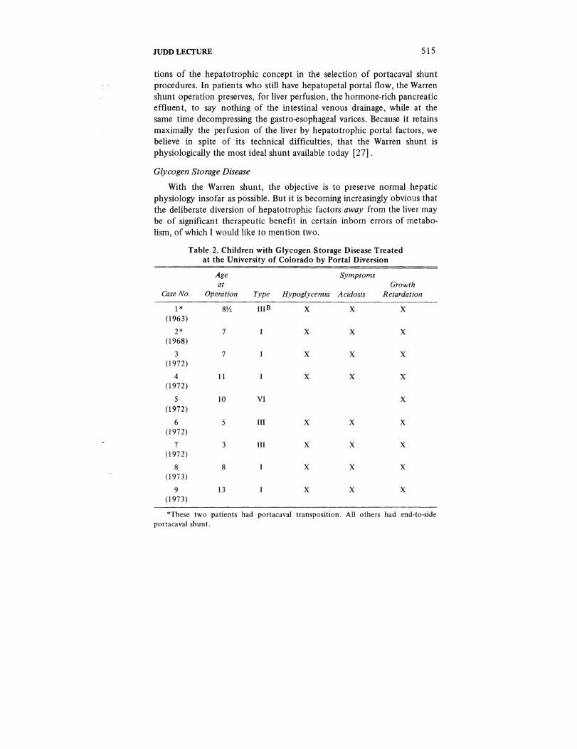
With the Warren shunt, the objective is to preserve normal hepatic physiology insofar as possible. But it is becoming increasingly obvious that the deliberate diversion of hepatotrophic factors away from the liver may be of significant therapeutic benefit in certain inborn errors of metabolism, of which I would like to mention two.
Table 2. Children with Glycogen Storage Disease Treated at the University of Colorado by Portal Diversion
Age Symptoms at Growth
Case No. Operation Type Hypoglycemia Acidosis Retardation
1* 8Y2 IIIB X X X (1963)
2* 7 X X X (1968)
3 7 X X X (1972)
4 11 X X X (1972)
5 10 VI X (1972)
6 5 III X X X (1972)
7 3 III X X X (1972)
8 8 X X X (1973)
9 13 X X X (1973)
*These two patients had portacaval transposition. All others had end-to-side portacaval shunt.

516 T. E. STARZL
The first is glycogen storage disease, a disorder for which we recommended and first performed portal diversion more than ten years ago [7]. We have now treated a total of nine patients (Table 2). The second patient died of a technical surgical accident following the unnecessarily complicated procedure of portacaval transposition, but the other eight are still alive after six months to more than a decade. The glycogen storage diseases have been Type 1, in which the enzyme deficiency responsible for hepatic glycogen accumulation is glucose-6-phosphatase, Type III (or amylo-1-6-glucosidase deficiency) and Type VI in which the deficient enzyme is phosphorylase. Stunting of growth was present in all the cases (Table 2). The patients with Types I and IJI all had metabolic acidosis and episodic hypoglycemia that required frequent night feedings (Table 2).
After portal diversion, the peripheral blood sugar response to a glucose meal was only a little prolonged in comparison to the preoperative study [28] . Thus, it would be a mistake in many of these cases, such as the child with Type I disease shown in Figure 15, to try to discontinue dietary management or to eliminate night feedings. Perhaps the reason that portacaval shunt did not produce a more dramatic glucose response what
GLUCOSE'
I"Q IIOOmO J60
FIG. 15. Peripheral venous insulin and glucose values during oral glucose tolerance tests preoperatively (top) and two weeks after portacaval shunt (bottom). The arrow indicates the time of glucose ingestion. Note that the glucose curve is only slightly prolonged by the shunt, but that peripheral insulin levels have increased tremendously.

JUDD LECTURE 517
1000 PLASMA
TRIGLYCERIDES 900
mg "" {Normal Value 130)"" BOO
700
PLASMA 600
PHOSPHOLIPIDS mg % 500
(Normal RonQe 192-337) 400
PLASMA 300
CHOLESTEROL mg % 200
(Normal Range 138-242) 100
LV. HYP(RAUMENTATION
, ,
~~---~-------~ ,~--------- -------------- 2.0 UNESTERIFIED
1.0 FATTY ACIDS meq/L
.L--.-.-r---r--r-.----,---,-----r--.---...,----1- 0 (Normal Range 0.45~O,90) -15 0 20 40 60 80 120 160 200 240 280 320
t TIME IN DAYS PORTA-CAVAL
SHUNT
FIG. 16. Effect of parenteral hyperalimentation and end-to~ide portacaval shunt on the plasma lipids of a patient whose diagnosis was Type I glycogen storage disease (glucose-6-phosphatase deficiency). Note the rapid and relatively complete reversal of all abnormalities. (By permission of Annals of Surgery, 178 :525, 1973.)
that nutritional diversion was accompanied by a concomitant increase in circulating insulin (Fig. 15). It is well known [29] that patients with glycogen storage disease have low venous insulins. as in the preoperative study shown in Figure 15. Presumably, bypassing the liver, which normally extracts more than half of the portal venous insulin content, accounts for the augmentation of peripheral insulin which, in tum, we believe is responsible for many of the beneficial effects of portacaval shun t in these patients.
In contrast to the incomplete relief of hypoglycemia, all components of the hyperlipidemia which is characteristic of Type I disease are rapidly relieved by the procedure. In addition, there has been correction of other metabolic defects, including abnormal bleeding. uric acid elevations and deranged calcium metabolism as we have described fully in an article [28].
One of the most interesting findings in these patients has been the effect of portal diversion on body growth. In our original case, a remarkable growth spurt was noted which ended ten years later in a "super-sized" teenager. Accelerated height increases have been seen in all of our patients followed for longer times. After operation, these previously dwarfed children have grown at a rate of 5/10 to one full centimeter every month (Table 3).

518 T. E. STARZL
Table 3.
Chronologie Age Time of Postopera tive at Operation Follow-up Height Increase
Case No. (Years) (Months) (cm)
1* 8Yz 113 47%
3 7 11 Yz 11%
4 11 11 5%
5 10 8% 5%
*These data have been published [28].
Comparison of the wrist and hand films before and 11\6 months after operation show the effects of doubling the bone age in the child shown in Figure 17. The bracket shown was 5 cm in length. In addition to the size change, note that mineralization has occurred and that new bones have appeared in the wrists. Circulating somatrophins were normal. The growth spurts may have been at least partially attributable to the increased distribution of insulin to the periphery, as I discussed earlier. In recent years, insulin has been recognized to be a major growth hormone comparable in potency to somatotrophin [29].
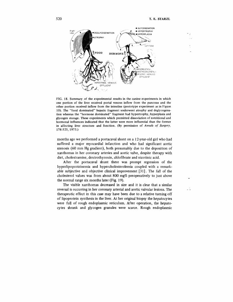
Earlier, I provided some data from dog experiments which I have summarized in Figure 18 in terms of human anatomy. This work, which permitted the delineation of nutritional from hormonal effects in portal blood, has indicated that the hormonal influences are the more profound. Of course, in the children we have been discussing, nutritional as well as hormonal bypass was achieved and both factors may have contributed to the outcome.
In the dog experiments, there was shrinkage of the individual hepatocytes deprived of pancreatic blood. In the liver biopsies of our glycogen storage patients taken before and after portal diversion, there was a similar diminution in hepatocyte and lobular size. This observation explains, at least in part, why portal diversion has resulted in relief of hepatomegaly without, at the same time, resulting in a change in actual hepatic glycogen concentration.
Idiopathic Hyperlipidemia
Another example of the profound metabolic effects of portal diversion is its truly astonishing amelioration of homozygous Type II hyperlipidemia, a disease for which available therapy, including the ileal bypass of Buchwald and Varco [30], has not been very satisfactory. Sixteen

FIG
. 17
. T
he d
ram
atic
wri
st a
nd h
and
bo
ne
grow
th in
a p
atie
nt w
ith
Typ
e I
glyc
ogen
st
orag
e di
seas
e du
ring
the
ftr
st 1
1 Y,
post
oper
ativ
e m
onth
s af
ter
port
acav
al s
hunt
. The
br
acke
t o
n t
he
left
ind
ex f
tnge
r is
5 c
m i
n le
ngth
. In
add
itio
n to
th
e si
ze c
hang
e, n
ote
th
e m
iner
aliz
atio
n th
at
has
occu
rred
, as
w
ell
as
the
appe
aran
ce
of
new
bo
nes,
pa
rtic
ular
ly o
f th
e w
rist
.
.....
c::: g t"' ~ VI
I-'
\0
520
I I I I I I .,,1
\\ \t ..
1HI0RMONEt \
T. E. STARZL
• GLYCOGENATION
FIG. 18. Summary of the experimental results in the canine experiments in which one portion of the liver received portal venous inflow from the pancreas and the other portion received inflow from the intestine (prototype experiment as in Figure 10). The "food dominated" hepatic fragment underwent atrophy and deglycogenation whereas the "hormone dominated" fragment had hypertrophy, hyperplasia and glycogen storage. These experiments which permitted dissociation of nutritional and hormonal influences indicated that the latter were more influential than the former in affecting liver structure and function. (By permission of Annals of Surgery, 178:525,1973.)
months ago we performed a portacaval shunt on a l2-year-old girl who had suffered a major myocardial infarction and who had significant aortic ~tenosis (60 mm Hg gradient), both presumably due to the deposition of xanthomas in her coronary arteries and aortic valve, despite therapy with diet, cholestramine, dextrothyroxin, chlofibrate and nicotinic acid.
After the portacaval shunt there was prompt regression of the hyperlipoproteinemia and hypercholesterolemia coupled with a remarkable subjective and objective clinical improvement [31]. The fall of the cholesterol values was from about 800 mg% preoperatively to just above the normal range six months later (Fig. 19).
The visible xanthomas decreased in size and it is clear that a similar reversal is occurring in her coronary arterial and aortic valvular lesions. The therapeutic effect in this case may have been due to a relative turning off of lipoprotein synthesis in the liver. At her original biopsy the hepatocytes were full of rough endoplasmic reticulum. After operation, the hepatocytes shrank and glycogen granules were scarce. Rough endoplasmic

JUDD LECTURE
800
...J 600 o~
5 E f-o Cflo ~" 400 o '" ::r::.5 <.>
200
PORTACAVAL SHUNT ..
O~~-L~-,--,--, __ .--, __ .-~\ ~" --.--r--~~I ope~:t~~e 0 4 8 12 16 20 24 28 2 3 4 5 6
DAYS MONTHS
521
FIG. 19. Cholesterol concentrations before and for six months after portal diversion. The preoperative value represents the mean of nine determinations ± standard deviation.
FIG. 20. (A) Complete portacaval shunt by which all nonhepatic splanchnic blood is diverted around the liver. In dogs, this procedure always lowers the serum concentration of cholesterol. (B) Selective diversion of the splanchnic blood. A t the .first stage the nutrient-rich intestinal blood is bypassed and subsequently the hormone-rich blood from upper splanchnic organs is rerouted.

522 T. E. STARZL
reticulum was greatly decreased, to one-fourth or one-third of its original amount, as judged by a quantitative technique. Since rough endoplasmic reticulum is involved in lipoprotein synthesis, curtailment of this synthesis could be at least partly responsible for the improvement.
The role of hepatotrophic factors in this antilipidemia effect is suggested by the very simple experiment shown in Figure 20 which diverts intestinal nutrients around the liver by mesenteric-vena caval shunt. Later, hormone diversion can be added by a central portacaval shunt. Thus, in two stages the same thing is achieved as with a one-stage portal diversion. The same kind of result is almost always obtained in dogs and we think also in baboons. The mesenteric venous bypass has no effect on serum cholesterol concentrations, whereas when the pancreatic hormones are shunted at a second stage, the cholesterol starts down. Thus, we think the lipid lowering effect of portacaval shunt is from the shunting around the liver of hormones, particularly insulin.
The remarkable effects of portal diversion on lipid metabolism have implications far beyond the treatment of a few children with inborn errors of metabolism. At stake may be the chance to treat many adults with premature atherosclerosis.
Summary
The conclusion from these experiments and observations of the last 15 years is that hepatotrophic factors previously reported from our laboratories and by other investigators to be in splanchnic venous blood are, in fact, mainly trace quantities of hormones and, probably most importantly, insulin. However, it is the interrelationship of these hormones to each other and to nutritional substrate which is of key importance in the moment-to-moment regUlation of nutrient and hepatic homeostasis. These interrelationships and their profound effects constitute a central and previously undefined fact of liver physiology that should reconcile a number of previously divergent opinions about portoprival syndromes, mechanisms of hepatic atrophy and hyperplasia, control of liver regeneration, and the effects of portal diversion for glycogen storage disease and idiopathic hyperlipidemia to mention only a few random examples.
References
1. Eck, N. V.: Ligature of the portal vein. Voen. Med. J., St. Petersburg 130 (2): 1-2, 1877. (Translated and discussed by Child, C. G., III: Eck's fistula. Surg. Gynec. Obstet., 96:375, 1953.)
JUDD LECTURE 523
2. Hahn, M., Massen, 0., Nenchi, M. et al: Die Eck'sche Fistel zwischen der unteren Hohlvene und der Pfortader und ihre Folgen fiir den Organismus. Arch. Exp. Path. Pharmakol., 32:161,1893.
3. Rous, P. and Larimore, L. D.: Relation of the portal blood to liver maintenance; a demonstration of liver atrophy conditional on compensation. J. Exp. Med., 31:609,1920.
4. Mann, F. C.: Restoration and pathological reactions of the liver. J. Mount Sinai Hosp. N.Y., 11:65, 1944.
5. Child, C. G., Barr, D., Holswade, G. R. et al: Liver regeneration following portacaval transposition in dogs. Ann. Surg., 138:600,1953.
6. Fisher, B., Russ, C., Updegraff, H. et al: Effect of increased hepatic blood flow upon liver regeneration. Arch. Surg., 69:263, 1954.
7. Starzl, T. E., Marchioro, T. L., Sexton, A. W. et al: The effect of portacaval transposition upon carbohydrate metabolism: Experimental and clinical observations. Surgery, 57:687,1965.
8. Welch, C. S.: A note on transplantation of the whole liver in dogs. Transplantation Bull., 2:54, 1955.
9. Starzl, T. E., Marchioro, T. L., Rowlands, D. T., Jr. et al: Immunosuppression after experimental and clinical homotransplantation of the liver. Ann. Surg., 160:411,1964.
10. Daloze, P. M., Huguet, C., Groth, C. G. et al: Blood flow in auxiliary canine liver homografts. J. Surg. Res., 9:10,1969.
11. Marchioro, T. L., Porter, K. A., Dickinson, T. C. et al: Physiologic requirements for auxiliary liver homotransplantation. Surg. Gynec. Obstet., 121: 17, 1965.
12. Halgrimson, C. G., Marchioro, T. L., Faris, T. D. et al: Auxiliary liver homotransplantation: Effect of host portacaval shunt. Arch. Surg., 93: 107, 1966.
13. Faris, T. D., Dickhaus, A. J., Marchioro, T. L. et al: Liver radioisotope scanning in auxiliary hepatic homografts. Surg. Gynec. Obstet., 123:1261, 1966.
14. Thomford, N. R., Shorter, R. G. and Hallenbeck, G. A.: Homotransplantation of the canine liver. Arch. Surg., 90:527,1965.
15. Tretbar, L. L., Beven, E. G. and Hermann, R. E.: The effects of portacaval shunt and portal flow occlusion on canine auxiliary liver homo transplants. Surgery, 61 :733, 1967.
16. Marchioro, T. L., Porter, K. A., Illingworth, B. 1. et al: The specific influence of nonhepatic splanchnic venous blood flow upon the liver. Surg. Forum, 16:280, 1965.
17. Price, J. B., Jr., Voorhees, A. B., Jr. and Britton, R. C.: The role of portal blood in regeneration and function of completely revascularized partial hepatic autografts. Surgery, 62:195,1967.
18. Lee, S., Keiter, J. E., Rosen, H. et al: Influence of blood supply on regeneration of liver transplants. Surg. Forum, 20:369, 1969.
19. Chandler, J. G., Lee, S., Krubel, R. et al: The roles of inter-liver competition and portal blood in regeneration of auxiliary liver transplants. Surg. Forum, 22:341, 1971.
20. Fisher, B., Fisher, E. R. and Lee, S.: Experimental evaluation of liver atrophy and portacaval shunt. Surg. Gynec. Obstet., 125: 1253,1967.
21. Fisher, B., Fisher, E. R. and Saffer, E.: Investigations concerning the role of a

524 T. E. STARZL
humoral factor in liver regeneration. Cancer Res., 23:914, 1963. 22. Starzl, T. E., Francavilla, A., Halgrimson, C. G. et al: The origin, hormonal
nature and action of portal venous hepatotrophic substances. Surg. Gynec. Obstet., 137:179, 1973.
23. Popper, H.: Panel on portal hepatotrophic factors: Implications in hepatology. Gastroenterology. (In press.)
24. Sgro, J.-C., Charters, A. C., Chandler, J. G. et al: Site of origin of the hepatotrophic portal blood factor involved in liver regeneration. Surg. Forum, 24:377,1973.
25. Bucher, N. L. R. and Swaffield, M. N.: Regeneration of liver in rats in the absence of portal splanchnic organs and a portal blood supply. Cancer Res., 33:3189,1973.
26. Price, J. B., Jr., Takeshige, K., Max, M. H. et al: Glucagon as the portal factor modifying hepatic regeneration. Surgery, 72:74, 1972.
27. Warren, W. D., Zeppa, R. and Fomon, 1. J.: Selective trans-splenic decompression of gastroesophageal varices by distal splenorenal shunt. Ann. Surg., 166:437,1967.
28. Starzl, T. E., Putnam, C. W., Porter, K. A. et al: Portal diversion for the treatment of glycogen storage disease in humans. Ann. Surg., 178:525, 1973.
29. Lockwood, D. H., Merimee, T. J., Edgar, P. J. et al: Insulin secretion in Type I glycogen storage disease. Diabetes, 18:755, 1969.
30. Moore, R. B., Varco, R. L. and Buchwald, H.: Metabolic surgery in the hyperlipoproteinemias. Amer. J. Cardio!., 31: 148, 1973.
31. Starzl, T. E., Chase, H. P., Putnam, C. W. et al: Portacaval shunt in hyperiipoproteinemia. Lancet, 2:940, 1973.
The work was supported by research grants from the Veterans Administration; by grants AI-AM-08898 and AM-07772 from the National Institutes of Health; by grants RR-00051 and RR-00069 from the General Clinical Research Centers Program of the Division of Research Resources, National Institutes of Hef1lth.


![[Free Scores.com] Tchaikovsky Piotr Ilitch Romeo and Juliet Overture Fantasia 3728](https://img.pdfslide.us/doc/110x75/577d1d811a28ab4e1e8c6904/free-scorescom-tchaikovsky-piotr-ilitch-romeo-and-juliet-overture-fantasia.jpg)