Embed Size (px)
Citation preview
Role of MYC in Medulloblastoma
Martine F. Roussel1 and Giles W. Robinson2
1Department of Tumor Cell Biology, St. Jude Children’s Research Hospital, Memphis, Tennessee 381052Division of Neuro-Oncology, St. Jude Children’s Research Hospital, Memphis, Tennessee 38105
Correspondence: [email protected]
Since its discovery as an oncogene carried by the avian acute leukemia virus MC29 inmyelocytomatosis (Roussel et al. 1979) and its cloning (Vennstrom et al. 1982), c-MYC(MYC), as well as its paralogs MYCN and MYCL1, has been shown to play essential rolesin cycling progenitorcells born from proliferating zones during embryonic development, andin all proliferating cells after birth. MYC deletion induces cell-cycle exit or cell death,depending on the cell type and milieu, whereas MYC and MYCN amplification or over-expression promotes cell proliferation and occurs in many cancers. Here, we review therelationship of MYC family proteins to the four molecularly distinct medulloblastomasubgroups, discuss the possible roles MYC plays in each of these subgroups and in thedeveloping cells of the posterior fossa, and speculate on possible therapeutic strategiestargeting MYC.
Medulloblastoma (MB), the most commonmalignant brain tumor of childhood, is a
diverse and heterogeneous disease. Most fre-quent in children between the ages 0 and 9,this aggressive tumor can occur at any age (Louiset al. 2007). Histopathological variants also existand are associated with different outcomes(Rutkowski et al. 2005; Ellison et al. 2011b).Most recently, transcriptional profiling identi-fied four major MB subgroups: two associatedwith pathogenic abnormalities in the Wingless(WNT) and Sonic Hedgehog (SHH) develop-mental signaling pathways (the WNT and SHHsubgroups), and two that are less well molecu-larly characterized and referred to as group 3and group 4 tumors. These transcriptional sub-groups relate to differences in age at diagnosis,sex, histopathology, incidence of metastatic dis-
ease, somatic variations, and provide an im-proved prediction of clinical outcome (Fig.1A) (Thompson et al. 2006; Kool et al. 2008;Cho et al. 2011; Northcott et al. 2011; Tayloret al. 2012). Despite this heterogeneity, medul-loblastomas are still treated uniformly. Patientsreceive surgery, radiation, and adjuvant chemo-therapy, and although curative at about 70%,this regimen leaves survivors with debilitatingside effects and fails to cure all comers (Mulhernet al. 2005; Gajjar et al. 2006; Packer et al. 2006).Before this suboptimal but broadly effectivetreatment can be changed, an improved under-standing of the pathogenesis of the subtypes isneeded. If therapy is to become truly tailored toeach molecular subgroup, bona fide tumorigen-ic targets for each subgroup must be identifiedand drugs developed to hit those targets.
Editors: Chi V. Dang and Robert N. Eisenman
Additional Perspectives on MYC and the Pathway to Cancer available at www.perspectivesinmedicine.org
Copyright # 2013 Cold Spring Harbor Laboratory Press; all rights reserved; doi: 10.1101/cshperspect.a014308
Cite this article as Cold Spring Harb Perspect Med 2013;3:a014308
1
ww
w.p
ersp
ecti
vesi
nm
edic
ine.
org
Press on December 26, 2019 - Published by Cold Spring Harbor Laboratoryhttp://perspectivesinmedicine.cshlp.org/Downloaded from
One such target is the MYC family of pro-teins (MYC, MYCN, and to a lesser degreeMYCL1). These proteins are of interest becausethey appear to play different roles in each of themedulloblastoma subgroups. This relationshipto the different subtypes underscores the diver-
sityof these proteins and promises to reveal cluesabout medulloblastoma tumorigenesis, espe-cially if we can gain a better understanding ofthe relationship of MYC protein function tonormal and transformed cells of the posteriorfossa, the intracranial cavity that houses the cer-
WNTA
B
SHH G3 G4
Age group:
Gender:
Classic;rarely LCA
Rarely M+ UncommonlyM+
Veryfrequently M+ Frequently M+
IntermediatePoorVery goodInfants good,
othersintermediate
Desmoplastic/nodular,
classic, LCAClassic, LCA Classic, LCA
Infa
ntChil
dAdu
lt
Histology
Metastasis
Prognosis
12
10
MY
C(L
og2
mR
NA
expr
essi
on)
8
6
4
12
10
MY
CL
(Log
2 m
RN
Aex
pres
sion
)
8
6
4
12
10
MY
CN
(Log
2 m
RN
Aex
pres
sion
)
8
6
4
WNT SHH G3 G4
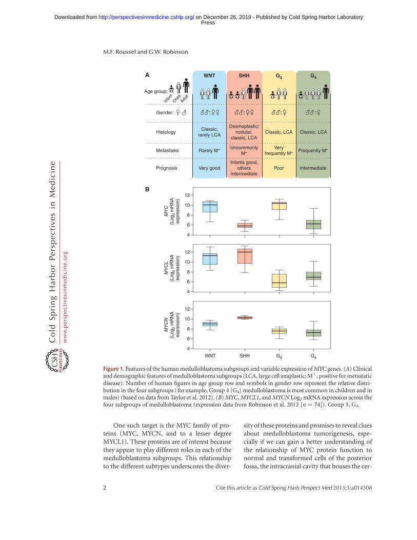
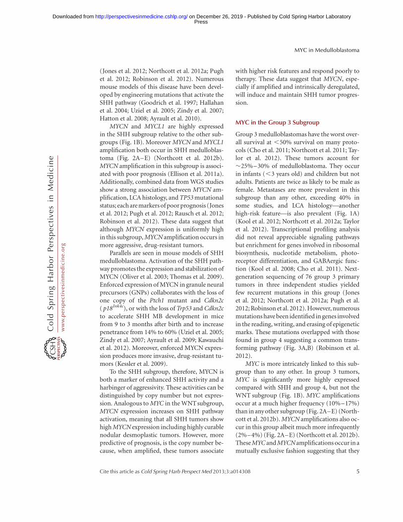
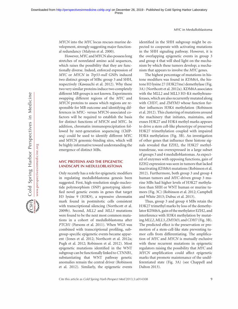
Figure 1. Features of the human medulloblastoma subgroups and variable expression of MYC genes. (A) Clinicaland demographic features of medulloblastoma subgroups (LCA, large cell anaplastic; Mþ, positive for metastaticdisease). Number of human figures in age group row and symbols in gender row represent the relative distri-bution in the four subgroups (for example, Group 4 [G4] medulloblastoma is most common in children and inmales) (based on data from Taylor et al. 2012). (B) MYC, MYCL1, and MYCN Log2 mRNA expression across thefour subgroups of medulloblastoma (expression data from Robinson et al. 2012 [n ¼ 74]). Group 3, G3.
M.F. Roussel and G.W. Robinson
2 Cite this article as Cold Spring Harb Perspect Med 2013;3:a014308
ww
w.p
ersp
ecti
vesi
nm
edic
ine.
org
Press on December 26, 2019 - Published by Cold Spring Harbor Laboratoryhttp://perspectivesinmedicine.cshlp.org/Downloaded from
ebellum and the brain stem within which me-dulloblastomas arise.
MYC AND THE MEDULLOBLASTOMASUBGROUPS
MYC proteins are associated with many cancersand medulloblastoma is no exception. MYC,MYCN, and MYCL1 amplifications have allbeen described in medulloblastomas (Northcottet al. 2012b). MYC and MYCN amplificationand expression have been intensely scrutinizedin medulloblastoma because highly aggressivetumors frequently harbor MYC or MYCN am-plification and/or overexpression (McManamyet al. 2007; Pfister et al. 2009; Cho et al. 2011).Classification schemes correlating expressionand amplification of these proteins to poor out-come have been proposed (de Haas et al. 2008;Park et al. 2012). However inconsistencies, suchas high expression in a subset of good respond-ers, have made these criteria difficult to apply.These discrepancies arise because MYC proteinsrelate differently to each subgroup. Whenviewed in this context, the relationship of MYCto prognosis becomes clearer.
MYC in the WNT Subgroup
The WNT subgroup of medulloblastoma is themost curable with .90% of patients survivingon current therapy (Ellison et al. 2005; Cliffordet al. 2006; Gajjar et al. 2006; Ellison et al. 2011a).It is also the least common medulloblastomasubtype, accounting for only 10%. Patients aregenerally older, with an average age of about 10years. There is an even distribution of females tomales. The histology is overwhelmingly classic,and the tumors are very infrequently metastatic(Fig. 1A) (Kool et al. 2012; Taylor et al. 2012).Transcriptional profiling reveals a high expres-sion of WNT pathway genes in these tumorscompared with the other subgroups (Thomp-son et al. 2006; Kool et al. 2008; Northcottet al. 2011). Next-generation sequencing studiesshow that �90% of these tumors harbor acti-vating mutations in b-CATENIN (CTNNB1),the central orchestrator of the canonical WNTpathway (Jones et al. 2012; Northcott et al.
2012a; Pugh et al. 2012; Robinson et al. 2012).The only mouse model of this disease requires anactivating mutation in Ctnnb1 and loss of Trp53(Gibson et al. 2010). Partners of CTNNB1 andmembers of the canonical WNT pathway arealso frequently mutated in this subgroup (Ro-binson et al. 2012). These data strongly supportthe constitutive activation of the WNT pathwayas causal to this medulloblastoma subgroup.
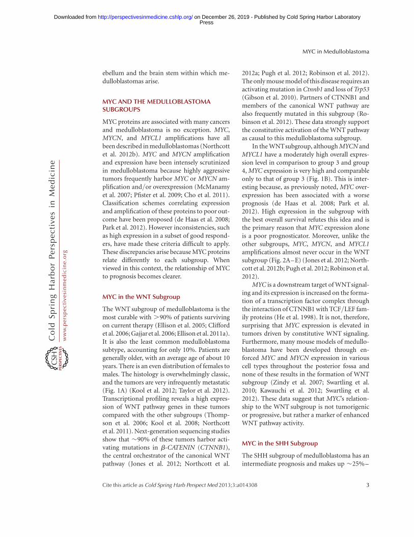
In the WNTsubgroup, although MYCN andMYCL1 have a moderately high overall expres-sion level in comparison to group 3 and group4, MYC expression is very high and comparableonly to that of group 3 (Fig. 1B). This is inter-esting because, as previously noted, MYC over-expression has been associated with a worseprognosis (de Haas et al. 2008; Park et al.2012). High expression in the subgroup withthe best overall survival refutes this idea and isthe primary reason that MYC expression aloneis a poor prognosticator. Moreover, unlike theother subgroups, MYC, MYCN, and MYCL1amplifications almost never occur in the WNTsubgroup (Fig. 2A–E) (Jones et al. 2012; North-cott et al. 2012b; Pugh et al. 2012; Robinson et al.2012).
MYC is a downstream target of WNTsignal-ing and its expression is increased on the forma-tion of a transcription factor complex throughthe interaction of CTNNB1 with TCF/LEF fam-ily proteins (He et al. 1998). It is not, therefore,surprising that MYC expression is elevated intumors driven by constitutive WNT signaling.Furthermore, many mouse models of medullo-blastoma have been developed through en-forced MYC and MYCN expression in variouscell types throughout the posterior fossa andnone of these results in the formation of WNTsubgroup (Zindy et al. 2007; Swartling et al.2010; Kawauchi et al. 2012; Swartling et al.2012). These data suggest that MYC’s relation-ship to the WNT subgroup is not tumorigenicor progressive, but rather a marker of enhancedWNT pathway activity.
MYC in the SHH Subgroup
The SHH subgroup of medulloblastoma has anintermediate prognosis and makes up �25%–
MYC in Medulloblastoma
Cite this article as Cold Spring Harb Perspect Med 2013;3:a014308 3
ww
w.p
ersp
ecti
vesi
nm
edic
ine.
org
Press on December 26, 2019 - Published by Cold Spring Harbor Laboratoryhttp://perspectivesinmedicine.cshlp.org/Downloaded from
30% of all medulloblastomas. SHH subgrouppatients tend to be very young (,5 years old)or older (.16 years old). Both males and fe-males are equally likely to be diagnosed withSHH medulloblastoma. All three major histo-logic variants (nodular desmoplastic, classic,and large cell/anaplastic [LCA]) are describedwithin this group (Northcott et al. 2011; Koolet al. 2012; Taylor et al. 2012). Outcome corre-lates with histology, and nodular desmoplasticSHH tumors have a good overall survival,whereas LCA tumors show a worse outcome
(McManamy et al. 2007; Ellison et al. 2011a).Metastatic disease is uncommon but associateswith other poor prognostic features such as LCAhistology and older age at diagnosis (Fig. 1A)(Ellison et al. 2011a). Transcriptional profilingshows significant overexpression of SHH signal-ing genes in this group relative to the other sub-groups (Thompson et al. 2006; Kool et al. 2008;Cho et al. 2011; Northcott et al. 2011). WGS andother studies have identified numerous muta-tions in genes that regulate the SHH pathway,all of which result in its constitutive activation
SMARCA4
PTCH1
WNT
WNT
A
B
C
D
E
SHH Group 3 Group 4
SUFU
SMO
CTNNB1
KDM6A
MYCN
WNT
SHH
Group 3
Group 4
MYC
20
10
WGS
MYCN
MYCN
0
7 (11%)
3 (4%)
7 (6.5)
MYC
0
0
6 (8%)
0
MYC
Tum
or n
umbe
r (%
)
0
20
10
Northcott
MYCN MYC MYCL
Tum
or n
umbe
r (%
)
0
SHH
G3
G4
WNT
MYCN
0
22 (8.3%)
4 (2.3%)
20 (6.3%)
MYC
0
0
28 (16.7%)
4 (1.3%)
MYCL
0
6 (2.3%)
0
0
SHH
G3
G4
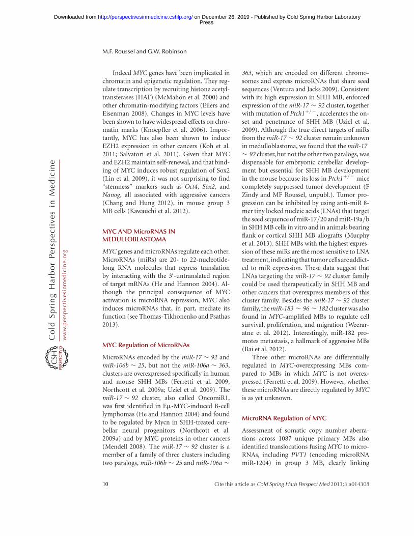
Figure 2. Amplifications of MYC genes in human medulloblastoma. (A) MYC and MYCN amplifications, shownin red, and the most common recurrent subgroup mutations (missense, indels, frameshift mutations), shown inblack, relative to the four medulloblastoma subgroups. Results are reported across three genomic studies (Joneset al. 2012; Pugh et al. 2012; Robinson et al. 2012). (B,C) MYCN and MYC amplifications relative to fourmedulloblastoma subgroups across three whole genome sequencing (WGS) studies (Jones et al. 2012; Pugh et al.2012; Robinson et al. 2012). (D,E) MYCN and MYC amplifications relative to four medulloblastoma subgroupsin 827 medulloblastomas (Northcott et al. 2012b).
M.F. Roussel and G.W. Robinson
4 Cite this article as Cold Spring Harb Perspect Med 2013;3:a014308
ww
w.p
ersp
ecti
vesi
nm
edic
ine.
org
Press on December 26, 2019 - Published by Cold Spring Harbor Laboratoryhttp://perspectivesinmedicine.cshlp.org/Downloaded from
(Jones et al. 2012; Northcott et al. 2012a; Pughet al. 2012; Robinson et al. 2012). Numerousmouse models of this disease have been devel-oped by engineering mutations that activate theSHH pathway (Goodrich et al. 1997; Hallahanet al. 2004; Uziel et al. 2005; Zindy et al. 2007;Hatton et al. 2008; Ayrault et al. 2010).
MYCN and MYCL1 are highly expressedin the SHH subgroup relative to the other sub-groups (Fig. 1B). Moreover MYCN and MYCL1amplification both occur in SHH medulloblas-toma (Fig. 2A–E) (Northcott et al. 2012b).MYCN amplification in this subgroup is associ-ated with poor prognosis (Ellison et al. 2011a).Additionally, combined data from WGS studiesshow a strong association between MYCN am-plification, LCA histology, and TP53 mutationalstatus; each are markers of poor prognosis (Joneset al. 2012; Pugh et al. 2012; Rausch et al. 2012;Robinson et al. 2012). These data suggest thatalthough MYCN expression is uniformly highin this subgroup, MYCN amplification occurs inmore aggressive, drug-resistant tumors.
Parallels are seen in mouse models of SHHmedulloblastoma. Activation of the SHH path-way promotes the expression and stabilization ofMYCN (Oliver et al. 2003; Thomas et al. 2009).Enforced expression of MYCN in granule neuralprecursors (GNPs) collaborates with the loss ofone copy of the Ptch1 mutant and Cdkn2c( p18Ink4c), or with the loss of Trp53 and Cdkn2cto accelerate SHH MB development in micefrom 9 to 3 months after birth and to increasepenetrance from 14% to 60% (Uziel et al. 2005;Zindy et al. 2007; Ayrault et al. 2009; Kawauchiet al. 2012). Moreover, enforced MYCN expres-sion produces more invasive, drug-resistant tu-mors (Kessler et al. 2009).
To the SHH subgroup, therefore, MYCN isboth a marker of enhanced SHH activity and aharbinger of aggressivity. These activities can bedistinguished by copy number but not expres-sion. Analogous to MYC in the WNTsubgroup,MYCN expression increases on SHH pathwayactivation, meaning that all SHH tumors showhigh MYCN expression including highly curablenodular desmoplastic tumors. However, morepredictive of prognosis, is the copy number be-cause, when amplified, these tumors associate
with higher risk features and respond poorly totherapy. These data suggest that MYCN, espe-cially if amplified and intrinsically deregulated,will induce and maintain SHH tumor progres-sion.
MYC in the Group 3 Subgroup
Group 3 medulloblastomas have the worst over-all survival at ,50% survival on many proto-cols (Cho et al. 2011; Northcott et al. 2011; Tay-lor et al. 2012). These tumors account for�25%–30% of medulloblastoma. They occurin infants (,3 years old) and children but notadults. Patients are twice as likely to be male asfemale. Metastases are more prevalent in thissubgroup than any other, exceeding 40% insome studies, and LCA histology—anotherhigh-risk feature—is also prevalent (Fig. 1A)(Kool et al. 2012; Northcott et al. 2012a; Tayloret al. 2012). Transcriptional profiling analysisdid not reveal appreciable signaling pathwaysbut enrichment for genes involved in ribosomalbiosynthesis, nucleotide metabolism, photo-receptor differentiation, and GABAergic func-tion (Kool et al. 2008; Cho et al. 2011). Next-generation sequencing of 76 group 3 primarytumors in three independent studies yieldedfew recurrent mutations in this group (Joneset al. 2012; Northcott et al. 2012a; Pugh et al.2012; Robinson et al. 2012). However, numerousmutations have been identified in genes involvedin the reading, writing, and erasing of epigeneticmarks. These mutations overlapped with thosefound in group 4 suggesting a common trans-forming pathway (Fig. 3A,B) (Robinson et al.2012).
MYC is more intricately linked to this sub-group than to any other. In group 3 tumors,MYC is significantly more highly expressedcompared with SHH and group 4, but not theWNT subgroup (Fig. 1B). MYC amplificationsoccur at a much higher frequency (10%–17%)than in anyother subgroup (Fig. 2A–E) (North-cott et al. 2012b). MYCN amplifications also oc-cur in this group albeit much more infrequently(2%–4%) (Fig. 2A–E) (Northcott et al. 2012b).These MYC and MYCNamplifications occur in amutually exclusive fashion suggesting that they
MYC in Medulloblastoma
Cite this article as Cold Spring Harb Perspect Med 2013;3:a014308 5
ww
w.p
ersp
ecti
vesi
nm
edic
ine.
org
Press on December 26, 2019 - Published by Cold Spring Harbor Laboratoryhttp://perspectivesinmedicine.cshlp.org/Downloaded from
may have overlapping functions in this setting(Fig. 2A) (Northcott et al. 2012a).
Two MYC-driven mouse models of group 3MBs have been generated. Our model was gen-erated by enforced expression of the wild-typeMYC gene in GNPs purified from Trp53-nullmice (Kawauchi et al. 2012); the other reliedon enforced expression of a partially stabilizedmutant of MYC (MYCT58A) with a dominant-negative form of Trp53 in CD133/prominin 1-positive neural stem cells (NSCs) (Pei et al.2012). The resultant tumors showed that theLCA phenotypes were highly aggressive, andtranscriptionally resembled group 3 MB. MYCwithdrawal caused complete tumor regression(Pei et al. 2012). These data suggest that MYC isa significant contributor to the initiation, main-tenance, and progression of this disease (seeGabay et al. 2013).
Of note, many mouse models of MB, in-cluding these MYC-driven group 3 models, re-
quire the loss of Trp53 function; yet somaticmutations of TP53 occur mostly in WNT andSHH medulloblastoma and are associated withMYCN rather than MYC amplification (Pfaffet al. 2010; Rausch et al. 2012). The absence ofthese mutations in group 3 tumors means thatTP53 loss is not required for human tumor ini-tiation, that another cooperative genetic eventassists tumor initiation, or that the TP53 path-way is in some other way compromised. To clar-ify this question, an analysis of TP53 proteinexpression and function and its regulation inhuman primary medulloblastoma samplesgrown as neurospheres or xenografts is warrant-ed (Milde et al. 2012; Zhao et al. 2012).
MYC in the Group 4 Subgroup
Group 4 medulloblastomas have a similar prog-nosis to SHH tumors. They account for �40%–50% of medulloblastoma, occur mainly in chil-
Group 3
A
B C
MLL2
SMARCA4
CHD7
HDAC2
KDM1A
KDM4C
KDM5A
KDM6A
MLL3
SETD2
ZMYM3
MYCN
H3K
27m
e3
Mouse medulloblastoma
MYC
HDAC9
Group 4
Mutation
Amplification
EZH2HDAC
MLL3
= MeMYC
K4K27
Stem cell state Differentiated state
CHD7
WNT SHH Group 3
MLL2KDM6AZMYM3KDM1A
H3H3 H3H3
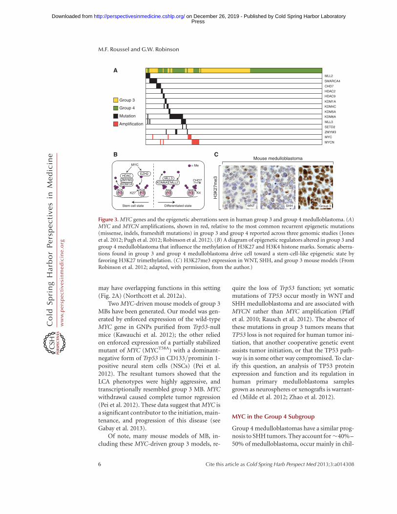
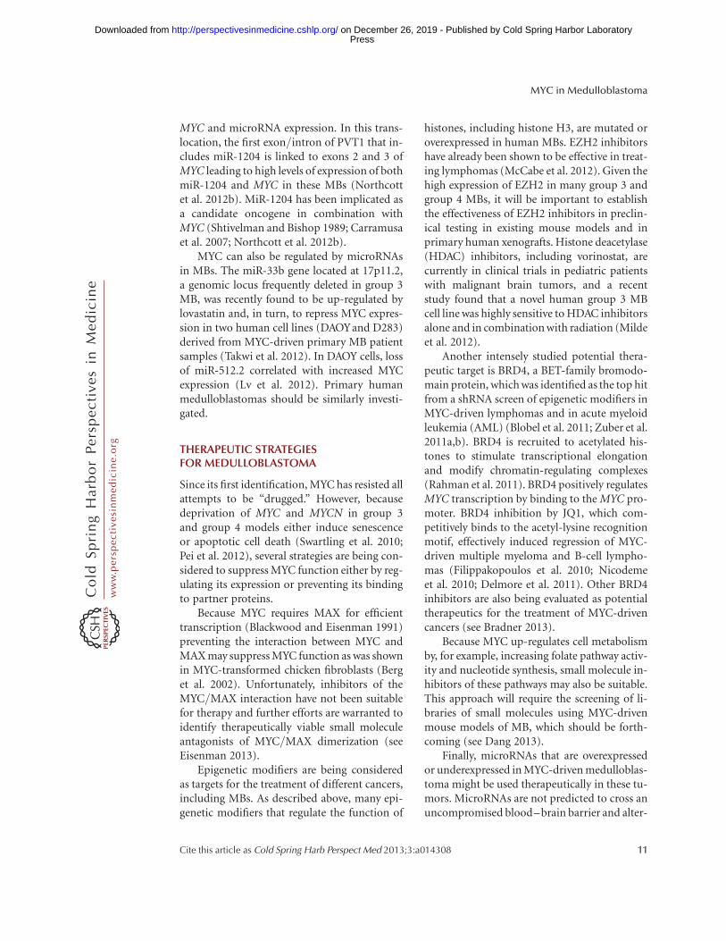
Figure 3. MYC genes and the epigenetic aberrations seen in human group 3 and group 4 medulloblastoma. (A)MYC and MYCN amplifications, shown in red, relative to the most common recurrent epigenetic mutations(missense, indels, frameshift mutations) in group 3 and group 4 reported across three genomic studies (Joneset al. 2012; Pugh et al. 2012; Robinson et al. 2012). (B) A diagram of epigenetic regulators altered in group 3 andgroup 4 medulloblastoma that influence the methylation of H3K27 and H3K4 histone marks. Somatic aberra-tions found in group 3 and group 4 medulloblastoma drive cell toward a stem-cell-like epigenetic state byfavoring H3K27 trimethylation. (C) H3K27me3 expression in WNT, SHH, and group 3 mouse models (FromRobinson et al. 2012; adapted, with permission, from the author.)
M.F. Roussel and G.W. Robinson
6 Cite this article as Cold Spring Harb Perspect Med 2013;3:a014308
ww
w.p
ersp
ecti
vesi
nm
edic
ine.
org
Press on December 26, 2019 - Published by Cold Spring Harbor Laboratoryhttp://perspectivesinmedicine.cshlp.org/Downloaded from
dren but can occur in adults, and, similar togroup 3, predominantly occur in males. Mostgroup 4 MBs have a classic histology (Fig. 1A)(Kool et al. 2012; Northcott et al. 2012a; Tayloret al. 2012). Transcriptional profiling revealedenrichment for genes of neuronal differentia-tion and glutamatergic receptors (Kool et al.2008; Cho et al. 2011). Next-generation se-quencing identified recurrent mutations in themolecular machinery that reads, writes, anderases epigenetic marks (described below),some overlapping with those found in group 3tumors (Fig. 3A,B) (Jones et al. 2012; Northcottet al. 2012a; Pugh et al. 2012; Robinson et al.2012).
Group 4 tumors generally have low MYC andMYCN expression compared with the other sub-groups (Fig. 1B). However, mean MYCN expres-sion levels are still comparatively high whencompared with mature cerebellum and exist atcomparable levels to fetal cerebella (Swartlinget al. 2010). This implies that a certain level ofMYCN may be required for tumor maintenance.In addition, MYCN amplifications occur insome group 4 tumors and these, like group 3,appear to be mutually exclusive with epigeneticmutations (Fig. 3A) and to other amplifications(Northcott et al. 2012a; Robinson et al. 2012).This suggests that, although rare, MYCN ampli-fication may initiate group 4 medulloblastoma.
Mouse cerebellar NSCs from postnatal day0, but not from embryonic day 16, transducedwith a partially stabilized form of MYCN(MYCNT58A) produce SHH-independent me-dulloblastoma (Swartling et al. 2012). Similar-ly, when conditional MYCNT58A expression istargeted to neural cells from postnatal day 1through adulthood, mice develop group 4-likeMBs (Swartling et al. 2010, 2012). These tumorsenter senescence on removal of stabilizedMYCNT58A (Swartling et al. 2010). These exper-iments show the influence of the timing of theoncogenic stimulus and its requirement for tu-morigenesis (see Gabay et al. 2013).
This temporal association draws parallels tothe peak age of this disease occurring in olderchildren rather than infants and implies thatthere may be a finite period during which cer-tain cell pools are particularly vulnerable to tu-
morigenesis. These data suggest that MYCNplays a role in initiation and maintenance ofgroup 4 medulloblastoma.
MYC IN THE DEVELOPING POSTERIORFOSSA
The cerebellum is a unique brain structure be-cause, unlike the rest of the brain, granule neu-ral progenitors actively proliferate after birth(Roussel and Hatten 2011). Cerebellar progen-itor neurons arise during embryogenesis fromthe ventricular zone and the upper rhombic lip(URL) to form the cerebellar anlage (Fig. 4).During this time, neural progenitors are alsoborn in the lower rhombic lip (LRL) on the floorof the fourth ventricle and express the Blbp gene(Gibson et al. 2010). The neural progenitorswithin the ventricular zone of the cerebellar an-lage migrate radially outward and give rise toBergman glia and Purkinje cells, which are post-mitotic by birth. Conversely neural progenitorsfrom the upper rhombic lip give rise to GNPsthat rapidly proliferate after birth in the externalgranule layer (EGL) of the developing cerebel-lum, exit cycle, and migrate inward. Once theGNPs cross the Purkinje cell layer they cometo rest and form the internal granule layer (IGL)as postmitotic neurons (Roussel and Hatten2011). The white matter within the core of themature cerebellum contains neuronal stem cellsexpressing CD133/prominin that are capable ofgenerating new neurons during the life of theorganism.
MYC, MYCN, and MYCL expression in thedeveloping posterior fossa appears to be cellcontext dependent (Fig. 4). GNPs expressMYCN, but not MYC, in their proliferativephase in response to SHH. Neuronal progeni-tors in the Purkinje cell layer and in the ventric-ular zone express MYCL but not MYCN orMYC (www.cdtdb.neuroinf.jp/CDT/Top.jsp).MYC is expressed in brain lipid-binding protein(BLBP)-positive glial and LRL neural progeni-tors and possibly in CD133/prominin NSCsthat reside in the white matter (Wey et al.2010). This differential expression of MYC andMYCN by distinct cerebellar progenitors lendscredence to the argument that the different me-
MYC in Medulloblastoma
Cite this article as Cold Spring Harb Perspect Med 2013;3:a014308 7
ww
w.p
ersp
ecti
vesi
nm
edic
ine.
org
Press on December 26, 2019 - Published by Cold Spring Harbor Laboratoryhttp://perspectivesinmedicine.cshlp.org/Downloaded from
dulloblastoma subgroups arise from distinctcerebellar progenitors. Indeed SHH subgroupmedulloblastomas originate from GNPs andoverexpress MYCN, whereas WNT subgroupmedulloblastomas emanate from LRL precur-sors and overexpress MYC. However, attemptsto identify precursors of group 3 and group 4medulloblastomas through enforced expressionof MYC or MYCN into identical cerebellar pre-cursor cells yielded some intriguing differentialeffects depending on the gene used, which sug-gests that the two paralogs influence the cellu-lar program in different ways (Kawauchi et al.2012).
Rapid proliferation of GNPs is mediated bySHH, which is produced by Purkinje cells. Bybinding to its receptor PATCHED (PTCH1),SHH directly activates MYCN transcription viaGLI transcription factors (Kenney et al. 2003).The MYCN requirement for cerebellum devel-opment was shown in a Nestin-Cre transgenicmouse. Conditional deletion of Mycn in earlyneural stem and progenitor cells of this mouse
led to profound cerebellum defects, in part ow-ing to the failure to suppress two cyclin-depen-dent kinase inhibitory proteins (CKIs), p18Ink4c
and p27Kip1 (Knoepfler et al. 2002). Remark-ably, these cerebellar defects could be partiallyrescued by the loss of the two CKIs suggestingthat MYCN inhibition of p18Ink4c and p27Kip1
expression is required for proper cerebellar de-velopment (Knoepfler et al. 2002; Zindy et al.2006; Hurlin 2013).
Although MYC is not expressed in GNPs,the conditional loss of Mycn in neural progen-itors induces Myc in GNPs, which partiallycompensates for the loss of Mycn, whereas lossof both Myc and Mycn leads to the completeabsence of the EGL and GNPs (Zindy et al.2006). This functional redundancy impliesthat MYC and MYCN may have similar roles.These homologs contain conserved MYC boxes,and similar basic helix-loop-helix (bHLH)-zip-per structures that recognize the same DNA mo-tif when dimerized with MAX (see Eisenman2013; Rahl and Young 2013). Knock-in of
LRLPrecerebellar neuron progenitors
(BLBP+) MYC
GABAergic neuronprogenitors
(PTF1A+, BLBP+)MYCN
EGLGNPs (ATOH1+)
MYCN
EGLGNPs (ATOH1+)
MYCN
Postmitoticneurons (ATOH1–)
MAD
Purkinjecell layerMYCL
Purkinjecell layerMYCL
IGLMYCL
P21/adultP7
Cerebral development
E13.5
Bergmannglia
MYCL
Choroidplexus
Ventricular zone URL
Multipotent progenitors(CD133/Prom1+) MYC?
Multipotent progenitors(CD133/Prom1+) MYC?
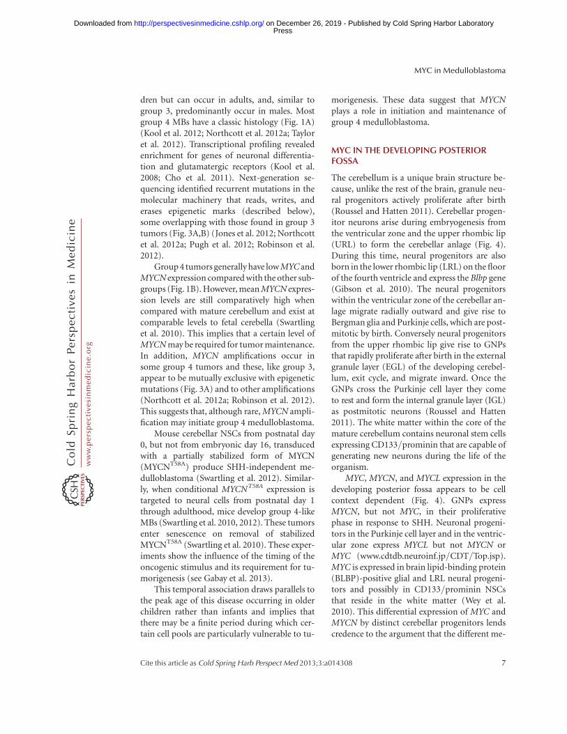
Figure 4. Expression of MYC genes during mouse cerebellar development. At embryonic day E13.5, GNPs thatmigrate from the upper rhombic lip (URL) and form the external granule layer (EGL) and GABAergic neuralprogenitors in the cerebellar ventricular zone (VZ) express MYCN. MYC is found expressed in precerebellarneuron progenitors in the lower rhombic lip. At P7, whereas the GNPs in the EGL continue to express MYCN,the Purkinje cell layer, composed of Bergman glia and Purkinje cells, expresses MYCL and CD133/prominin-positive multipotent progenitors. At P21, all GNPs have exited cycle, no longer express MYCN but do expressMXD (see Eisenman 2013), and have migrated through the Purkinje cell layer to reside as postmitotic neuronsin the internal granule layer (IGL) that, like Purkinje cells, express MYCL (www.gensat.org; www.cdtdb.neuroinf.jp/CDT/Top.jsp; Allen Brain Atlas). CD133/prominin-positive multipotent progenitors are marked with a“MYC?” because this is suspected but not known. Prom1, prominin 1.
M.F. Roussel and G.W. Robinson
8 Cite this article as Cold Spring Harb Perspect Med 2013;3:a014308
ww
w.p
ersp
ecti
vesi
nm
edic
ine.
org
Press on December 26, 2019 - Published by Cold Spring Harbor Laboratoryhttp://perspectivesinmedicine.cshlp.org/Downloaded from
MYCN into the MYC locus rescues murine de-velopment, strongly suggesting major function-al redundancy (Malynn et al. 2000).
However, MYC and MYCN also possess longstretches of nonrelated amino acid sequences,which raises the possibility that they are func-tionally diverse. Indeed, enforced expression ofMYC or MYCN in Trp53-null GNPs inducedtwo distinct groups of MBs: group 3 and SHH,respectively (Kawauchi et al. 2012). Why thesetwo very similar proteins induce two completelydifferent MB groups is not known. Experimentsswapping different regions of the MYC andMYCN proteins to assess which regions are re-sponsible for MB outcome and identifying dif-ferences in MYC- versus MYCN-associated co-factors will be required to establish the basisfor distinct functions of MYCN and MYC. Inaddition, chromatin immunoprecipitation fol-lowed by next-generation sequencing (ChIP-seq) could be used to identify different MYCand MYCN genomic-binding sites, which willbe highly informative toward understanding theemergence of distinct MBs.
MYC PROTEINS AND THE EPIGENETICLANDSCAPE IN MEDULLOBLASTOMA
Only recently has a role for epigenetic modifiersin regulating medulloblastoma genesis beensuggested. First, high-resolution single-nucleo-tide polymorphism (SNP) genotyping identi-fied novel genetic events in genes that targetH3 lysine 9 (H3K9), a repressive chromatinmark found in postmitotic cells consistentwith transcriptional silencing (Northcott et al.2009b). Second, MLL2 and MLL3 mutationswere found to be the next most common muta-tions in a cohort of medulloblastoma afterPTCH1 (Parsons et al. 2011). When WGS wascombined with transcriptional profiling, sub-group-specific epigenetic events became appar-ent (Jones et al. 2012; Northcott et al. 2012a;Pugh et al. 2012; Robinson et al. 2012). Mostepigenetic mutations identified in the WNTsubgroup can be functionally linked to CTNNB1,substantiating that WNT pathway geneticanomalies remain the central driver (Robinsonet al. 2012). Similarly, the epigenetic events
identified in the SHH subgroup might be ex-pected to cooperate with activating mutationsin the SHH signaling pathway. However, it isthe overlapping epigenetic events in group 3and group 4 that will shed light on the mecha-nism by which these tumors develop; a mecha-nism that appears to involve the MYC genes.
The highest percentage of mutations in his-tone modifiers was found in KDM6A, the his-tone H3 lysine 27 (H3K27me) demethylase (Fig.3A) (Northcott et al. 2012a). KDM6A associateswith the MLL2 and MLL3 H3-K4 methyltrans-ferases, which are also recurrently mutated alongwith CHD7, and ZMYM3 whose function fur-ther influences H3K4 methylation (Robinsonet al. 2012). This clustering of mutations aroundthe machinery that initiates, maintains, anderases H3K27 and H3K4 methyl marks appearsto drive a stem cell-like phenotype of preservedH3K27 trimethylation coupled with impairedH3K4 methylation (Fig. 3B). An investigationof other genes that influence these histone sig-nals revealed that EZH2, the H3K27 methyl-transferase, was overexpressed in a large subsetof groups 3 and 4 medulloblastomas. As expect-ed of enzymes with opposing functions, gain ofEZH2 expression was seen in tumors that lackedinactivating KDM6A mutations (Robinson et al.2012). Furthermore, both group 3 and group 4human tumors and MYC-driven group 3 mu-rine MBs had higher levels of H3K27 methyla-tion than SHH or WNT human or murine tu-mors (Fig. 3C) (Robinson et al. 2012; Campbelland White 2013; Dubuc et al. 2013).
Thus, group 3 and group 4 MBs retain theH3K27 trimethyl marks by loss of the demethy-lator KDM6A, gain of the methylator EZH2, andinterference with H3K4 methylation by mutat-ing MLL2, MLL3, ZMYM3, and CDH7 (Fig. 3B).The predicted effect is the preservation or pro-motion of a stem-cell-like state preventing tu-mor cells from differentiating. The amplifica-tion of MYC and MYCN is mutually exclusivewith these recurrent mutations in epigeneticregulators raising the possibility that MYC andMYCN amplification could affect epigeneticmarks that promote maintenance of the undif-ferentiated state (Fig. 3A) (see Chappell andDalton 2013).
MYC in Medulloblastoma
Cite this article as Cold Spring Harb Perspect Med 2013;3:a014308 9
ww
w.p
ersp
ecti
vesi
nm
edic
ine.
org
Press on December 26, 2019 - Published by Cold Spring Harbor Laboratoryhttp://perspectivesinmedicine.cshlp.org/Downloaded from
Indeed MYC genes have been implicated inchromatin and epigenetic regulation. They reg-ulate transcription by recruiting histone acetyl-transferases (HAT) (McMahon et al. 2000) andother chromatin-modifying factors (Eilers andEisenman 2008). Changes in MYC levels havebeen shown to have widespread effects on chro-matin marks (Knoepfler et al. 2006). Impor-tantly, MYC has also been shown to induceEZH2 expression in other cancers (Koh et al.2011; Salvatori et al. 2011). Given that MYCand EZH2 maintain self-renewal, and that bind-ing of MYC induces robust regulation of Sox2(Lin et al. 2009), it was not surprising to find“stemness” markers such as Oct4, Sox2, andNanog, all associated with aggressive cancers(Chang and Hung 2012), in mouse group 3MB cells (Kawauchi et al. 2012).
MYC AND MicroRNAS INMEDULLOBLASTOMA
MYC genes and microRNAs regulate each other.MicroRNAs (miRs) are 20- to 22-nucleotide-long RNA molecules that repress translationby interacting with the 30-untranslated regionof target mRNAs (He and Hannon 2004). Al-though the principal consequence of MYCactivation is microRNA repression, MYC alsoinduces microRNAs that, in part, mediate itsfunction (see Thomas-Tikhonenko and Psathas2013).
MYC Regulation of MicroRNAs
MicroRNAs encoded by the miR-17 � 92 andmiR-106b � 25, but not the miR-106a � 363,clusters are overexpressed specifically in humanand mouse SHH MBs (Ferretti et al. 2009;Northcott et al. 2009a; Uziel et al. 2009). ThemiR-17 � 92 cluster, also called OncomiR1,was first identified in Em-MYC-induced B-celllymphomas (He and Hannon 2004) and foundto be regulated by Mycn in SHH-treated cere-bellar neural progenitors (Northcott et al.2009a) and by MYC proteins in other cancers(Mendell 2008). The miR-17 � 92 cluster is amember of a family of three clusters includingtwo paralogs, miR-106b � 25 and miR-106a �
363, which are encoded on different chromo-somes and express microRNAs that share seedsequences (Ventura and Jacks 2009). Consistentwith its high expression in SHH MB, enforcedexpression of the miR-17 � 92 cluster, togetherwith mutation of Ptch1þ/2, accelerates the on-set and penetrance of SHH MB (Uziel et al.2009). Although the true direct targets of miRsfrom the miR-17 � 92 cluster remain unknownin medulloblastoma, we found that the miR-17� 92 cluster, but not the other two paralogs, wasdispensable for embryonic cerebellar develop-ment but essential for SHH MB developmentin the mouse because its loss in Ptch1þ/2 micecompletely suppressed tumor development (FZindy and MF Roussel, unpubl.). Tumor pro-gression can be inhibited by using anti-miR 8-mer tiny locked nucleic acids (LNAs) that targetthe seed sequence of miR-17/20 and miR-19a/bin SHH MB cells in vitro and in animals bearingflank or cortical SHH MB allografts (Murphyet al. 2013). SHH MBs with the highest expres-sion of these miRs are the most sensitive to LNAtreatment, indicating that tumorcells are addict-ed to miR expression. These data suggest thatLNAs targeting the miR-17 � 92 cluster familycould be used therapeutically in SHH MB andother cancers that overexpress members of thiscluster family. Besides the miR-17 � 92 clusterfamily, the miR-183 � 96 � 182 cluster was alsofound in MYC-amplified MBs to regulate cellsurvival, proliferation, and migration (Weerar-atne et al. 2012). Interestingly, miR-182 pro-motes metastasis, a hallmark of aggressive MBs(Bai et al. 2012).
Three other microRNAs are differentiallyregulated in MYC-overexpressing MBs com-pared to MBs in which MYC is not overex-pressed (Ferretti et al. 2009). However, whetherthese microRNAs are directly regulated by MYCis as yet unknown.
MicroRNA Regulation of MYC
Assessment of somatic copy number aberra-tions across 1087 unique primary MBs alsoidentified translocations fusing MYC to micro-RNAs, including PVT1 (encoding microRNAmiR-1204) in group 3 MB, clearly linking
M.F. Roussel and G.W. Robinson
10 Cite this article as Cold Spring Harb Perspect Med 2013;3:a014308
ww
w.p
ersp
ecti
vesi
nm
edic
ine.
org
Press on December 26, 2019 - Published by Cold Spring Harbor Laboratoryhttp://perspectivesinmedicine.cshlp.org/Downloaded from
MYC and microRNA expression. In this trans-location, the first exon/intron of PVT1 that in-cludes miR-1204 is linked to exons 2 and 3 ofMYC leading to high levels of expression of bothmiR-1204 and MYC in these MBs (Northcottet al. 2012b). MiR-1204 has been implicated asa candidate oncogene in combination withMYC (Shtivelman and Bishop 1989; Carramusaet al. 2007; Northcott et al. 2012b).
MYC can also be regulated by microRNAsin MBs. The miR-33b gene located at 17p11.2,a genomic locus frequently deleted in group 3MB, was recently found to be up-regulated bylovastatin and, in turn, to repress MYC expres-sion in two human cell lines (DAOYand D283)derived from MYC-driven primary MB patientsamples (Takwi et al. 2012). In DAOY cells, lossof miR-512.2 correlated with increased MYCexpression (Lv et al. 2012). Primary humanmedulloblastomas should be similarly investi-gated.
THERAPEUTIC STRATEGIESFOR MEDULLOBLASTOMA
Since its first identification, MYC has resisted allattempts to be “drugged.” However, becausedeprivation of MYC and MYCN in group 3and group 4 models either induce senescenceor apoptotic cell death (Swartling et al. 2010;Pei et al. 2012), several strategies are being con-sidered to suppress MYC function either by reg-ulating its expression or preventing its bindingto partner proteins.
Because MYC requires MAX for efficienttranscription (Blackwood and Eisenman 1991)preventing the interaction between MYC andMAX may suppress MYC function as was shownin MYC-transformed chicken fibroblasts (Berget al. 2002). Unfortunately, inhibitors of theMYC/MAX interaction have not been suitablefor therapy and further efforts are warranted toidentify therapeutically viable small moleculeantagonists of MYC/MAX dimerization (seeEisenman 2013).
Epigenetic modifiers are being consideredas targets for the treatment of different cancers,including MBs. As described above, many epi-genetic modifiers that regulate the function of
histones, including histone H3, are mutated oroverexpressed in human MBs. EZH2 inhibitorshave already been shown to be effective in treat-ing lymphomas (McCabe et al. 2012). Given thehigh expression of EZH2 in many group 3 andgroup 4 MBs, it will be important to establishthe effectiveness of EZH2 inhibitors in preclin-ical testing in existing mouse models and inprimary human xenografts. Histone deacetylase(HDAC) inhibitors, including vorinostat, arecurrently in clinical trials in pediatric patientswith malignant brain tumors, and a recentstudy found that a novel human group 3 MBcell line was highly sensitive to HDAC inhibitorsalone and in combination with radiation (Mildeet al. 2012).
Another intensely studied potential thera-peutic target is BRD4, a BET-family bromodo-main protein, which was identified as the top hitfrom a shRNA screen of epigenetic modifiers inMYC-driven lymphomas and in acute myeloidleukemia (AML) (Blobel et al. 2011; Zuber et al.2011a,b). BRD4 is recruited to acetylated his-tones to stimulate transcriptional elongationand modify chromatin-regulating complexes(Rahman et al. 2011). BRD4 positively regulatesMYC transcription by binding to the MYC pro-moter. BRD4 inhibition by JQ1, which com-petitively binds to the acetyl-lysine recognitionmotif, effectively induced regression of MYC-driven multiple myeloma and B-cell lympho-mas (Filippakopoulos et al. 2010; Nicodemeet al. 2010; Delmore et al. 2011). Other BRD4inhibitors are also being evaluated as potentialtherapeutics for the treatment of MYC-drivencancers (see Bradner 2013).
Because MYC up-regulates cell metabolismby, for example, increasing folate pathway activ-ity and nucleotide synthesis, small molecule in-hibitors of these pathways may also be suitable.This approach will require the screening of li-braries of small molecules using MYC-drivenmouse models of MB, which should be forth-coming (see Dang 2013).
Finally, microRNAs that are overexpressedor underexpressed in MYC-driven medulloblas-toma might be used therapeutically in these tu-mors. MicroRNAs are not predicted to cross anuncompromised blood–brain barrier and alter-
MYC in Medulloblastoma
Cite this article as Cold Spring Harb Perspect Med 2013;3:a014308 11
ww
w.p
ersp
ecti
vesi
nm
edic
ine.
org
Press on December 26, 2019 - Published by Cold Spring Harbor Laboratoryhttp://perspectivesinmedicine.cshlp.org/Downloaded from
native modes of delivery, such as nanoparticlesor intraventricular administration, should beinvestigated.
CONCLUDING REMARKS
The relationship of the MYC family of genes tomedulloblastoma is widespread but subgroupspecific. Although promoting proliferation inall the subgroups, the MYC genes play differentroles in each. Fittingly, these differences appearto reflect the distinct roles MYC genes play inhindbrain development. The role in the initia-tion, maintenance, and progression of the ag-gressive group 3 medulloblastomas as well astoward the progression of SHH subgroupmake the MYC genes and their regulators veryinviting therapeutic targets for MBs for whichcurrent therapies are inadequate.
ACKNOWLEDGMENTS
We thank Susan Watson for scientific editing;Betsy Williford from the Department of Bio-medical Communication for illustrations; andDavid Solecki, Mary-Elizabeth Hatten, and PaulKnoepfler for helpful discussions. We apologizeto colleagues whose work was not includedhere owing to space limitations. This work isfunded by a National Institutes of Health grantCA-096832 (M.F.R.), a Cancer Core GrantCA-21765 (M.F.R. and G.W.R.), and the Amer-ican Lebanese-Syrian Associated Charities (AL-SAC) of St. Jude Children’s Research Hospital(SJCRH).
REFERENCES�Reference is also in this collection.
Ayrault O, Zindy F, Rehg J, Sherr CJ, Roussel MF. 2009. Twotumor suppressors, pp27Kip1 and patched-1, collaborateto prevent medulloblastoma. Mol Cancer Res 7: 33–40.
Ayrault O, Zhao H, Zindy F, Qu C, Sherr CJ, Roussel MF.2010. Atoh1 inhibits neuronal differentiation and collab-orates with Gli1 to generate medulloblastoma-initiatingcells. Cancer Res 70: 5618–5627.
Bai AH, Milde T, Remke M, Rolli CG, Hielscher T, Cho YJ,Kool M, Northcott PA, Jugold M, Bazhin AV, et al.2012. MicroRNA-182 promotes leptomeningeal spreadof non-sonic hedgehog-medulloblastoma. Acta Neuropa-thol 123: 529–538.
Berg T, Cohen SB, Desharnais J, Sonderegger C, Maslyar DJ,Goldberg J, Boger DL, Vogt PK. 2002. Small-moleculeantagonists of Myc/Max dimerization inhibit Myc-in-duced transformation of chicken embryo fibroblasts.Proc Natl Acad Sci 99: 3830–3835.
Blackwood EM, Eisenman RN. 1991. Max: A helix-loop-helix zipper protein that forms a sequence-specificDNA-binding complex with Myc. Science 251: 1211–1217.
Blobel GA, Kalota A, Sanchez PV, Carroll M. 2011. Shorthairpin RNA screen reveals bromodomain proteins asnovel targets in acute myeloid leukemia. Cancer Cell 20:287–288.
� Bradner J. 2013. Inhibiting Myc. Cold Spring Harb PerspectMed doi: 10.1101/cshperspect.a014266.
� Campbell KJ, White RJ. 2013. MYC regulation of cell growththrough control of RNA polymerae I and III activities.Cold Spring Harb Perspect Med doi: 10.1101/cshper-spect.a018408.
Carramusa L, Contino F, Ferro A, Minafra L, Perconti G,Giallongo A, Feo S. 2007. The PVT-1 oncogene is a Mycprotein target that is overexpressed in transformed cells. JCell Physiol 213: 511–518.
Chang CJ, Hung MC. 2012. The role of EZH2 in tumourprogression. Br J Cancer 106: 243–247.
� Chappell J, Dalton S. 2013. Roles for MYC in the establish-ment and maintenance of pluripotency. Cold Spring HarbPerspect Med doi: 10.1101/cshperspect.a014381.
Cho YJ, Tsherniak A, Tamayo P, Santagata S, Ligon A, Greu-lich H, Berhoukim R, Amani V, Goumnerova L, EberhartCG, et al. 2011. Integrative genomic analysis of medullo-blastoma identifies a molecular subgroup that drivespoor clinical outcome. J Clin Oncol 29: 1424–1430.
Clifford SC, Lusher ME, Lindsey JC, Langdon JA, GilbertsonRJ, Straughton D, Ellison DW. 2006. Wnt/Wingless path-way activation and chromosome 6 loss characterize adistinct molecular sub-group of medulloblastomas asso-ciated with a favorable prognosis. Cell Cycle 5: 2666–2670.
� Dang CV. 2013. MYC, metabolism, cell growth, and tumor-igenesis. Cold Spring Harb Perspect Med 3: a014217.
de Haas T, Hasselt N, Troost D, Caron H, Popovic M, Za-dravec-Zaletel L, Grajkowska W, Perek M, Osterheld MC,Ellison D, et al. 2008. Molecular risk stratification of me-dulloblastoma patients based on immunohistochemicalanalysis of MYC, LDHB, and CCNB1 expression. ClinCancer Res 14: 4154–4160.
Delmore JE, Issa GC, Lemieux ME, Rahl PB, Shi J, JacobsHM, Kastritis E, Gilpatrick T, Paranal RM, Qi J, et al.2011. BET bromodomain inhibition as a therapeuticstrategy to target c-Myc. Cell 146: 904–917.
Dubuc AM, Remke M, Korshunov A, Northcott PA, ZhanSH, Mendez-Lago M, Kool M, Jones DT, Unterberger A,Morrissy AS, et al. 2013. Aberrant patterns of H3K4 andH3K27 histone lysine methylation occur across sub-groups in medulloblastoma. Acta Neuropathol 125:373–384.
Eilers M, Eisenman RN. 2008. Myc’s broad reach. Genes Dev22: 2755–2766.
Ellison DW, Onilude OE, Lindsey JC, Lusher ME, WestonCL, Taylor RE, Pearson AD, Clifford SC. 2005. b-Catenin
M.F. Roussel and G.W. Robinson
12 Cite this article as Cold Spring Harb Perspect Med 2013;3:a014308
ww
w.p
ersp
ecti
vesi
nm
edic
ine.
org
Press on December 26, 2019 - Published by Cold Spring Harbor Laboratoryhttp://perspectivesinmedicine.cshlp.org/Downloaded from
status predicts a favorable outcome in childhood medul-loblastoma: The United Kingdom Children’s CancerStudy Group Brain Tumour Committee. J Clin Oncol23: 7951–7957.
Ellison DW, Dalton J, Kocak M, Nicholson SL, Fraga C,Neale G, Kenney AM, Brat DJ, Perry A, Yong WH, et al.2011a. Medulloblastoma: Clinicopathological correlatesof SHH, WNT, and non-SHH/WNT molecular sub-groups. Acta Neuropathol 121: 381–396.
Ellison DW, Kocak M, Dalton J, Megahed H, Lusher ME,Ryan SL, Zhao W, Nicholson SL, Taylor RE, Bailey S, et al.2011b. Definition of disease-risk stratification groups inchildhood medulloblastoma using combined clinical,pathologic, and molecular variables. J Clin Oncol 29:1400–1407.
Ferretti E, De Smaele E, Po A, Di Marcotullio L, Tosi E,Espinola MS, Di Rocco C, Riccardi R, Giangaspero F,Farcomeni A, et al. 2009. MicroRNA profiling in humanmedulloblastoma. Int J Cancer 124: 568–577.
Filippakopoulos P, Qi J, Picaud S, Shen Y, Smith WB, Fe-dorov O, Morse EM, Keates T, Hickman TT, Felletar I, etal. 2010. Selective inhibition of BET bromodomains. Na-ture 468: 1067–1073.
� Gabay M, Li Y, Felsher DW. 2013. MYC activation is a hall-mark of cancer initiation and maintenance. Cold SpringHarb Perspect Med doi: 10.1101/cshperspect.a014241.
Gajjar A, Chintagumpala M, Ashley D, Kellie S, Kun LE,Merchant TE, Woo S, Wheeler G, Ahern V, Krasin MJ,et al. 2006. Risk-adapted craniospinal radiotherapy fol-lowed by high-dose chemotherapy and stem-cell rescuein children with newly diagnosed medulloblastoma (StJude Medulloblastoma-96): Long-term results from aprospective, multicentre trial. Lancet Oncol 7: 813–820.
Gibson P, Tong Y, Robinson G, Thompson MC, Currle DS,Eden C, Kranenburg TA, Hogg T, Poppleton H, Martin J,et al. 2010. Subtypes of medulloblastoma have distinctdevelopmental origins. Nature 468: 1095–1099.
Goodrich LV, Milenkovic L, Higgins KM, Scott MP. 1997.Altered neural cell fates and medulloblastoma in mousepatched mutants. Science 277: 1109–1113.
Hallahan AR, Pritchard JI, Hansen S, Benson M, Stoeck J,Hatton BA, Russell TL, Ellenbogen RG, Bernstein ID,Beachy PA, et al. 2004. The SmoA1 mouse model revealsthat notch signaling is critical for the growth and survivalof sonic hedgehog-induced medulloblastomas. CancerRes 64: 7794–7800.
Hatton BA, Villavicencio EH, Tsuchiya KD, Pritchard JI,Ditzler S, Pullar B, Hansen S, Knoblaugh SE, Lee D,Eberhart CG, et al. 2008. The Smo/Smo model: Hedge-hog-induced medulloblastoma with 90% incidence andleptomeningeal spread. Cancer Res 68: 1768–1776.
He L, Hannon GJ. 2004. MicroRNAs: Small RNAs with a bigrole in gene regulation. Nat Rev Genet 5: 522–531.
He TC, Sparks AB, Rago C, Hermeking H, Zawel L, da CostaLT, Morin PJ, Vogelstein B, Kinzler KW. 1998. Identifica-tion of c-MYC as a target of the APC pathway. Science281: 1509–1512.
� Hurlin PJ. 2013. Control of vertebrate development byMYC. Cold Spring Harb Perspect Med doi: 10.1101/cshperspect.a014332.
Jones DT, Jager N, Kool M, Zichner T, Hutter B, Sultan M,Cho YJ, Pugh TJ, Hovestadt V, Stutz AM, et al. 2012.
Dissecting the genomic complexity underlying medullo-blastoma. Nature 488: 100–105.
Kawauchi D, Robinson G, Uziel T, Gibson P, Rehg J, Gao C,Finkelstein D, Qu C, Pounds S, Ellison DW, et al. 2012. Amouse model of the most aggressive subgroup of humanmedulloblastoma. Cancer Cell 21: 168–180.
Kenney AM, Cole MD, Rowitch DH. 2003. Nmyc upregu-lation by sonic hedgehog signaling promotes prolifera-tion in developing cerebellar granule neuron precursors.Development 130: 15–28.
Kessler JD, Hasegawa H, Brun SN, Emmenegger BA, YangZJ, Dutton JW, Wang F, Wechsler-Reya RJ. 2009. N-mycalters the fate of preneoplastic cells in a mouse model ofmedulloblastoma. Genes Dev 23: 157–170.
Knoepfler PS, Cheng PF, Eisenman RN. 2002. N-myc isessential during neurogenesis for the rapid expansionof progenitor cell populations and the inhibition of neu-ronal differentiation. Genes Dev 16: 2699–2712.
Knoepfler PS, Zhang XY, Cheng PF, Gafken PR, McMahonSB, Eisenman RN. 2006. Myc influences global chroma-tin structure. EMBO J 25: 2723–2734.
Koh CM, Iwata T, Zheng Q, Bethel C, Yegnasubramanian S,De Marzo AM. 2011. Myc enforces overexpression ofEZH2 in early prostatic neoplasia via transcriptionaland post-transcriptional mechanisms. Oncotarget 2: 669–683.
Kool M, Koster J, Bunt J, Hasselt NE, Lakeman A, van Sluis P,Troost D, Meeteren NS, Caron HN, Cloos J, et al. 2008.Integrated genomics identifies five medulloblastomasubtypes with distinct genetic profiles, pathway signa-tures and clinicopathological features. PloS ONE 3:e3088.
Kool M, Korshunov A, Remke M, Jones DT, Schlanstein M,Northcott PA, Cho YJ, Koster J, Schouten-van Meete-ren A, van Vuurden D, et al. 2012. Molecular subgroupsof medulloblastoma: An international meta-analysisof transcriptome, genetic aberrations, and clinical dataof WNT, SHH, Group 3, and Group 4 medulloblastomas.Acta Neuropathol 123: 473–484.
Lin CH, Lin C, Tanaka H, Fero ML, Eisenman RN. 2009.Gene regulation and epigenetic remodeling in murineembryonic stem cells by c-Myc. PLoS ONE 4: e7839.
Louis DN, Ohgaki H, Wiestler OD, Cavenee WK, Burger PC,Jouvet A, Scheithauer BW, Kleihues P. 2007. The 2007WHO classification of tumours of the central nervoussystem. Acta Neuropathol 114: 97–109.
Lv SQ, Kim YH, Giulio F, Shalaby T, Nobusawa S, Yang H,Zhou Z, Grotzer M, Ohgaki H. 2012. Genetic alterationsin microRNAs in medulloblastomas. Brain Pathol 22:230–239.
Malynn BA, de Alboran IM, O’Hagan RC, Bronson R, Da-vidson L, DePinho RA, Alt FW. 2000. N-myc can func-tionally replace c-myc in murine development, cellulargrowth, and differentiation. Genes Dev 14: 1390–1399.
McCabe MT, Ott HM, Ganji G, Korenchuk S, Thompson C,Van Aller GS, Liu Y, Graves AP, Della Pietra A 3rd, Diaz E,et al. 2012. EZH2 inhibition as a therapeutic strategy forlymphoma with EZH2-activating mutations. Nature 492:108–112.
McMahon SB, Wood MA, Cole MD. 2000. The essentialcofactor TRRAP recruits the histone acetyltransferasehGCN5 to c-Myc. Mol Cell Biol 20: 556–562.
MYC in Medulloblastoma
Cite this article as Cold Spring Harb Perspect Med 2013;3:a014308 13
ww
w.p
ersp
ecti
vesi
nm
edic
ine.
org
Press on December 26, 2019 - Published by Cold Spring Harbor Laboratoryhttp://perspectivesinmedicine.cshlp.org/Downloaded from
McManamy CS, Pears J, Weston CL, Hanzely Z, Ironside JW,Taylor RE, Grundy RG, Clifford SC, Ellison DW. 2007.Nodule formation and desmoplasia in medulloblasto-mas-defining the nodular/desmoplastic variant and itsbiological behavior. Brain Pathol 17: 151–164.
Mendell JT. 2008. miRiad roles for the miR-17–92 cluster indevelopment and disease. Cell 133: 217–222.
Milde T, Lodrini M, Savelyeva L, Korshunov A, Kool M,Brueckner LM, Antunes AS, Oehme I, Pekrun A, PfisterSM, et al. 2012. HD-MB03 is a novel Group 3 medullo-blastoma model demonstrating sensitivity to histone de-acetylase inhibitor treatment. J Neurooncol 110: 335–348.
Mulhern RK, Palmer SL, Merchant TE, Wallace D, Kocak M,Brouwers P, Krull K, Chintagumpala M, Stargatt R, Ash-ley DM, et al. 2005. Neurocognitive consequences of risk-adapted therapy for childhood medulloblastoma. J ClinOncol 23: 5511–5519.
Murphy BL, Obad S, Bihannic L, Ayrault O, Zindy F, Kau-pinnen S, Roussel MF. 2013. Silencing of the miR-17�92cluster family inhibits medulloblastoma progression.Cancer Res (to be published).
Nicodeme E, Jeffrey KL, Schaefer U, Beinke S, Dewell S,Chung CW, Chandwani R, Marazzi I, Wilson P, CosteH, et al. 2010. Suppression of inflammation by a synthetichistone mimic. Nature 468: 1119–1123.
Northcott PA, Fernandez LA, Hagan JP, Ellison DW, Graj-kowska W, Gillespie Y, Grundy R, Van Meter T, Rutka JT,Croce CM, et al. 2009a. The miR-17/92 polycistron is up-regulated in sonic hedgehog-driven medulloblastomasand induced by N-myc in sonic hedgehog-treated cere-bellar neural precursors. Cancer Res 69: 3249–3255.
Northcott PA, Nakahara Y, Wu X, Feuk L, Ellison DW, CroulS, Mack S, Kongkham PN, Peacock J, Dubuc A, et al.2009b. Multiple recurrent genetic events converge oncontrol of histone lysine methylation in medulloblasto-ma. Nat Genet 41: 465–472.
Northcott PA, Korshunov A, Witt H, Hielscher T, EberhartCG, Mack S, Bouffet E, Clifford SC, Hawkins CE, FrenchP, et al. 2011. Medulloblastoma comprises four distinctmolecular variants. J Clin Oncol 29: 1408–1414.
Northcott PA, Jones DT, Kool M, Robinson GW, GilbertsonRJ, Cho YJ, Pomeroy SL, Korshunov A, Lichter P, TaylorMD, et al. 2012a. Medulloblastomics: The end of thebeginning. Nat Rev Cancer 12: 818–834.
Northcott PA, Shih DJ, Peacock J, Garzia L, Morrissy AS,Zichner T, Stutz AM, Korshunov A, Reimand J, Schu-macher SE, et al. 2012b. Subgroup-specific structural var-iation across 1,000 medulloblastoma genomes. Nature488: 49–56.
Oliver TG, Grasfeder LL, Carroll AL, Kaiser C, GillinghamCL, Lin SM, Wickramasinghe R, Scott MP, Wechsler-ReyaRJ. 2003. Transcriptional profiling of the Sonic hedgehogresponse: A critical role for N-myc in proliferation ofneuronal precursors. Proc Natl Acad Sci 100: 7331–7336.
Packer RJ, Gajjar A, Vezina G, Rorke-Adams L, Burger PC,Robertson PL, Bayer L, LaFond D, Donahue BR, Mary-mont MH, et al. 2006. Phase III study of craniospinalradiation therapy followed by adjuvant chemotherapyfor newly diagnosed average-risk medulloblastoma. JClin Oncol 24: 4202–4208.
Park AK, Lee SJ, Phi JH, Wang KC, Kim DG, Cho BK, Hab-erler C, Fattet S, Dufour C, Puget S, et al. 2012. Prognos-
tic classification of pediatric medulloblastoma basedon chromosome 17p loss, expression of MYCC andMYCN, and Wnt pathway activation. Neurooncol 14:203–214.
Parsons DW, Li M, Zhang X, Jones S, Leary RJ, Lin JC, BocaSM, Carter H, Samayoa J, Bettegowda C, et al. 2011. Thegenetic landscape of the childhood cancer medulloblas-toma. Science 331: 435–439.
Pei Y, Moore CE, Wang J, Tewari AK, Eroshkin A, Cho YJ,Witt H, Korshunov A, Read TA, Sun JL, et al. 2012. Ananimal model of MYC-driven medulloblastoma. CancerCell 21: 155–167.
Pfaff E, Remke M, Sturm D, Benner A, Witt H, Milde T, vonBueren AO, Wittmann A, Schottler A, Jorch N, et al. 2010.TP53 mutation is frequently associated with CTNNB1mutation or MYCN amplification and is compatiblewith long-term survival in medulloblastoma. J Clin On-col 28: 5188–5196.
Pfister S, Remke M, Benner A, Mendrzyk F, Toedt G, FelsbergJ, Wittmann A, Devens F, Gerber NU, Joos S, et al. 2009.Outcome prediction in pediatric medulloblastoma basedon DNA copy-number aberrations of chromosomes 6qand 17q and the MYC and MYCN loci. J Clin Oncol 27:1627–1636.
� Psathas JN, Thomas-Tikhonenko A. 2013. MYC and the artof microRNA maintenance. Cold Spring Harb PerspectMed doi: 10.1101/cshperspect.a014175.
Pugh TJ, Weeraratne SD, Archer TC, Pomeranz KrummelDA, Auclair D, Bochicchio J, Carneiro MO, Carter SL,Cibulskis K, Erlich RL, et al. 2012. Medulloblastomaexome sequencing uncovers subtype-specific somaticmutations. Nature 488: 106–110.
� Rahl PB, Young RA. 2013. Myc and transcription elonga-tion. Cold Spring Harb Perspect Med doi: 10.1101/cshper-spect.a014340.
Rahman S, Sowa ME, Ottinger M, Smith JA, Shi Y, HarperJW, Howley PM. 2011. The Brd4 extraterminal domainconfers transcription activation independent of pTEFbby recruiting multiple proteins, including NSD3. MolCell Biol 31: 2641–2652.
Rausch T, Jones DT, Zapatka M, Stutz AM, Zichner T,Weischenfeldt J, Jager N, Remke M, Shih D, NorthcottPA, et al. 2012. Genome sequencing of pediatric medul-loblastoma links catastrophic DNA rearrangements withTP53 mutations. Cell 148: 59–71.
Robinson G, Parker M, Kranenburg TA, Lu C, Chen X, DingL, Phoenix TN, Hedlund E, Wei L, Zhu X, et al. 2012.Novel mutations target distinct subgroups of medullo-blastoma. Nature 488: 43–48.
Roussel MF, Hatten ME. 2011. Cerebellum development andmedulloblastoma. Curr Topics Dev Biol 94: 235–282.
Roussel M, Saule S, Lagrou C, Rommens C, Beug H, Graf T,Stehelin D. 1979. Three new types of viral oncogene ofcellular origin specific for haematopoietic cell transfor-mation. Nature 281: 452–455.
Rutkowski S, Bode U, Deinlein F, Ottensmeier H, Warmuth-Metz M, Soerensen N, Graf N, Emser A, Pietsch T, WolffJE, et al. 2005. Treatment of early childhood medulloblas-toma by postoperative chemotherapy alone. N Engl J Med352: 978–986.
Salvatori B, Iosue I, Djodji Damas N, Mangiavacchi A,Chiaretti S, Messina M, Padula F, Guarini A, Bozzoni I,
M.F. Roussel and G.W. Robinson
14 Cite this article as Cold Spring Harb Perspect Med 2013;3:a014308
ww
w.p
ersp
ecti
vesi
nm
edic
ine.
org
Press on December 26, 2019 - Published by Cold Spring Harbor Laboratoryhttp://perspectivesinmedicine.cshlp.org/Downloaded from
Fazi F, et al. 2011. Critical role of c-Myc in acute myeloidleukemia involving direct regulation of miR-26a and his-tone methyltransferase EZH2. Genes Cancer 2: 585–592.
Shtivelman E, Bishop JM. 1989. The PVT gene frequentlyamplifies with MYC in tumor cells. Mol Cell Biol 9: 1148–1154.
� Sorrell MC, McFerrin L, Eisenman RN. 2013. An overview ofMYC and its interactome. Cold Spring Harb Perspect Meddoi: 10.1101/cshperspect.a014357.
Swartling FJ, Grimmer MR, Hackett CS, Northcott PA, FanQW, Goldenberg DD, Lau J, Masic S, Nguyen K, Yako-venko S, et al. 2010. Pleiotropic role for MYCN in me-dulloblastoma. Genes Dev 24: 1059–1072.
Swartling FJ, Savov V, Persson AI, Chen J, Hackett CS,Northcott PA, Grimmer MR, Lau J, Chesler L, Perry A,et al. 2012. Distinct neural stem cell populations give riseto disparate brain tumors in response to N-MYC. CancerCell 21: 601–613.
Takwi AA, Li Y, Becker Buscaglia LE, Zhang J, Choudhury S,Park AK, Liu M, Young KH, Park WY, Martin RC, et al.2012. A statin-regulated microRNA represses human c-Myc expression and function. EMBO Mol Med 4: 896–909.
Taylor MD, Northcott PA, Korshunov A, Remke M, Cho YJ,Clifford SC, Eberhart CG, Parsons DW, Rutkowski S,Gajjar A, et al. 2012. Molecular subgroups of medullo-blastoma: The current consensus. Acta Neuropathol 123:465–472.
Thomas WD, Chen J, Gao YR, Cheung B, Koach J, Sekyere E,Norris MD, Haber M, Ellis T, Wainwright B, et al. 2009.Patched1 deletion increases N-Myc protein stability as amechanism of medulloblastoma initiation and progres-sion. Oncogene 28: 1605–1615.
Thompson MC, Fuller C, Hogg TL, Dalton J, Finkelstein D,Lau CC, Chintagumpala M, Adesina A, Ashley DM, Kel-lie SJ, et al. 2006. Genomics identifies medulloblastomasubgroups that are enriched for specific genetic alter-ations. J Clin Oncol 24: 1924–1931.
Uziel T, Zindy F, Xie S, Lee Y, Forget A, Magdaleno S, RehgJE, Calabrese C, Solecki D, Eberhart CG, et al. 2005. Thetumor suppressors Ink4c and p53 collaborate indepen-dently with Patched to suppress medulloblastoma forma-tion. Genes Dev 19: 2656–2667.
Uziel T, Karginov FV, Xie SQ, Parker JS, Wang YD, Gajjar A,He L, Ellison D, Gilbertson RJ, Hannon G, et al. 2009. The
miR-17 similar to 92 cluster collaborates with the SonicHedgehog pathway in medulloblastoma. Proc Natl AcadSci 106: 2812–2817.
Vennstrom B, Sheiness D, Zabielski J, Bishop JM. 1982.Isolation and characterization of c-myc, a cellular homo-log of the oncogene (v-myc) of avian myelocytomatosisvirus strain 29. J Virol 42: 773–779.
Ventura A, Jacks T. 2009. MicroRNAs and cancer: ShortRNAs go a long way. Cell 136: 586–591.
Weeraratne SD, Amani V, Teider N, Pierre-Francois J, WinterD, Kye MJ, Sengupta S, Archer T, Remke M, Bai AH, et al.2012. Pleiotropic effects of miR-183�96�182 convergeto regulate cell survival, proliferation and migration inmedulloblastoma. Acta Neuropathol 123: 539–552.
Wey A, Martinez Cerdeno V, Pleasure D, Knoepfler PS. 2010.c- and N-myc regulate neural precursor cell fate, cell cy-cle, and metabolism to direct cerebellar development.Cerebellum 9: 537–547.
Zhao X, Liu Z, Yu L, Zhang Y, Baxter P, Voicu H, Guru-siddappa S, Luan J, Su JM, Leung HC, et al. 2012. Globalgene expression profiling confirms the molecular fidelityof primary tumor-based orthotopic xenograft mousemodels of medulloblastoma. Neuro-oncol 14: 574–583.
Zindy F, Knoepfler PS, Xie S, Sherr CJ, Eisenman RN, Rous-sel MF. 2006. N-Myc and the cyclin-dependent kinaseinhibitors pp18Ink4c and p27Kip1 coordinately regulatecerebellar development. Proc Natl Acad Sci 103: 11579–11583.
Zindy F, Uziel T, Ayrault O, Calabrese C, Valentine M, RehgJE, Gilbertson RJ, Sherr CJ, Roussel MF. 2007. Geneticalterations in mouse medulloblastomas and generationof tumors de novo from primary cerebellar granule neu-ron precursors. Cancer Res 67: 2676–2684.
Zuber J, Rappaport AR, Luo W, Wang E, Chen C, Vaseva AV,Shi J, Weissmueller S, Fellmann C, Taylor MJ, et al. 2011a.An integrated approach to dissecting oncogene addictionimplicates a Myb-coordinated self-renewal program asessential for leukemia maintenance. Genes Dev 25:1628–1640.
Zuber J, Shi J, Wang E, Rappaport AR, Herrmann H, SisonEA, Magoon D, Qi J, Blatt K, Wunderlich M, et al. 2011b.RNAi screen identifies Brd4 as a therapeutic target inacute myeloid leukaemia. Nature 478: 524–528.
MYC in Medulloblastoma
Cite this article as Cold Spring Harb Perspect Med 2013;3:a014308 15
ww
w.p
ersp
ecti
vesi
nm
edic
ine.
org
Press on December 26, 2019 - Published by Cold Spring Harbor Laboratoryhttp://perspectivesinmedicine.cshlp.org/Downloaded from
2013; doi: 10.1101/cshperspect.a014308Cold Spring Harb Perspect Med Martine F. Roussel and Giles W. Robinson Role of MYC in Medulloblastoma
Subject Collection MYC and the Pathway to Cancer
Diverse Biological OutcomesMYC Cofactors: Molecular Switches Controlling
Stephen R. Hann
MYC and the Control of ApoptosisSteven B. McMahon
Model of MYC-Mediated RepressionMYC Association with Cancer Risk and a New
Michael D. Cole
Therapeutic Strategies to Inhibit MYCMichael R. McKeown and James E. Bradner
MYC and the Art of MicroRNA MaintenanceJames N. Psathas and Andrei Thomas-Tikhonenko
MYC and the Control of DNA ReplicationDavid Dominguez-Sola and Jean Gautier
and MaintenanceMYC Activation Is a Hallmark of Cancer Initiation
Meital Gabay, Yulin Li and Dean W. FelsherTranscription by RNA Polymerases I and IIIMYC Regulation of Cell Growth through Control of
Kirsteen J. Campbell and Robert J. WhiteMYC and Mitochondrial Biogenesis
Fionnuala Morrish and David HockenberyMYC Degradation
Amy S. Farrell and Rosalie C. Sears
Understand and Treat MYC-Driven CancersSynthetic Lethal Screens as a Means to
Silvia Cermelli, In Sock Jang, Brady Bernard, et al.
MYC and Transcription ElongationPeter B. Rahl and Richard A. Young
An Overview of MYC and Its Interactome
N. EisenmanMaralice Conacci-Sorrell, Lisa McFerrin and Robert
c-MYC-Induced Genomic InstabilityAlexandra Kuzyk and Sabine Mai
CancerDevelopment and as a Model for Premalignant Socializing with MYC: Cell Competition in
Laura A. Johnston
Oncogenic Mechanisms in Burkitt Lymphoma
Pittaluga, et al.Roland Schmitz, Michele Ceribelli, Stefania
http://perspectivesinmedicine.cshlp.org/cgi/collection/ For additional articles in this collection, see
Copyright © 2013 Cold Spring Harbor Laboratory Press; all rights reserved
Press on December 26, 2019 - Published by Cold Spring Harbor Laboratoryhttp://perspectivesinmedicine.cshlp.org/Downloaded from
![Medulloblastoma: [Print] - eMedicine Neurology · accounts for approximately 7-8% of all intracranial tumors and 30% of ... Incidence of medulloblastoma is 1.5-2 cases per ... Medulloblastoma:](https://img.pdfslide.us/doc/110x75/5b7fc2317f8b9ae6088caa0e/medulloblastoma-print-emedicine-accounts-for-approximately-7-8-of-all.jpg)