Embed Size (px)
Citation preview
1
Guidelines for the management of
Medulloblastoma (children >3years of age)
2
Oncology Dr Naureen Mushtaq (PI)
Dr Afia Arif
Dr Syed Ahmer Hamid
Dr Eric Bouffet
Neurosurgery Dr Syed Ather Enam
Dr Gohar Javed
Radiology Dr Fatima Mubarak
Radiation Oncology Dr Bilal Mazhar Qureshi
Neuro Pathalogy Dr Khurram Minhas
Endocrinology Disorder in children with brain tumor Dr Salman Kirmani
Paediatric Palliative care Physician Dr Shahzadi Resham
3
Acknowledgement:
These guidelines are supported by generous support of sponsor Sanofi Espoir Fondation for
Capacity building of Pediatric Neuro-oncology in Pakistan. We are also thankful to all our
coordinators of Pakistan`s Pediatric Neuro Oncology group from different institutions for their
contributions.
AEMC: Dr Hina Hashmi, Dr Raheela Mehmood, Dr Muhammad Ali Memon and Dr Ayesha
Siddique, Children Hospital Lahore: Dr Shazia Riaz, Dr Alia Ahmed, Dr Samina Zaman, Dr
Amber Goraya. Children Hospital Multan: Dr Zulfikar Rana. CMH Rawalpindi: Dr Tariq
Ghafoor. Indus Children Cancer Hospital: Dr Nida Zia. INMOL: Dr Ahmed Farooq and Dr Abu
Bakr Shahid. Jinnah Hospital Lahore: Dr Shahzad Hussain. JPMC Karachi : Dr LalRehman, Dr
Kamran Saeed, Dr Shazia Kadri. Liaquat University of Medical and Health Sciences Jamshoro:
Prof Dr Raja Riaz, Dr Madiha Memon. Mayo Hospital: Dr Shahzad Shams, Dr Hassan. Mukhtar
A Sheikh Hospital: Dr Aneela Darbar. NICH: Dr Uzma Imam, Dr Syed Habib. NIMRA-Dr
Ameen Abbasi. North West General Hospital Peshawar: Dr Tariq Khan, Dr Atif Munawar.
Pakistan Institute of Medical Sciences, Islamabad: Dr Nuzhat Yasmeen. Pakistan Institute of
Neurosciences.Lahore: Prof Dr Khalid Mehmood , Dr Adeeb ul Hassan. Patel Hospital: Dr
Yaseen Rauf Mushtaq. Shaukat Khanum Cancer Memorial Hospital: Dr Najma Shaheen, Dr Saad
Bin Anees, Dr Irfan Yousuf. Sidra Medical Center: Dr Ata Ur Rehman Maaz. Tawam Hospital
John Hopkins Medicine International Al-Ain: Dr Mohammad Saghir.
4
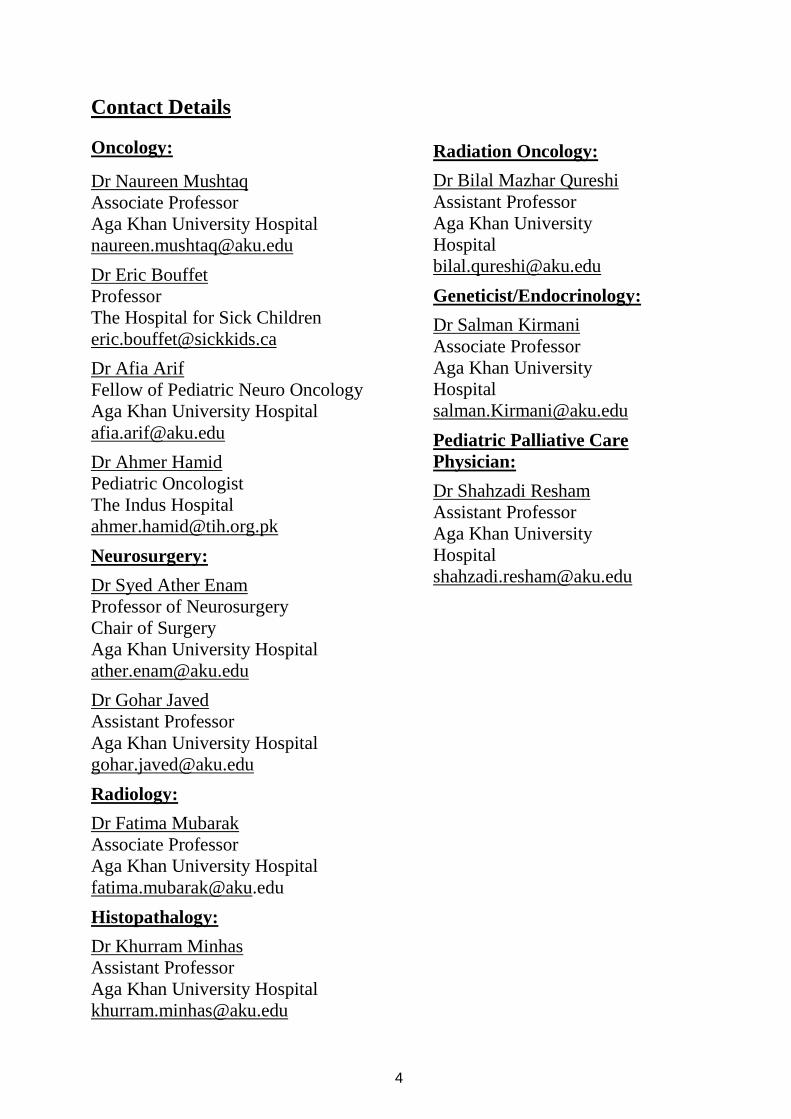
Contact Details
Oncology:
Dr Naureen Mushtaq
Associate Professor
Aga Khan University Hospital
Dr Eric Bouffet
Professor
The Hospital for Sick Children
Dr Afia Arif
Fellow of Pediatric Neuro Oncology
Aga Khan University Hospital
Dr Ahmer Hamid
Pediatric Oncologist
The Indus Hospital
Neurosurgery:
Dr Syed Ather Enam
Professor of Neurosurgery
Chair of Surgery
Aga Khan University Hospital
Dr Gohar Javed
Assistant Professor
Aga Khan University Hospital
Radiology:
Dr Fatima Mubarak
Associate Professor
Aga Khan University Hospital
Histopathalogy:
Dr Khurram Minhas
Assistant Professor
Aga Khan University Hospital
Radiation Oncology:
Dr Bilal Mazhar Qureshi
Assistant Professor
Aga Khan University
Hospital
Geneticist/Endocrinology:
Dr Salman Kirmani
Associate Professor
Aga Khan University
Hospital
Pediatric Palliative Care
Physician:
Dr Shahzadi Resham
Assistant Professor
Aga Khan University
Hospital
5
Table of Contents
Introduction: ............................................................................................................................... 8
Eligibility criteria: ........................................................................................................................ 8
Patient Criteria ......................................................................................................................... 8
Performance Level ................................................................................................................... 9
Prior Therapy ........................................................................................................................... 9
Patient Criteria for Standard Risk Patients .......................................................................... 9
Patient Criteria for High risk patients: ................................................................................ 10
Radiology Guidelines ................................................................................................................. 10
Prior to surgery ...................................................................................................................... 10
Imaging Technique ............................................................................................................. 10
Imaging interpretation ....................................................................................................... 10
2- During Surgery ............................................................................................................... 11
3-After Surgery and Prior to Radiotherapy ..................................................................... 11
Weekly During Radiotherapy ............................................................................................ 12
Four Weeks Post Radiotherapy and Prior to Chemotherapy ........................................ 12
Surgical Guidelines .................................................................................................................... 13
Medulloblastoma with hydrocephalus .................................................................................. 13
Surgery for Medulloblastoma ............................................................................................... 13
Post-Operative Care ............................................................................................................... 14
Neuropathology Guidelines ....................................................................................................... 15
Definition ................................................................................................................................. 15
Subgroups: .............................................................................................................................. 15
WNT subgroup: .................................................................................................................. 15
SHH subgroup: ................................................................................................................... 15
Group 3: ............................................................................................................................... 15
Group 4: ............................................................................................................................... 16
Radiation Therapy Guidelines; ................................................................................................. 17
1. Timing of Radiation Therapy: ....................................................................................... 18
2. Equipment: ...................................................................................................................... 18
a. Modality ....................................................................................................................... 18
b. Calibration ................................................................................................................... 18
c. Equipment .................................................................................................................... 18
3. 3-D Target volume and organ at risk definition ........................................................... 18
a. Gross Tumor Volume (GTV) ..................................................................................... 19
b. Clinical and Planning Target Volumes (CTV and PTV) ......................................... 19
c. Organs at Risk (OAR) ................................................................................................. 21
4. Dosimetry ......................................................................................................................... 21
6
a. Prescription Point ........................................................................................................... 21
b. Dose Definition ................................................................................................................ 22
c. Prescribed Dose and Fractionation STANDARD RISK PATIENTS ........................ 22
d. Dose Fractionation .......................................................................................................... 22
e. Dose Uniformity .............................................................................................................. 22
f. Treatment Interruptions ................................................................................................. 23
5. Treatment Technique ..................................................................................................... 23
Craniospinal Axis Irradiation............................................................................................ 23
6. Normal tissue sparing ..................................................................................................... 24
Spinal Cord .......................................................................................................................... 24
Optic Apparatus .................................................................................................................. 24
Vertebral Body .................................................................................................................... 25
7. Supportive Care During Irradiation ............................................................................. 25
Hematologic ......................................................................................................................... 25
Gastrointestinal ................................................................................................................... 25
Pneumocystis ....................................................................................................................... 25
Varicella ............................................................................................................................... 25
8. Dose calculation and reporting ...................................................................................... 25
Prescribed Dose ................................................................................................................... 25
Isodose Distributions .......................................................................................................... 26
Dose Volume Histograms ................................................................................................... 26
9. Quality Assurance and documentation .......................................................................... 26
Chemotherapy ........................................................................................................................... 28
a. Aerage Risk Medulloblastoma concurrent chemotherapy during radiation 28
b. Average Risk Medulloblastoma (Maintenance Chemotherapy) ........................... 28
c. High Risk Medulloblastoma (Maintenance Chemotherapy) ................................. 29
d. Dose Modification for Toxicities .................................................................................... 30
Vincristine Toxicity............................................................................................................. 30
Hematopoietic Toxicity....................................................................................................... 31
Nephrotoxicity ..................................................................................................................... 31
Ototoxicity ........................................................................................................................... 31
Hypomagnesemia ................................................................................................................ 32
Supportive Care Guidelines ...................................................................................................... 32
Supprotive Care Guidelines during Chemoradiotherapy. ................................................. 32
Venous Access: .................................................................................................................... 32
Antiemetics: ......................................................................................................................... 32
Filgrastim (G-CSF): ............................................................................................................ 32
7
Fever and Neutropenia:...................................................................................................... 32
Prophylactic Antibiotics: .................................................................................................... 32
Blood Products: ...................................................................................................................... 33
Nutritional Support: ............................................................................................................... 33
Endocrine Guidelines: ............................................................................................................ 33
Required Evaluations Following Completion of Protocol Therapy ...................................... 35
References ................................................................................................................................... 36
8
Guidelines for the management of Medulloblastoma (children
>3years of age)
Introduction: Medulloblastoma is the most common malignant brain tumour in children and is a major cause
of mortality and morbidity, particularly in low- and middle-income countries. Up to nw,
medulloblastoma has been risk-stratified on the basis of clinical (age, metastasis and extent of
resection) and histological subtypes (classic, desmoplastic and anaplastic). However, recently
medulloblastoma has been sub-grouped by using a variety of different genomic approaches, such
as gene expression profiling, micro-ribonucleic acid profiling and methylation array into 4
groups, namely Wingless, Sonic hedgehog, Group 3 and Group 4. This new sub-grouping has
important therapeutic and prognostic implications. After acute leukaemia, brain tumour is the
second most common malignancy in the paediatric age group. The improvement in outcome of
acute lymphoblastic leukaemia in low- and middle-income countries reflects the relative
simplicity of diagnostic procedures and management. Unlike leukaemia, the management of brain
tumours requires a complex multidisciplinary approach, including neuro-radiologists,
neurosurgeons with a paediatric expertise, neuropathologists, radiation oncologists and neuro-
oncologists. In addition, the equipment required for the diagnosis (magnetic resonance imaging
scan, histological, molecular and genetic techniques) and the management (operating room,
radiation facilities) is a limiting factor in countries with limited resources. In Pakistan, there are
very few centres able to treat children with brain tumours. The current literature review was
planned to provide an update on the management of this tumour.
Key Words: Childhood brain tumours, Medulloblastoma, WNT, SHH.
Eligibility criteria:
Patient Criteria
Age:
Patients must be greater than or equal to 3 years and less than 22 years at the time of diagnosis.
Diagnosis:
The presence of a posterior fossa medulloblastoma as determined by institutional pathologic
evaluation.
Preoperative and postoperative cranial MRI with and without contrast must be available. Pre
(better) or early postoperative MRI scan of the spine as well.
Assessment must include a pre-operative (within 5 days prior to surgery) or postoperative
enhanced MRI of the spine within 28 days after surgery.
Cytological examination of CSF performed after surgery but before the time of enrollment.
(minimum 14 days after tumour resection)
False positive cytology can occur within 10 days of surgery. Patients with positive CSF cytology
obtained before 10 days after surgery may have cytology repeated to determine eligibility.
9
Performance Level
Patients must have a Karnofsky performance level of ≥ 50 for patients > 16 years of age or a
Lansky performance scale of ≥ 30 for patients ≤ 16 years of age.
Prior Therapy
Patients must have no previous radiotherapy or chemotherapy other than corticosteroids.Organ
Function Requirements:
a- Adequate renal function defined as:
- Creatinine clearance or radioisotope GFR ≥ 70ml/min/1.73m2 OR
- A serum creatinine based on age/gender as follows:
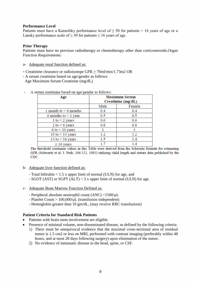
Age Maximum Serum Creatinine (mg/dL)
b- Adequate liver function defined as:
- Total bilirubin < 1.5 x upper limit of normal (ULN) for age, and
- SGOT (AST) or SGPT (ALT) < 3 x upper limit of normal (ULN) for age.
c- Adequate Bone Marrow Function Defined as:
- Peripheral absolute neutrophil count (ANC) >1500/μL
- Platelet Count > 100,000/μL (transfusion independent)
- Hemoglobin greater than 10 gm/dL, (may receive RBC transfusions)
Patient Criteria for Standard Risk Patients
Patients with brain stem involvement are eligible.
Presence of minimal volume, non-disseminated disease, as defined by the following criteria:
1) There must be unequivocal evidence that the maximal cross-sectional area of residual
tumor is 1.5 cm2 or less on MRI, performed with contrast imaging (preferably within 48
hours, and at most 28 days following surgery) upon elimination of the tumor.
2) No evidence of metastatic disease in the head, spine, or CSF.
10
Patient Criteria for High risk patients:
Newly diagnosed, previously untreated:
1) M0 Medulloblastoma with >1.5 cm2 residual;
2) M+ Medulloblastoma;
3) M0 or M+).
Patients with diffusely anaplastic medulloblastoma are eligible regardless of M-stage or residual
tumor.
Radiology Guidelines;
Prior to surgery (when possible)
1- MRI Brain and Spine
2- MRI brain and spine (with contrast) should be done in all patients suspected of having
medulloblastoma, who are referred to your center prior to surgery. In case surgery had
been done in an outside hospital, all pre-surgery scans should be obtained and reviewed
in your center.
Imaging Technique
All patients should underwent brain MR imaging atleast at 0.5T.
Following sequences should be obtained: axial and coronal T2 FSE (TR/TE, 2700/100 ms),
axial or Coronol FLAIR (TR/TE, 9000/120 ms; TI, 2200 ms), precontrast T1 spin-echo and
contrast-enhanced T1 spoiled gradient-recalled echo (TR/TE, 8/3 ms; 1-mm section thickness, 0
skip), followed by 2 planes of contrast-enhanced T1 spin-echo (TR/TE, 600–700/20 ms; 5-mm
section thickness, 0.5 skip).
All, patients should undergo DWI; b-value of 1000 s/mm2; 3 directions; 4-mm thickness, 0 skip)
SWI/GRE/T2* is optional.
Imaging interpretation:
All reports should comment on:
1.tumor location, 2.enhancement pattern,3. cysts/cavities, 4.hemorrhage/ mineralization,
5.intracranial or leptomeningeal seeding, 6.tumor margin, 7.necrosis as suggested by ring-
enhancement
“tumor location” should be defined as midline vermian/fourth ventricle, cerebellar hemisphere,
or cerebellar peduncle/cerebellopontine angle cistern (CP/CPA).
“Tumor margin” should be characterized as ill-defined if >50% of the margin could not be
distinguished from the surrounding cerebellar parenchyma on the basis of all imaging
sequences.
“Enhancement pattern” should be defined as minimal/none if <10% was estimated to enhance,
solid if >90% of the tumor volume was estimated to enhance, and heterogeneous if varying
degrees of enhancement were seen in 10%–90% of the tumor volume on the basis of
radiologist’s visual assessments.
11
Low signal on 2D gradient recalled-echo or bright on T1W should be used to detect
hemorrhage/mineralization.
Tumour size should be given in three dimensions and try best to give volume. Formula for
tumour volume is: Tumor volume= length x width2/2, where length represents the
largest tumor diameter and width represents the perpendicular tumor diameter.
Measurements should be taken on postcontrast or T2W/FLAIR.
Immediate postop scan should be performed in 24-48 hours and should be MRI Brain.
2- During Surgery
At surgery, every effort should be made to remove the tumor completely. If not possible, then
the neurosurgeon should describe, in detail, sites where tumor is believed to remain. Tumor
materials from the biopsy, or from materials collected by the surgical vacuum sucker in a sterile
trap should be submitted to Pathology for frozen section (if possible) and.histopathology.
3-After Surgery and Prior to Radiotherapy
MRI brain with contrast (MRI is preferred) and MRI spine with gadolinium if possible these
examinations should be performed within 72 hours (if not done before) , or between 18-21 days
post-op. This is believed to minimize the chances of post-op change being confused with residual
tumor. Lumbar CSF cytology examination must be obtained pre-operatively or within 31 days
following surgery. The optimal time for obtaining CSF is 2-3 weeks following surgery.
Ventricular CSF (either pre and post-op) may be used only if a post-operative spinal tap is
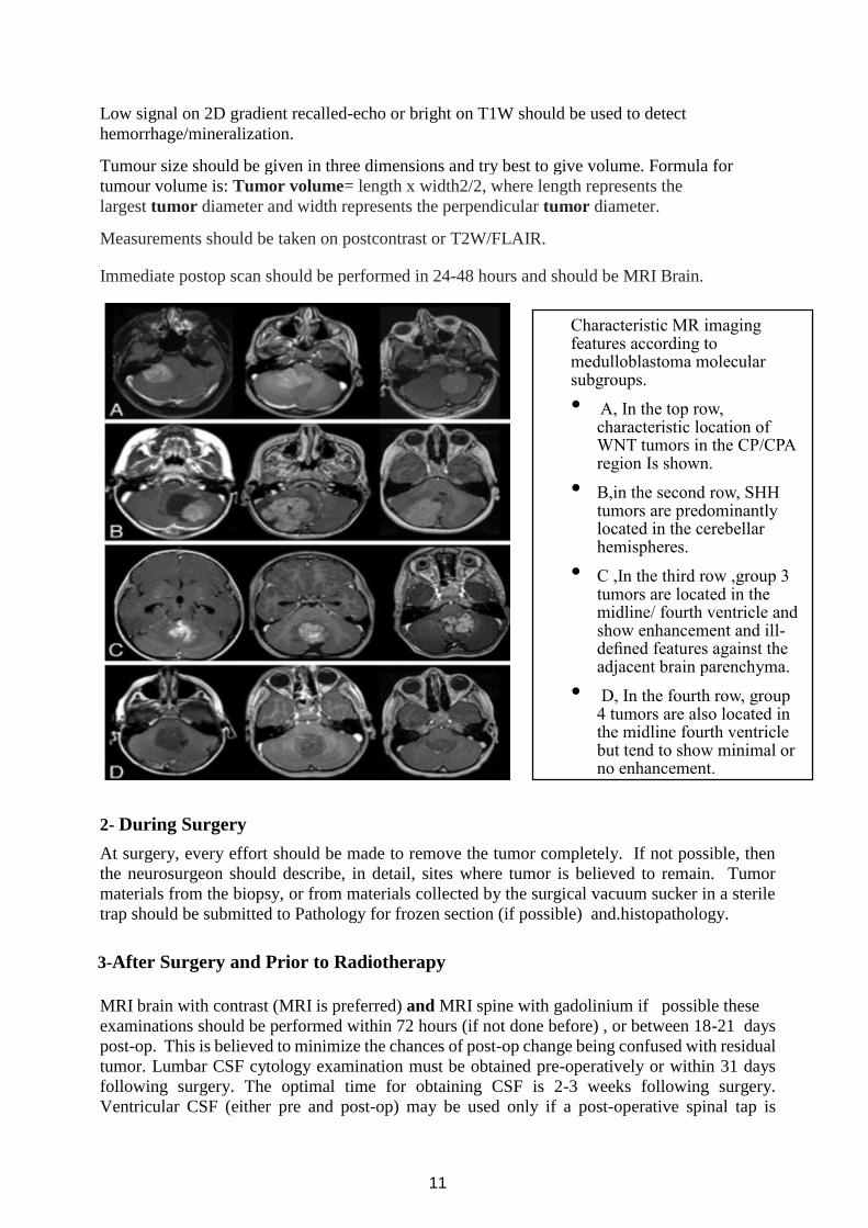
Characteristic MR imaging features according to medulloblastoma molecular subgroups.
• A, In the top row, characteristic location of WNT tumors in the CP/CPA region Is shown.
• B,in the second row, SHH tumors are predominantly located in the cerebellar hemispheres.
• C ,In the third row ,group 3 tumors are located in the midline/ fourth ventricle and show enhancement and ill-defined features against the adjacent brain parenchyma.
• D, In the fourth row, group 4 tumors are also located in the midline fourth ventricle but tend to show minimal or no enhancement.
12
contraindicated. CSF should be sampled post op and prior to starting radiotherapy, for cell count,
cytology, glucose and protein (if not already performed at the time of surgery).
1. CBC, differential and platelet count
2. Neurological examination
Weekly During Radiotherapy
ii. Neurological examination including monitoring for signs of vincristine toxicity.
iii. CBC, differential and platelet count (where bone marrow depletion occurs, these
must be carried out more frequently). It is essential that interruptions to
treatment are kept as infrequent, and for as short duration as possible.
Four Weeks Post Radiotherapy and Prior to Chemotherapy
iv. Neurological examination
v. CBC and differential count, SGOT, SGPT, bilirubin, creatinine and BUN, urine
analysis.
vi. MRI brain (CT of the brain may be used if this was the initial diagnostic tool;
consistency of diagnostic examinations during follow up is important).
vii. MRI spine with gadolinium, only if prior evidence of spinal mets, a positive CSF
or new symptoms suggestive of spinal mets.
viii. CSF should be sampled for cell count, cytology, glucose and protein if
previously positive.
ix. Audiogram before start of chemotherapy and after every other chemotherapy
cycle.
13
Surgical Guidelines:
Presentation of medulloblastoma in pediatric population may be due to local mass effect of the
lesion in the posterior fossa, but more commonly it presents with hydrocephalus and raised
intracranial pressure.
Medulloblastoma with hydrocephalus:
The hydrocephalus is most commonly due to blockage of the CSF pathway in the fourth ventricle
or at the cerebral aqueduct or the outflow foramen. The best strategy would be a definitive
procedure with resection of tumor and concomitant opening of the CSF pathway. CSF pathway
can be opened up even without gross total resection of the tumor in most of the cases.
A shunt such as ventriculoperitoneal shunt (VPS) should be avoided, if possible. If the patient
presents with acute hydrocephalus and logistics do not allow urgent surgery for the tumor, an
external ventriculostomy drain (EVD), preferably with long subcutaneous tunnel, can be
considered. Alternatively, Endoscopic Third Ventriculostomy (ETV) can also be considered. The
problem with VPS is that the child is then committed to a foreign object for the rest of its life. In
rare instances, it may lead to reverse herniation or seeding of the abdominal cavity with the tumor.
One unique problem seen in Pakistan (probably seen in other LMICs too) is the regression of the
parents into denial about the tumor. This happens because treating hydrocephalus leads to
remarkable improvement of symptoms. In these cases, the parents tend to ignore the primary
disease and fail to follow up. When patient does present back to the oncologist or the surgeon,
the tumor is grown tremendously or has seeded into CSF spaces. Therefore, putting VPS is not
a good strategy. In rare instances when adequate neurosurgical facility is not within reasonable
reach, or nutritional status of the child prohibits tumor surgery, VPS can be considered to avoid
any sudden herniation of the brain but then the neurosurgeon has the obligation to keep a close
eye on the patient and to ensure that the patient eventually gets definitive surgical treatment.
Wherever possible, ETV should be preferred over VPS.
Surgery for Medulloblastoma:
Pre-op screening of the spine for metastases is highly recommended. Pre-op counselling of the
patient’s parents with pediatric neuro-oncologist is one of the most important steps in preparation
of surgery.
The surgery should be performed by a neurosurgeon with experience and expertise in posterior
fossa surgery. The misconception that a pediatric neurosurgeon should do the surgery of this
tumor needs to be rectified. What is required, is a neurosurgeon with adequate experience in
brain tumor surgery and particularly in posterior fossa tumor surgery.
The surgery for tumor should be carried out in institutions that have a suitable team to handle this
case in the OR and during post-operative care. The team of anesthesiology should be experienced
in pediatric neurosurgical cases to manage volume loss intraoperatively and to use intravenous
fluid and blood transfusion prudently. A team of pediatricians should be available to co-manage
the patient during post-operative period.
For surgical technique, prone position with head and neck flexed (Concorde position) and a
midline incision works reasonably well in most of the cases. Sitting position can be considered
based on the experience and familiarity of the surgeon, the anesthesiologist and the OR team with
14
that position. If a surgeon is meticulous in her/his technique, there is no additional significant
benefit of sitting position, but there is definitely a higher risk of air embolism and other risks with
the sitting position. Use of Mayfield pins is advisable, and the torque and depth of the pins have
to be adjusted based on the age of the child. The surgeon can opt to avoid Mayfield pins and use
padded horseshoe, in children 3 years or below.
If the CSF diversion was not performed before tumor surgery, a decision to place an EVD
temporarily is reasonable. This can be done preferably through Frazier’s point in the occipital
region. A sample of 20-30 cc of CSF for cytology can be considered at this juncture.
Dissection of the muscle tissue has to be carried out in the midline raphe to avoid excessive blood
loss. Every attempt should be made to leave a cuff of muscles at the level of inion extending
laterally and avoiding exposing the skull to the point where the aponeurosis ends. This cuff of
muscle provides a good closure to prevent post-operative CSF leak or formation of
pseudomeningocele. Besides posterior fossa craniotomy, the surgeon should decide about the
removal of C-1 arch, depending on the extent of the disease and need of exposure for
visualization.
Every effort should be made to achieve gross total resection (GTR) of the tumor if possible, but
in many cases attachment of the tumor to the obex or the floor of the fourth ventricle may prevent
GTR, in these cases the strategy should be to attempt Maximum Safe Resection (MSR). It is best
to define the extent of the tumor initially, and to temporarily plug the opening of cerebral aqueduct
to prevent blood entering into rest of the ventricular system. For very large tumors, defining the
extent of the tumor may have to be delayed until significant tumor debulking has been achieved.
To minimize the chance of cerebellar mutism, it is best to avoid splitting the vermis and removal
of tumor from the roof of the fourth ventricle and the cerebellar peduncles is done with a lot of
caution and deliberation. With appropriate positioning of the cranium, the tumor can be excised
through the foramen of Magendie, which is usually enlarged by this time.
Closure of the dura is best done with the help of a patch obtained from the aponeurosis obtained
by sub-galeal dissection further cranial to the muscle cuff at the inion. Water-tight closure is the
primary objective at closure.
Post-Operative Care:
It is best if the patient is extubated in the OR post-operatively and is taken care of in a high
dependency unit for 24 to 48 hours. The EVD should be drained at 10-15 cm. It should be
pulled out after 48 hours if possible once it is ascertained that there is not much blood in the
CSF.
The management of the patient during the post-operative period should be done by the team of
neurosurgeon as well as the pediatrician. Pediatric neuro-oncologist, should be involved in the
care too.
An MR with contrast should be obtained within first 48 hours. If it is delayed beyond 72 hours
then it is best delayed for 3 weeks but should not be delayed more than 4 weeks.
15
Neuropathology Guidelines:
Medulloblastoma is the most common CNS embryonal tumor and the most common malignant
tumors of childhood. Medulloblastoma falls under CNS embryonal tumors and is classified
according to molecular characteristics in addition to histopathological features. Histopathological
classification has been retained, due to its clinical utility when mocular nalysis is limited or not
feasible.
Definition
These are embryonal tumors arising in cerebellum or dorsal brain stem, presenting mainly in
childhood and consisting of densely packed small round undifferentiated cells with mild to
moderate nuclear pleomorphism and a high mitotic count.
Subgroups:
With the advances in genomics, gene expression profiling, and DNA methylation analysis the
medulloblastomas have been divided into subgroups. The current integrated classification of
medulloblastoma takes into account histological subtype and molecular subgrouping. Following
are the subgroups with prognostic implications:
WNT subgroup:
The WNT subgroup represents 10% of all MBs. These are mostly located in the fourth
ventricle near the brainstem. Nearly all the medulloblastoma in this subgroup are of classic
type This subgroup has the most favourable outcome and these are rarely metastatic. WNT
MBs are characterized by activation of the WNT signaling pathway, often caused by
activating mutations in exon 3 of the CTNNB1 gene. They also show loss of chromosome 6.
SHH subgroup:
SHH MBs represent approximately 30% of all MB cases and are characterized by aberrant
activation of the SHH signaling pathway. Most SHH MBs are located in the cerebellar
hemispheres. Histology in 50% cases is demoplastic/nodular while most of the rest are classic
type. Common alterations in SHH MBs include germline or somatic mutations in components
of the SHH pathway, such as PATCHED1 (PTCH1) and SUPPRESSOR OF FUSED (SUFU).
Focal amplifications of MYCN and GLI2 are also reported. Mutations in the telomerase
reverse transcriptase (TERT) promoter are frequently found in adult SHH MBs.
Recent studies have identified heterogeneity within the SHH subgroup, further dividing it into
four subtypes. The SHH α subtype is enriched in TP53 mutations, as well as MYCN and GLI2
amplification, and is associated with an extremely poor prognosis. SHH β tumors affect
mainly infants and are often metastatic at the time of diagnosis, resulting in poor outcomes.
The SHH γ subtype is also found mainly in infants but has a relatively quiet genome and better
outcomes. Most MBs with extensive nodularity (MBENs) belong to the SHH γ subtype. The
SHHδ subtype is foundmainly in adults and frequently contains TERT promoter mutations.
Group 3:
Group 3 MBs account for approximately 25% of all MBs and are the most aggressive of the
four subgroups. They mostly have classic or large cell morphology. They are characterized
by transcriptional signatures resembling photoreceptors and gamma aminobutyric acid–
secreting (GABAergic) neurons. Group 3 tumors are often located in the fourth ventricle near
16
the brainstem, but nearly 50% of Group 3MB patients exhibit metastatic dissemination at
diagnosis.
The most common genetic alteration is amplification of the MYC oncogene, found in
approximately 20% of Group 3 MB patients. Group 3 tumors often have unstable genomes,
with multiple chromosomal gains and losses. Among these, one of themost common is
coordinate loss of chromosome 17p and gain of chromosome 17q—called isochromosome
17q (i17q). i17q is found in 40% of Group 3 MB patients and is associated with poor
outcomes.
Recent integrative analysis has suggested that there may be three distinct subtypes within
Group 3 MB. Group 3α tumors are often found in infants and frequently exhibit metastasis at
diagnosis. Group 3β has a high frequency of GFI1 family oncogene activation and
orthodenticle homeobox 2 (OTX2) amplification.Group 3γ is also associated with a high
incidence of metastasis and often exhibitsMYC amplification; it has theworst prognosis
among the three Group 3 subtypes.
Group 4:
Group 4 is the most common subgroup, accounting for approximately 35% of all MBs. These
are mostly classic on histology. Group 4 tumors are frequently metastatic at diagnosis and
have intermediate outcomes. Similar to Group 3, Group 4 MB also shows intertumoral
heterogeneity. Groups 4α and 4γ have focal CDK6 amplification, chromosome 8p loss, and
chromosome 7q gain; however, Group 4α also exhibits MYCN amplification, whereas Group
4γ does not. Group 4β is enriched in SNCAIP duplication and PRDM6 overexpression.
Molecular profiling studies are the gold standard for accurate characterization of
medulloblastoma subgroups. However these are not available in all regions. Fortunately,
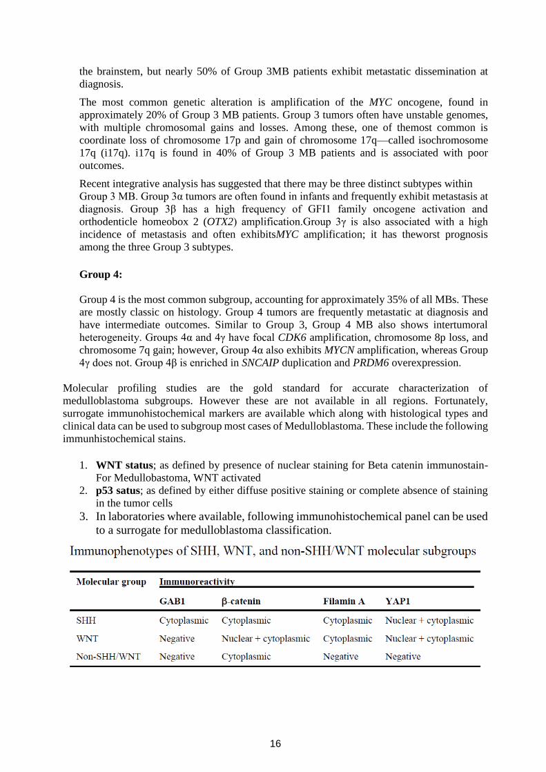
surrogate immunohistochemical markers are available which along with histological types and
clinical data can be used to subgroup most cases of Medulloblastoma. These include the following
immunhistochemical stains.
1. WNT status; as defined by presence of nuclear staining for Beta catenin immunostain-
For Medullobastoma, WNT activated
2. p53 satus; as defined by either diffuse positive staining or complete absence of staining
in the tumor cells
3. In laboratories where available, following immunohistochemical panel can be used
to a surrogate for medulloblastoma classification.
17
The Non-SHH/WNT group can only be separated into group 3 or group 4 based on moclecular
studies only.
4. Medulloblastoma, NOS is appropriate when an embryonal neural tumors is located in the
fourth ventricle or cerebellum and the nature of biopsied tissue prevents classification of
the tumor.
Radiation Therapy Guidelines;
Radiation therapy for medulloblastoma consists of craniospinal axis irradiation (CSI) followed
by boost to the primary site. Craniospinal irradiation itself is complex due to multiple fields to
irradiate the cranium and spine with matching of these fields to cover the clinical target volume
and at the same time avoid overdose to the spine. Three-dimensional image-based radiation
therapy treatment planning and computer-controlled delivery systems (conformal radiation
therapy) improves disease control and functional outcome for children with brain tumors. The
availability of tools necessary to perform conformal radiation therapy with sufficient experience
of the centre who treat these patients is required. The allowed treatment methods are restricted to
conformal or intensity-modulate radiation therapy using photons.
For treatment planning and delivery, the treating radiotherapy center should have
Conformal radiation therapy capabilities with adequate hardware and application system
Quality assurance system which includes peer review of all treatment plan with a site specific
team
General anesthesia and/or deep-sedation capabilities by specialized anesthesia team as
needed,
Customized immobilization that provides for treatment that is both reproducible and safe.
Required Benchmark
Radiation therapy shall be administered using photons. Required photon methods include 3D
conformal radiation therapy (3D-CRT), intensity modulated radiation therapy (IMRT) and
craniospinal radiation therapy. Centers participating in this protocol using 3D-CRT are required
to complete the 3D benchmark; those using IMRT must complete the IMRT standards and
benchmark on phantom. The QARC craniospinal radiation therapy benchmark form can be used
in the absence of national quality standards. Radiation therapy center shall complete the QARC
CT/MR image fusion benchmark. Benchmark materials and questionnaires may be obtained from
the Quality Assurance Review Center (www.qarc.org) For information regarding the IMRT
phantoms, resources like RPC (http://rpc.mdanderson.org/rpc) can be used.
Guidelines and Requirements for the Use of IMRT:
Radiation oncologists are required to comply with the ICRU reports (ICRU report 50,63 & 83)
guidelines. There is need of a national quality assurance system for recording of treatment setup
and accessories, planning and diagnostic imaging used, dose to the targets and normal tissue,
dosimetry etc. In our scenario, for the use of IMRT, National Cancer Institute sponsored
cooperative group trials guidelines are available through www.qarc.org. These guidelines
require that the protocol explicitly state their requirements and methods for localization and
immobilization; the use of volumetric imaging; target and organ motion management;
nomenclature, definitions and rationale for targets and organs at risk; target volume coverage
and normal tissue dose constraints; effects of heterogeneity in tissues; and quality assurance.
18
1. Timing of Radiation Therapy:
All patients shall receive irradiation to the craniospinal axis followed by a boost to the posterior
fossa. Patients shall begin radiation treatments within four weeks of definitive surgery and shall
not be delayed beyond 7 weeks. Delay in start of radiotherapy has shown inferior outcome.
Patients who start radiotherapy beyond 7 weeks of surgery are considered high risk, requiring
higher dose of craniospinal irradiation of 36 Gy.
2. Equipment:
Radiation therapy shall be delivered using photons on Linear Accelerator. CT based planning
shall be done on all patients with adequate immobilization devices. If IMRT or VMAT is used,
photon energy should be no greater than 10 MV.
a. Modality
X-rays with a nominal energy 4 MV. Craniospinal axis irradiation is best done with x-rays
between 4-6 MV. The boost volume may be treated with a nominal energy 4 MV, as long as
dosimetric Constraints are accomplished.
b. Calibration
The calibration of therapy machines used shall be calibrated and verified by the pertinent
authority.
c. Equipment
Patients treated with this study must be treated using conformal radiation therapy treatment
planning and delivery techniques at a minimum. IMRT or VMAT shall be used if appropriate
quality benchmarks have been attained and maintained as per international standards (QARC,
AAPC etc). All patients must be treated on isocentric machines. For treatment to be conformal as
per protocol, the following criteria must be met:
Three-dimensional imaging data (CT or MR) are acquired with the patient in the treatment
position. Three-dimensional treatment planning software must be used for planning.
Double checks of treatment plan shall be done on standardized reporting form e.g. QARC
[QARC website (www.QARC.org)]
Image data are used to delineate and reconstruct a gross target volume, clinical target volume,
planning target volume, and normal or critical structures in 3-dimensions.
All contours and treatment planning volumes shall be peer reviewed by a second radiation
oncologist.
Radiation beams can be freely oriented in 3-dimensions for both the planning and delivery
process, and structures traversed by the beam can be visualized with the eye of the beam
(beam’s eye view – BEV)
The distribution of dose relative to the target volume or any structure is computable on a
point-by-point basis in 3-dimensional space.
Institutions not equipped to perform conformal radiation therapy according to these guidelines
should refer the patient to a center with proven capabilities to comply with the outlined
parameters.
3. 3-D Target volume and organ at risk definition
The definitions for the target volumes and treatment dosimetry will adhere as closely as possible
to the ICRU Reports 62 and 83. RT volumes for treatment shall be determined by the collective
information that delineates the extent of disease at the time of diagnosis and prior to radiation
therapy. RT Planning volumes and organ at risk contours shall be peer reviewed with paediatric
radiotherapy team of doctors.
19
These guidelines are meant to be comprehensive and include commonly anticipated treatment
scenarios. If for any reason the guidelines do not match the characteristics of a given patient, the
treating physicians shall contact colleague from other specialized centers having experience of
pediatric CNS radiotherapy.
a. Gross Tumor Volume (GTV)
The GTV includes all gross residual tumor and/or the walls of the resection cavity at the primary
site based on the initial imaging examination that defines the tissues initially involved with
disease anatomically and the post-operative and pre-irradiation neuroimaging examinations that
identify residual disease and the tumor bed.
In accordance with ICRU report, the Gross Tumor Volume (GTV) is defined as the contrast-
enhanced tumor in the Brain and the Spine, unless the preoperative tumor is predominantly non-
enhancing, in which case this represents the preoperative tumor extent as defined by the most
informative MR imaging sequence.
b. Clinical and Planning Target Volumes (CTV and PTV)
The CTV includes the GTV with an added margin that is meant to treat subclinical microscopic
disease and is anatomically confined (i.e., the CTV is limited to the confines of the bony
calvarium and tentorium where applicable). There are multiple CTVs to be treated.
The Clinical Target Volume1 (CTV1) is the entire craniospinal axis. The craniospinal axis
Planning Target Volume (PTV1) is institution-defined according to immobilization techniques
and their inherent setup uncertainties. The margin defining the PTV may range from 0.5 cm to
1.0 cm. For the Craniospinal volume, a larger PTV1 margin should be considered due to
inexactness of repositioning for the spinal fields.
CT based or 3D treatment planning for craniospinal axis irradiation offers advantages over
conventional simulation methods. A better appreciation of the cribriform plate and middle cranial
fossa can be gained with a CT simulation. In most circumstances, the same CT simulation data
used for planning the craniospinal axis RT can be used to plan the 3-D conformal boost.
Whole Brain (CTV1): The whole-brain field shall extend anteriorly to include the entire
frontal lobe and cribriform plate region. The volume shall cover the superior orbital tissue (but
not the posterior globe as in leukemia protocols). Inferiorly, the CTV1 shall be at least 0.5 cm
below the base of the skull at the foramen magnum. PTV1 should be defined to account for
setup error. The radiation fields to cover this target volume should follow established guidelines
for cranial irradiation in medulloblastoma. There will be a junction with the spinal field.
For standard risk patients, the dose prescription for the brain and spine (CSI) will be 23.40 Gy in
13 daily fractions of 1.80 Gy. However, for high risk patients, the dose prescription for CSI will
be 36.0 Gy in 20 fractions of 1.80 Gy.
Spine (CTV1): The spinal target volume will be the entire thecal sac. The field to cover this
volume should extend laterally on both sides to cover the recesses of the entire vertebral bodies,
with at least a 1 cm margin on either side. The superior border will be the junction with the
whole brain field. The inferior border of the treatment volume will be placed after review of the
spinal MRI. The border will be 2 cm below the termination of the subdural space. This will
extend at least to the inferior border of the second sacral segment (S2-S3 interspace), but may
be as low as the inferior border of S4. If this cannot be accomplished in a single field, there will
be a junction between the two spinal fields. PTV1 should be defined to account for setup error
as per institution.

20
For standard risk patients, the dose prescription for the brain and spine (CSI) will be 23.40 Gy in
13 daily fractions of 1.80 Gy. However, for high risk patients, the dose prescription for CSI will
be 36.0 Gy in 20 fractions of 1.80 Gy.
Supratentorial Boost (CTVST): This volume refers to children with supratentorial primaries
(ST-PNET). The CTVST for a supratentorial boost will be defined using a 1 cm margin around
the presurgical MRI defined tumor volume. In the event of proximity to normal tissue
structures, this margin may be reduced to 0.5 cm to allow for sparing of critical structures. As
the total dose to the optic chiasm and both optic nerves should not exceed 50.4 Gy, these
structures should be excluded accordingly. The Planning Target Volume (PTVST) is a 0.3cm to
0.5cm margin around the CTVST to account for day-to-day setup variation.
Limited Target Volume Boost (CTVboost) [for standard risk patients only: 3D-based
treatment planning is mandatory for this volume. IMRT or VMAT is allowed for the PTVboost
The Gross Target Volume (GTV) is based upon the T1 signal changes with and without
Gadolinium contrast. Identification of the GTV shall be based upon pre-operative extent and
anatomic shifts or changes after surgery. The GTV should include any residual enhancing tumor
mass and the wall of the resection cavity. The Clinical Target Volume (CTVboost) is defined as
the GTV plus a 1.5-cm margin except at bone or tentorial interface (where it remains within the
confines of the posterior fossa). The PTVboost margin should be an additional 0.3 to 0.5 cm
around the CTVboost. In treatment planning, shielding of critical structures should be
attempted, however, coverage of the PTVboost must not be less than 50 Gy. The cumulative
dose to PTVboost will be 54.0 Gy. At least 95% of the prescribed dose (54Gy) must encompass
at least 95% of the PTVboost as shown by DVH. No part of the PTVboost should receive less
than 50 Gy.
Posterior Fossa Boost (CTVPF): 3D-based treatment planning is mandatory for these
volumes. IMRT is allowed for the PTVPF. The CTVpf should encompass the entire posterior
fossa. The posterior fossa must be defined on the planning CT scan. It is strongly recommended
that a sagittal MRI be used to assist in identification of the position of the tentorium. The CTVPF
extends inferiorly from C1 vertebral canal through the foramen magnum, laterally to the bony
walls of the occiput and temporal bones and superiorly to the tentorium cerebelli. Generally, the
sigmoid sinuses define the lateral-superior extent of the bony confines of the posterior fossa that
attach contiguously to the tentorium above. The folia of the cerebellum and the anterior border
of the brainstem and midbrain bound the CNS contents of the posterior fossa. The Planning
Target Volume (PTVPF) is a 0.3cm to 0.5cm (or more depending upon institutional setup error)
margin around the CTV to account for day-to-day setup variation. The PTVPF should not extend
beyond the external bony confines of the skull except at the foramen magnum to C1-C2. The
PTVPF should extend anteriorly to the posterior clinoids (the pituitary is not included) and
inferiorly to the C1-C2 junction. In treatment planning, shielding of critical structures should be
attempted. At least 95% of the prescription study dose of 55.8 Gy must encompass at least 95%
of the PTVPF (the posterior fossa) as shown by DVH. No part of the PTVPF should receive less
than 50 Gy. Treatment techniques for the PTVPF may include parallel opposed laterals or other
3D CRT methods to limit dose to the supratentorial brain, hypothalamus, pituitary or middle
ear.
Metastasis Site Boost (CTVM): Patients with M1 disease will receive no additional boost.
Patients with M2 disease (intracranial subarachnoid disease) will receive boosts to areas of
supratentorial or posterior fossa metastatic disease.
21
Patients with M3 disease (spinal deposits of disease) are subdivided into those with diffuse
disease and those with focal disease.
Diffuse spinal disease is defined as radiographically visible multiple sites of disease in each of at
least 3 out of 4 spinal regions (i.e., cervical, thoracic, lumbar or sacral). If there is diffuse
involvement of the spine, the entire spine will be treated in the boost volume.
The CTVm margin for boosting metastatic deposits will be 0.5 to 1.0 cm encompassing the lesion
within the anatomic compartment. Another 0.3 to 0.5 cm margin will be added for the PTVM.
Field shaping. may be conformal and consideration may be given for normal organ sparing and
abutment of other high dose or boost sites.
c. Organs at Risk (OAR)
The following organs must be defined for 3-D conformal radiation therapy or IMRT planning:
Supratentorial brain (left and right)
Cochlea (left and right)
Hypothalamus/pituitary
Lacrimal apparatus
Eyes (left and right) including lens
Optic nerves (left and right)
Optic chiasm
Cervical spinal cord (foramen magnum to C2)
Skin (non-specified tissues)
Thyroid gland
Heart
Bilateral Lungs
Bilateral kidneys
Both ovaries in females
Vertebral bodies
Details contouring guidelines have be published and illustrated as following:
1. Clinical Oncology Group (COG) atlas for contouring of CSI
http://www.qarc.org/cog/protocol%20resources/ACNS0331Atlas.pdf
2. Brain Tumor Study Group of Siop Europe (SIOP-E) guideline on craniospinal target
volume delineation for high-precision radiotherapy
https://www.thegreenjournal.com/article/S0167-8140(18)30204-4/fulltext
3. Management of vertebral radiotherapy dose in paediatric patients with cancer: consensus
recommendations from the SIOPE radiotherapy working group
https://www.thelancet.com/journals/lanonc/article/PIIS1470-2045(19)30034-8/fulltext
4. Dosimetry
a. Prescription Point
The prescription point for the neuroaxis (PTV1, whole brain and spine) is at or near the center of
the targets. For the brain this may be a point other than the central axis. Consideration should be
made to using a point midway of the biparietal diameter. The spinal axis dose should be prescribed
to the anterior aspect of the spinal canal. In many cases, the depth of the anterior spinal canal will
22
vary by vertebral level. An average depth may be used that keeps the dose uniformity within the
constraints defined below. For the posterior fossa and limited target boost volumes, the doses
shall be prescribed in order to have at least 95% of the 54 Gy isodose covers at least 95% of the
respective planning target volumes. The minimum dose to the PTVPF or PTVboost shall not be
< 50 Gy.
b. Dose Definition
Dose is to be specified in Gray (Gy)-to-muscle.
c. Prescribed Dose and Fractionation STANDARD RISK PATIENTS
23.40 Gy Craniospinal radiotherapy (CSRT) (PTV1) followed by:
30.60 Gy Local boost (PTVboost)
Cumulative Dose: 54Gy
High Risk Patients:
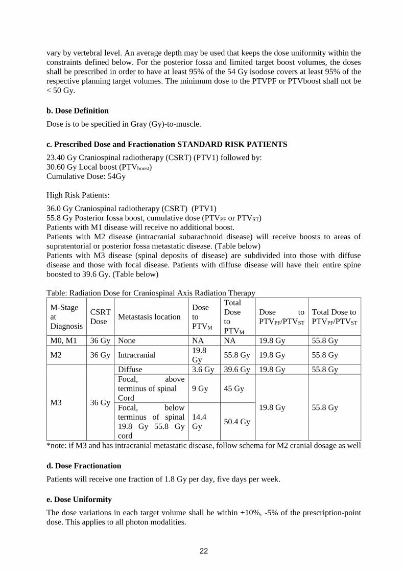
36.0 Gy Craniospinal radiotherapy (CSRT) (PTV1)
55.8 Gy Posterior fossa boost, cumulative dose (PTVPF or PTVST)
Patients with M1 disease will receive no additional boost.
Patients with M2 disease (intracranial subarachnoid disease) will receive boosts to areas of
supratentorial or posterior fossa metastatic disease. (Table below)
Patients with M3 disease (spinal deposits of disease) are subdivided into those with diffuse
disease and those with focal disease. Patients with diffuse disease will have their entire spine
boosted to 39.6 Gy. (Table below)
Table: Radiation Dose for Craniospinal Axis Radiation Therapy
M-Stage
at
Diagnosis
CSRT
Dose Metastasis location
Dose
to
PTVM
Total
Dose
to
PTVM
Dose to
PTVPF/PTVST
Total Dose to
PTVPF/PTVST
M0, M1 36 Gy None NA NA 19.8 Gy 55.8 Gy
M2 36 Gy Intracranial 19.8
Gy 55.8 Gy 19.8 Gy 55.8 Gy
M3 36 Gy
Diffuse 3.6 Gy 39.6 Gy 19.8 Gy 55.8 Gy
Focal, above
terminus of spinal
Cord
9 Gy 45 Gy
19.8 Gy 55.8 Gy Focal, below
terminus of spinal
19.8 Gy 55.8 Gy
cord
14.4
Gy 50.4 Gy
*note: if M3 and has intracranial metastatic disease, follow schema for M2 cranial dosage as well
d. Dose Fractionation
Patients will receive one fraction of 1.8 Gy per day, five days per week.
e. Dose Uniformity
The dose variations in each target volume shall be within +10%, -5% of the prescription-point
dose. This applies to all photon modalities.
23
At least 95% of the PTVboost should be encompassed within 95% of the 54 /55.8 Gy isodose
surface and no more than 5% of the volume within this isodose surface should receive greater
than 110% of the prescription dose as evaluated by DVH. These targets should not receive less
than 50 Gy. Treatment should be planned to spare the spinal cord, brainstem, optic chiasm and
optic nerves from the highest doses resulting from dose inhomogeneity. An effort should be made
to spare the cochlea and middle ear contents.
f. Treatment Interruptions
Treatment will not be interrupted for anemia, leukopenia, or thrombocytopenia unless life
threatening. Blood product support should be instituted according to institutional/protocol
guidelines. For interruptions of more that 2 treatment days it should be discussed with pediatric
oncologist.
5. Treatment Technique
Craniospinal Axis Irradiation
Patient Position
For cranio-spinal irradiation the patient may be treated prone or supine. Supine position is feasible
for patients being treated with general anesthesia. The neck should be extended sufficiently to
keep the mandible out of the exit beam of the spinal field but not so much as to exceed the dose
uniformity specifications of the spinal field. Reproducible setups are critical. Immobilization
devices such as head holders or custom molds are highly recommended. Deep sedation or general
anesthesia is strongly encouraged for young children. For the posterior fossa boost the patient
may be in either the prone or supine position. It is recommended to have a uniform setup through
all phases of treatment
Whole Brain Irradiation
Conformal treatment planning is recommended. Regardless, dose from this component of the
radiation therapy shall be included in the 3D CRT / IMRT plan of the limited boost volume,
including DVHs of the targets and adjacent organs at risk.
Parallel opposed fields may be used. Alternatively, the field center can be placed near the match
line with the spinal field and an independent jaw or half-beam block technique utilized. This
method decreases overlap at the match line. The collimation of the brain field should be rotated
to match the divergence of the spinal field.
If symmetric collimator jaws are used:
The angle = tan-1 spine length/2
SAD
If asymmetric collimator jaws are used:
The angle = tan-1 upperspine length
SAD
The lateral fields may be angled posteriorly to spare the collateral lens, but, if this is done, great
care must be taken to assure adequate coverage of the cribriform plate. Custom divergent blocking
of at least 5 HVL should be used to shape the brain field at the base of the skull and around the
eyes. The brain field should extend to at least 1 cm beyond the periphery of the scalp.
24
Spine Irradiation
Preferably, the spinal volume should be treated with a single posterior field. An extended SSD is
preferable to the use of adjacent ports. If adjacent ports are necessary, the 50% decrement should
cross at the posterior margins of the vertebral body. It is preferable that the match line be placed
inferior to the spinal cord (below L2) and should be moved every 900 cGy. Custom blocking may
be required at the inferior border of the spine.
Abutting Fields
With the use of collimator rotation and an independent jaw technique, the cranial and spinal fields
may be directly abutted (light fields). Many radiation oncologists, though, are more comfortable
with a gap between the cranial and spinal light fields. A gap of 0.5 cm is allowed on this protocol.
The match line should be moved at least twice during treatment of the cranio-spinal axis (e.g.
after each 9Gy). Also, a penumbra broadening “match line wedge” or a dynamic wedge may be
used. The match line should never overlap the posterior fossa boost. Therefore, it is recommended
that the first match line lie just above the shoulder, and the last 2 cm higher. Alternatively, the
first match point could begin at the superior point and end at the inferior point.
Conformal Boost Treatment
Conformal (three-dimensional) planning is required for all phases of treatment. Beam
arrangements and treatment techniques should be used that minimize the dose to the auditory
apparatus (cochlea), hypothalamicpituitary unit and supratentorial brain providing that they do
not compromise treatment of the intended PTV. Examples of 3-D conformal beam arrangements
can be found at the QARC (www.qarc.org) or ITC (itc.wustl.edu) website.
Field Shaping
Field-shaping is required. Shielding shall be at least 5 HVL thick. Multi-leaf collimation may be
used.
Example Cases
http://www.qarc.org/cog/protocol%20resources/ACNS0331Atlas.pdf
Imaging
CT (3 mm - 5 mm section thickness from the thoracic inlet to the base of the skull, 3 mm for the
entire skull) should be performed for treatment planning. Preoperative and postoperative MR is
used primarily (co-registered with CT planning data) or adjunctively in the treatment planning
process. Surgical guidelines encourage postoperative imaging within 72 hours post-operatively.
6. Normal tissue sparing
Normal tissue dose recommendations are the same for photons
Spinal Cord
No more than 50% of the cervical spinal cord between C-1 and C-2 should receive more than 54
Gy. DVH of this volume shall be submitted.
Tolerance of rest of the spinal cord is to be followed, i.e. <45Gy.
Optic Apparatus
Dose to the bilateral optic nerves and optic chiasm shall be <54Gy
25
Vertebral Body
Dose gradient over the vertebral body shall be avoided. SIOPE guidelines in this regard is helpful
and shall be followed:
https://www.thelancet.com/journals/lanonc/article/PIIS1470-2045(19)30034-8/fulltext
7. Supportive Care During Irradiation
Hematologic
CBC’s should be obtained weekly. If ANC ≤ 500/l, either a break in treatment or the use of
growth factors may be considered. The option of continuing therapy with the posterior fossa boost
field may alsobe considered in patients who have lowered counts during the cranio-spinal therapy.
Cranio-spinal radiation may be resumed when the ANC has risen over 10% on 2 consecutive
tests, or is above 750/l. If ANC is less than 1000/l and the patient is febrile (≥ 38º C), a break
may be instituted while the patient is being evaluated. During this time, the posterior fossa boost
may be treated, but cranio-spinal irradiation should be avoided.
Platelets should be transfused as clinically indicated when counts are < 30,000/l. Irradiated and
Pall filtered blood products should be used. Cranio-spinal irradiation should be reinstituted when
platelet counts exceeds 50,000/l or have risen > 10% on 2 consecutive tests.
Hemoglobin: Transfusions are recommended when hemoglobin falls below 9 gm/dL. Radiated
blood
products should be used. Growth factors can also be utilized.
Gastrointestinal
Patients should be weighed weekly. If there is greater than 10% weight loss, aggressive nutritional
support, either enteral or parenteral, should be given. Prophylactic anti-emetic therapy, with e.g.
a selective 5-HT3 receptor antagonist, should be considered.
Pneumocystis
Pneumocystis prophylaxis with trimethoprim/sulfamethoxazole (5 mg/kg/d of trimethoprim in
two divided doses given 2 consecutive days per week) is recommended for all patients during
radiotherapy and should be continued for 2 months after completion of radiotherapy.
Varicella
If patients are seronegative for varicella, VZIG should be administered within 72 hours if history
of exposure to chickenpox or zoster is elicited.
8. Dose calculation and reporting
Quality assurance measures, plan parameter and dosimetry record shall be maintained as per
international standards both for three dimensional conformal and IMRT / VMAT planning.
The sample Benchmark material can be obtained from the Quality Assurance Review Center
(www.QARC.org ) Contact the RPC (http://rpc.mdanderson.org/rpc), AAPM or IAEA resources.
Prescribed Dose
For standard or 3D conformal techniques: The monitor units required to deliver the prescribed
dose shall be calculated and submitted using the standard documentation, like “RT-1 Dosimetry
Summary” form. A separate form shall be filled and recorded for each of the planning target
volumes.
26
For IMRT techniques: The monitor units required to deliver the prescribed dose shall be
calculated and submitted using the IMRT Dosimetry Summary Form. The monitor units
generated by the IMRT
planning system must be independently checked prior to the patient’s first treatment.
Measurements in a QA phantom can suffice for a check as long as the plan’s fluence distributions
can be recomputed for a phantom geometry.
Isodose Distributions
Color hard copies of the isodose distributions must be maintained for each of the treatment sites
including the craniospinal axis and primary site boost at the start of radiation therapy. A
composite isodose distribution must also be saved. The isodose distributions will display the
actual dose to be delivered for a particular phase of treatment (e.g., CSA isodose distributions
will show 23.4 Gy, primary site boost isodose distributions will show 30.6 Gy). The cumulative
dose (54.0 or 55.8 Gy) will be displayed on the composite isodose distribution. It is understood
that some patients may be treated in 2 consecutive phases and have the CSA radiation therapy
delivered in a position that is different from the boost radiation therapy (not recommended) .
While it is sometimes not possible to accurately add radiation doses due to the change in patient
shape and position, the institution must submit a plan that closely approximates the cumulative
radiation dose that is delivered as part of both the cranial fields from the CSA and the boost
treatments. This can generally be done by creating a cranial field in the boost plan that is dose-
weighted and shaped similarly to that used in the CSA phase of therapy.
For conformal planning, the following isodose distributions are required by treatment site and for
the composite and will include the gross tumor volume, clinical target volume and planning target
volume and normal tissue structures listed in section earlier. Axial, sagittal, and coronal isodose
distributions through the treatment isocenter will be overlayed on reconstruction of the CT scan
in the same plane.
Isodose distributions corresponding to each CT image that shows protocol-specified normal tissue
structures or tumor/target volume contours.
The following isodose curves should be shown to determine that the dose distributions conform
to the protocol guidelines: dose-maximum, 110%, 105%, 100%, 95%, 90%, 70%, 50%, 30%,
10%.
Dose Volume Histograms
Dose volume histograms shall be calculated and submitted for organs at risk (section 18.3.3)
including right and left optic nerves, optic chiasm, right and left cochlea, pituitary/hypothalamus,
vertebral bodies, heart, thyroid, kidneys, supratentorial brain, and spinal cord (foramen magnum
to top of C2). Dose volume histograms will be calculated and submitted for the GTV, CTV and
PTV of each volume treated. Dose volume histograms will be calculated and submitted in
composite form whenever technically possible.
9. Quality Assurance and documentation
For quality assurance purpose, the radiation therapy treatment plan should be available in digital
format (either Dicom RT, RTOG format or other standard format) if possible. One can see the
QARC website (www.QARC.org) for digital data submission information.
It is encouraged to compile the following documents:
Copies of all diagnostic materials and surgical reports used in defining the target volume
including (i) preoperative MRI and postoperative cranial MRI with and without contrast; (ii) pre
or postoperative spinal MRI with and without contrast; sagittal imaging should be included for
both brain and spine; (iii) operative reports.
27
Copies of isodose distributions to demonstrate that the dose variation is within specification.
The target volume, and the prescription point must be clearly shown
Prescription Sheet for Entire Treatment
Copies of simulator films and /or digitally reconstructed radiographs (DRR’s) for each field.
Copies of verification (portal) films (or hard copy of real time portal images) for each field.
Photographs of the patient in the treatment position with the fields marked.
Beam’s Eye Views (BEV’s) for all fields and showing the PTV (boost) and critical structures.
BEV hard copies must be in color to enable reviewers to identify structures.
A room view display of all fields should be submitted.
Dose volume histograms as specified in section 18.8.4. If IMRT is used, a DVH shall also be
submitted for a category of tissue called “unspecified tissue,” which is defined as tissue
contained within the skin, but which is not otherwise identified by containment within any
other structure.
Documentation of an independent check of the calculated dose if IMRT is used.
Color copies of isodose distributions to demonstrate that the dose variation is within
specification.
The target volume and the prescription point must be clearly shown.
Documents verifying peer review and double checks from radiation oncologist, medical
physicist and RTTs for all the phases of treatment for each patient.
RT-1 Dosimetry Summary Form
RT-2 Radiotherapy Total Dose Record form.
28
Chemotherapy
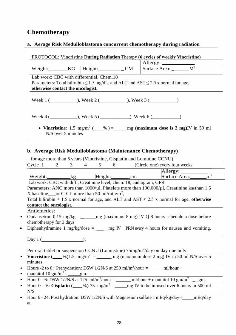
a. Aerage Risk Medulloblastoma concurrent chemotherapy during radiation
PROTOCOL: Vincristine During Radiation Therapy (6 cycles of weekly Vincristine)
Allergy:
Weight: KG Height: _ CM Surface Area: _ M2
Lab work: CBC with differential, Chem.18
Parameters: Total bilirubin ≤ 1.5 mg/dL, and ALT and AST ≤ 2.5 x normal for age,
otherwise contact the oncologist.
Week 1 (___________), Week 2 (___________), Week 3 (___________)
Week 4 (___________), Week 5 (____________), Week 6 (___________)
Vincristine: 1.5 mg/m2 ( % ) = mg (maximum dose is 2 mg)IV in 50 ml
N/S over 5 minutes
b. Average Risk Medulloblastoma (Maintenance Chemotherapy)
– for age more than 5 years (Vincristine, Cisplatin and Lomutine CCNU) Cycle 1 2 3 4 5 6 (Circle one) every four weeks
Allergy:
Weight: kg Height: _cm Surface Area: m2
Lab work: CBC with diff., Creatinine level, chem. 18, audiogram, GFR
Parameters: ANC more than 1000/µl, Platelets more than 100,000/µl, Creatinine less than 1.5
X baseline or CrCL more than 50 ml/min/m2,
Total bilirubin ≤ 1.5 x normal for age, and ALT and AST ≤ 2.5 x normal for age, otherwise
contact the oncologist. Antiemetics:
• Ondansetron 0.15 mg/kg = _ mg (maximum 8 mg) IV Q 8 hours schedule a dose before
chemotherapy for 3 days
Diphenhydramine 1 mg/kg/dose =_ __mg IV PRN every 4 hours for nausea and vomiting.
Day 1 ( ):
Per oral tablet or suspension CCNU (Lomustine) 75mg/m2/day on day one only. ▪ Vincristine (____%)1.5 mg/m2 = ______ mg (maximum dose 2 mg) IV in 50 ml N/S over 5
minutes ▪ Hours -2 to 0: Prehydration: D5W 1/2N/S at 250 ml/m2/hour = ______ml/hour +
▪ mannitol 10 gm/m2= _____gm. ▪ Hour 0 - 6: D5W 1/2N/S at 125 ml/m2/hour = ______ ml/hour + mannitol 10 gm/m2= _ gm. ▪ Hour 0 - 6: Cisplatin (____%) 75 mg/m2 = _____mg IV to be infused over 6 hours in 500 ml
N/S ▪ Hour 6 - 24: Post hydration: D5W 1/2N/S with Magnesium sulfate 1 mEq/kg/day=_____mEq/day
at
29
125 ml/m2/hour= _ ____ml/hr. ▪ After finishing post hydration ,start PO magnesium oxide 20 mg/kg/day=_____ mg/day divided
twice daily for 5 days. ▪ Strict urine out is required once cisplatin infusion started
▪ If urine output is less than 3 ml/kg/hr at _____ kg = ____ ml/hr for 2 hours,
▪ Administer Mannitol 0.5 gm/kg x ______kg = _ ___ gm in D5W 10 ml/kg x __ kg
= ml over 30 minutes
▪ If urine output is not increased within one hour, give furosemide 0.5 mg/kg
= _ mg IV push. Fellow Name Signature _____ Date__________ Time _______
Attending Name _ Signature _ Date ________Time _______
Clinical pharmacist Name_________ Signature _______Date______Time_____
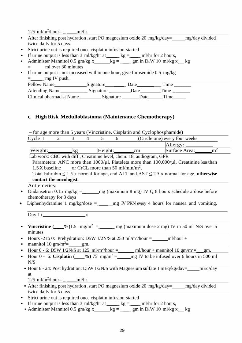
c. High Risk Medulloblastoma (Maintenance Chemotherapy)
– for age more than 5 years (Vincristine, Cisplatin and Cyclophosphamide) Cycle 1 2 3 4 5 6 (Circle one) every four weeks
Allergy:
Weight: kg Height: _cm Surface Area: m2
Lab work: CBC with diff., Creatinine level, chem. 18, audiogram, GFR
Parameters: ANC more than 1000/µl, Platelets more than 100,000/µl, Creatinine less than
1.5 X baseline or CrCL more than 50 ml/min/m2,
Total bilirubin ≤ 1.5 x normal for age, and ALT and AST ≤ 2.5 x normal for age, otherwise
contact the oncologist. Antiemetics:
• Ondansetron 0.15 mg/kg = _ mg (maximum 8 mg) IV Q 8 hours schedule a dose before
chemotherapy for 3 days
Diphenhydramine 1 mg/kg/dose =_ __mg IV PRN every 4 hours for nausea and vomiting.
Day 1 ( ):
▪ Vincristine (____%)1.5 mg/m2 = ______ mg (maximum dose 2 mg) IV in 50 ml N/S over 5
minutes ▪ Hours -2 to 0: Prehydration: D5W 1/2N/S at 250 ml/m2/hour = ______ml/hour +
▪ mannitol 10 gm/m2= _____gm. ▪ Hour 0 - 6: D5W 1/2N/S at 125 ml/m2/hour = ______ ml/hour + mannitol 10 gm/m2= _ gm. ▪ Hour 0 - 6: Cisplatin (____%) 75 mg/m2 = _____mg IV to be infused over 6 hours in 500 ml
N/S ▪ Hour 6 - 24: Post hydration: D5W 1/2N/S with Magnesium sulfate 1 mEq/kg/day=_____mEq/day
at
125 ml/m2/hour= _ ____ml/hr. ▪ After finishing post hydration ,start PO magnesium oxide 20 mg/kg/day=_____ mg/day divided
twice daily for 5 days. ▪ Strict urine out is required once cisplatin infusion started
▪ If urine output is less than 3 ml/kg/hr at _____ kg = ____ ml/hr for 2 hours,
▪ Administer Mannitol 0.5 gm/kg x ______kg = _ ___ gm in D5W 10 ml/kg x __ kg
30
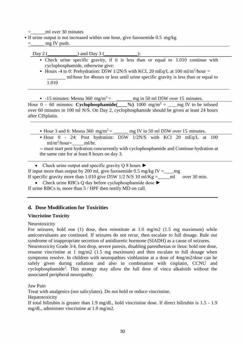
= ml over 30 minutes
▪ If urine output is not increased within one hour, give furosemide 0.5 mg/kg
= _ mg IV push.
Day 2 ( ) and Day 3 ( ): ▪ Check urine specific gravity, if it is less than or equal to 1.010 continue with
cyclophosphamide, otherwise give:
▪ Hours -4 to 0: Prehydration: D5W 1/2N/S with KCL 20 mEq/L at 100 ml/m2/hour =
ml/hour for 4hours or less until urine specific gravity is less than or equal to
1.010
▪ -15 minutes: Mesna 360 mg/m2 = ______ mg in 50 ml D5W over 15 minutes. Hour 0 - 60 minutes: Cyclophosphamide(____%) 1000 mg/m2 = ____mg IV to be infused
over 60 minutes in 100 ml N/S. On Day 2, cyclophosphamide should be given at least 24 hours
after CISplatin.
▪ Hour 3 and 6: Mesna 360 mg/m2 = ______ mg IV in 50 ml D5W over 15 minutes. ▪ Hour 0 - 24: Post hydration: D5W 1/2N/S with KCl 20 mEq/L at 100
ml/m2/hour=_____ml/hr. -- must start post hydration concurrently with cyclophosphamide and Continue hydration at the same rate for at least 8 hours on day 3.
Check urine output and specific gravity Q 8 hours ►
If input more than output by 200 ml, give furosemide 0.5 mg/kg IV =____mg
If specific gravity more than 1.010 give D5W 1/2 N/S 10 ml/Kg =_____ml over 30 min.
Check urine RBCs Q day before cyclophosphamide dose ►
If urine RBCs is, more than 5 / HPF then notify MD on call.
d. Dose Modification for Toxicities
Vincristine Toxicity
Neurotoxicity
For seizures, hold one (1) dose, then reinstitute at 1.0 mg/m2 (1.5 mg maximum) while
anticonvulsants are continued. If seizures do not recur, then escalate to full dosage. Rule out
syndrome of inappropriate secretion of antidiuretic hormone (SIADH) as a cause of seizures.
Neurotoxicity Grade 3/4, foot drop, severe paresis, disabling paresthesias or ileus: hold one dose,
resume vincristine at 1 mg/m2 (1.5 mg maximum) and then escalate to full dosage when
symptoms resolve. In children with neuropathies vinblastine at a dose of 4mg/m2/dose can be
safely given during radiation and also in combination with cisplatin, CCNU and
cyclophosphamide3. This strategy may allow the full dose of vinca alkaloids without the
associated peripheral neuropathy.
Jaw Pain
Treat with analgesics (not salicylates). Do not hold or reduce vincristine.
Hepatotoxicity
If total bilirubin is greater than 1.9 mg/dL, hold vincristine dose. If direct bilirubin is 1.5 - 1.9
mg/dL, administer vincristine at 1.0 mg/m2.
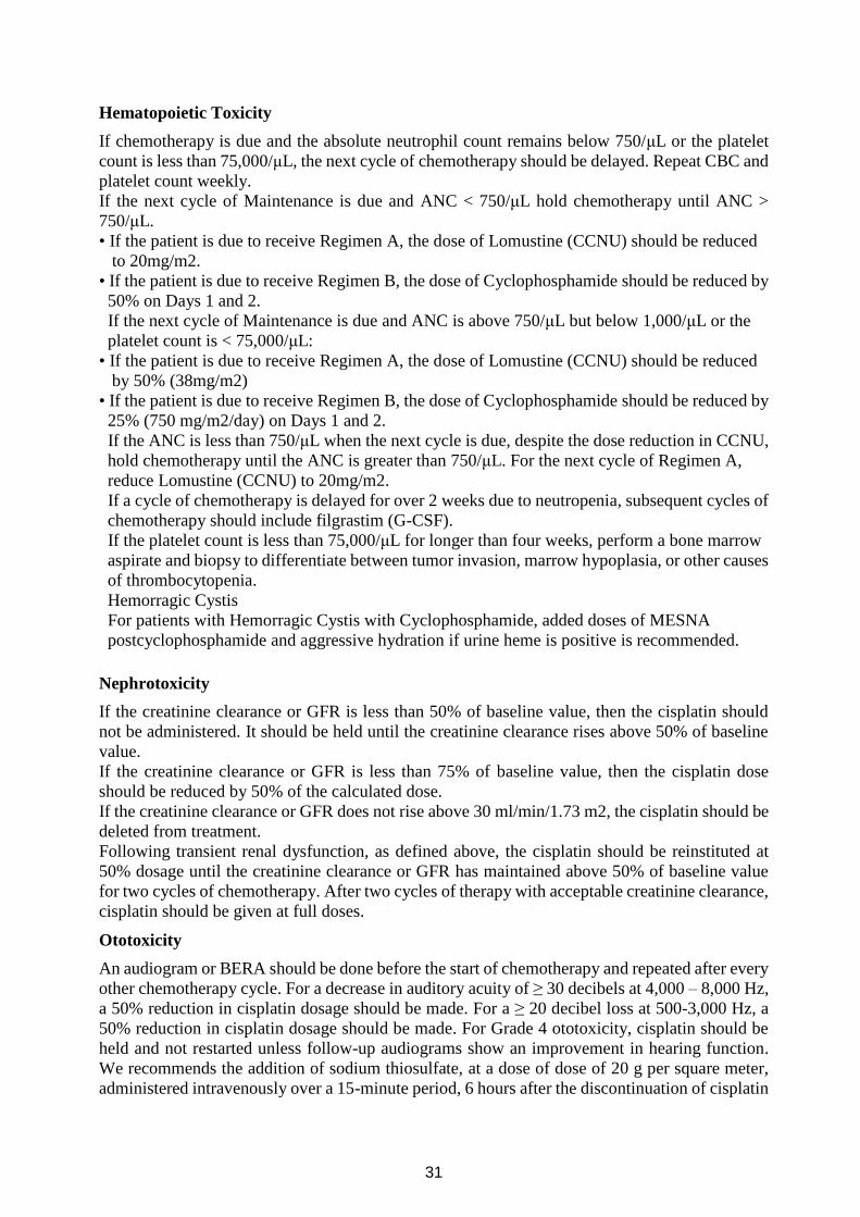
31
Hematopoietic Toxicity
If chemotherapy is due and the absolute neutrophil count remains below 750/μL or the platelet
count is less than 75,000/μL, the next cycle of chemotherapy should be delayed. Repeat CBC and
platelet count weekly.
If the next cycle of Maintenance is due and ANC < 750/μL hold chemotherapy until ANC >
750/μL.
• If the patient is due to receive Regimen A, the dose of Lomustine (CCNU) should be reduced
to 20mg/m2.
• If the patient is due to receive Regimen B, the dose of Cyclophosphamide should be reduced by
50% on Days 1 and 2.
If the next cycle of Maintenance is due and ANC is above 750/μL but below 1,000/μL or the
platelet count is < 75,000/μL:
• If the patient is due to receive Regimen A, the dose of Lomustine (CCNU) should be reduced
by 50% (38mg/m2)
• If the patient is due to receive Regimen B, the dose of Cyclophosphamide should be reduced by
25% (750 mg/m2/day) on Days 1 and 2.
If the ANC is less than 750/μL when the next cycle is due, despite the dose reduction in CCNU,
hold chemotherapy until the ANC is greater than 750/μL. For the next cycle of Regimen A,
reduce Lomustine (CCNU) to 20mg/m2.
If a cycle of chemotherapy is delayed for over 2 weeks due to neutropenia, subsequent cycles of
chemotherapy should include filgrastim (G-CSF).
If the platelet count is less than 75,000/μL for longer than four weeks, perform a bone marrow
aspirate and biopsy to differentiate between tumor invasion, marrow hypoplasia, or other causes
of thrombocytopenia.
Hemorragic Cystis
For patients with Hemorragic Cystis with Cyclophosphamide, added doses of MESNA
postcyclophosphamide and aggressive hydration if urine heme is positive is recommended.
Nephrotoxicity
If the creatinine clearance or GFR is less than 50% of baseline value, then the cisplatin should
not be administered. It should be held until the creatinine clearance rises above 50% of baseline
value.
If the creatinine clearance or GFR is less than 75% of baseline value, then the cisplatin dose
should be reduced by 50% of the calculated dose.
If the creatinine clearance or GFR does not rise above 30 ml/min/1.73 m2, the cisplatin should be
deleted from treatment.
Following transient renal dysfunction, as defined above, the cisplatin should be reinstituted at
50% dosage until the creatinine clearance or GFR has maintained above 50% of baseline value
for two cycles of chemotherapy. After two cycles of therapy with acceptable creatinine clearance,
cisplatin should be given at full doses.
Ototoxicity
An audiogram or BERA should be done before the start of chemotherapy and repeated after every
other chemotherapy cycle. For a decrease in auditory acuity of ≥ 30 decibels at 4,000 – 8,000 Hz,
a 50% reduction in cisplatin dosage should be made. For a ≥ 20 decibel loss at 500-3,000 Hz, a
50% reduction in cisplatin dosage should be made. For Grade 4 ototoxicity, cisplatin should be
held and not restarted unless follow-up audiograms show an improvement in hearing function.
We recommends the addition of sodium thiosulfate, at a dose of dose of 20 g per square meter,
administered intravenously over a 15-minute period, 6 hours after the discontinuation of cisplatin
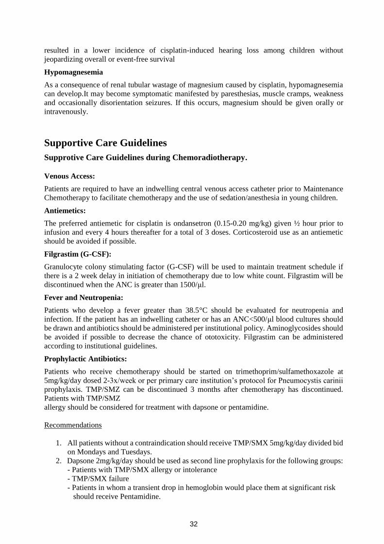
32
resulted in a lower incidence of cisplatin-induced hearing loss among children without
jeopardizing overall or event-free survival
Hypomagnesemia
As a consequence of renal tubular wastage of magnesium caused by cisplatin, hypomagnesemia
can develop.It may become symptomatic manifested by paresthesias, muscle cramps, weakness
and occasionally disorientation seizures. If this occurs, magnesium should be given orally or
intravenously.
Supportive Care Guidelines
Supprotive Care Guidelines during Chemoradiotherapy.
Venous Access:
Patients are required to have an indwelling central venous access catheter prior to Maintenance
Chemotherapy to facilitate chemotherapy and the use of sedation/anesthesia in young children.
Antiemetics:
The preferred antiemetic for cisplatin is ondansetron (0.15-0.20 mg/kg) given ½ hour prior to
infusion and every 4 hours thereafter for a total of 3 doses. Corticosteroid use as an antiemetic
should be avoided if possible.
Filgrastim (G-CSF):
Granulocyte colony stimulating factor (G-CSF) will be used to maintain treatment schedule if
there is a 2 week delay in initiation of chemotherapy due to low white count. Filgrastim will be
discontinued when the ANC is greater than 1500/μl.
Fever and Neutropenia:
Patients who develop a fever greater than 38.5°C should be evaluated for neutropenia and
infection. If the patient has an indwelling catheter or has an ANC<500/μl blood cultures should
be drawn and antibiotics should be administered per institutional policy. Aminoglycosides should
be avoided if possible to decrease the chance of ototoxicity. Filgrastim can be administered
according to institutional guidelines.
Prophylactic Antibiotics:
Patients who receive chemotherapy should be started on trimethoprim/sulfamethoxazole at
5mg/kg/day dosed 2-3x/week or per primary care institution’s protocol for Pneumocystis carinii
prophylaxis. TMP/SMZ can be discontinued 3 months after chemotherapy has discontinued.
Patients with TMP/SMZ
allergy should be considered for treatment with dapsone or pentamidine.
Recommendations
1. All patients without a contraindication should receive TMP/SMX 5mg/kg/day divided bid
on Mondays and Tuesdays.
2. Dapsone 2mg/kg/day should be used as second line prophylaxis for the following groups:
- Patients with TMP/SMX allergy or intolerance
- TMP/SMX failure
- Patients in whom a transient drop in hemoglobin would place them at significant risk
should receive Pentamidine.
33
3. Pentamidine can be given IV or aerosolized and is generally efficacious once monthly.
Pentamidine should be considered prophylaxis for the following groups:
- Patients without prescription plans who are unable to afford TMP/SMX or Dapsone,
- Patients who are non-adherent with oral TMP/SMX or Dapsone,
- Patients with TMP/SMX and Dapsone allergy or intolerance,
- Patients for whom a mild hemolytic anemia would not be tolerated.
Blood Products:
Platelets
Patients will be transfused as necessary with platelets. It is suggested that the platelet count be
maintained > 30,000/μL. All blood products will be irradiated to prevent graft-versus-host
disease. Filters to remove leukocytes should be used to prevent WBC sensitization. CMV
seronegative patients should receive CMV negative blood products.
Red Blood Cells
Therapy-induced anemia and reticulocytopenia are expected with this protocol. Patients will be
transfused as necessary with irradiated packed red blood cells to maintain a hematocrit > 20-25%.
Blood products will be irradiated to prevent graft-versus-host disease. Filters should be used to
prevent WBC sensitization.
Nutritional Support:
Any patient with greater than 10% weight loss should begin nutritional support. Aggressive
nutritional support will be provided either enterally or via a central venous catheter with
parenteral hyperalimentation. All patients should have their magnesium checked prior to each
cycle and be supported with magnesium supplementation if necessary in a minimum dose of 3
mEq/m2/day p.o. or IV in three divided doses to maintain serum levels in the normal range.
Endocrine Guidelines:
Obtain endocrine evaluations at diagnosis, completion of radiation therapy, completion of
treatment, 6 months following the completion of treatment and then annually unless otherwise
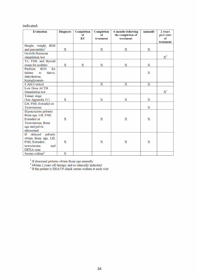
34
indicated.
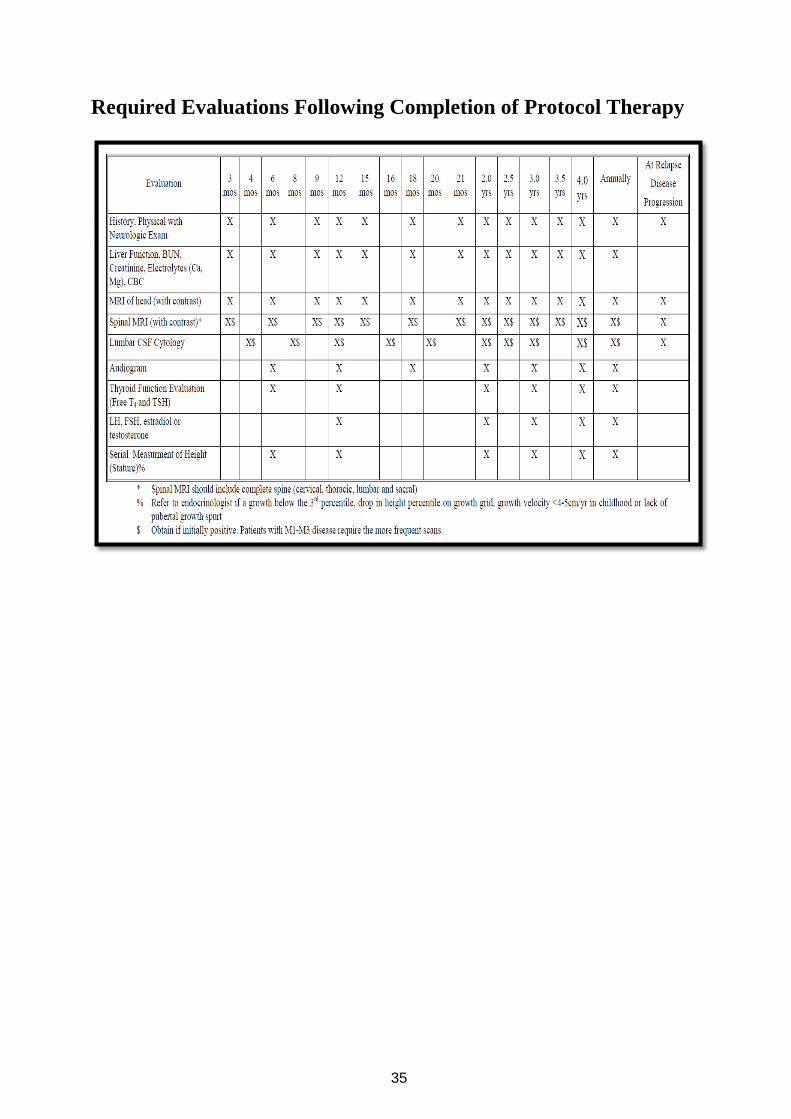
35
Required Evaluations Following Completion of Protocol Therapy
36
References
“These guidelines are drafted on treatment strategy of Clinical Oncology Group based upon the
findings of ACNS0331 & ACNS0332 prospective studies.
1. Olson, J.M. Chemotherapy and Radiation Therapy in Treating Young Patients with
Newly Diagnosed, Previously Untreated, High-Risk Medulloblastoma.
ClinicalTrials.gov Identifier: NCT00392327; 2006. Available online:
https://clinicaltrials.gov/ct2/show/NCT00392327 (Accessed September 01, 2020).
2.
3. Michalski, A.; Janss, A.; Vezina, G.; Gajjar, A.; Pollack, I.; Merchant, T.E.; FitzGerald,
T.J.; Booth, T.; Tarbell, N.J.; Billups, C.A.; et al. Results of COG ACNS0331: A Phase
III Trial of Involved-Field Radiotherapy (IFRT) and Low Dose Craniospinal Irradiation
(LD-CSI) with Chemotherapy in Average-Risk Medulloblastoma: A Report from the
Children’s Oncology Group. Int. J. Radiat. Oncol. Biol. Phys. 2016, 96, 937–938.
4. Liana Nobre, David Pauck, Brian Golbourn, Mara Maue,Eric Bouffet1 Marc Remke,
Vijay Ramaswamy,Effective and safe tumor inhibition using vinblastine in
medulloblastoma. Pediatr Blood Cancer. 2019;66:e27694.


![Medulloblastoma: [Print] - eMedicine Neurology · emedicine.medscape.com eMedicine Specialties > Neurology > Pediatric Neurology Medulloblastoma George I Jallo, MD, Associate Professor](https://img.pdfslide.us/doc/110x75/5d472c3c88c993527c8b60e5/medulloblastoma-print-emedicine-neurology-emedicinemedscapecom-emedicine.jpg)

![Medulloblastoma: [Print] - eMedicine Neurology · accounts for approximately 7-8% of all intracranial tumors and 30% of ... Incidence of medulloblastoma is 1.5-2 cases per ... Medulloblastoma:](https://img.pdfslide.us/doc/110x75/5b7fc2317f8b9ae6088caa0e/medulloblastoma-print-emedicine-accounts-for-approximately-7-8-of-all.jpg)