Embed Size (px)
Citation preview
Perspectives in Pharmacology
Mitochondrial Function and Dysfunction: An Update
Robert E. Davis and Michael Williams3-D Pharmaceutical Consultants, La Jolla, California (R.E.D.); and Department of Molecular Pharmacology and BiologicalChemistry, Feinberg School of Medicine, Northwestern University, Chicago, Illinois (M.W.)
Received January 17, 2012; accepted March 23, 2012
ABSTRACTWith the current explosion of knowledge on the role of mito-chondrial dysfunction in the genesis of various human diseasestates, there is an increased interest in targeting mitochondrialprocesses, pathways, and proteins for drug discovery efforts incancer and cardiovascular, metabolic, and central nervous sys-
tem diseases, the latter including autism and neurodegenera-tive diseases. We provide an update on understanding thecentral role of the mitochondrion in ATP and reactive oxygenspecies production and in controlling cell death pathways.
IntroductionFor many pharmacologists, the mitochondrion is probably
last remembered as a major topic in their undergraduateefforts in biochemistry where the importance of this keyintracellular organelle was assessed almost exclusively inthe context of its key role in ATP production, some 40 to 50 kgeach day, and calcium homeostasis (McBride et al., 2006;
Schatz, 2007; Lax et al., 2011). Since then, studies on the roleof mitochondria in cell function have evolved considerably witha veritable explosion in knowledge on their role as rheostats orbiosensors for oxidative stress and as a focal point for cellularsignaling platforms especially those involved in modulating celldeath, the latter including necrosis, apoptosis, and autophagy(Edinger and Thompson, 2004; McBride et al., 2006; Kroemer etal., 2009; Huang and Figueiredo-Pereira, 2010; Kitsis andMolkentin, 2010; Martin et al., 2011; Koopman et al., 2012)together with their mitochondrial-specific variations, mitopto-sis and mitophagy (Youle and Narendra, 2011).
Article, publication date, and citation information can be found athttp://jpet.aspetjournals.org.
http://dx.doi.org/10.1124/jpet.112.192104.
ABBREVIATIONS: mtDNA, mitochondrial DNA; ��m, mitochondrial membrane potential; A�, amyloid � peptide; AD, Alzheimer’s disease; AIF,apoptosis-inducing factor; ALS, amyotrophic lateral sclerosis; ANT, adenine nucleotide translocator; Bak, Bcl-2 homologous antagonist killer; Bax,proapoptotic Bcl-2-associated X protein; BcL, B-cell lymphoma protein; BH3, proapopotoic Bcl-2 family members; CytC, cytochrome c; Cyp-D,cyclophilin D; DRP1, dynamin-related protein 1; ETC, electron transport chain; FAD, flavin adenine dinucleotide; HD, Huntington’s disease; IAP,inhibitor of apoptosis; IMM, inner mitochondrial membrane; MLKL, mixed lineage kinase-domain-like protein; MPT, mitochondrial permeabilitytransition; MPTP, MPT pore; NCE, new chemical entity; Nix/BNip3L, BCL2/adenovirus E1B 19 kDa protein-interacting protein 3-like; nDNA,nuclear DNA; NSAID, nonsteroidal anti-inflammatory drug; OMM, outer mitochondrial membrane; Omi/HtrA2, homotrimeric serine proteasehigh-temperature requirement A2; OXPHOS, oxidative phosphorylation; PARP, poly(ADP) ribose polymerase; PD, Parkinson’s disease; PGAM5S,phosphoglycerate mutase/protein phosphatase 5, short form; PGC-1�, peroxisome proliferator-activated receptor-� coactivator 1�; PKA, proteinkinase A; RCT, randomized clinical trial; ROS, reactive oxygen species; RIPK, receptor interacting protein kinase; sAC, soluble adenylyl cyclase;Smac/DIABLO, second mitochondria-derived activator of caspases/direct IAP-associated binding protein with low pI; TNF, tumor necrosis factor;TRAF2, TNF receptor-associated factor 2; VDAC, voltage-dependent anion channel; ABT-737, 4-{4-[(4�-chlorobiphenyl-2-yl)methyl]piperazin-1-yl}-N-{[4-({(1R)-3-(dimethylamino)-1-[(phenylsulfanyl)methyl]propyl}amino)-3-nitrophenyl]sulfonyl}benzamide; AT-101, (�)-1,1�,6,6�,7,7�-hexahy-droxy-3,3�-dimethyl-5,5�-bis(1-methylethyl)-[2,2�-binaphthalene]-8,8�-dicarboxaldehyde; CD437, 6-(3-(1-adamantyl)-4-hydroxyphenyl)-2-naph-thalenecarboxylic acid; PK 11195, N-butan-2-yl-1-(2-chlorophenyl)-N-methylisoquinoline-3-carboxamide; ATN-224, choline tetrathiomolybdate;STA-4783, N�1, N�3-dimethyl-N�1, N�3- bis(phenylcarbonothioyl)propanedihydrazide; PI-H71, 6-amino-8-[(6-iodo-1,3-benzodioxol-5-yl)thio]-N-(1-methylethyl)-9H-purine-9-propanamine; KNS-760704, (R)-N6-propyl-4,5,6,7-tetrahydrobenzo[d]thiazole-2,6-diamine; CGP 37157, 7-chloro-5-(2-chlorophenyl)-1,5-dihydro-4,1-benzothiazepin-2(3H)-one; SS-31, arginyl-2,�6�-dimethyltyrosyl-lysyl-phenylalaninamide; TRO19622,(NZ)-N-[(8S,9S,10R,13R,14S,17R)-10,13-dimethyl-17-[(2R)-6-methylheptan-2-yl]-1,2,6,7,8,9,11,12,14,15,16,17-dodecahydrocyclopenta[a]phenanthren-3-ylidene]hydroxylamine.
1521-0103/12/3423-598–607$25.00THE JOURNAL OF PHARMACOLOGY AND EXPERIMENTAL THERAPEUTICS Vol. 342, No. 3Copyright © 2012 by The American Society for Pharmacology and Experimental Therapeutics 192104/3789467JPET 342:598–607, 2012
598
at ASPE
T Journals on M
ay 26, 2018jpet.aspetjournals.org
Dow
nloaded from
Deficiencies in energy metabolism, the bioenergetic failurecharacteristic of both mitochondrial and epigenomic diseasestates (Wallace and Fan, 2010), have been implicated in avariety of human disease states, especially in those organs inwhich there is a high level of energy consumption, e.g., thebrain, which with only 2% of total body weight represents20% of the total oxygen consumption in the body. Diseasesspecifically linked to mitochondrial dysfunction vary fromthe well known (glaucoma, inflammation, neurodegenerativediseases, type 2 diabetes, cancers, especially those involvingprostate and colon, cardiomyopathies, and dysrhythmias) tothe less well known (Freiderich’s ataxia) to a group of rela-tively obscure disease states [Kearns-Sayre syndrome (KSS),Leber hereditary optic neuropathy (LHON), mitochondrialencephalopathy lactic acidosis and strokes (MELAS), myoclo-nic epilepsy with ragged red fibers (MERRF), and mitochon-drial neuro-gastrointestinal encephalomyopathy (MNGIE)](Haas et al., 2008).
These various disease states have been associated in someor all of their manifestations with mutations in both mito-chondrial DNA (mtDNA) and nuclear DNA (nDNA) that re-sult in defects in mitochondrial function (Wallace, 1999;Schapira, 2006; Copeland, 2008; Finsterer, 2010) or with aninability to accommodate the consequences of oxidativestress (Poljsak, 2011). While an excess of free radical, e.g.,ROS (reactive oxygen species), production leads to both mu-tations of DNA and the degradation of proteins, lipids, and
nucleic acids, the view that ROS is causal to mitochondrially-related diseases has been challenged in the context of “oxi-dative shielding” (Naviaux, 2012). This concept, albeit con-troversial, views ROS production as a form of innate immunityto protect the cell with ROS production being the responseto tissue trauma or disease, a view similar to that evolvingfor the role of A� in Alzheimer’s disease (Castellani et al.,2009). The spatial proximity of mtDNA to the free radicalsproduced by the electron transport chain (ETC) (Fig. 1) makesit uniquely susceptible to mutations, especially when theETC is dysfunctional. This has led to the heuristically engag-ing, albeit controversial, mitochondrial oxidative stress/freeradical/genotoxic stress theory of aging that reflects the neg-ative impact of chronic, accumulating damage to DNA andcellular proteins from free radicals as a function of age (Ku-joth et al., 2005; Wallace, 2005; Dagda et al., 2009; Swerdlowand Kahn, 2009; Lapointe and Hekimi, 2010; Durieux et al.,2011; Pamplona, 2011). This involves a progressive loss offunctional telomeres that contribute to replicative senes-cence and apoptosis via decreased mitochondria and mtDNAcopy numbers, increased ROS production, and decreasedATP production (Sahin et al., 2011).
With the current evolution in understanding of the contri-bution of mitochondrial dysfunction to the genesis of humandisease states, the majority of them chronic, there is in-creased interest in targeting mitochondrial processes andproteins for drug discovery efforts in cancer (Fulda et al.,
∆ψ
β
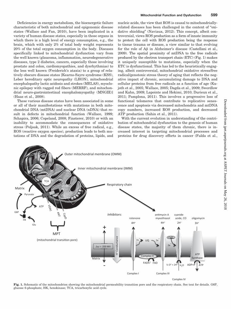
Fig. 1. Schematic of the mitochondrion showing the mitochondrial permeability transition pore and the respiratory chain. See text for details. G6P,glucose 6-phosphate; HK, hexokinase; TCA, tricarboxylic acid cycle.
Mitochondrial Function and Dysfunction 599
at ASPE
T Journals on M
ay 26, 2018jpet.aspetjournals.org
Dow
nloaded from
2010; Maldonado and LeMasters, 2012), cardiovascular dis-ease (Ballinger, 2005; Akar and O’Rourke, 2011; Ong andHausenloy, 2010), metabolic diseases (Gilliam and Neufer,2012; Szendroedi et al., 2012), central nervous system dis-eases including autism (Rossignol and Frye, 2012), and neu-rodegenerative diseases including Alzheimer’s disease (AD),Parkinson’s disease (PD), Huntington’s disease (HD), multi-ple sclerosis, amyotrophic lateral sclerosis (ALS), and pain(Moreira et al., 2010; Reyes et al., 2010; Witte et al., 2010;Ferrari et al., 2011; Lax et al., 2011; Johri and Beal, 2012;Cooper et al., 2012). Progress in these efforts from a tradi-tional small-molecule perspective has, however, been chal-lenging (Finsterer, 2010; Kerr, 2010; Stacpoole, 2011; Davis,2012).
The present review highlights the current state of knowl-edge on the role of mitochondrial dysfunction in variousdisease states and identifies potential drug targets.
The Role of Mitochondria in Cell FunctionMitochondrial Genetics. Mitochondria are unique in
that they have their own DNA pool (mtDNA) distinct fromthat of nDNA. mtDNA is almost exclusively maternally in-herited and has independent evolutionary origins fromnDNA that date back to the time when mitochondria wereseparate organisms before forming a symbiotic relationshipwith eukaryotes (Schapira, 2006; Wallace and Fan, 2010).Human mtDNA is approximately 16.6 base pairs long, form-ing a closed, double-stranded structure (Legros et al., 2004).Each mitochondrion contains between 2 and 10 mtDNA cop-ies that consist of 37 genes coding for 22 transfer and 2ribosomal DNAs and 13 proteins, the latter including theenzymes involved in the oxidative phosphorylation (OXPHOS)pathway involved in ATP production. OXPHOS units arecoded by both nDNA and mtDNA, with the former contrib-uting somewhere in excess of 1000 proteins that are essentialfor mitochondrial function (Wallace and Fan, 2010; Eichnerand Giguere, 2011). Of these, 705 are under the transcrip-tional control of estrogen-related receptors, �, �, and �, thatare responsible for the integrated control of mitochondrialmetabolism (Eichner and Giguere, 2011). Although themtDNA sequence in most cells is identical and is conse-quently termed homoplasmic, the coexistence of wild-typeand mutant mtDNA in the same mitochondrion and/or cell isknown as heteroplasmy.
The OXPHOS pathway consists of five different ETC com-plexes located on the inner mitochondrial membrane thattogether contribute to the generation of the mitochondrialelectrochemical gradient (Fig. 1). These complexes are com-posed of proteins that originate from both nDNA and mtDNA(Schon et al., 2010; Wallace and Fan, 2010). Complex I con-sists of 45 peptide subunits, 7 originating from mtDNA withthe remainder from nDNA. Complex II has four subunits, allof which are derived from nDNA, and Complex III has 11subunits, only one of which originates from mtDNA. ComplexIV has 12 subunits, 3 of which are derived from mtDNA, andcomplex IV has approximately 16 subunits, 2 of which arefrom mtDNA. The fifth ETC complex, Complex V, is ATPsynthase.
Mutation rates in mtDNA are generally 2- to 3-foldhigher than those occurring in nDNA (although some ex-perts estimate a 10-fold or more difference), a consequence,
as already noted, of the proximity of mtDNA to the ROSproduced by electron leakage from complexes I and III ofthe ETC (Fig. 1), coupled with inefficient DNA repairmechanisms and a lack of protective histones on mtDNA.To date, some 270 disease-related mtDNA point mutationshave been identified (http://www.mitomap.org/bin/view.pl/MITOMAP/MutationsCodingControl) that are thought toaffect mitochondrial protein synthesis, protein-encodinggenes and mRNA, and ultimately mitochondrial function.These are complemented by rearrangements, deletions,and insertions in mtDNA and their altered interactionswith nDNA, the latter reflecting defects in mitochondrialtransport processes (Schon et al., 2010).
In heteroplasmic situations, the percentage of mutantmtDNA dictates the degree of mitochondrial dysfunction anddisease occurrence. Thus the age-related accumulation ofsomatic mtDNA mutations that can lead in time to decreasedmitochondrial function has been associated with an in-creased rate of aging and cancer incidence (Wallace, 2005;Schapira, 2006; Wallace and Fan, 2010). A variety of condi-tions (hypoxia, stress, trauma, blood glucose levels, aberrantcircadian rhythms, etc.) and agents/mechanisms [phosphor-ylation, DNA methylation/acetylation, Akt/protein kinase Bsignaling, calcium homeostasis, estrogen-related receptorsignaling, heat shock proteins, soluble adenylyl cyclase(sAC), receptor-interacting protein 3 kinase, Target of Rapa-mycin kinases, peroxisome proliferator-activated receptor-�coactivator 1� (PGC-1�), Signal Transducer and Activator ofTranscription 3, AMP-activated protein kinase, PGAM5S(phosphoglycerate mutase/protein phosphatase 5, shortform), �-amyloid, sirtuin-1, etc.] are involved in both modu-lating transcription of the mitochondrial genome and thefunction of the transcribed proteins. Mutated proteinssuch as huntingtin in HD, amyloid (A�) in AD, superoxidedismutase 1 in ALS, and parkin, DJ1, and �-synuclein inPD have been localized to mitochondrial membranes(Reddy, 2009) where they can alter ETC function to in-crease ROS production.
The increased interest in mtDNA as a risk factor and/orcausative to human disease states parallels the renewedfocus on noncoding or “junk” nuclear DNA that was originallydismissed as lacking importance when the map of the humangenome was finally annotated. Far from being unimportant,junk DNA has been found to contain key regulatory se-quences that modify gene expression and activity (Biemont,2010), adding an additional level of complexity to under-standing gene function and disease risk. This has the poten-tial to negate the validity of many of the genomewide asso-ciation studies (GWAS) conducted to date that sought toestablish the relationship between specific genes and specificdisease states (Mullane and Williams, 2012). The superim-position of mtDNA as yet another overlooked/underesti-mated component of the human genome coupled with itspotential interactions with nDNA adds yet another level ofcomplexity to deciphering gene-driven risk factors and cau-sality. It is noteworthy that, more than a decade ago, Wallace(1999) noted that a specific mtDNA mutation could producevery different human disease phenotypes, whereas differentmutations could result in the same phenotype. This insight isnot limited to the mitochondrial genome and seems equallyapplicable to the total cellular genome, a conclusion that issupported by the identification of multiple, and often concep-
600 Davis and Williams
at ASPE
T Journals on M
ay 26, 2018jpet.aspetjournals.org
Dow
nloaded from

tually puzzling, gene candidates/associations for diseasestates such as asthma, schizophrenia, and AD with the lattercurrently numbering in excess of 130 and still growing (Mul-lane and Williams, 2012).
ATP Production. ATP is produced in mitochondria viaOXPHOS, a complex process involving mitochondrial respi-ration and the generation of a proton (or electrochemical)gradient [mitochondrial membrane potential (��m)] acrossthe mitochondrial inner membrane (Bertram et al., 2006) viathe ETC (Fig. 1). Approximately 90% of ATP arises frommitochondria. In complex I (NADH dehydrogenase) two elec-trons are removed from NADH and transferred to the lipid-soluble carrier, ubiquinone (Q) forming the reduced product,ubiquinol (QH2) that can freely diffuse in the membrane.Complex I thus leads to the translocation of four protons (H�)across the membrane to produce a proton gradient (Fig. 1). Incomplex II (succinate dehydrogenase) additional electronsare delivered from succinate via flavin adenine dinucleotide(FAD) to the quinone pool (Q) and transferred via FAD to Q.In complex III [ubiquinol-cytochrome c (CytC) reductase] sixelectrons are removed from QH2, two of which are sequen-tially transferred to two molecules of CytC, a water-solubleelectron carrier located in the intermembrane space and fourto the Qi site where the quinone moiety in ubiquinone isreduced to quinol contributing to the proton gradient. Incomplex IV (cytochrome c oxidase), four electrons contributedby four CytC molecules are transferred to molecular oxygen(O2), resulting in two molecules of water. Concomitantly, fourprotons translocate across the membrane, adding further tothe proton gradient. The latter is then used in complex V, theF0/F1 ATP synthase complex to produce ATP via OXPHOS.The ��m is normally in the range of 80 to 140 mV. Theoptimal ��m for ATP production is 100 to 120 mV with ��m
values more than 140 mV leading to increased ROS produc-tion at the expense of ATP generation (Huttemann et al.,2011).
The function of CytC, other key OXPHOS proteins, andnecrosis signaling pathways (Wang et al., 2012) can be dy-namically modulated by phosphorylation. One example is thenegative feedback effects of ATP to control ETC functioninvolve phosphorylation-dependent changes that alter theability of CytC to bind to cytochrome c oxidase, which isdetermined by the ATP/ADP ratio. ATP is also a key sub-strate in generically determining kinase activity (Dagda etal., 2009).
Mitochondrial Dynamics and Cell DeathSignaling
Mitochondria are dynamic organelles that form networksthroughout the cell via the opposing processes of fission andfusion (Sheridan and Martin, 2010). The latter is critical tothe maintenance of mitochondrial function because it affectsthe repair of dysfunctional and damaged mitochondria inaddition to intermixing DNA and proteins between mitochon-dria (Chan, 2006). Fusion involves the merging of the innerand outer membranes from two mitochondria to facilitate theGTPase-dependent exchange of materials to aid in mitochon-drial repair. Fission occurs when a mitochondrion splits intwo. When this process occurs in the presence of decreasedfusion, it can lead to a fragmented mitochondrial phenotypethat is widespread in both necrosis and apoptosis. Deficient
fission and fusion mechanisms are thus key events inmitochondrial disease causality. In HD, fission is facili-tated via the action of dynamin-related protein 1 (DRP1),leading to fragmented mitochondria that are fewer in num-ber (Song et al., 2011). The mutant form of huntintin, aprotein associated with HD, enhances DRP-1 activity. Al-though fission seems to be involved in mitoptosis, there isconsiderable debate as to whether this is a primary orsecondary event, in the former instance being causative tomitochondrial permeability transition (MPT) pore (MPTP)(see below) formation with a secondary, passive role inpromoting mitochondrial network disassembly (Sheridanand Martin, 2010).
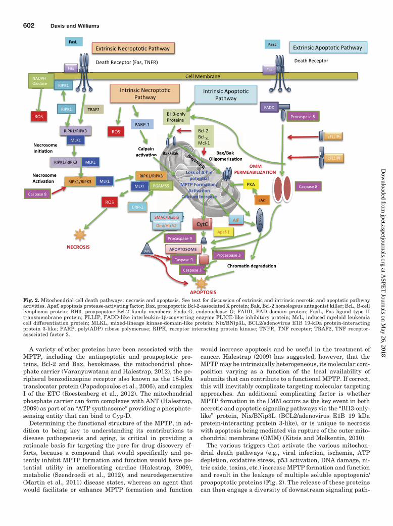
Mitochondria can promote both necrotic and apoptotic celldeath via an abrupt increase in the permeability of the innermitochondrial membrane (IMM) that allows the passage ofmolecules with molecular masses below 1.5 kDa (Zamzami etal., 2005; Baines, 2010). The MPT event results in the decou-pling of OXPHOS, resulting in the dissipation of the protonelectrochemical gradient with decreased ATP production, in-creased ROS production, calcium overload, and mitochon-drial swelling (Rodriguez-Enriquez et al., 2004). The degreeto which the level of mitochondrial ATP is depleted is thoughtto be the major determinant as to whether cell death pro-ceeds by necrosis or apoptosis, with very low ATP levelsleading to necrosis. The relationship between apoptosis andnecrosis is complex with data suggesting that: 1) necrosis ismore important in cell death than apoptosis; 2) necrosis is analternative death pathway to apoptosis when caspases areinhibited; and 3) necrosis is engaged as a cell death pathwaywhen mitochondria form a complex with the endoplasmicreticulum (Baines, 2010). Until recently, necrosis was thoughto be a random, uncontrolled process (Kitsis and Molkentin,2010) that like apopotosis produced its effects via MPTPformation and mitochondrial membrane permeabilization.However, necrosis has now been recognized as a programmedprocess, the effects of which are mediated through pathwaysthat, although distinct from those mediating apoptosis, mayinvolve common pathway members (Sun et al., 2012; Wanget al., 2012) (Fig. 2) with canonical apoptoic moleculesbeing involved in programmed necrosis (Baines, 2010). Theeffects of these common proteins may be antagonistic. Forinstance, caspase 8, which is involved in chromatin degra-dation and apoptosome formation, can inhibit necrosomefunction (Fig. 2).
Mitochondrial Membrane Permeability. The increasein mitochondrial membrane permeability in the IMM is me-diated via the MPTP, the composition of which remains asubject of active debate (Halestrap, 2009; Javadov et al.,2011). Although early studies had indicated that the MPTPwas comprised of three subunits, a voltage-dependent anionchannel (VDAC) (Shoshan-Barmatz and Ben-Hail, 2012), theadenine nucleotide translocator (ANT) (Kunji and Crichton,2010), and mitochondrial cyclophilin D (Cyp-D) (Schinzel etal., 2005), the latter a matrix peptidyl-prolyl cis-transisomerase, gene knockout studies have questioned the in-volvement of VDAC while relegating ANT to a modulatoryrole because MPT can still occur in mitochondria lackingVDAC or ANT (Baines, 2010). ANT also exists in severalforms that have different and opposing functions. ANT-1 andANT-3 are proapoptotic, whereas ANT-2 is antiapoptotic(Fulda et al., 2010).
Mitochondrial Function and Dysfunction 601
at ASPE
T Journals on M
ay 26, 2018jpet.aspetjournals.org
Dow
nloaded from
A variety of other proteins have been associated with theMPTP, including the antiapoptotic and proapoptotic pro-teins, Bcl-2 and Bax, hexokinase, the mitochondrial phos-phate carrier (Varanyuwatana and Halestrap, 2012), the pe-ripheral benzodiazepine receptor also known as the 18-kDatranslocator protein (Papadopoulos et al., 2006), and complexI of the ETC (Roestenberg et al., 2012). The mitochondrialphosphate carrier can form complexes with ANT (Halestrap,2009) as part of an “ATP synthasome” providing a phosphate-sensing entity that can bind to Cyp-D.
Determining the functional structure of the MPTP, in ad-dition to being key to understanding its contributions todisease pathogenesis and aging, is critical in providing arationale basis for targeting the pore for drug discovery ef-forts, because a compound that would specifically and po-tently inhibit MPTP formation and function would have po-tential utility in ameliorating cardiac (Halestrap, 2009),metabolic (Szendroedi et al., 2012), and neurodegenerative(Martin et al., 2011) disease states, whereas an agent thatwould facilitate or enhance MPTP formation and function
would increase apoptosis and be useful in the treatment ofcancer. Halestrap (2009) has suggested, however, that theMPTP may be intrinsically heterogeneous, its molecular com-position varying as a function of the local availability ofsubunits that can contribute to a functional MPTP. If correct,this will inevitably complicate targeting molecular targetingapproaches. An additional complicating factor is whetherMPTP formation in the IMM occurs as the key event in bothnecrotic and apoptotic signaling pathways via the “BH3-only-like” protein, Nix/BNip3L (BCL2/adenovirus E1B 19 kDaprotein-interacting protein 3-like), or is unique to necrosiswith apoptosis being mediated via rupture of the outer mito-chondrial membrane (OMM) (Kitsis and Molkentin, 2010).
The various triggers that activate the various mitochon-drial death pathways (e.g., viral infection, ischemia, ATPdepletion, oxidative stress, p53 activation, DNA damage, ni-tric oxide, toxins, etc.) increase MPTP formation and functionand result in the leakage of multiple soluble apoptogenic/proapoptotic proteins (Fig. 2). The release of these proteinscan then engage a diversity of downstream signaling path-
Ψ
Fig. 2. Mitochondrial cell death pathways: necrosis and apoptosis. See text for discussion of extrinsic and intrinsic necrotic and apoptotic pathwayactivities. Apaf, apoptosis protease-activating factor; Bax, proapoptotic Bcl-2-associated X protein; Bak, Bcl-2 homologous antagonist killer; BcL, B-celllymphoma protein; BH3, proapopotoic Bcl-2 family members; Endo G, endonuclease G; FADD, FAD domain protein; FasL, Fas ligand type IItransmembrane protein; FLLIP, FADD-like interleukin-1�-converting enzyme FLICE-like inhibitory protein; McL, induced myeloid leukemiacell differentiation protein; MLKL, mixed-lineage kinase-domain-like protein; Nix/BNip3L, BCL2/adenovirus E1B 19-kDa protein-interactingprotein 3-like; PARP, poly(ADP) ribose polymerase; RIPK, receptor interacting protein kinase; TNFR, TNF receptor; TRAF2, TNF receptor-associated factor 2.
602 Davis and Williams
at ASPE
T Journals on M
ay 26, 2018jpet.aspetjournals.org
Dow
nloaded from
ways, the composition of which has increased in complexityas new members, and their interactions, continue to beidentified.
Apoptotic Cell Death Pathway. Proteins released via acombination of MPTP formation and OMM collapse includeCytC, Bcl-2, Smac/DIABLO [second mitochondria-derived ac-tivator of caspases/direct inhibitor of apoptosis (IAP)-associ-ated binding protein with low pI], Omi/HtrA2 (homotrimericserine protease high-temperature requirement A2), apopto-sis-inducing factor (AIF), and endonuclease G. CytC is thekey protein in the initiation of apoptosis. Together with theprotein APAF-1 (apoptosis protease-activating factor) andpro-caspase-9, CytC forms an “apoptosome” that facilitatesactivation of the cysteine protease, caspase-9, which thenactivates effector caspases to enable apoptosis. AIF and en-donuclease G are key mediators in the DNA fragmentationand chromosomal condensation that occurs in apoptosis.
The function of CytC, like many of the other proteins in thecell death pathways, depends on its state of phosphorylation,a point that was not fully appreciated in early studies whenit was isolated and studied in its dephosphorylated state(Huttemann et al., 2011). Phosphorylation of tyrosines inCytC inhibits interactions with cytochrome c oxidase, sup-porting the concept that under normal conditions when thereis adequate ATP OXPHOS runs at a reduced activity, a“controlled’ state, to maintain ��m below those leading tofree-radical formation (Huttemann et al., 2011).Smac/DIABLO and Omi/HtrA2 are antagonists of the pro-tein IAPs that promote caspase activation. Mitochondrialmembrane permeabilization can also occur independentlyof pore formation and involves Bcl-2 family members thatinclude both proapoptotic (Bax, Bak, and Bok) and anti-apoptotic (Bcl-2 and Bcl-xL) members.
Apoptosis in mitochondria comprises the intrinsic apopto-tic or Type I pathway as contrasted to the extrinsic pathway(Type II) that involves activation of the cell surface deathreceptor family, a subclass of the tumor necrosis factor (TNF)superfamily. The intrinsic and extrinsic apopototic pathwaysare linked by the Bcl2 family protein, Bid (Bcl-2 interactingdomain), a BH-3 proapoptotic regulator. sAC is a proapop-totic mediator that translocates to mitochondria under con-ditions of acidic stress. The effects of sAC are mediated viaactivation of Protein Kinase A (PKA), which facilitates trans-location of Bax from the cytosol to the mitochondrion whereBax is involved in OMM permeabilization. PKA is alsothought to block the effects of Akt on inactivating Bax, thusattenuating apoptosis (Kumar et al., 2009).
Necrotic Cell Death Pathway. Like apoptosis, necrosishas both extrinsic and intrinsic components, the former in-volving death receptor activation and the latter involvingROS production and PARP-1/calpain activation (Fig. 2). Thereceptor interacting protein kinases (RIPKs) together withTRAF2 and MLKL (mixed lineage kinase-domain-like pro-tein) initiate necrosome formation that is then activated bysequential phosphorylation events (Sun et al., 2012). It thenforms a complex with the mitochondrial protein phosphatase,PGAM5S, which in turn recruits the mitochondrial fissionfactor, DRP1. The resultant necrosome complex can thenactivate DRP-1 GTPase to induce mitochondrial fragmenta-tion, initiating necrosis execution (Wang et al., 2012). Mito-chondrial PGAM5S is also involved in ROS-induced necrosis
and may thus represent a major convergence point for ne-crotic pathways.
Autophagy. Mitophagy, an organelle-specific autophagicelimination, is responsible for both the elimination of dam-aged mitochondria and the regulation of their number andinvolves ubiquitination that recruits the ubiquitin-bindingautophagic components histone deacetylase (HDAC) 6 andp62 to facilitate mitochondrial clearance (Lee et al., 2010).Mitophagy can be regulated by parkin and PTEN-inducedputative kinase protein 1 (PINK1) (Youle and Narendra,2011).
Mitochondrial Disease StatesAs noted, the tissues that are most susceptible to mito-
chondrial-driven disease states are those with a high meta-bolic demand. These include brain, eye, liver, heart, andskeletal muscle. Mitochondrial disease states include themitochondrial myopathies, a group of neuromuscular dis-eases that includes KSS, MELSAS, MERRF, and MNGIEthat have genetic origins (Schapira, 2006; Wallace and Fan,2010), disorders of mitochondrial ETC that affect ETC as-sembly and/or stability and function and involve both geneticfactors and cofactor deficiencies (coenzyme Q10) that canlead to decreased ATP production and increased free-rad-ical production, the latter potentially leading to neurode-generative diseases (AD, PD, HD, and ALS) (Johri andBeal, 2012). LHON, which is associated with visual failurecaused by the degeneration of retinal ganglion cells, is themost common disease associated with mtDNA mutationswith a prevalence of approximately 12 cases per 100,000 inthe population (Schapira, 2006).
A unifying enabling theme in neurodegenerative diseasestates involves the misfolding of key cellular proteins thatlead to the amyloidopathies (AD), tauopathies (AD, PD,Pick’s disease, progressive supranuclear palsy, corticobasaldegeneration, and argyrophilic grain disease), �-synucleopa-thies (PD, dementia with Lewy bodies, multiple system atro-phy, and some instances of AD), and the Tar DNA-bindingprotein 43 proteinopathies/ubiquinopathies (ALS, frontotem-poral dementias, and argyrophilic grain disease) (Geser etal., 2009). In PD, defects in complex I activity involve mtDNAmutations, alterations in mitochondrial kinase signaling(e.g., PTEN-induced kinase I, Akt/PKB, JNK, and ERK;Dagda et al., 2009), and can be caused by the effects ofenvironmental toxins (rotenone) that lead to increased free-radical production and reduced activity in complex IV. In PD,dysregulation of the ubiquitin-proteasomal system, which isenergy sensitive, leads to destruction of dopamine cells in thesubstantia nigra. In HD, the mutant form of huntingtinprotein (mHtt) alters mitochondrial function, leading to aloss of membrane potential, decreased expression of OX-PHOS enzymes (Mochel and Haller, 2011), and increasedfission events that lead to decreases in the number, size, anddistribution of mitochondria (Song et al., 2011). Alterationsin ETC function also occur in AD where the major culpritthought to be responsible for disease causation, A�, can in-hibit OXPHOS and specifically inhibit the mitochondrial en-zyme, ABAD (A�-binding alcohol dehydrogenase) also knownas ERAB (ER amyloid-�-peptide binding protein) that exac-erbates A�-induced cell stress, leading to mitochondrial andneuronal dysfunction (Lustbader et al., 2004; Tillement et
Mitochondrial Function and Dysfunction 603
at ASPE
T Journals on M
ay 26, 2018jpet.aspetjournals.org
Dow
nloaded from

al., 2011). ABAD inhibition in a mouse transgenic APP (am-yloid precursor protein) model of AD reduces A� accumula-tion and improves mitochondrial function (Yao et al., 2011).A� also binds to ANT in the MPTP and to complexes IV andV of the ETC, resulting in changes in calcium homeostasis,OXPHOS efficiency, decreases in DRP-1, enhancement ofnitric oxide production, ROS-induced oxidative stress, tautoxicity, cytokine production, and inflammation (Moreira etal., 2010; Tillement et al., 2011). Alterations in XIAP (X-linked inhibitor of apoptosis), caspase-3, and lipofuscin accu-mulation are also observed in AD, the latter decreasing au-tophagy and reducing mitochondrial recycling. Nonetheless,mitochondrial autophagocyotosis is increased in AD and mayreflect differential roles for autophagy depending on thestage of the disease (Moreira et al., 2010). Mitochondrialdysfunction has also been associated with multiple sclerosis(Witte et al., 2010) and autism spectrum disorders (Rossignoland Frye, 2012).
Type II diabetes (T2DM) is associated with reductions inOXPHOS capacity and decreased mitochondrial plasticityand numbers in skeletal muscle and liver, resulting in insu-lin resistance (Szendroedi et al., 2012). Mitochondrial dys-function appears to be a key link between AD and diabetes(Moreira et al., 2007) having been described as “type 3 dia-betes” (de la Monte et al., 2006). Changes in cardiac mito-chondrial morphology that are linked to changes in mito-chondrial metabolism have been associated with heartfailure, coronary artery disease, and responses to ischemicepisodes (Ong and Hausenloy, 2010).
Mitochondria as a Target for Drug DiscoveryThe explosion of knowledge regarding the key role of mi-
tochondria in human disease states has led to efforts todevelop drugs based on the considerable knowledge base.Given the exquisite complexity of the structural proteins andpathways associated with mitochondrial function, there is noshortage of potential targets, although the majority of thoseof current interest involve modulation of MPTP formationand function (Eichner and Giguere, 2011).
Seminal efforts in addressing inherited and acquired ETCdiseases have focused on replacing deficient components ofthe ETC chain or adding membrane penetrating antioxidantsand free-radical scavengers. The former include vitamins (Dand E) and supplements that include carnitine, coenzymeQ10 and its analogs, thiamine pyrophosphate, mitoquinoneand the SKQs (Skulachev ions), the SS-peptide arginyl-2,�6�-dimethyltyrosyl-lysyl-phenylalaninamide (SS-31), 7-chloro-5-(2-chlorophenyl)-1,5-dihydro-4,1-benzothiazepin-2(3H)-one(CGP 37157), riboflavin, trolox, thiamine, creatine, pyruvate,the pyruvate analog dichloracetate, succinate, folate, omega-3fatty acids [docosahexaenoic acid (DHA) and eicosapenta-enoic acid (EPA)], and methylene blue that can improve theefficiency of the ETC, increase ATP production, and reduceROS production (Reddy, 2009; Kerr, 2010; Schon et al., 2010;Roestenberg et al., 2012). Removal of noxious metabolites,such as lactate, by using bicarbonate and/or dichloraceticacid (Finsterer, 2010) and free-radical scavenging entities,both dietary and synthetic, are other approaches to improv-ing ETC function, although the latter, while effective in celllines and animal models, have questionable efficacy in theclinical setting (Halliwell, 2011; Poljsak, 2011).
Such agents have shown varying levels of success in treat-ing mitochondrial disorders, and work continues to improvetheir access to, and selectivity for, their mitochondrial sites ofaction. Much of the current effort is focused on finding newchemical entities (NCEs) that facilitate or block mitochon-drial cell death pathways. This represents the yin and yangof cell death-related disease treatment where accelerating/facilitating apoptosis to develop more effective anticancerdrugs is contraindicated in cardiovascular, metabolic, andneurodegenerative disease states where abrogating celldeath processes is the target for therapeutics to address andimprove mitochondrial energetics in these disease states (Ja-vadov et al., 2011). These drug discovery efforts are focusedprimarily on small molecules, including peptides, that canmodulate MPTP formation and function (Finsterer, 2010;Kerr, 2010; Stacpoole, 2011; Davis, 2012) and calcium ho-meostasis (Giorgi et al., 2012). A number of compounds havebeen found to interact with the putative MPTP-constituentprotein VDAC and include the antisense 18mer G3139(oblimersen, TCTCCCAGCGTGCGCCAT), the avicin class ofplant stress metabolites, the antidepressant fluoxetine, cis-platin, and endostatin (Shosnan-Barmatz and Ben-Hail,2012).
A major issue in mitochondrial-targeted drug discovery isthe challenge of delivering NCEs at sufficient levels to betherapeutically useful to targets located inside an intracellu-lar organelle, requiring effective passage through cell mem-brane, cytosol, and the mitochondrial membrane. Analogs ofCoQ10, like MitoQ and SKQ1 (Fink et al., 2012), are prefer-entially absorbed in the IMM, whereas the peptide SS-31shows a 5000-fold accumulation in mitochondrial fractions(Roestenberg et al., 2012). Functionalized polymeric and me-tallic nanoparticles are also being explored as potential mi-tochondrial delivery systems (Durazo and Komplella, 2012)as are novel approach proteomimetic polyanionic or amphi-pathic cell-penetrating peptides. The latter contain epitopesthat act as vectors for the highly efficient delivery of bioactivecargoes into the intracellular milieu (Jones et al., 2010).Cell-penetrating peptides from human CytC, specificallyCytC77–101 and CytC86–101, can mimic the apopotogenic ef-fects of CytC to induce tumor cell apoptosis. Nup153-CytC, achimeric N-terminal extension of CytC77–101 with a targetmimetic of FG nucleoporin, enhanced the apopotogenic po-tency of the parent compound (LD50 CytC77–101 80.6 M;LD50 Nup153-CytC 730 nM) by facilitating redistributionof nuclear pore complex proteins and targeting inositol tris-phosphate receptors on the endoplasmic reticulum involvedin calcium homeostasis to amplify apoptotic signaling events(Jones et al., 2010). Other mitochondrially targeted antican-cer NCEs that are focused on enhancing apoptosis includemodulators of BCL-2 family function [4-{4-[(4�-chlorobiphe-nyl-2-yl)methyl] piperazin-1-yl}-N-{[4-({(1R)-3-(dimethylamino)-1-[(phenylsulfanyl) methyl] propyl}amino)-3-nitrophenyl]sulfonyl}benzamide (ABT-737), (�)-1,1�,6,6�,7,7�-hexahydroxy-3,3�-dimethyl-5,5�-bis(1-methylethyl)-[2,2�-binaphthalene]-8,8�-dicarboxaldehyde(AT-101)], metabolic inhibitors (dichloracetate,orlistat), ANT/VDAC ligands [lonidamine, 6-(3-(1-adaman-tyl)-4-hydroxyphenyl)-2-naphthalenecarboxylic acid (CD437),N-butan-2-yl-1-(2-chlorophenyl)-N-methylisoquinoline-3-carboxamide (PK 11195), arsenite trioxide, clodronate], ROSregulators [choline tetrathiomolybdate (ATN-224), N�1, N�3-dimethyl-N�1, N�3- bis(phenylcarbonothioyl) propanedihydrazide
604 Davis and Williams
at ASPE
T Journals on M
ay 26, 2018jpet.aspetjournals.org
Dow
nloaded from
(STA-4783)], Hsp-90 inhibitors [6-amino-8-[(6-iodo-1,3-benzodioxol-5-yl)thio]-N-(1-methylethyl)-9H-purine-9-pro-panamine (PI-H71), phenethyl isothiocyanates], and F1-ATPase inhibition (resveratrol) (Fulda et al., 2010). Thesirtuin resveratrol, an NAD�-dependent deacetylase withmany diverse and controversial biological effects (Couzin-Frankel, 2011), can improve mitochondrial function byinducing the genes for OXPHOS and mitochondrial biogen-esis. In addition to acting as sensors for the redox/nutritionalstate of mitochondria, the sirtuins have the potential to mod-ulate the acetylation state of mitochondrial proteins and,consequently, their functions (Pereira et al., 2012).
The effects of resveratrol are mediated by an increase inPGC-1� activity (Lagouge et al., 2006; Roestenberg et al.,2012). PGC-1� is a master regulator of mitochondrial biogen-esis and function, ensuring tight coupling between mitochon-drial respiration and ROS production (Austin et al., 2011).
Nonsteroidal anti-inflammatory drugs (NSAIDs), e.g., as-pirin and indomethacin, in addition to their ability to inhibitthe cyclooxygenase enzymes responsible for prostaglandinproduction affect mitochondrial function by uncoupling OX-PHOS, decreasing ATP production, and inducing MPTP for-mation and apoptosis. Although these effects may be respon-sible for many of the side effects of NSAIDs, they are alsothought to mediate the beneficial prophylactic effects ofNSAIDs in preventing colorectal cancer (Suzuki et al., 2010).
Blocking the formation and/or function of the MPTP is aconceptually promising approach to treating metabolic,cardiac, and neurodegenerative diseases. The immunosu-pressants cyclosporin A and sangliferin A block MPTPfunction by binding to Cyp-D, an effect independent oftheir immunosuppressant actions. Cyclosporin A has ben-eficial effects in reducing cardiac hypertrophy and coun-teracting the adverse effects of ischemia (Szewczyk andWojtczak, 2002). There are also various reports of benefi-cial actions in preclinical models of AD, PD, HD, and ALS.Antamanide, a cyclic decapeptide from the fungus Aman-ita phalloides, also blocks the MPTP by targeting Cyp-Dand inhibiting its cis-trans isomerase activity (Azzolin etal., 2011). Olesoxime [(NZ)-N-[(8S,9S,10R,13R,14S,17R)-10,13-dimethyl-17-[(2R)-6-methylheptan-2-yl]-1,2,6,7,8,9,11,12,14,15,16,17-dodecahydrocyclopenta[a]phenan-thren-3-ylidene]hydroxylamine (TRO19622)], an orally ac-tive cholesterol-oxime that crosses the blood-brain barrier,targets proteins in the OMM to prevent MPTP formationin response to oxidative stress, resulting in neuroprotec-tion (Bordet et al., 2010). Blockade of apoptosis is benefi-cial in animal models of ALS (Reyes et al., 2010), andolesoxime is currently in clinical trials for this indicationand being developed for use in the treatment of spinalmuscular atrophy. Dexpramipexole [(R)-N6-propyl-4,5,6,7-tetrahydrobenzo[d]thiazole-2,6-diamine (KNS-760704)],the “inactive” isomer of the dopamine agonist, pramipexole,which has neuroprotectant activity via blockade of ROS pro-duction and the activation of apoptotic pathways, has shownpositive outcomes in phase II trials in ALS (Cudkowicz et al.,2011). Dimebon (latrepirdine), another modulator of MPTPpore formation/function that can enhance mitochondrialfunction (Zhang et al., 2010), had major therapeutic benefitsin a phase II AD trial (Doody et al., 2008) but showed nobeneficial effects in a subsequent pivotal phase III trial,leading to concerns regarding the use of this generic antihis-
tamine as a selective MPTP blocker, the depth and quality ofits preclinical pharmacological and pharmaceutical charac-terization especially given its known poly-pharmic actions, and the execution of the phase II trial inEastern Europe (Sabbagh and Berk, 2010; Williams andCoyle, 2012). Whether these misgivings can be extrapolatedto questioning a key role for the MPTP in AD etiology re-mains to be determined.
Noninvasive approaches to improving mitochondrial func-tion in AD are also being evaluated and include transcraniallaser therapy, which normalized A� neuropathology in an ADtransgenic mouse model while improving mitochondrial func-tion and brain ATP levels (De Taboada et al., 2011).
The cholesterol-lowering statins can also activate cardiacmitochondrial biogenesis and increase antioxidant capacityvia effects that involve the ROS/PGC-1 signaling pathway(Bouitbir et al., 2011).
Other approaches to restoring mitochondrial function in-clude blood transfusions, diet, somatic stem cell, germ line,and gene therapy, the latter involving the introduction ofengineered mitochondrial genes, the manipulation of hetero-plasmy levels, and rescue of mtDNA mutations (Finsterer,2010; Poljsak, 2011).
Translational Aspects of MitochondrialDisease Therapy
Drug discovery effects targeting mitochondria haveevolved through two distinct research eras: the first, treatinginherited and acquired ETC disorders and the second, nowunderway, focused on modulating MPTP function and under-standing the role of mtDNA-based genetics in disease etiol-ogy and the “drugability” of key protein products.
Identification of therapeutics for mitochondrial diseaseshas, however, proved to be a challenge with clinical trials inthe area being characterized as “generally ineffective . . .inadequately designed, often anecdotal and underpowered”(Schon et al., 2010). In a 2011 PubMed analysis of clinicaltrials related to mitochondrial diseases, Stacpoole (2011)identified 75 trials of which 43 (57%) were double-blind,placebo-controlled, randomized clinical trials (RCTs). Ofthese, only 10 were conducted in patients with identifiedmitochondrial cytopathies. The entities evaluated were des-oxycorticosterone acetate, several natural products, and amixture of nutraceuticals that together led to concerns re-garding both the limited number of RCTs tested and the factthat they did not represent the diversity of potential thera-peutics. In addition,Stacpoole (2011) expressed concerns re-garding the approach to the funding, design, and endpointdesignation of RCTs for mitochondrial diseases that werethought to reflect “a persistence of clinical anecdotes as sub-stitutes for scientifically and ethically rigorous clinical tri-als,” echoing similar concerns that had been raised previ-ously by Kerr (2010). Clearly these concerns do not apply totrials for NCEs targeted at the MPTP being tested as anti-cancer agents or for the treatment of cardiovascular, meta-bolic, or neurodegenerative disorders where the format ofRCTs is well established, although these are not withouttheir concerns. As in other areas of drug discovery, the de-velopment of reliable biomarkers for mitochondrially-associ-ated disease states will be key in facilitating translationalefforts.
Mitochondrial Function and Dysfunction 605
at ASPE
T Journals on M
ay 26, 2018jpet.aspetjournals.org
Dow
nloaded from

ConclusionsAdvances in understanding mitochondrial function and the
role of these intracellular organelles presents a novel para-digm for drug discovery, “a dawn for evolutionary medicine”(Wallace, 2005) that although in its infancy has considerablepotential for identifying drugs for a diversity of chronic hu-man disease states. An increased appreciation of the com-plexity of putative drug targets in the mitochondrion andtheir associated signaling pathways together with drug dis-covery efforts that are specifically focused on mitochondrialtargets and improved translational paradigms will facilitatethe discovery of novel compounds, which, on their own or incombination with drugs acting at other complementary tar-gets, have the potential to treat a myriad of human diseasestates for which there are currently no effective treatments.
Authorship Contributions
Wrote or contributed to the writing of the manuscript: Davis andWilliams.
ReferencesAkar FG and O’Rourke B (2011) Mitochondria are sources of metabolic sink and
arrhythmias. Pharmacol Ther 131:287–294.Austin S, Klimcakova E, and St-Pierre J (2011) Impact of PGC-1� on the topology
and rate of superoxide production by the mitochondrial electron transport chain.Free Radic Biol Med 51:2243–2248.
Azzolin L, Antolini N, Calderan A, Ruzza P, Sciacovelli M, Marin O, Mammi S,Bernardi P, and Rasola A (2011) Antamanide, a derivative of Amanita phalloides,is a novel inhibitor of the mitochondrial permeability transition pore. PLoS One6:e16280.
Baines CP (2010) Role of the mitochondrion in programmed necrosis. Front Physiol1:156.
Ballinger SW (2005) Mitochondrial dysfunction in cardiovascular disease. Free RadicBiol Med 38:1278–12795.
Bertram R, Gram Pedersen M, Luciani DS, and Sherman A (2006) A simplifiedmodel for mitochondrial ATP production. J Theor Biol 243:575–586.
Biemont C (2010) A brief history of the status of transposable elements: from junkDNA to major players in evolution. Genetics 186:1085–1093.
Bordet T, Berna P, Abitbol J-L, and Pruss RM (2010) Olesoxime (TRO19622): a novelmitochondrial-targeted neuroprotective compound. Pharmaceuticals 3:345–368.
Bouitbir J, Charles A-L, Echaniz-Laguna A, Kindo M, Daussin F, Auwerx J, PiquardF, Geny B, and Zoll J (2011) Opposite effects of statins on mitochondria of cardiacand skeletal muscles: a ‘mitohormesis’ mechanism involving reactive oxygen spe-cies and PGC-1. Eur Heart J 33:1397–1407.
Castellani RJ, Lee HG, Siedlak SL, Nunomura A, Hayashi T, Nakamura M, Zhu X,Perry G, and Smith MA (2009) Reexamining Alzheimer’s disease: evidence for aprotective role for amyloid-beta protein precursor and amyloid-beta. J AlzheimersDis 18:447–452.
Chan DC (2006) Mitochondrial fusion and fission in mammals. Annu Rev Cell DevBiol 22:79–99.
Copeland WC (2008) Inherited mitochondrial diseases of DNA replication. Annu RevMed 59:131–146.
Cooper O, Seo H, Andrabi S, Guardia-Laguarta C, Graziotto J, Sundberg M, McLeanJR, Carrillo-Reid L, Xie Z, Osborn T. et al., (2012) Pharmacological rescue ofmitochondrial deficits in iPSC-derived neural cells from patients with familialParkinson’s disease. Sci Transl Med 4:141ra90.
Couzin-Frankel J (2011) Aging genes: the sirtuin story unravels. Science 334:1194–1198.
Cudkowicz M, Bozik ME, Ingersoll EW, Miller R, Mitsumoto H, Shefner J, MooreDH, Schoenfeld D, Mather JL, Archibald D, et al. (2011) The effects of dex-pramipexole (KNS-760704) in individuals with amyotrophic lateral sclerosis. NatMed 17:1652–1656.
Dagda RK, Zhu J, and Chu CT (2009) Mitochondrial kinases in Parkinson’s disease:converging insights from neurotoxin and genetic models. Mitochondrion 9:289–298.
de la Monte SM, Tong M, Lester-Coll N, Plater M Jr, and Wands JR (2006) Thera-peutic rescue of neurodegeneration in experimental type 3 diabetes: relevance toAlzheimer’s disease. J Alzheimers Dis 10:89–109.
De Taboada L, Yu J, El-Amouri S, Gattoni-Celli S, Richieri S, McCarthy T, StreeterJ, and Kindy MS (2011) Transcranial laser therapy attenuates amyloid-� peptideneuropathology in amyloid-� protein precursor transgenic mice. J Alzheimers Dis23:521–535.
Doody RS, Gavrilova SI, Sano M, Thomas RG, Aisen PS, Bachurin SO, Seely L, HungD, and dimebon investigators (2008) Effect of dimebon on cognition, activities ofdaily living, behaviour, and global function in patients with mild-to-moderateAlzheimer’s disease: a randomised, double-blind, placebo-controlled study. Lancet372:207–215.
Durazo SA and Kompella UB (2012) Functionalized nanosystems for targeted mito-chondrial delivery. Mitochondrion 12:190–201.
Durieux J, Wolff S, and Dillin A (2011) The cell-non-autonomous nature of electrontransport chain-mediated longevity. Cell 144:79–91.
Edinger AL and Thompson CB (2004) Death by design: apoptosis, necrosis andautophagy. Curr Opin Cell Biol 16:663–669.
Eichner LJ and Giguere V (2011) Estrogen related receptors (ERRs): a new dawn intranscriptional control of mitochondrial gene networks. Mitochondrion 11:544–552.
Ferrari LF, Chum A, Bogen O, Reichling DB, and Levine JD (2011) Role of Drp1, akey mitochondrial fission protein, in neuropathic pain. J Neurosci 31:11404–11410.
Fink BD, Herlein JA, Yorek MA, Fenner AM, Kerns RJ, and Sivitz WI (2012)Bioenergetic effects of mitochondrial-targeted coenzyme Q analogs in endothelialcells. J Pharmacol Ex Ther 342:709–719.
Finsterer J (2010) Treatment of mitochondrial disorders. Eur J Paediatr Neurol14:29–44.
Fulda S, Galluzzi L, and Kroemer G (2010) Targeting mitochondria for cancertherapy. Nat Rev Drug Discov 9:447–464.
Geser F, Martinez-Lage M, Kwong LK, Lee VM, and Trojanowski JQ (2009) Amyo-trophic lateral sclerosis, frontotemporal dementia and beyond: the TDP-43 dis-eases. J Neurol 256:1205–1214.
Gilliam LAA and Neufer PD (2012) Transgenic mouse models resistant to diet-induced metabolic disease: is energy balance the key? J Pharmacol Exp Ther342:631–636.
Giorgi C, Agnoletto C, Bononi A, Bonora M, De Marchi E, Marchi S, Missiroli S,Patergnani S, Poletti F, Rimessi A, et al., (2012) Mitochondrial calcium homeo-stasis as potential target for mitochondrial medicine. Mitochondrion 12:77–85.
Haas RH, Parikh S, Falk, MJ, Saneto RP, Wolff NI, Darin N, Wong LJ, Cohen BH,and Naviaux RK (2008) The in-depth evaluation of suspected mitochondrial dis-ease. Mol Genet Metab 94:16–37.
Halestrap AP (2009) What is the mitochondrial permeability transition pore? J MolCell Cardiol 46:821–831.
Halliwell B (2011) Free radicals and antioxidants–quo vadis? Trends Pharmacol Sci32:125–130.
Huang Q and Figueiredo-Pereira ME (2010) Ubiquitin/proteasome pathway impair-ment in neurodegeneration: therapeutic implications. Apoptosis 15:1292–1311.
Huttemann M, Pecina P, Rainbolt M, Sanderson TH, Kagan VE, Samavati L, DoanJW, and Lee I (2011) The multiple functions of cytochrome c and their regulationin life and death decisions of the mammalian cell: From respiration to apoptosis.Mitochondrion 11:369–381.
Javadov S, Hunter JC, Barreto-Torres G, and Parodi-Rullan R (2011) Targeting themitochondrial permeability transition: cardiac ischemia-reperfusion versus carci-nogenesis. Cell Physiol Biochem 27:179–190.
Johri A and Beal MF (2012) Mitochondrial dysfunction in neurodegenerative dis-eases. J Pharmacol Exp Ther 342:619–630.
Jones S, Holm T, Mager I, Langel U, and Howl J (2010) Characterization of bioactivecell penetrating peptides from human cytochrome c: protein mimicry and thedevelopment of a novel apoptogenic agent. Chem Biol 17:735–744.
Kerr DS (2010) Treatment of mitochondrial electron transport chain disorders: areview of clinical trials over the past decade. Mol Genet Metab 99:246–255.
Kitsis RN and Molkentin JD (2010) Apoptotic cell death “Nixed” by an ER mitochon-drial necrotic pathway. Proc Natl Acad Sci U S A 107:9031–9032.
Koopman WJ, Willems PH, and Smettink JA (2012) Monogenic mitochondrial dis-orders. N Engl J Med 366:1132–1141.
Kroemer G, Galluzzi L, Vandenabeele P, Abrams J, Alnemri ES, Baehrecke EH,Blagosklonny MV, El-Deiry WS, Golstein P, Green DR, et al. (2009) Classificationof cell death: recommendations of the Nomenclature Committee on Cell Death2009. Cell Death Differ 16:3–11.
Kujoth GC, Hiona A, Pugh TD, Someya S, Panzer K, Wohlgemuth SE, Hofer T, SeoAY, Sullivan R, Jobling WA, et al. (2005) Mitochondrial DNA mutations, oxidativestress, and apoptosis in mammalian aging. Science 309:481–484.
Kumar S, Kostin S, Flacke JP, Reusch HP, and Ladilov Y (2009) Soluble adenylylcyclase controls mitochondria-dependent apoptosis in coronary endothelial cells.J Biol Chem 284:14760–14768.
Kunji ER and Crichton PG (2010) Mitochondrial carriers function as monomers.Biochim Biophys Acta 1797:817–831.
Lagouge M, Argmann C, Gerhart-Hines Z, Meziane H, Lerin C, Daussin F, MessadeqN, Milne J, Lambert P, Elliott P, et al., (2006) Resveratrol improves mitochondrialfunction and protects against metabolic disease by activating SIRT1 and PGC-1�.Cell 127:1109–1122.
Lapointe J and Hekimi S (2010) When a theory of aging ages badly. Cell Mol Life Sci67:1–8.
Lax NZ, Turnbull DM, and Reeve AK (2011) Mitochondrial mutations: newly dis-covered players in neuronal degeneration. Neuroscientist 17:645–658.
Lee JY, Nagano Y, Taylor JP, Lim KL, and Yao TP (2010) Disease-causing mutationsin parkin impair mitochondrial ubiquitination, aggregation, and HDAC6-dependent mitophagy. J Cell Biol 189:671–679.
Legros F, Malka F, Frachon P, Lombes A, and Rojo M (2004) Organization anddynamics of human mitochondrial DNA. J Cell Sci 117:2653–2662.
Lustbader JW, Cirilli M, Lin C, Xu HW, Takuma K, Wang N, Caspersen C, Chen X,Pollak S, Chaney M, et al. (2004) ABAD directly links Abeta to mitochondrialtoxicity in Alzheimer’s disease. Science 304:448–452.
Maldonado EN and LeMasters JL (2012) Warburg revisited: regulation of mitochon-drial metabolism by voltage-dependent anion channels in cancer cells. J Pharma-col Exp Ther 342:637–641.
Martin LJ, Adams NA, Pan Y, Price A, and Wong M (2011) The mitochondrialpermeability transition pore regulates nitric oxide-mediated apoptosis of neuronsinduced by target deprivation. J Neurosci 31:359–370.
McBride HM, Neuspiel M, and Wasiak S (2006) Mitochondria: more than just apowerhouse. Curr Biol 16:R551–R560.
606 Davis and Williams
at ASPE
T Journals on M
ay 26, 2018jpet.aspetjournals.org
Dow
nloaded from
Mochel F and Haller RG (2011) Energy deficit in Huntington’s disease: why itmatters. J Clin Invest 121:493–499.
Moreira PI, Carvalho C, Zhu X, Smith MA, and Perry G (2010) Mitochondrialdysfunction is a trigger of Alzheimer’s disease pathophysiology. Biochim BiophysActa 1802:2–10.
Moreira PI, Santos MS, Seica R, and Oliveira CR (2007) Brain mitochondrial dys-function as a link between Alzheimer’s disease and diabetes. J Neurol Sci 257:206–214.
Mullane K and Williams M (2012) Translational semantics and infrastructure:another search for the emperor’s new clothes? Drug Discov Today 17:459–468.
Naviaux RK (2012) Oxidative shielding or oxidative stress? J Pharmacol Exp Ther342:608–618.
Ong SB and Hausenloy DJ (2010) Mitochondrial morphology and cardiovasculardisease. Cardiovasc Res 88:16–29.
Pamplona R (2011) Mitochondrial DNA damage and animal longevity: insights fromcomparative studies. J Aging Res 2011:807108.
Papadopoulos V, Baraldi M, Guilarte TR, Knudsen TB, Lacapere JJ, Lindemann P,Norenberg MD, Nutt D, Weizman A, Zhang MR, et al. (2006) Translocator protein(18kDa): new nomenclature for the peripheral-type benzodiazepine receptor basedon its structure and molecular function. Trends Pharmacol Sci 27:402–409.
Pereira CV, Lebiedzinska M, Wieckowski MR, and Oliveira PJ (2012) Regulation andprotection of mitochondrial physiology by sirtuins. Mitochondrion 12:66–76.
Poljsak B (2011) Strategies for reducing or preventing the generation of oxidativestress. Oxid Med Cell Longev 2011:194586.
Reddy PH (2009) The role of mitochondria in neurodegenerative diseases: mitochon-dria as a therapeutic target in Alzheimer’s disease. CNS Spectr 14:8–13; discus-sion 16–18.
Reyes NA, Fisher JK, Austgen K, VandenBerg S, Huang EJ, and Oakes SA (2010)Blocking the mitochondrial apoptotic pathway preserves motor neuron viabilityand function in a mouse model of amyotrophic lateral sclerosis. J Clin Invest120:3673–3679.
Rodriguez-Enriquez S, He L, and Lemasters JJ (2004) Role of mitochondrial perme-ability transition pores in mitochondrial autophagy. Int J Biochem Cell Biol36:2463–2472.
Roestenberg P, Manjeri GR, Valsecchi F, Smeitink JA, Willems PH, and KoopmanWJ (2012) Pharmacological targeting of mitochondrial complex I deficiency: thecellular level and beyond. Mitochondrion 12:57–65.
Rossignol DA and Frye RE (2012) A review of research trends in physiologicalabnormalities in autism spectrum disorders: immune dysregulation, inflamma-tion, oxidative stress, mitochondrial dysfunction and environmental toxicant ex-posures. Mol Psychiatry 17:389–401.
Sabbagh MN and Berk C (2010) Latrepirdine for Alzheimer’s disease: trials andtribulations. Future Neurol 5:645–665.
Sahin E, Colla S, Liesa M, Moslehi J, Muller FL, Guo M, Cooper M, Kotton D, FabianAJ, Walkey C, et al. (2011) Telomere dysfunction induces metabolic and mitochon-drial compromise. Nature 470:359–365.
Schapira AH (2006) Mitochondrial disease. Lancet 368:70–82.Schatz G (2007) The magic garden. Annu Rev Biochem 76:673–678.Schinzel AC, Takeuchi O, Huang Z, Fisher JK, Zhou Z, Rubens J, Hetz C, Danial NN,
Moskowitz MA, and Korsmeyer SJ (2005) Cyclophilin D is a component of mito-chondrial permeability transition and mediates neuronal cell death after focalcerebral ischemia. Proc Natl Acad Sci U S A 102:12005–12010.
Schon EA, DiMauro S, Hirano M, and Gilkerson RW (2010) Therapeutic prospects formitochondrial disease. Trends Mol Med 16:268–276.
Sheridan C and Martin SJ (2010) Mitochondrial fission/fusion dynamics and apo-ptosis. Mitochondrion 10:640–648.
Shoshan-Barmatz V and Ben-Hail D (2012) VDAC, a multi-functional mitochondrialprotein as a pharmacological target. Mitochondrion 12:24–34.
Song W, Chen J, Petrilli A, Liot G, Klinglmayr E, Zhou Y, Poquiz P, Tjong J, PouladiMA, Hayden MR, et al. (2011) Mutant huntingtin binds the mitochondrial fissionGTPase dynamin-related protein-1 and increases its enzymatic activity. Nat Med17:377–382.
Stacpoole PW (2011) Why are there no proven therapies for genetic mitochondrialdiseases? Mitochondrion 11:679–685.
Sun L, Wang H, Wang Z, He S, Chen S, Liao D, Wang L, Yan J, Liu W, Lei X, et al.(2012) Mixed lineage kinase domain-like protein mediates necrosis signalingdownstream of RIP3 kinase. Cell 148:213–227.
Suzuki Y, Inoue T, and Ra C (2010) NSAIDs, mitochondria and calcium signaling:special focus on aspirin/salicylates. Pharmaceuticals 3:1594–1613.
Swerdlow RH and Khan SM (2009) The Alzheimer’s disease mitochondrial cascadehypothesis: an update. Exp Neurol 218:308–315.
Szendroedi J, Phielix E, and Roden M (2012) The role of mitochondria in insulinresistance and type 2 diabetes mellitus. Nat Rev Endocrinol 8:92–103.
Szewczyk A and Wojtczak L (2002) Mitochondria as a pharmacological target. Phar-macol Rev 54:101–127.
Tillement L, Lecanu L, and Papadopoulos V (2011) Alzheimer’s disease: effects of�-amyloid on mitochondria. Mitochondrion 11:13–21.
Varanyuwatana P and Halestrap AP (2012) The roles of phosphate and the phos-phate carrier in the mitochondrial permeability transition pore. Mitochondrion12:120–125.
Wallace DC (1999) Mitochondrial diseases in man and mouse. Science 283:1482–1488.
Wallace DC (2005) A mitochondrial paradigm of metabolic and degenerative dis-eases, aging, and cancer: a dawn for evolutionary medicine. Annu Rev Genet39:359–407.
Wallace DC and Fan W (2010) Energetics, epigenetics, mitochondrial genetics.Mitochondrion 10:12–31.
Wang C and Youle RJ (2009) The role of mitochondria in apoptosis*. Annu Rev Genet43:95–118.
Wang Z, Jiang H, Chen S, Du F, and Wang X (2012) The mitochondrial phosphatasePGAM5 functions at the convergence point of multiple necrotic death pathways.Cell 148:228–243.
Williams M and Coyle JT (2012) Historical perspectives on the discovery and devel-opment of drugs to treat neurological disorders, in Translational NeuroscienceApplications in Psychiatry, Neurology and Neurodevelopmental Disorders (BarrettJE, Coyle JT and Williams M eds) pp 129–148, Cambridge University Press,Cambridge, UK.
Witte ME, Geurts JJ, de Vries HE, van der Valk P, and van Horssen J (2010)Mitochondrial dysfunction: a potential link between neuroinflammation and neu-rodegeneration? Mitochondrion 10:411–418.
Yao J, Du H, Yan S, Fang F, Wang C, Lue LF, Guo L, Chen D, Stern DM, GunnMoore FJ, et al.. (2011) Inhibition of amyloid-� (A�) peptide-binding alcoholdehydrogenase-A� interaction reduces A� accumulation and improves mitochon-drial function in a mouse model of Alzheimer’s disease. J Neurosci 31:2313–2320.
Youle RJ and Narendra DP (2011) Mechanisms of mitophagy. Nat Rev Mol Cell Biol12:9–14.
Zamzami N, Larochette N, and Kroemer G (2005) Mitochondrial permeability tran-sition in apoptosis and necrosis. Cell Death Differ 12:1478–1480.
Zhang S, Hedskog L, Petersen CA, Winblad B, and Ankarcrona M (2010) Dimebon(latrepirdine) enhances mitochondrial function and protects neuronal cells fromdeath. J Alzheimers Dis 21:389–402.
Address correspondence to: Michael Williams, Feinberg School of Medicine,Northwestern University, Chicago, IL, 60611. E-mail: [email protected]
Mitochondrial Function and Dysfunction 607
at ASPE
T Journals on M
ay 26, 2018jpet.aspetjournals.org
Dow
nloaded from


![[Mike Davis, Robert Morrow] City of Quartz Excava(BookFi.org)](https://img.pdfslide.us/doc/110x75/577cdb111a28ab9e78a73e06/mike-davis-robert-morrow-city-of-quartz-excavabookfiorg.jpg)