Embed Size (px)
Citation preview
Proc. Natl. Acad. Sci. USAVol. 93, pp. 8175-8182, August 1996
Review
Molecular control of vertebrate iron metabolism: mRNA-basedregulatory circuits operated by iron, nitric oxide, and oxidative stressMatthias W. Hentze* and Lukas C. Kuhnt*Gene Expression Programme, European Molecular Biology Laboratory, Meyerhofstrasse 1, D-69117 Heidelberg, Germany; and tGenetics Unit, Swiss Institutefor Experimental Cancer Research, Chemin des Boveresses, CH- 1066 Epalinges, Switzerland
Communicated by Fotis C. Kafatos, European Molecular Biology Laboratory, Heidelberg, Germany, March 29, 1996 (received for reviewJanuary 5, 1996)
ABSTRACT As an essential nutrient and a potentialtoxin, iron poses an exquisite regulatory problem in biologyand medicine. At the cellular level, the basic molecularframework for the regulation of iron uptake, storage, andutilization has been defined. Two cytoplasmic RNA-bindingproteins, iron-regulatory protein-i (IRP-1) and IRP-2, re-spond to changes in cellular iron availability and coordinatethe expression of mRNAs that harbor IRP-binding sites,iron-responsive elements (IREs). Nitric oxide (NO) and oxi-dative stress in the form of H202 also signal to IRPs andthereby influence cellular iron metabolism. The recent dis-covery of two IRE-regulated mRNAs encoding enzymes of themitochondrial citric acid cycle may represent the beginningsof elucidating regulatory coupling between iron and energymetabolism. In addition to providing insights into the regu-lation of iron metabolism and its connections with othercellular pathways, the IRE/IRP system has emerged as aprime example for the understanding of translational regu-lation and mRNA stability control. Finally, IRP-1 has high-lighted an unexpected role for iron sulfur clusters as post-translational regulatory switches.
Coordinate Posttranscriptional Regulation in Cellular IronMetabolism
Iron present in heme, iron-sulfur clusters, or directly associ-ated with proteins plays a central role in a large number ofessential cellular functions, such as oxygen transport, mito-chondrial energy metabolism, electron transport, deoxynucle-otide synthesis, or detoxification. On the other hand, non-protein bound "free" iron is thought to catalyze the formationof highly reactive radicals that damage membranes and DNA.Therefore, it is of utmost importance for both the cells and theorganism to maintain iron homeostasis to ensure iron supplybut to prevent accumulation of excess iron. Insufficient ironuptake impairs cell growth in culture, and nutritional depri-vation or malabsorption in whole organisms provokes anemia.In contrast, transfusional iron overload and pathologicallyincreased iron uptake in genetic hemochromatosis will exceedthe extracellular iron binding capacity of transferrin as well asthe intracellular iron storage capacity of ferritin, leading topermanent cell and tissue damage.The expression of key proteins in the iron metabolism of
vertebrate cells is controlled by intracellular iron levels. Thisregulation is mediated by specific mRNA-protein interactions inthe cytoplasm. Particular hairpin structures, called iron-responsive elements (IREs) in the respective mRNAs, are rec-ognized by trans-acting proteins, known as iron-regulatory pro-teins (IRPs) [formerly referred to as IRE-binding protein (IRE-
BP), iron regulatory factor (IRF), or ferritin repressor protein(FRP)], that control the rate of mRNA translation or stability.Two closely related IRPs (IRP-1 and IRP-2) have been identifiedto date. Both display IRE-binding under conditions of irondeprivation, but become posttranslationally inactivated (IRP-1)or degraded (IRP-2) when the iron supply to cells is increased.The posttranscriptional control mechanisms that result from theIRE-IRP interactions have provided a frame for our currentthinking about cellular iron homeostasis and the maintenance ofan adequate steady state level of "free" cellular iron at thecrossroads between iron uptake, iron storage, and iron incorpo-ration into proteins (Table 1). Because IREs are present invarious mRNAs that encode proteins functioning in either ofthese pathways, IRE-IRP interactions affect virtually all majoraspects of iron metabolism.IREs were first identified in the 5' untranslated regions (UTR)
of ferritin H- and L-chain mRNAs (1-4) and found to mediateinhibition of ferritin mRNA translation in iron-deprived cells.However, ferritin synthesis is not repressed when cellular iron isplentiful. Under such conditions, newly made ferritin basketsassemble, increasing the iron storage capacity of the cell. Thephysiological significance resides in a feedback regulationwhereby a chelatable "free" cytoplasmic iron pool controls theformation of its own deposition site. This notion is fully supportedby the finding that inhibition of ferritin mRNA translation in vitrodepends directly on the binding of IRP-1 to the ferritin H- andL-chain mRNA 5' IREs (5, 6).
Shortly after the discovery of IREs in ferritin mRNAs, fivesimilar motifs were identified within the 2.7 kb 3' UTR oftransferrin receptor (TfR) mRNA (7). The precise location ofthese IREs coincided with two regions of about 200 bases each,which are known to confer differential stability to transferrinreceptor mRNA as a function of cellular iron levels (8-10).The predicted interaction of IRP-1 with the TfR IREs wasreadily demonstrated in cells treated with an iron chelator andcorrelates perfectly with the induction of TfR mRNA andprotein after iron deprivation (11, 12). Likewise, iron additioninhibits the IRE-binding activity of IRP-1 within 2 hr and leadsto the degradation of TfR mRNA. Based on numerous dele-tions and mutations in the regulatory region, bound IRP-1appeared to protect TfR mRNA from rapid degradation.Hence, iron deficiency is compensated by increased receptorlevels, permitting cells to absorb more iron by endocytosis oftransferrin. Apparently, like iron storage, iron uptake is ad-justed by a feedback control loop in which an intracellular"free" iron pool controls its own size.A more extended regulatory network operating through
IRPs appears to connect the synthesis of protoporphyrin IX inerythroid cells and of certain mitochondrial iron sulfur pro-
Abbreviations: IRP, iron-regulatory protein; IRE, iron-responsiveelement; UTR, untranslated region; TfR, transferrin receptor; 5-ALA-synthase, 5-aminolaevulinic acid synthase.
The publication costs of this article were defrayed in part by page chargepayment. This article must therefore be hereby marked "advertisement" inaccordance with 18 U.S.C. §1734 solely to indicate this fact.
8175
Dow
nloa
ded
by g
uest
on
Oct
ober
16,
202
1
Proc. Natl. Acad. Sci. USA 93 (1996)
Table 1. Physiological effects of IRE-IRP interactions
Gene Low iron:IRP active High iron:IRP inactive
Single IRE in 5' UTR Translation inhibited Translation not inhibitedFerritin H-chain Iron storage low Iron storage increasedFerritin L-chain Iron storage low Iron storage increasedErythroid 5-ALA-synthase Porphyrin synthesis diminished? .Porphyrin synthesis normalMitochondrial aconitase ? ?Mitochondrial succinate dehydrogenase
subunit b* ?Multiple IREs in 3' UTR mRNA degradation inhibited Rapid mRNA degradation
Transferrin receptor Iron uptake increased Iron uptake low
*Present in Drosophila melanogaster, but not evident in man.
teins to iron availability. Notably, mRNAs encoding erythroid5-aminolaevulinic acid synthase (5-ALA-synthase) (13-15),mammalian mitochondrial aconitase (15, 16), and mitochon-drial succinate dehydrogenase subunit b (SDHb) ofDrosophilamelanogaster (17, 18) each harbor an IRE in their 5' UTRs.These elements seem to function akin to those of ferritinmRNA: they act as translational control elements in cellsand/or in vitro (17-21). The IRE in 5-ALA-synthase mRNArepresents a regulatory connection between iron availabilityand heme synthesis for hemoglobin, which constitutes themajor iron utilization pathway. The inhibitory effect of acti-vated IRPs on the synthesis of 5-ALA-synthase, the firstenzyme in heme synthesis, can be viewed as a way to reduceexcessive protoporphyrin production under conditions of irondeprivation (13-15, 19, 20). However, direct experimentalevidence for this prediction is still missing. Moreover, withIREs present in mRNAs encoding citric acid cycle enzymes, ahitherto unexpected connection between iron and mitochon-drial energy metabolism seems to emerge. Why this connec-tion has evolved remains largely unknown. One possibleexplanation may relate to a preventive mechanism wherebycells could limit the accumulation of apoprotein subunits ofFe-S proteins, which may perturb the function of multienzy-matic pathways. Another possible reason discussed further inthis review may relate to the prevention of "oxidative stress."The discovery of these new IREs confirms their early
evolutionary origin, which can be traced back to arthropodsand mollusks (22, 23). This corroborates previous resultsshowing the conservation of IRE-binding activity in annelidsand insects as well as vertebrates, and the lack of such activityin yeast, bacteria, or plants (5, 24, 25). It is quite possible thatthe known network of IRE-regulated mRNAs may grow in thefuture. Transferrin mRNA has also been suggested as apossible candidate, because it contains a 5' sequence thatvaguely resembles an IRE and that seems to bind a cellularprotein with properties similar to IRPs in vitro (26, 27).However, this proposed interaction was not confirmed withrecombinant human IRP-1 (28).IRP-1 and IRP-2 Binding To IREs
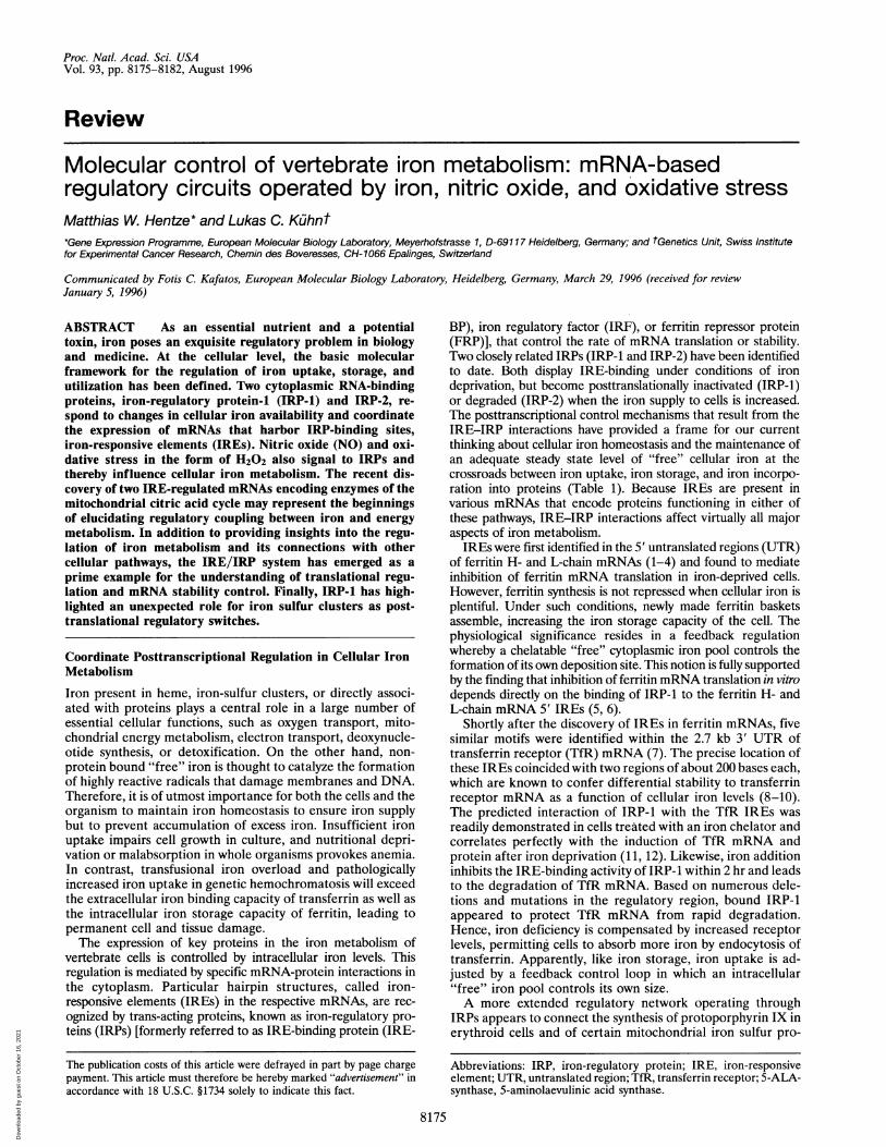
The IRE is highly conserved in evolution for any given geneand is remarkably similar between different genes harboringsuch elements, suggesting precise structural constraints in thebinding to IRPs. The canonical IRE consists of a stem-loopstructure with an upper double-stranded, 5-bp-long helix ofvariable sequence and a six-nucleotide loop with the consensussequence 5'-CAGUGN-3'. Below the paired stem, there isinvariantly a small asymmetrical bulge with an unpaired cyto-sine as the first nucleotide 5' of the stem (29). In certain IREs,this bulge is best drawn with a single unpaired C nucleotide; inother IREs, the C nucleotide and two additional 5' nucleotidesseem to oppose one free 3' nucleotide. The bulge may serveto adopt a specific bend in the IRE structure. Beyond thebulge, a second base paired region without evident sequenceconstraints appears to stabilize the IRE hairpin (Fig. 1).
Several studies have elucidated the structure and sequenceconstraints within which this motif serves as a recognition sitefor IRP-1. Whereas wild-type IREs show a very high affinitybinding to IRP-1 (Kd -10-30 pM) (30, 31), deletion of nucle-otides in the loop and the bulge region severely impair ordestroy the high affinity (4, 28, 32). Moreover, point mutantsdisrupting the upper helix were found to be nonfunctional,whereas complementary mutations restoring the doublestrandedness are tolerated (31, 33, 34). Exchanges in the firstfive loop nucleotides and the bulge nucleotides usually causea marked decrease in binding to IRP-1 (28, 31-33). However,such mutations may dramatically affect regulation in vivo, asdocumented in a case of dominant familial hyperferritinemiathat leads to early-onset cataract (35). In this family, theferritin L-chain gene carries an A2 -* G2 mutation in theIRE-loop, which lowers its affinity to IRP provoking excessiveferritin synthesis in affected individuals.The best IRP-1-binding sequences among IREs were se-
lected from a pool of 16,384 different IRE variants, where thesix nucleotides in the loop and the free 5' C nucleotides werepermutated randomly (28). The selected consensus corre-sponded precisely to the phylogenetically conserved IREmotif. However, a certain number of single or double muta-tions were tolerated at virtually every permutated position,with mutant IREs showing 2- to 10-fold diminished affinities.Most interestingly, this study provided evidence that the loop
A2 U4 A2 U4 loopCl. G5 Cl- G5
N6 N6N-N N.NN.N N-NN"N N'N upperN.N N-NNoN N-NC C
G C N - N bulgeU N-NN.N N-NN.N N.NN.N N.NN * N N * N lower
5'-N N-3' 5'-NN-3'
consensus IREwith *ferritin
bule
stem
stem
consensus IREwith single C
bulgeFIG. 1. IREs in eukaryotic mRNAs. A comparative analysis of the
different IREs reveals their conservation of sequence and structuredeterminants. Instead of the previously suggested structural represen-tation with a six nucleotide loop, the IRE is depicted in two alternativestructures that both feature hydrogen bonding between nucleotidesone and five of the loop region. N6 is less well-conserved, but is neverfound as a G nucleotide. This presumably avoids the formation oftetraloops through pairing with C1.
8176 Review: Hentze and Kiihn
Dow
nloa
ded
by g
uest
on
Oct
ober
16,
202
1
Proc. Natl. Acad. Sci. USA 93 (1996) 8177
is likely to be structured with an interaction between nucleo-tides one and five (Fig. 1). Double mutations at loop residuesCl and G5 altered to U1A5 were frequently selected; this classof mutants bound IRP-1 with an affinity comparable to that ofwild-type IRE (28). Likewise, a G'C5 (but not an A1U5) pairprovides a high affinity IRE (36). Base interactions in the loophave also been observed byNMR analysis of the IRE structure(37). The selex approach also yielded IRE mutants thatselectively bind to IRP-1, but not to IRP-2 (28), and a set ofdouble mutations that is specific for IRP-2 was recentlyidentified by a direct screening method (36). Thus, the con-straints for IRE recognition are closely related in the tworegulatory proteins, but are not precisely the same. Theexistence of IREs with private specificities for IRP-1 or IRP-2opens an opportunity to evaluate the contribution of eitherprotein to iron-dependent regulation. It remains of greatinterest to identify cellular mRNAs that are regulated vianoncanonical IREs. Computer searches for mutant IRE se-quences (15, 38) did thus far not reveal candidate mRNAs ofinterest (28, 36). Considering the relatively high affinity ofcertain IRE mutants, hitherto unrecognized mRNAs couldnevertheless be regulated through such noncanonical IREs,perhaps exclusively either by IRP-1 or by IRP-2.
Mechanism of Translational Control by IRPs
As early as 1966, iron regulation of ferritin expression wasconsidered to involve translational control (39). Munro andcolleagues showed that the activation of ferritin synthesis iniron-treated rats is insensitive to transcription inhibitors and isassociated with a shift of ferritin mRNA from translationallyinactive messenger ribonucleoprotein particles to polyribo-somes (40). They suggested that a protein (perhaps a ferritinsubunit) binds to the 5' UTR of ferritin mRNA and suppressesits translation in iron-deficient cells. Likewise, iron regulationof ferritin synthesis in reticulocytes of bullfrog tadpoles wasshown to be translational (41, 42). By the end of the 1980s, theregulation of ferritin translation by iron-controlled binding ofIRP-1 to an IRE in the 5' UTRs of ferritin H- and L-chainmRNAs had emerged as an extensively studied example oftranslational regulation of a mammalian gene (1, 3-6, 29,43-46). The IRE is necessary for the posttranscriptionalregulation of ferritin expression by iron (3, 45) and suffices toconfer IRP-mediated translational control to reporter mRNAsin transfected cells (1, 3, 4).
Plant and yeast cells lack endogenous IRP activity (5, 24, 25,46). Because the moderately stable hairpin of the IRE (c-7kcal/mol) does not impede translation per se, IRE-containingmRNAs are efficiently translated in Saccharomyces cerevisiae (25)and in cell-free extracts from wheat germ (40, 42). The translationof suitable reporter mRNAs is specifically repressed by coexpres-sion of IRP-1 in S. cerevisiae (25) or by addition of purified IRP-1to wheat germ extract (5, 46, 47). At least in vitro, IRP-mediatedtranslational repression does not require polyadenylylation of themRNA template (6). These findings indicate that the binding ofIRP-1 [or IRP-2 (48)] to the IRE suffices to regulate translationwithout requirement for additional cis-acting sequences or trans-acting factors. Nevertheless, adjacent non-IRE sequences mayaffect the function of an IRE (49, 50), and pre- and posttrans-lational mechanisms in addition to IRE-mediated translationalcontrol can quantitatively contribute to the iron regulation offerritin expression in vivo (39, 51-53).How does an IRE/IRP complex inhibit mRNA translation?
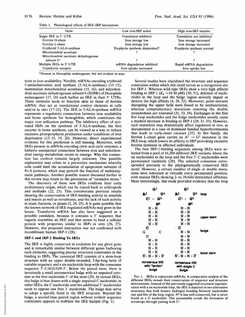
Using cell-free translation systems and sucrose gradient analyses,this complex was shown to prevent the stable association of thesmall ribosomal subunit (the 43S translation preinitiation com-plex) with mRNA (21). This result is in excellent accord with theposition dependence of IRE function: placement of an IRE >60nucleotides downstream from the cap structure permits 43Spreinitiation complex binding and profoundly diminishes trans-lational control by IRE/IRP complexes in transfected cells (refs.
52 and 54; N. K. Gray, N. U. Gray, and M.W.H., unpublishedwork). Not surprisingly, ferritin, erythroid 5-ALA-synthase, andmitochondrial aconitase IREs are all located .40 nucleotidesfrom the 5' end of the vertebrate mRNAs. The lack of efficienttranslational repression by downstream IRE/IRP complexesimplies that the translation initiation apparatus acquires theability to overcome otherwise inhibitory RNA/protein com-plexes after the 43S preinitiation complex is bound to the mRNA.The function of an IRE/IRP complex can also be exerted by thespliceosomal UlA protein or the bacteriophage MS2 coat proteinwhen bound to cognate binding sequences replacing the IRE.This experimental acquisition of translational repressor activityby RNA-binding proteins with physiological functions unrelatedto the control of eukaryotic mRNA translation was shown inmammalian and yeast cells (55) as well as in cell-free translationsystems from rabbit reticulocytes and wheat germ (56). As withthe IRE/IRP interaction, the function of the complex is positiondependent (56) and prevents the stable association of the smallribosomal subunit (21). These results suggest that IRE/IRPcomplexes act as steric inhibitors of 43S preinitiation complexbinding (Fig. 2). Moreover, these results argue that this mechanismreflects a more general mode for translational control that couldalso explain the translational regulation of other mRNAs (57).Mechanisms Controlling Transferrin Receptor mRNAStability
TfR protein expression in proliferating cells was reported inthe early 1980s to respond to variations in iron availability (58,59). After the isolation of TfR cDNAs (60-62), it was notedthat iron chelators increase and iron salts or hemin diminishTfR mRNA levels in cultured cells (63). Surprisingly, theregulation was not primarily the result of transcriptionalcontrol as initially expected (64), but could be ascribed tosequences in the 3' UTR of the TfR mRNA (8). Deletion ofthe 3' UTR yields a high, nonregulated expression of TfR intransfectant cell lines. The TfR mRNA 3' UTR is also suffi-cient to confer iron-dependent regulation to a chimeric tran-script of HLA-A2 (9) or human growth hormone (7). Deletionmapping of the regulatory sequences identified two necessaryareas of about 200 nucleotides each, separated by some 250nucleotides (9, 10). These relevant regulatory sequences, butnot the adjacent 3' UTR regions, display -94% sequenceidentity between human and rat (65) mRNAs and 89% be-tween human and chicken TfR mRNAs (12, 66), a conserva-tion that exceeds that of the coding regions.
This bipartite regulatory region of TfR mRNA is more com-plex than that of ferritin mRNA, and both RNA-stabilizing and-destabilizing elements have been identified (Fig. 3). Because it israther large, its structure cannot be predicted with certainty. Inone model, the two regions of 200 nucleotides each interact
aoc. ternary
FIG. 2. Translational regulation by IRE/IRP interactions in the 5'UTR. The 5' UTR of an mRNA is depicted with the IRE locatedwithin the 5' proximal 50-60 nucleotides, which represent the entrysite for the 43S complex (40S ribosomal subunit plus ternary complexand associated translation initiation factors). The 43S complex andIRP-1/2 are shown to compete for binding to their respective bindingsites; thus, IRP-1/2 bound to the IRE prevents the stable associationof the 43S complex and translation of this mRNA.
Review: Hentze and Kiihn
Dow
nloa
ded
by g
uest
on
Oct
ober
16,
202
1
8178 Review: Hentze and Kuhn
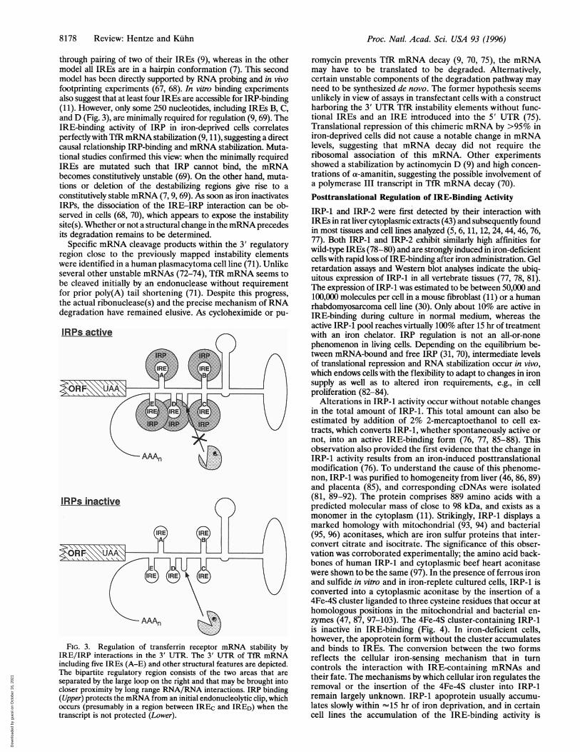
through pairing of two of their IREs (9), whereas in the othermodel all IREs are in a hairpin conformation (7). This secondmodel has been directly supported by RNA probing and in vivofootprinting experiments (67, 68). In vitro binding experimentsalso suggest that at least four IREs are accessible for IRP-binding(11). However, only some 250 nucleotides, including IREs B, C,and D (Fig. 3), are minimally required for regulation (9, 69). TheIRE-binding activity of IRP in iron-deprived cells correlatesperfectly with TfR mRNA stabilization (9, 1 1), suggesting a directcausal relationship IRP-binding and mRNA stabilization. Muta-tional studies confirmed this view: when the minimally requiredIREs are mutated such that IRP cannot bind, the mRNAbecomes constitutively unstable (69). On the other hand, muta-tions or deletion of the destabilizing regions give rise to aconstitutively stable mRNA (7, 9, 69). As soon as iron inactivatesIRPs, the dissociation of the IRE-IRP interaction can be ob-served in cells (68, 70), which appears to expose the instabilitysite(s). Whether or not a structural change in themRNA precedesits degradation remains to be determined.
Specific mRNA cleavage products within the 3' regulatoryregion close to the previously mapped instability elementswere identified in a human plasmacytoma cell line (71). Unlikeseveral other unstable mRNAs (72-74), TfR mRNA seems tobe cleaved initially by an endonuclease without requirementfor prior poly(A) tail shortening (71). Despite this progress,the actual ribonuclease(s) and the precise mechanism of RNAdegradation have remained elusive. As cycloheximide or pu-
IRPs active
AAAn
FIG. 3. Regulation of transferrin receptor mRNA stability byIRE/IRP interactions in the 3' UTR. The 3' UTR of TfR mRNAincluding five IREs (A-E) and other structural features are depicted.The bipartite regulatory region consists of the two areas that are
separated by the large loop on the right and that may be brought intocloser proximity by long range RNA/RNA interactions. IRP binding(Upper) protects the mRNA from an initial endonucleolytic clip, whichoccurs (presumably in a region between IREc and IRED) when thetranscript is not protected (Lower).
romycin prevents TfR mRNA decay (9, 70, 75), the mRNAmay have to be translated to be degraded. Alternatively,certain unstable components of the degradation pathway mayneed to be synthesized de novo. The former hypothesis seemsunlikely in view of assays in transfectant cells with a constructharboring the 3' UTR TfR instability elements without func-tional IREs and an IRE introduced into the 5' UTR (75).Translational repression of this chimeric mRNA by >95% iniron-deprived cells did not cause a notable change in mRNAlevels, suggesting that mRNA decay did not require theribosomal association of this mRNA. Other experimentsshowed a stabilization by actinomycin D (9) and high concen-trations of a-amanitin, suggesting the possible involvement ofa polymerase III transcript in TfR mRNA decay (70).Posttranslational Regulation of IRE-Binding Activity
IRP-1 and IRP-2 were first detected by their interaction withIREs in rat liver cytoplasmic extracts (43) and subsequently foundin most tissues and cell lines analyzed (5, 6, 11, 12, 24, 44, 46, 76,77). Both IRP-1 and IRP-2 exhibit similarly high affinities forwild-type IREs (78-80) and are strongly induced in iron-deficientcells with rapid loss of IRE-binding after iron administration. Gelretardation assays and Western blot analyses indicate the ubiq-uitous expression of IRP-1 in all vertebrate tissues (77, 78, 81).The expression of IRP-1 was estimated to be between 50,000 and100,000 molecules per cell in a mouse fibroblast (11) or a humanrhabdomyosarcoma cell line (30). Only about 10% are active inIRE-binding during culture in normal medium, whereas theactive IRP-1 pool reaches virtually 100% after 15 hr of treatmentwith an iron chelator. IRP regulation is not an all-or-nonephenomenon in living cells. Depending on the equilibrium be-tween mRNA-bound and free IRP (31, 70), intermediate levelsof translational repression and RNA stabilization occur in vivo,which endows cells with the flexibility to adapt to changes in ironsupply as well as to altered iron requirements, e.g., in cellproliferation (82-84).
Alterations in IRP-1 activity occur without notable changesin the total amount of IRP-1. This total amount can also beestimated by addition of 2% 2-mercaptoethanol to cell ex-tracts, which converts IRP-1, whether spontaneously active ornot, into an active IRE-binding form (76, 77, 85-88). Thisobservation also provided the first evidence that the change inIRP-1 activity results from an iron-induced posttranslationalmodification (76). To understand the cause of this phenome-non, IRP-1 was purified to homogeneity from liver (46, 86, 89)and placenta (85), and corresponding cDNAs were isolated(81, 89-92). The protein comprises 889 amino acids with apredicted molecular mass of close to 98 kDa, and exists as amonomer in the cytoplasm (11). Strikingly, IRP-1 displays amarked homology with mitochondrial (93, 94) and bacterial(95, 96) aconitases, which are iron sulfur proteins that inter-convert citrate and isocitrate. The significance of this obser-vation was corroborated experimentally; the amino acid back-bones of human IRP-1 and cytoplasmic beef heart aconitasewere shown to be the same (97). In the presence of ferrous ironand sulfide in vitro and in iron-replete cultured cells, IRP-1 isconverted into a cytoplasmic aconitase by the insertion of a4Fe-4S cluster liganded to three cysteine residues that occur athomologous positions in the mitochondrial and bacterial en-zymes (47, 87, 97-103). The 4Fe-4S cluster-containing IRP-1is inactive in IRE-binding (Fig. 4). In iron-deficient cells,however, the apoprotein form without the cluster accumulatesand binds to IREs. The conversion between the two formsreflects the cellular iron-sensing mechanism that in turncontrols the interaction with IRE-containing mRNAs andtheir fate. The mechanisms by which cellular iron regulates theremoval or the insertion of the 4Fe-4S cluster into IRP-1remain largely unknown. IRP-1 apoprotein usually accumu-lates slowly within "15 hr of iron deprivation, and in certaincell lines the accumulation of the IRE-binding activity is
Proc. Natl. Acad. Sci. USA 93 (1996)
Dow
nloa
ded
by g
uest
on
Oct
ober
16,
202
1
Proc. Natl. Acad. Sci. USA 93 (1996) 8179
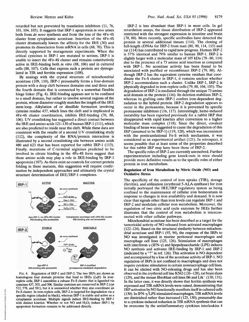
retarded but not prevented by translation inhibitors (11, 76,101, 104, 105). It suggests that IRP-1 apoprotein in vivo arisesboth from de novo synthesis and frotn the loss of the 4Fe-4Scluster from cytoplasmic aconitase. Insertion of the 4Fe-4Scluster dramatically lowers the affinity for the IRE (30) andpromotes its dissociation from mRNA in cells (68, 70). This isdirectly supported by mutagenesis experiments. When thecritical cysteines in IRP-1 are mutated to serines, IRP-1 isunable to insert the 4Fe-4S cluster and remains constitutivelyactive in IRE-binding both in vitro (88, 106) and in culturedcells (88, 107). Cells that express such a protein are dysregu-lated in TfR and ferritin expression (108).By analogy with the crystal structure of mitochondrial
aconitase (109, 110), IRP-1 presumably forms a four-domainprotein with a deep cleft between domains one and three andthe fourth domain that is connected by a somewhat flexiblehinge linker (Fig. 4). IRE-binding appears not to be confinedto a small domain, but rather to involve several regions of theprotein, whose diameter roughly matches the length of the IREstem-loop. Alkylation of or disulfide formation involvingcysteine residue 437, which lines the cleft and is implicated in4Fe-4S cluster coordination, inhibits IRE-binding (76, 88,106). UV crosslinking has suggested a direct contact betweenthe IRE and amino acids 121-130 of human IRP-1 (111), whichare also predicted to reside near the cleft. While these data areconsistent with the results of a second UV crosslinking study(112), the complexity of the RNA/protein interaction isillustrated by a second crosslinking site between amino acids480 and 623 that has been reported for rabbit IRP-1 (113).Finally, mutations of C-terminal arginines predicted to beinvolved in citrate binding in the 4Fe-4S form suggest thatthese amino acids may play a role in IRE-binding by IRP-1apoprotein (107). As there exist no controls for correct proteinfolding in these mutants, this suggestion will require confir-mation by independent approaches and ultimately the crystalstructure determination of IRE/IRP-1 complexes.
RP-1] ACTIVE INACTIVE
Apo-IRP-1: no [4Fe-4S] cluster Cytoplasmic aconitase with [4Fe-4S] cluster
IRE-binding site accessible IRE-binding site not accessible
STABLE UNSTABLE
7mG denovo synthesis
U L Fe2+(2-4h)'G'
tDe
Apo-IRP-2 IRP-2 modified
IRE-binding site accessible (proteasome-mediated) degradation
FIG. 4. Regulation of IRP-1 and IRP-2. The two IRPs are shown as
homologous four domain proteins that bind to IREs (Left). In iron-replete cells, IRP-1 assembles a cubane Fe-S cluster that is liganded viacysteines 437, 503, and 506. Similar cysteines are conserved in IRP-2 (cys512, 578, and 581), but it is unresolved whether they also coordinate an
Fe-S cluster. In iron-replete cells, IRP-2 is targeted for degradation via a
specific region (shaded in black), whereas IRP-1 is stable and active as a
cytoplasmic aconitase. Multiple signals induce IRE-binding by IRP-1with distinct kinetics. Whether or not NO and H202 induce IRP-1 byapoprotein formation remains to be addressed directly.
IRP-2 is less abundant than IRP-1 in most cells. In gelretardation assays, the tissue distribution of IRP-2 appearedrestricted with the strongest expression in intestine and brain(78, 80). More recently, specific antibodies have detected theprotein in several additional tissues (114). The cloning offull-length cDNAs for IRP-2 from man (80, 90, 114, 115) andrat (114) has contributed to rapid new progress. Human IRP-2is 57% identical and 79% similar to human IRP-1. IRP-2 isslightly larger with a molecular mass of 105 kDa (78-80, 114)due to the presence of a 73 amino acid insertion as comparedwith IRP-1. No aconitase activity has been found to beassociated with purified or recombinant IRP-2 (79, 80). Al-though IRP-2 has the equivalent cysteine residues that coor-dinate the Fe-S cluster in IRP-1, it remains unclear whetherIRP-2 accommodates such a cluster. Unlike IRP-1, IRP-2 isphysically degraded in iron-replete cells (79, 80, 104, 105). Thedegradation of IRP-2 is mediated through the unique 73 aminoacid domain in the protein (116). Its deletion stabilizes IRP-2,whereas its grafting onto IRP-1 confers iron-dependent deg-radation to the hybrid protein. IRP-2 degradation appears tooccur in the proteasome, because it is prevented by specificproteasome inhibitors (116, 117). Interestingly, heme-inducedinstability has been reported previously for a rabbit IRP thatdisappeared with rapid kinetics after conversion to a highermolecular mass complex (118). Since the direct, covalentbinding of heme was suggested to cause the degradation of thisIRP (assumed to be IRP-1) (119, 120), which was inconsistentwith the posttranslational Fe-S switch mechanism, it wasconsidered as an experimental artifact (121). In retrospect, itseems possible that at least some of the properties describedfor this rabbit IRP may have been those of IRP-2.The specific roles of IRP-2 are currently unresolved. Further
experimentation including gene knock-outs in mice shouldprovide more definitive results as to the specific roles of eitherprotein in iron metabolism.
Regulation of Iron Metabolism by Nitric Oxide (NO) andOxidative Stress
The specificity of the control of iron uptake (TfR), storage(ferritin), and utilization (erythroid 5-ALA-synthase) for ironinitially portrayed the IRE/IRP regulatory system as beingconfined to the maintenance of cellular iron homeostasis inresponse to changes in iron availability and demand. It is nowclear that signals other than iron levels can regulate IRP-1 andIRP-2 and modulate cellular iron metabolism. Moreover, theregulation of two citric acid cycle enzymes by IREs furtherillustrates that the control of iron metabolism is intercon-nected with other cellular pathways.
Mitochondrial aconitase has been described as a target for thetumoricidal activity of NO released from activated macrophages(122-124). Based on the structural similarity between mitochon-drial aconitase and IRP-1 (93, 94), the response of the IRPs toNO was investigated in murine peritoneal macrophages andmacrophage cell lines (125, 126). Stimulation of macrophageswith interferon--y (IFN--y) and lipopolysaccharide (LPS) inducesNO synthesis and activates IRE-binding by IRP-1 and IRP-2(indicated by a "?" in ref. 126). This activation is NO dependentand accompanied by a loss of the aconitase activity of IRP-1. NOregulation of IRPs is not confined to macrophages and does notrequire cytokine stimulation in certain nonmacrophage cell lines.It can be elicited with NO-releasing drugs and has also beenobserved in the erythroid cell line K562 (126-128), rat brain slices(129), and the mouse fibroblast cell lines B6 and Ltk- (130, 131).In the latter case, it was directly shown that ferritin synthesis wasrepressed and TfR mRNA levels were raised, demonstrating thatIRP activation byNO functionally manifests itself in cultured cells(130). In IFN-,y/LPS-stimulated macrophages, TfRmRNA levelsare diminished rather than increased (125, 130), presumably dueto a cytokine-induced reduction in TfR mRNA synthesis that canbe overcome by the antiinflammatory cytokines interleukins 4
Review: Hentze and Kiihn
I|ERP 2l
Dow
nloa
ded
by g
uest
on
Oct
ober
16,
202
1
Proc. Natl. Acad. Sci. USA 93 (1996)
and 13 (G. Weiss, C. Bogdan, and M.W.H., unpublished results).The regulatory crosstalk between iron metabolism and NO inmacrophages is further highlighted by the transcriptional regu-lation of the inducible NO synthase gene (NOS 2) by iron (132).Thus, in macrophages, the regulation of iron metabolism and NOis tightly connected. Future work will have to address the role ofthis regulatory connection in animal models, where macrophagesare exposed to the influence of multiple, partially antagonisticcytokines.Another important biological link exists between iron me-
tabolism and oxidative stress. This link is most directly illus-trated by Fenton chemistry, which yields the highly reactiveand toxic hydroxyl radical from the reaction of Fe2+ (or Cu+)with H202. Much of the damage inflicted to cells by oxidativestress is mediated by iron, as has been suggested for athero-sclerotic lesions (133) and in hypoxia-reperfusion injury (134,135). In bacteria, oxidative stress and the regulation of ironmetabolism are tightly coupled (136, 137). In mammalian celllines, IRP-1 establishes a direct regulatory linkage betweeniron metabolism and oxidative stress (138, 139). WhereasEscherichia coli aconitase is inactivated by superoxide andperoxynitrite (140), the cytoplasmic aconitase (IRP-1) re-sponds strongly to H202 in living cells but not in vitro (138,139). This response involves the inactivation of its aconitaseactivity (139) and the rapid stimulation of IRE-binding inseveral mammalian cell lines (130, 138, 139), leading toincreased TfR mRNA levels and repressed ferritin synthesis(139). The H202 responsiveness of IRP-1 may also explain theincrease in ferritin synthesis observed after ascorbic acidaddition to cultured K562 cells (141), because the antioxidantvitamin may counteract the effects of H202 on IRP-1. H202activates almost exclusively IRP-1 (131), possibly because thecellular pool of preformed activatable IRP-2 is very small.How do the small diffusible molecules NO and H202 trigger
IRP activation? Both molecules activate IRP-1 by a cyclohexim-ide-insensitive posttranslational mechanism (139), whereas IRP-2activation by NO requires de novo protein synthesis (131). Aftertreatment of cells with NO or H202, the 4Fe-4S cluster-dependent aconitase activity is inhibited (125, 139). Citrate thatcan bind to a 3Fe-4S or a 4Fe-4S cluster of IRP-1 and preventIRE-binding after treatment of extract with reducing agents (87)fails to inhibit IRE-binding ofNO- or H202-activated IRP-1 (131,139), suggesting that the cluster is disassembled in NO- orH202-treated cells beyond the 3Fe-4S state and perhaps absent.However, direct proof for the conversion of IRP-1 into theapoprotein form by NO and H202 is currently still lacking.
Surprisingly, the activation of IRP-1 by NO differs significantlyfrom H202 activation and more closely resembles the pattern ofactivation observed in iron-deficient cells. Iron deficiency and NOrequire up to 15 hr for complete activation of IRP-1 (and IRP-2)(11, 76, 131), whereas H202 achieves maximal IRP-1 activationwithin '60 min (138, 139). Furthermore, IRP-1 activation byH202 is diminished by the type I/Ila protein phosphatase inhib-itor okadaic acid in B6 cells, whereas the activation by irondeficiency and NO is insensitive to this drug (131, 139). Finally,iron deficiency or NO has to be continuously present for IRPactivation, whereas H202 treatment of cells for as little as 10-15min induces a cellular response program that leads to completeand sustained IRP-1 activation within 60 min even after H202 hasbeen removed (131). These results suggest that the activation byH202 does not simply result from a direct oxidant attack onIRP-1, but rather involves additional (phosphorylation depen-dent) cellular activities for IRP-1 activation, probably by accel-erated cluster removal. Future studies will have to directlycompare the turnover rates of the 4Fe-4S cluster of IRP-1 in cellsexposed to H202, NO, and iron deficiency. Although the H202response has been observed in several different cell lines (131,138, 139), the stress-induced IRP-1 activation pathway may notexist in all cells.
Treatment of HL-60 cells with phorbol 12-myristate 13-acetate (PMA) was reported to phosphorylate IRP-1 viaprotein kinase C and to double its IRE-binding activity within30 min (142). Given that PMA can induce the formation ofreactive oxygen intermediates (143) and is known to activatethe oxidant stress-responsive transcription factor NFKB (144),the roles of H202, PMA, and protein kinase C in IRP-1activation may be related.How then does NO activate IRP-1 and IRP-2? Drapier et al.
(125) found that exposure of recombinant IRP-1 to NO gas underanaerobic conditions completely inhibits its aconitase activity andpartially (-20%) activates IRE-binding. In contrast, lack ofinactivation of the aconitase activity of recombinant humanIRP-1 by NO gas under apparently similar conditions has alsobeen claimed (140), leading to the suggestion that peroxynitrite,a reaction product of NO and superoxide, represents the chem-ically relevant effector. However, the authors did not assay forchanges in IRE-binding or describe the purity of the IRP-1preparations that were used. In vivo, activation of IRE-binding byIRP-1 is favored when cells are treated with agents that releaseNO (SNAP, NOC-18) as compared with treatments that favorperoxynitrite formation (SIN-1, SNAP plus paraquat) (127, 131).Moreover, while the in vitro studies have focused attention on adirect effect ofNO (or a chemical derivative) on (the Fe-S clusterof) IRP-1, the slow, iron deficiency-like kinetics of IRP-1 activa-tion in vivo raise the possibility that NO may act more indirectlyby reducing the availability of cellular iron or sulfide used forcluster assembly. This interpretation would offer a commonmechanism for the regulation of IRP-1 and IRP-2 by NO, butmore work is needed before we understand whether the ironsulfur cluster of IRP-1 senses or merely responds to changes inH202 and NO concentration.Perspectives
This summary has illustrated two different aspects of theIRE/IRP regulatory system: (i) its utility as a model system tounravel mechanisms of posttranscriptional gene regulationand (ii) its biological role as a master regulator for the controlof cellular iron homeostasis as well as its more recentlyconsidered role as a possible regulatory node for interconnec-tions between iron metabolism and other cellular pathways.Further analysis of these interconnections will likely also leadto a better understanding of pathological processes in whichiron may participate, such as radical-mediated tissue damageor neurodegenerative processes (134, 135, 145).To further dissect the translational control mechanism, it will
be important to understand how RNA/protein complexes such asIRE/IRP interfere with the binding of the small ribosomalsubunit. Do IRPs stabilize the secondary structure of the IRE toprevent its unwinding or does the complex impede the functionof translation initiation factors (such as those binding to the capstructure) required for the subsequent binding of the 43S preini-tiation complex? How does the translation initiation apparatusovercome a downstream IRE/IRP complex? This may yieldinteresting information on how mRNA translation can occur inan environment abounding with RNA-binding proteins. Regard-ing the regulation of TfR mRNA stability, the two critical futurequestions pertain to the identification and characterization of thenuclease(s) against which IRE/IRP complexes protect the RNAas well as the secondary and tertiary structure of the TfR mRNAsubstrate that this nuclease recognizes and cuts. Since it is unlikelythat a complete nuclease system evolved solely for the purpose ofdegrading TfR mRNA, it will also be interesting to identify thesubstrate spectrum of this nuclease.While the role of iron sulfur clusters in electron transfer or
enzymatic reactions is well-documented, we are still just begin-ning to understand how iron sulfur clusters act as regulatoryswitches. Regarding IRP-1, two aspects must be distinguished: (i)how the presence of the cluster inhibits IRE-binding and (ii) howdifferent cellular signals operate the switch between 4Fe-4S
8180 Review: Hentze and Kiihn
Dow
nloa
ded
by g
uest
on
Oct
ober
16,
202
1
Proc. Natl. Acad. Sci. USA 93 (1996) 8181
protein and apoprotein. The former question is intimately con-nected with the elucidation of the structure of IRP-1 and of theIRE/IRP-1 complex. Answering the latter question will neces-sitate a biochemical definition factors involved in cluster assemblyand disassembly. Is cluster formation a "spontaneous" processdriven by the availability of ferrous iron and sulfide or is itcofactor mediated? At what rate does the cluster come apart?Can this process be accelerated? Is the cluster a sensor of thesignals that affect it or merely a respondent to processes triggeredby these signals? Finding the answers to these questions will likelyalso impact on other regulatory systems, as additional examplesof iron sulfur cluster-controlled regulatory proteins have recentlybegun to emerge; this group includes the prokaryotic oxidantstress response factor soxR (a DNA-binding protein) (146), theoxygen-regulated transcription factor FNR from E. coli (147),and a family of transcription factors that harbors so-called LIMdomains (for the founding members lin-11, Isl-1, and mec-3) (148).There is also much left to be learned about the regulation of IRP-2.Is the formation of an iron sulfur cluster part of the process leadingto its degradation? How does the 73 amino acid domain unique toIRP-2 mark the protein for degradation in iron-replete cells? Howis degradation prevented in iron-deficient cells?The IRE/IRP system is beginning to emerge from its confines
of representing a closed circuit "housekeeping" system for ironhomeostasis, as illustrated by its interconnections with the citricacid cycle or the cytokine/NO regulation in activated macro-phages. Much information will have to be gathered on the rolesthat the two diffusible messengers, NO and H202, play inregulating IRE-containing mRNAs. IRP-1 activation by H202decreases ferritin and increases TfR expression (139). An in-crease in iron uptake and a decreased potential to store intra-cellular iron in ferritin should raise the level of "free" iron that canreact with H202 and yield hydroxyl radicals. Thus, this regulatoryloop may catalyze tissue damage rather than be protective. Thepathophysiological implications of this situation, particularly invascular disorders such as reperfusion injury where iron-derivedreactive oxygen species appear to play an important role (134,135), will warrant further investigations. Similarly, we have tolearn how tissues initiate counterbalancing protective mecha-nisms, as indicated by the induction of ferritin expression >12 hrafter an oxidant injury (149-151). What are the physiologicalbenefits of H202-mediated IRP-1 activation?A rather speculativelead may be offered by the IREs that serve to repress thetranslation of two citric acid cycle enzymes after IRP activationin mammals and invertebrates (17, 18). The citric acid cycle fuelsthe respiratory chain with reducing equivalents and thus contrib-utes to the formation of reactive oxygen intermediates duringmitochondrial ATP synthesis. Could the activation of IRP-1 byexcess H202 serve as a metabolic homeostat to balance respira-tory ATP synthesis and oxidative stress by exerting negativefeedback on the citric acid cycle? The regulation of IRP-1 byendogenously generated reactive oxygen intermediates afterpharmacological modulation of respiratory chain activity is atleast consistent with this possibility (131).
Finally, the appreciation of the role of transferrin in extracel-lular iron transport is in stark contrast to the lack of molecularunderstanding of intracellular iron transport. Neither the processby which iron leaves the endosome nor the traffic of iron betweenthe cytoplasm and mitochondria are well-understood. Does ci-trate play a role in intracellular iron transport and, if so, does thisprovide a rationale for the regulation of both cellular aconitases[IRP-1 (via the Fe-S cluster switch) and mitochondrial aconitase(via the 5' UTR IRE)] by iron? With so many importantquestions left to be answered and in light of the fundamental rolesthat iron plays in health and disease, the IRE/IRP systemremains a major challenge.
1. Aziz, N. & Munro, H. N. (1987) Proc. Natl. Acad. Sci. USA 84,8478-8482.2. Leibold, E. A. & Munro, H. N. (1987) J. Bio. Chem. 262, 7335-7341.
3. Hentze, M. W., Rouault, T. A., Caughman, S. W., Dancis, A., Harford,J. B. & Klausner, R. D. (1987) Proc. Natl. Acad. Sci. USA 84, 6730-6734.
4. Hentze, M. W., Caughman, S. W., Rouault, T. A., Barriocanal, J. G., Dancis,A., Harford, J. B. & Klausner, R. D. (1987) Science 238, 1570-1572.
5. Walden, W. E., Daniels-McQueen, S., Brown, P. H., Gaffield, L., Russell,D. A., Bielser, D., Bailey, L. C. & Thach, R. E. (1988) Proc. Natl. Acad.Sci. USA 85, 9503-9507.
6. Brown, P. H., Daniels-McQueen, S., Walden, W. E., Patino, M. M., Gaffield,L., Bielser, D. & Thach, R. E. (1989) . Biol. Chem. 264, 13383-13386.
7. Casey, J. L., Hentze, M. W., Koeller, D. M., Caughman, S. W., Rouault,T. A., Klausner, R. D. & Harford, J. B. (1988) Science 240, 924-928.
8. Owen, D. & Kuhn, L. C. (1987) EMBO J. 6, 1287-1293.9. Mullner, E. W. & Kuhn, L. C. (1988) Cell 53, 815-825.
10. Casey, J. L., Di Jeso, B., Rao, K., Klausner, R. D. & Harford, J. B. (1988)Proc. Natl. Acad. Sci. USA 85, 1787-1791.
11. Mullner, E. W., Neupert, B. & Kuhn, L. C. (1989) Cell 58, 373-382.12. Koeller, D. M., Casey, J. L., Hentze, M. W., Gerhardt, E. M., Chan, L.-N.,
Klausner, R. D. & Harford, J. B. (1989) Proc. Natl. Acad. Sci. USA 86,3574-3578.
13. May, B. K., Bhasker, C. R., Bawden,.M. J. & Cox, T. C. (1990) MoI. BioLMed. 7, 405-421.
14. Cox, T. C., Bawden, M. J., Martin, A. & May, B. K. (1991) EMBO J. 10,1891-1902.
15. Dandekar, T., Stripecke, R., Gray, N. K, Goossen, B., Constable, A.,Johansson, H. E. & Hentze, M. W. (1991) EMBO J. 10, 1903-1909.
16. Zheng, L., Kennedy, M. C., Blondin, G. A., Beinert, H. & Zalkin, H.(1992) Arch. Biochem. Biophys. 299, 356-360.
17. Kohler, S. A., Henderson, B. R. & Kuhn, L. C. (1995) J. Biol. Chem. 270,30781-30786.
18. Gray, N. K, Pantopoulos, K., Dandekar, T., Ackrell, B. A. C. & Hentze,M. W. (1996) Proc. Natl. Acad. Sci. USA 93, 4925-4930.
19. Melefors, O., Goossen, B., Johansson, H. E., Stripecke, R., Gray, N. K. &Hentze, M. W. (1993) J. Biol. Chem. 268, 5974-5978.
20. Bhasker, C. R., Burgiel, G., Neupert, B., Emery-Goodman, A., Kuhn,L. C. & May, B. K. (1993) J. Bio. Chem. 268, 12699-12705.
21. Gray, N. K & Hentze, M. W. (1994) EMBO J. 13, 3882-3891.22. Von Darl, M., Harrison, P. M. & Bottke, W. (1994) Eur. J. Biochem. 222,
353-366.23. Dunkov, B. C., Zhang, D., Choumarov, K., Winzerling, J. J. & Law, J. H.
(1995) Arch. Insect Biochem. Physiol. 29, 293-307.24. Rothenberger, S., Mullner, E. W. & Kuhn, L. C. (1990) NucleicAcids Res.
18, 1175-1179.25. Oliveira, C. C., Goossen, B., Zanchin, N. I. T., McCarthy, J. E. G., Hentze,
M. W. & Stripecke, R. (1993) Nucleic Acids Res. 21, 5316-5322.26. Cox, L. A. & Adrian, G. S. (1993) Biochemistry 32, 4738-4745.27. Cox, L. A., Kennedy, M. C. & Adrian, G. S. (1995) Biochem. Biophys. Res.
Commun. 212, 925-932.28. Henderson, B. R., Menotti, E., Bonnard, C. & Kuhn, L. C. (1994) J. Biol.
Chem. 269, 17481-17489.29. Hentze, M. W., Caughman, S. W., Casey, J. L., Koeller, D. M., Rouault,
T. A., Harford, J. B. & Klausner, R. D. (1988) Gene 72, 201-208.30. Haile, D. J., Hentze, M. W., Rouault, T. A., Harford, J. B. & Klausner,
R. D. (1989) Mol. Cell. Biol. 9, 5055-5061.31. Barton, H. A., Eisenstein, R. S., Bomford, A. & Munro, H. N. (1990)
J. Biol. Chem. 265, 7000-7008.32. Jaffrey, S. R., Haile, D. J., Klausner, R. D. & Harford, J. B. (1993) Nucleic
Acids Res. 21, 4627-4631.33. Leibold, E. A., Laudano, A. & Yu, Y. (1990) NucleicAcids Res. 18,1819-1824.34. Bettany, A. J. E., Eisenstein, R. S. & Munro, H. N. (1992) J. Biol. Chem.
267, 16531-16537.35. Beaumont, C., Leneuve, P., Devaux, I., Scoazec, J.-Y., Berthier, M., Loiseau,
M.-N., Grandchamp, B. & Bonneau, D. (1995) Nat. Genet. 11, 444-446.36. Henderson, B. R., Menotti, E. & Kuhn, L. C. (1996) J. BioL Chem. 271,
4900-4908.37. Sierzputowska-Gracz, H., McKenzie, R. A. & Theil, E. C. (1995) Nucleic
Acids Res. 23, 146-153.38. Dandekar, T. & Hentze, M. W. (1995) Trends Genet. 11, 45-50.39. Drysdale, J. W. & Munro, H. N. (1966) J. Biol. Chem. 241, 3630-3637.40. Zahringer, J., Baliga, B. S. & Munro, H. N. (1976) Proc. Natl. Acad. Sci.
USA 73, 857-861.41. Shull, G. E. & Theil, E. C. (1982) J. Biol. Chem. 257, 14187-14191.42. Shull, G. E. & Theil, E. C. (1983) J. Biol. Chem. 258, 7921-7923.43. Leibold, E. A. &-Munro, H. N. (1988) Proc. Natl. Acad. Sci. USA 85,
2171-2175.44. Rouault, T. A., Hentze, M. W., Caughman, S. W., Harford, J. B. &
Klausner, R. D. (1988) Science 241, 1207-1210.45. Caughman, S. W., Hentze, M. W., Rouault, T. A., Harford, J. B. &
Klausner, R. D. (1988) J. Biol. Chem. 263, 19048-19052.46. Walden, W. E., Patino, M. M. & Gaffleld, L. (1989) J. Biol. Chem. 264,
13765-13769.47. Gray, N. K., Quick, S., Goossen, B., Constable, A., Hirling, H., Kuhn, L. C.
& Hentze, M. W. (1993) Eur. J. Biochem. 218, 657-667.48. Kim, H. Y., Klausner, R. D. & Rouault, T. A. (1995) J. Biol. Chem. 270,
4983-4986.49. Dickey, L. F., Wang, Y.-H., Shull, G. E., Wortman III, I. A. & Theil, E. C.
(1988) J. Biol. Chem. 263, 3071-3074.
Review: Hentze and Kiihn
Dow
nloa
ded
by g
uest
on
Oct
ober
16,
202
1
8182 Review: Hentze and Kuhn
50. Dix, D. J., Lin, P. N., McKenzie, A. R., Walden, W. E. & Theil, E. C.(1993) J. Moi. Biol. 231, 230-240.
51. White, K. & Munro, H. N. (1988) J. Bio. Chem. 263, 8938-8942.52. Goossen, B. & Hentze, M. W. (1992) Moi. Cell. Bio. 12, 1959-1966.53. Coulson, R. M. R. & Cleveland, D. W. (1993) Proc. NatL. Acad. Sci. USA
90, 7613-7617.54. Goossen, B., Caughman, S. W., Harford, J. B., Klausner, R. D. & Hentze,
M. W. (1990) EMBO J. 9, 4127-4133.55. Stripecke, R., Oliveira, C. C., McCarthy, J. E. G. & Hentze, M. W. (1994)
Mol. Cell. Biol. 14, 5898-5909.56. Stripecke, R. & Hentze, M. W. (1992) Nucleic Acids Res. 20, 5555-5564.57. Gray, N. K & Hentze, M. W. (1994) Mol. Biol. Reports 19, 195-200.58. Pelicci, P. G., Tabilio, A., Thomopoulos, P., Titeux, M., Vainchenker, W.,
Rochant, H. & Testa, U. (1982) FEBS Lett. 145, 350-354.59. Ward, J. H., Kushner, J. P. & Kaplan, J. (1982) J. Biol. Chem. 257,
10317-10323.60. Kuhn, L. C., McClelland, A. & Ruddle, F. H. (1984) Cell 37, 95-103.61. McClelland, A., Kuhn, L. C. & Ruddle, F. H. (1984) Cell 39, 267-274.62. Schneider, C., Owen, M. J., Banville, D. & Williams, J. G. (1984) Nature
(London) 311, 675-678.63. Rao, K. K., Shapiro, D., Mattia, E., Bridges, K & Klausner, R. (1985) Mol.
Cell. Biol. 5, 595-600.64. Rao, K. K., Harford, J. B., Rouault, T., McClelland, A., Ruddle, F. H. &
Klausner, R. D. (1986) Mol. Cell. Biol. 6, 236-240.65. Roberts, K. P. & Griswold, M. D. (1990) Mol. Endocrinol. 4, 531-542.66. Chan, L.-N. L., Grammatikakis, N., Banks, J. M. & Gerhardt, E. M. (1989)
Nucleic Acids Res. 17, 3763-3771.67. Horowitz, J. A. & Harford, J. B. (1992) New Biol. 4, 330-338.68. Bertrand, E., Fromont Racine, M., Pictet, R. & Grange, T. (1993) Proc.
Natl. Acad. Sci. USA 90, 3496-3500.69. Casey, J. L., Koeller, D. M., Ramin, V. C., Klausner, R. D. & Harford,
J. B. (1989) EMBO J. 8, 3693-3699.70. Seiser, C., Posch, M., Thompson, N. & Kuhn, L. C. (1995) J. Biol. Chem.
270, 29400-29406.71. Binder, R., Horowitz, J. A., Basilion, J. P., Koeller, D. M., Klausner, R. D.
& Harford, J. B. (1994) EMBO J. 13, 1969-1980.72. Chen, C.-Y. A. & Shyu, A.-B. (1995) Trends Biochem. Sci. 20, 465-470.73. Shyu, A. B., Belasco, J. G. & Greenberg, M. E. (1991) Genes Dev. 5,221-231.74. Decker, C. & Parker, R. (1993) Genes Dev. 7, 1632-1643.75. Koeller, D. M., Horowitz, J. A., Casey, J. L., Klausner, R. D. & Harford,
J. B. (1991) Proc. Natl. Acad. Sci. USA 88, 7778-7782.76. Hentze, M. W., Rouault, T. A., Harford, J. B. & Klausner, R. D. (1989)
Science 244, 357-359.77. Mullner, E. W., Rothenberger, S., Muller, A. M. & Kuhn, L. C. (1992)
Eur. J. Biochem. 208, 597-605.78. Henderson, B. R., Seiser, C. & Kuhn, L. C. (1993) J. Biol. Chem. 268,
27327-27334.79. Guo, B., Yu, Y. & Leibold, E. A. (1994) J. Biol. Chem. 269, 24252-24260.80. Samaniego, F., Chin, J., Iwai, K., Rouault, T. A. & Klausner, R. D. (1994)
J. Bio. Chem. 269, 30904-30910.81. Patino, M. M. & Walden, W. E. (1992) J. BioL Chem. 267, 19011-19016.82. Testa, U., Kuhn, L. C., Petrini, M., Quaranta, M. T., Pelosi, E. & Peschle,
C. (1991) J. Biol. Chem. 266, 13925-13930.83. Seiser, C., Teixeira, S. & Kuhn, L C. (1993) J. Biol. Chem. 268, 13074-13080.84. Cairo, G. & Pietrangelo, A. (1994) J. Biol. Chem. 269, 1-5.85. Neupert, B., Thompson, N. A., Meyer, C. & Kuhn, L. C. (1990) Nucleic
Acids Res. 18, 51-55.86. Rouault, T. A., Hentze, M. W., Haile, D. J., Harford, J. B. & Klausner,
R. D. (1989) Proc. Natl. Acad. Sci. USA 86, 5768-5772.87. Haile, D. J., Rouault, T. A., Harford, J. B., Kennedy, M. C., Blondin,
G. A., Beinert, H. & Klausner, R. D. (1992) Proc. Natl. Acad. Sci. USA 89,11735-11739.
88. Hirling, H., Henderson, B. R. & Kuhn, L. C. (1994) EMBOJ. 13,453-461.89. Yu, Y., Radisky, E. & Leibold, E. A. (1992) J. Biol. Chem. 267,19005-19010.90. Rouault, T. A., Tang, C. K., Kaptain, S., Burgess, W. H., Haile, D. J.,
Samaniego, F., McBride, 0. W., Harford, J. B. & Klausner, R. D. (1990)Proc. Natl. Acad. Sci. USA 87, 7958-7962.
91. Philpott, C. C., Rouault, T. A. & Klausner, R. D. (1991) NucleicAcids Res.19, 6333.
92. Hirling, H., Emery-Goodman, A., Thompson, N., Neupert, B., Seiser, C.& Kuhn, L. C. (1992) Nucleic Acids Res. 20, 33-39.
93. Hentze, M. W. & Argos, P. (1991) Nucleic Acids Res. 19, 1739-1740.94. Rouault, T. A., Stout, C. D., Kaptain, S., Harford, J. B. & Klausner, R. D.
(1991) Cell 64, 881-883.95. Prodromou, C., Artymiuk, P. J. & Guest, J. R. (1992)Eur. J. Biochem. 204,
599-609.96. Gruer, M. J. & Guest, J. R. (1994) Microbiology 140, 2531-2541.97. Kennedy, M. C., Mende-Mueller, L., Blondin, G. A. & Beinert, H. (1992)
Proc. Natl. Acad. Sci. USA 89, 11730-11734.98. Haile, D. J., Rouault, T. A., Tang, C. K., Chin, J., Harford, J. B. &
Klausner, R. D. (1992) Proc. Natl. Acad. Sci. USA 89, 7536-7540.99. Kaptain, S., Downey, W. E., Tang, C., Philpott, C. C., Haile, D., Orloff,
D. G., Harford, J. B., Rouault, T. A. & Klausner, R. D. (1991) Proc. NatI.Acad. Sci. USA 88, 10109-10113.
100. Constable, A., Quick, S., Gray, N. K. & Hentze, M. W. (1992) Proc. Natl.Acad. Sci. USA 89, 4554-4558.
Proc. Natl. Acad. Sci. USA 93 (1996)
101. Tang, C. K., Chin, J., Harford, J. B., Klausner, R. D. & Rouault, T. A.(1992) J. Bio. Chem. 267, 24466-24470.
102. Emery-Goodman, A., Hirling, H., Scarpellino, L., Henderson, B. & Kuhn,L. C. (1993) Nucleic Acids Res. 21, 1457-1461.
103. Basilion, J. P., Kennedy, M. C., Beinert, H., Massinople, C. M., Klausner,R. D. & Rouault, T. A. (1994) Arch. Biochem. Biophys. 311, 517-522.
104. Pantopoulos, K., Gray, N. K. & Hentze, M. W. (1995) RNA 1, 155-163.105. Henderson, B. R. & Kuhn, L. C. (1995) J. Biol. Chem. 270, 20509-20515.106. Philpott, C. C., Haile, D., Rouault, T. A. & Klausner, R. D. (1993) J. Biol.
Chem. 268, 17655-17658.107. Philpott, C. C., Klausner, R. D. & Rouault, T. A. (1994) Proc. Natl. Acad.
Sci. USA 91, 7321-7325.108. DeRusso, P. A., Philpott, C. C., Iwai, K., Mostowski, H. S., Klausner,
R. D. & Rouault, T. A. (1995) J. Biol. Chem. 270, 15451-15454.109. Robbins, A. H. & Stout, C. D. (1989) Proteins 5, 289-312.110. Robbins, A. H. & Stout, C. D. (1989) Proc. Nad. Acad. Sci. USA 86,3639-3643.111. Basilion, J. P., Rouault, T. A., Massinople, C. M., Klausner, R. D. &
Burgess, W. H. (1994) Proc. Natl. Acad. Sci. USA 91, 574-578.112. Neupert, B., Menotti, E. & Kuhn, L. C. (1995) Nucleic Acids Res. 23,
2579-2583.113. Swenson, G. R. & Walden, W. E. (1994) NucleicAcids Res. 22, 2627-2633.114. Guo, B., Brown, F. M., Phillips, J. D., Yu, Y. & Leibold, E. A. (1995)
J. Biol. Chem. 270, 16529-16535.115. Rouault, T. A., Haile, D. J., Downey, W. E., Philpott, C. C., Tang, C.,
Samaniego, F., Chin, J., Paul, I., Orloff, D., Harford, J. B. & Klausner,R. D. (1992) BioMetals 5, 131-140.
116. Iwai, K., Klausner, R. D. & Rouault, T. A. (1995)EMBO J. 14,5360-5357.117. Guo, B., Phillips, J. D., Yu, Y. & Leibold, E. A. (1995) J. Biol. Chem. 270,
21645-21651.118. Goessling, L. S., Daniels-McQueen, S., Bhattacharyya-Pakrasi, M., Lin,
J.-J. & Thach, R. E. (1992) Science 256, 670-673.119. Lin, J.-J., Daniels-McQueen, S., Patino, M. M., Gaffield, L., Walden,
W. E. & Thach, R. E. (1990) Science 247, 74-77.120. Lin, J.-J., Patino, M. M., Gaffield, L., Walden, W. E., Smith, A. & Thach,
R. E. (1991) Proc. Natl. Acad. Sci. USA 88, 6068-6071.121. Haile, D. J., Rouault, T. A., Harford, J. B. & Klausner, R. D. (1990)
J. Biol. Chem. 265, 12786-12789.122. Lancaster,J. R. & Hibbs,J. B. (1990)Proc. Natl.Acad. Sci. USA 87,1223-1227.123. Pellat, C., Henry, Y. & Drapier, J.-C. (1990) Biochem. Biophys. Res.
Commun. 166, 119-125.124. Henry, Y., Lepoivre, M., Drapier, J.-C., Ducrocq, C., Boucher, J.-L. &
Guissani, A. (1993) FASEB J. 7, 1124-1134.125. Drapier, J.-C., Hirling, H., Wietzerbin, J., Kaldy, P. & Kuhn, L. C. (1993)
EMBO J. 12, 3643-3649.126. Weiss, G., Goossen, B., Doppler, W., Fuchs, D., Pantopoulos, K, Werner-
Felmayer, G., Wachter, H. & Hentze, M. W. (1993) EMBO J. 12, 3651-3657.127. Richardson, D. R., Neumannova, V., Nagy, E. & Ponka, P. (1995) Blood
86, 3211-3219.128. Oria, R., Sanchez, L., Houston, T., Hentze, M. W., Liew, F. Y. & Brock,
J. M. (1995) Blood 85, 2962-2966.129. Jaffrey, S. R., Cohen, N. A., Rouault, T. A., Klausner, R. D. & Snyder,
S. H. (1994) Proc. Natl. Acad. Sci. USA 91, 12994-12998.130. Pantopoulos, K. & Hentze, M. W. (1995) Proc. Natl. Acad. Sci. USA 92,
1267-1271.131. Pantopoulos, K., Weiss, G. & Hentze, M. W. (1996) Mol. Cell. Biol. 16,
3781-3788.132. Weiss, G., Werner-Felmayer, G., Werner, E. R., Grunewald, K., Wachter,
H. & Hentze, M. W. (1994) J. Exp. Med. 180, 969-976.133. Smith, C., Mitchinson, M. J., Aruoma, 0. I. & Halliwell (1992) Biochem.
J. 286, 901-905.134. Hershko, C. (1994) Baillieres Clin Haematol. 7, 965-1000.135. Morris, C. J., Earl, J. R., Trenam, C. W. & Blake, D. R. (1995) Int.
J. Biochem. Cell. Biol. 27, 109-122.136. Touati, D., Jacques, M., Tardat, B., Bouchard, L. & Despied, S. (1995) J.
Bacteriol. 177, 2305-2314.137. Tardat, B. & Touati, D. (1993) Mol. Microbiol. 9, 53-63.138. Martins, E. A. L., Robalinho, R. L. & Meneghini, R. (1995)Arch Biochem.
Biophys. 316, 128-134.139. Pantopoulos, K. & Hentze, M. W. (1995) EMBO J. 14, 2917-2924.140. Hausladen, A. & Fridovich, I. (1994) J. Biol. Chem. 269, 29405-29408.141. Toth, I. & Bridges, K. R. (1995) J. Biol. Chem. 270, 19540-19544.142. Eisenstein, R. S., Tuazon, P. T., Schalinske, K. L., Anderson, S. A. &
Traugh, J. A. (1993) J. Biol. Chem. 268, 27363-27370.143. Cerutti, P. A. (1985) Science 227, 375-381.144. Meyer, M., Schreck, R. & Baeuerle, P. A. (1993) EMBO J. 12, 2005-2015.145. Youdim, M. B. H., Ben-Shachar, D. & Riederer, P. (1993) Movement
Disorders 8, 1-12.146. Hidalgo, E. & Demple, B. (1994) EMBO J. 13, 138-146.147. Khoroshilova, N., Beinert, H. & Kiley, P. J. (1995) Proc. Natl. Acad. Sci.
USA 92, 2499-2503.148. Li, P. M., Reichert, J., Freyd, G., Horvitz, H. R. & Walsh, C. T. (1991)
Proc. Natl. Acad. Sci. USA 88, 9210-9213.149. Vile, G. F. & Tyrrell, R. M. (1993) J. Biol. Chem. 268, 14678-14681.150. Balla, J., Jacob, H. S., Balla, G., Nath, K, Eaton, J. W. & Vercellotti,
G. M. (1993) Proc. Natl. Acad. Sci. USA 90, 9285-9289.151. Cairo, G., Tacchini, L., Pogliaghi, G., Anzon, E., Tomasi, A. & Bernelli-
Zazzera, A. (1995) J. Biol. Chem. 270, 700-703.
Dow
nloa
ded
by g
uest
on
Oct
ober
16,
202
1
![PROCEEDINGS. · April, 1896.] Proceedings. PROCEEDINGS. SEMI-ANNUAL MEETING, APRIL 29, 1S96, AT THE HALL OF THE AMERICAN ACADEMY OF ARTS AND SCIENCES, BOSTON. THE Society was called](https://img.pdfslide.us/doc/110x75/604585c89b48c110275bd41e/-april-1896-proceedings-proceedings-semi-annual-meeting-april-29-1s96-at.jpg)