Embed Size (px)
Citation preview

Proc. Nati. Acad. Sci. USAVol. 85, pp. 9037-9041, December 1988Cell Biology
Beta-cell lines derived from transgenic mice expressing a hybridinsulin gene oncogene
(immortalization of rare cell types/insulin secretion/insulinoma lines)
SHIMON EFRAT*, SUSANNE LINDEt, HANS KOFODt, DAVID SPECTOR*, MICHAEL DELANNOY*, SETH GRANT*,DOUGLAS HANAHAN**, AND STEINUNN BAEKKESKOVt*Cold Spring Harbor Laboratory, Cold Spring Harbor, NY 11724; and tHagedorn Research Laboratory, Gentofte, Denmark
Communicated by James D. Watson, July 13, 1988
ABSTRACT Three pancreatic beta-cell lines have beenestablished from insulinomas derived from transgenic micecarrying a hybrid insulin-promoted simian virus 40 tumorantigen gene. The beta tumor cell (J3TC) lines maintain thefeatures of differentiated beta cells for about 50 passages inculture. The cells produce both proinsulin I and II andefficiently process each into mature insulin, in a mannercomparable to normal beta cells in isolated islets. Electronmicroscopy reveals typical beta-cell type secretory granules, inwhich insulin is stored. Insulin secretion is inducible up to30-fold by glucose, although with a lower threshold for max-imal stimulation than that for normal beta cells. (3TC lines canbe repeatedly derived from primary beta-cell tumors thatheritably arise in the transgenic mice. Thus, targeted expres-sion of an oncogene with a cell-specific regulatory element canbe used both to immortalize a rare cell type and to provide aselection for the maintenance of its differentiated phenotype.
Pancreatic beta cells synthesize and secrete insulin, a hor-mone involved in regulation of glucose homeostasis. Inrodents there are two nonallelic insulin genes (I and II), whichdiffer in the number of introns as well as in chromosomallocation. Both genes are expressed in beta cells (1). An adultmurine pancreas contains about 106 beta cells, clustered inthe islets of Langerhans, which are dispersed throughout theexocrine tissue. As a consequence, molecular analyses ofbeta-cell function has in large part depended on in vitrocultures. Cells from isolated islets do not grow well in culture,although they maintain viability for a few weeks (2). In recentyears, several lines of transformed beta cells have beengenerated (3-6). Two of these, RIN-m SF, derived from anx-ray-induced rat insulinoma, and HIT, from hamster isletstransformed by simian virus 40, have been used extensivelyfor characterization of insulin gene expression (4, 5, 7, 8).However, it is unclear to what extent they represent normalbeta cells, given that the levels of insulin secreted areconsiderably lower than those of beta cells in vivo.The ability to target expression of oncogenes to particular
cells in transgenic mice, by using cell-specific regulatoryelements, presents a method for immortalization of rare celltypes. We have reported that transgenic mice harboringinsulin-simian virus 40 tumor (T) antigen (RIP-Tag) hybridgenes heritably develop beta-cell tumors (9-11). Here wedescribe the characterization of several beta tumor cell (J3TC)lines obtained from transgenic mouse tumors and propagatedin culture for over 60 passages. These cells provide a usefultool for studies of beta-cell regulation and gene expression.
METHODS
Cell Cultures. Pancreatic insulinomas were excised fromtransgenic mice and disrupted in Dulbecco's modified Ea-gle's medium (DMEM). To minimize contamination byfibroblasts and other nontransformed cells, the tumors werenot trypsinized. Rather, the tumor capsule was gently re-moved, and the tumor cells were mechanically dispersed.After one wash, they were plated in 12-well plates (Coming)at about 106 cells per well, in DMEM containing 25 mMglucose and supplemented with 15% horse serum (GIBCO),2.5% fetal bovine serum (Armour, Kankakee, IL), penicillin(100 units/ml), and streptomycin (0.1 mg/ml) and incubatedin humidified 5% C02/95% air at 370C. When cells reachedabout 50% confluency, they were transferred to 100-mmplates (Falcon) by trypsinization with 0.05% trypsin/0.5 mMEDTA. The cells were then subcultured approximately every7 days and refed twice a week. The cells can be frozen in 90%6fetal bovine serum/10%o dimethyl sulfoxide and thawed withgood viability after storage in liquid nitrogen.HIT cells (6) (clone T15) were grown in DMEM containing
25 mM glucose and supplemented with 15% horse serum and2.5% fetal bovine serum. RINr cells (clone 1046-38) wereobtained from W. L. Chick (University of MassachusettsMedical School, Worcester) and were grown in either me-dium 199 (GIBCO) supplemented with 5% fetal bovine serumor in a serum-free RPMI 1640 medium supplemented withhormones and growth factors as described (12).HPLC Analysis of Insulin Peptides. Cells were incubated at
3-5 x 10⁵ cells per ml for 30 min in leucine- and methionine-free RPMI 1640 medium supplemented with 15% horseserum, 2.5% fetal bovine serum, and 11 mM glucose or inKrebs-Ringer bicarbonate medium supplemented with 20mM Hepes, 5 mM NaHCO3, 0.2% bovine serum albumin,penicillin (200 units/ml), streptomycin (0.2 mg/ml), and 2mM L-glutamic acid (KRB medium) containing either 0.5 or25 mM glucose. The cells were labeled with [3H]leucine (1mCi/ml; 1 Ci = 37 GBq) and [35S]methionine (1 mCi/ml)(Radiochemical Center, Amersham), followed by a chaseperiod of 30 min in nonradioactive complete RPMI. Isletsfrom both normal B6D2F1/J mice and from transgenic miceof the RIP-Tag lineages were isolated (13) and pulse-chaselabeled as above. Tumors were similarly labeled. The cellswere homogenized by sonication and fractionated by re-
versed-phase HPLC by using a LiChrosorb RP-18 (5 ,um),250-. x 4-mm column and eluted at 1 ml/min with a lineargradient of acetonitrile (25-30%) in 0.125 M triethyl-ammonium phosphate at pH 4.0. The column eluate was
collected in 0.3-ml fractions and monitored for 3H and 35Sradioactivity as well as for absorbance at 210 nm. Positive
Abbreviation: T antigen, tumor antigen.tTo whom reprint requests should be addressed.
9037
The publication costs of this article were defrayed in part by page chargepayment. This article must therefore be hereby marked "advertisement"in accordance with 18 U.S.C. §1734 solely to indicate this fact.

Proc. Natl. Acad. Sci. USA 85 (1988)
identification of the peaks was based both upon amino acidsequencing and RIA (S.L., J. H. Nielsen, B. Hansen, andB. S. Welinder, unpublished results).
Insulin RIA. Insulin was assayed by using guinea piganti-insulin serum (GP12), with monoiodinated porcine insu-lin as tracer (14) and rat insulin (NOVO Industries, Bags-vaerd, Denmark) as a standard. Bound and free insulin wereseparated by using ethanol as described (15). The inter- andintraassay coefficients of variation between duplicate sam-ples were <10%.
Insulin Secretion: Perifusion. The column perifusion systemhas been described (16). Cells at 30-50% confluency werecultured in RPMI 1640 containing either 0.5 or 5 mM glucosefor 16 hr prior to perifusion. Preincubation in 0.5 mM glucoseresulted in 2- to 3-fold higher insulin peaks and was thereforeused in all the following experiments. Cells were detachedfrom the plate by a rubber policeman, mixed with Bio-Gel P-2(200-400 mesh, Bio-Rad), placed in the columns, and peri-fused at 0.25 ml/min with KRB medium supplemented withdifferent concentrations of glucose. Fractions were collectedover 5- to 10-min periods and analyzed by an insulin RIA.
Insulin Secretion and Insulin Content: Static Incubation.Cells were passaged in 12-well plates 4-7 days prior to eachexperiment and allowed to reach 30-40o confluency. Thecells were then washed twice in medium (RPMI 1640 orDMEM) containing dialyzed serum and 0.5 mM glucose andcultured for 16 hr. The medium was replaced with freshmedium of the same type containing 5 mM glucose. Sampleswere collected 2, 4, and 12 hr later, centrifuged briefly,diluted 1:10 in NaFAM buffer [40 mM sodium phosphate, pH7.4/0.1 M NaCl/bovine serum albumin (5 mg/ml)], andstored at -20'C. The cells were detached with a rubberpoliceman, centrifuged, homogenized in a small volume ofH20 by sonication, and then analyzed for DNA and insulin.Samples for insulin analysis were extracted with 3 M aceticacid, followed by freeze-drying and solubilization in NaFAMbuffer. DNA was determined fluorimetrically as described(17).
Immunohistochemistry. Plates of /3TC cells were washedin phosphate-buffered saline, fixed for 10 min at -20°C withice-cold acetone/methanol, 1:1 (vol/vol), and immuno-stained essentially as described (10). Antibodies were used atthe following dilutions: guinea pig anti-insulin (Linco, Eureka,MO), 1:100; rabbit-anti-T antigen (10), 1:5000; horseradishperoxidase-conjugated goat anti-guinea-pig IgG and goat anti-rabbit IgG (Accurate Chemicals, Westbury, NY), 1:200.RNA Analysis. Isolation of RNA and Northern blotting
analysis have been described (10).Electron Microscopy. Cells were trypsinized, fixed in 2%
glutaraldehyde, postfixed in 2% osmium tetroxide, solidifiedin 2% agar, dehydrated, and embedded in PolyBed 812(Polysciences, Warrington, PA). Thin sections (100 nm) werepoststained with uranyl acetate and lead citrate. For immu-nogold staining, cells were fixed in 0.5% glutaraldehyde andwashed with sodium borohydride at 0.5 mg/ml. The cellswere pelleted in 2% agar, dehydrated, embedded, and sec-tioned. Grids were incubated with 10%o H202 followed by10% bovine serum albumin for 1 hr each. They were thenincubated with guinea pig anti-insulin antibody at a dilutionof 1:350 for 1 hr, washed, incubated with protein A-goldparticles [E-Y Laboratories (San Mateo, CA), 15 nmJ, diluted1:20, poststained, and analyzed by EM.
RESULTSAdaptation of Beta Tumor Cells to Growth in Culture. Cells
were initially dispersed from a tumor and plated at a relativelyhigh density in medium containing high serum concentra-tions. These conditions have been found to be important forthe cells to attach to the plate and start dividing. In addition,
the combination of mechanical release of tumor cells fromcarefully cleaned, encapsulated tumors with their subsequentculture at high density in high concentrations of serum hasbeen found to reduce contamination by fibroblasts and othernontransformed cells. The characterization of f3TC lines inthis report was performed with nonclonal cell cultures, ofwhich the best characterized thus far is 8TC1, which wasderived from a mouse in the RIP-Tag4 lineage. The cells growin clusters, which are relatively flat and well-attached initially(Fig. 1) but tend to round and loosen their attachment to theplate as cell mass in the cluster increases. After the first fewpassages, the doubling time for the ETC1 line stabilized at'-58 hr. The cells can be grown to about 50% confluency,after which increased cell mortality is observed. In addition,the cells do not proliferate well below 10o confluency.Consequently, the cells cannot be easily cloned. fTC1 cellsanalyzed for tumorigenicity in nude and histocompatibleB6D2F1/J mice after more than 30 passages have generatedsubcutaneous beta-cell tumors, although at a markedly de-creased incidence when compared to cell suspensions pre-pared directly from tumors (data not shown).The conditions developed during the establishment of the
/3TC1 line allow for reproducible derivation of new cell lines
Ni,.1
t It^
A> F*,s tg
Phase
Insulin
I
T antigen
FIG. 1. Immunohistochemical analysis of insulin and large Tantigen in 83TC1 cells. Cells grown on tissue culture plates werephotographed at passage 7 with a research microscope underphase-contrast (Top) or under bright-field illumination after immu-nohistochemical staining with antibodies directed against insulin(Middle) or large T antigen (Bottom). A different cluster of cells isshown in each panel. (x450.)
9038 Cell Biology: Efrat et al.

Proc. Natl. Acad. Sci. USA 85 (1988) 9039
from primary tumors. To date, about 10 cell lines have beenestablished, of which 3 are described in this report.Immunohistochemical Analysis of I3TC1 Cells. The expres-
sion of insulin and the insulin-promoted T-antigen transgenein 3TC1 cells was analyzed by using immunohistochemicaltechniques. As shown in Fig. 1, all the cells express both thecell-specific marker (insulin) and the hybrid oncogene prod-uct (T antigen). This indicates that other cell types do notcontaminate the cell population and therefore that the con-ditions of establishing the cell cultures do not select forsporadic activation of the hybrid oncogene in non-beta cells.Some decrease in the intensity of the signal for both proteinsis observed after a large number ofpassages (>50). Two othercell lines derived from the insulin-T-antigen transgenic mice,denoted /3TC2 and (3TC3, show a similar pattern of immu-nostaining (data not shown). The 8TC2 line is derived froma tumor that developed in a mouse of the RIR-Tag2 lineage(10). The I3TC3 line originated from a tumor that arose in amouse from a third independent lineage (RIP-Tag2).A weak glucagon immunoreactivity appears in all the cells
of these three lines after a few passages in culture (data notshown). Moreover, they secrete glucagon, which amounts to1% (molar ratio) ofthe insulin secretion (J. Habener, personalcommunication).
Analysis of Insulin Biosynthesis in BTC1 Cells. Synthesisand processing of insulin I and II in f3TC1 cells has beencompared to that of normal mouse islets by using reversed-phase HPLC analysis. This method allows the separation ofproinsulins, C-peptides, and mature insulins I and II. Theratio between newly synthesized proinsulin I and II was-1:2. However, the ratio between I and II was reversed forboth newly synthesized mature insulins and C-peptides,which indicates a slower conversion of proinsulin II (Fig. 2).Analysis of normal mouse islets (Fig. 2 Inset) and transgenic
islets and tumors (data not shown) revealed the same con-version rate and relative proportions between newly synthe-sized proinsulins, mature insulins, and C-peptides I and II asthose in ,TC1 cells.The HPLC fractions were also analyzed for absorption at
210 nm, to assess the relative amounts of peptides stored inthe cells (data not shown). In this analysis, only the matureinsulin I and II could be detected. The proinsulins were notdetected, which indicates that the majority of insulin storedin the cells is present in the mature form. The failure to detectstored C-peptides is consistent with previous studies (ref. 18;S.L., J. H. Nielsen, B. Hansen, and B. S. Welinder, unpub-lished results). The ratio between stored insulins I and II wasabout 1:2, which is similar to that of the newly synthesizedproinsulins and comparable to the pattern of stored insulinobserved in normal mouse islets. Therefore, it appears that,BTC1 cells have a normal pattern of insulin biosynthesis,conversion, and storage.
Glucose Induction ofInsulin Secretion from 8TC1 Cells. Theshort-term response ofisolated islets to an increase in glucoselevels in the culture medium is the release from the cells ofinsulin stored in secretory granules. To evaluate the responseof jTC1 cells to glucose, the cells were mixed with a gelmatrix and placed in a column for perifusion analyses. Thecells were first perifused for 40 min without glucose toestablish a basal level of insulin release (=0.2 microunits per,ug ofDNA per min). Stimulation by 5 mM glucose resultedin a 27.5 (+3.7)-fold (mean ±SD) increase in insulin releasein the first fraction sampled over a 5-min period (Fig. 3).Insulin secretion gradually decreased thereafter. Perifusionexperiments at 1.25, 2.5, and 3.75 mM glucose showed thata maximum stimulation of insulin secretion was reached at1.25 mM glucose. The extent of induction and the secretionprofile closely resemble those ofcultured normal mouse islets(16). However, the latter require 15 mM glucose for optimal
o mM glucose 5 mM glucose
30
x
E0.2-C.0
Fraction no.
FIG. 2. Biosynthesis and conversion of insulin in 83TC1 cells.Cells between passages 39 and 44 were pulse-chase labeled (30-minpulse, 30-min chase) with [35S]methionine and [3H]leucine in KRBmedium containing 25 mM glucose, and cell homogenates werefractionated by reversed-phase HPLC. The fractions were analyzedfor 3H and 31S radioactivity. The peaks are labeled as follows: Cl,C-peptide I; C2, C-peptide II; Il, insulin I; I2, insulin II; PI, proinsulinI; P2, proinsulin II. Positive identification of the peaks was basedboth upon amino acid sequencing and RIA. Insulins I and II haveidentical numbers of leucine residues. Only proinsulin II and insulinII contain methionine. Comparable results were obtained with cellslabeled in either RPMI 1640 medium with 11 mM glucose or KRBmedium containing 0.5 mM glucose. (Inset) Analyses of normalmouse islets. Islets have a major nonidentified peptide (containingmethionine but no leucine) that migrates between C-peptide I andC-peptide II. This peptide is not present in STC1 cells.
(amX 20a,enE
coco
-i
3 10coa
Time, min
FIG. 3. Induction of insulin release from fBTC1 cells by glucose.Cells between passages 42 and 50 were cultured for 16 hr in RPMI1640 medium containing 0.5 mM glucose and then perifused in a gelmatrix with KRB medium containing the indicated glucose concen-trations. Fractions (5 and 10 min) were collected for insulin RIA. Thedata shown are the mean + SD of three experiments.
Cell Biology: Efrat et al.
Proc. Natl. Acad. Sci. USA 85 (1988)
stimulation (2, 19), which is at least 12-fold higher than thelevels required by BTC1 cells.
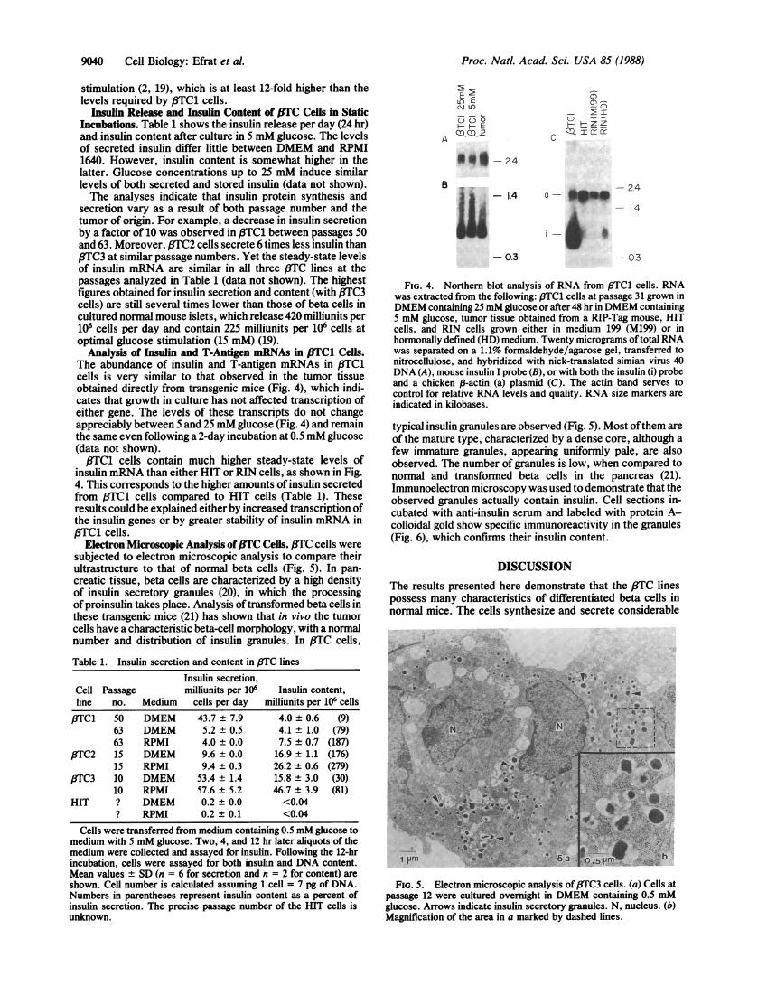
Insulin Release and Insulin Content of .lTC Cells in StaticIncubations. Table 1 shows the insulin release per day (24 hr)and insulin content after culture in 5 mM glucose. The levelsof secreted insulin differ little between DMEM and RPMI1640. However, insulin content is somewhat higher in thelatter. Glucose concentrations up to 25 mM induce similarlevels of both secreted and stored insulin (data not shown).The analyses indicate that insulin protein synthesis and
secretion vary as a result of both passage number and thetumor of origin. For example, a decrease in insulin secretionby a factor of 10 was observed in 3TC1 between passages 50and 63. Moreover, fTC2 cells secrete 6 times less insulin thanI3TC3 at similar passage numbers. Yet the steady-state levelsof insulin mRNA are similar in all three 83TC lines at thepassages analyzed in Table 1 (data not shown). The highestfigures obtained for insulin secretion and content (with ,8TC3cells) are still several times lower than those of beta cells incultured normal mouse islets, which release 420 milliunits per106 cells per day and contain 225 milliunits per 106 cells atoptimal glucose stimulation (15 mM) (19).
Analysis of Insulin and T-Antigen mRNAs in ,tTC1 Cells.The abundance of insulin and T-antigen mRNAs in f3TC1cells is very similar to that observed in the tumor tissueobtained directly from transgenic mice (Fig. 4), which indi-cates that growth in culture has not affected transcription ofeither gene. The levels of these transcripts do not changeappreciably between 5 and 25mM glucose (Fig. 4) and remainthe same even following a 2-day incubation at 0.5 mM glucose(data not shown).,3TC1 cells contain much higher steady-state levels of
insulin mRNA than either HIT or RIN cells, as shown in Fig.4. This corresponds to the higher amounts of insulin secretedfrom /3TC1 cells compared to HIT cells (Table 1). Theseresults could be explained either by increased transcription ofthe insulin genes or by greater stability of insulin mRNA in/3TC1 cells.
Electron Microscopic Analysis of 3TC Cells. 3TC cells weresubjected to electron microscopic analysis to compare theirultrastructure to that of normal beta cells (Fig. 5). In pan-creatic tissue, beta cells are characterized by a high densityof insulin secretory granules (20), in which the processingofproinsulin takes place. Analysis oftransformed beta cells inthese transgenic mice (21) has shown that in vivo the tumorcells have a characteristic beta-cell morphology, with a normalnumber and distribution of insulin granules. In J3TC cells,
Table 1. Insulin secretion and content in /3TC lines
Insulin secretion,Cell Passage milliunits per 106 Insulin content,line no. Medium cells per day milliunits per 106cells
,8TC1 50 DMEM 43.7 ± 7.9 4.0 ± 0.6 (9)63 DMEM 5.2 ± 0.5 4.1 ± 1.0 (79)63 RPMI 4.0 ± 0.0 7.5 ± 0.7 (187)
I3TC2 15 DMEM 9.6 ± 0.0 16.9 ± 1.1 (176)15 RPMI 9.4 ± 0.3 26.2 ± 0.6 (279)
fTC3 10 DMEM 53.4 ± 1.4 15.8 ± 3.0 (30)10 RPMI 57.6 ± 5.2 46.7 ± 3.9 (81)
HIT ? DMEM 0.2 ± 0.0 <0.04? RPMI 0.2 ± 0.1 <0.04
Cells were transferred from medium containing 0.5 mM glucose tomedium with 5 mM glucose. Two, 4, and 12 hr later aliquots of themedium were collected and assayed for insulin. Following the 12-hrincubation, cells were assayed for both insulin and DNA content.Mean values ± SD (n = 6 for secretion and n = 2 for content) areshown. Cell number is calculated assuming 1 cell = 7 pg of DNA.Numbers in parentheses represent insulin content as a percent ofinsulin secretion. The precise passage number of the HIT cells isunknown.
A
LENW)EE- ECLC_
51-H-ZZr-I t t
c
9 O#-2.4
B1.4 a- in
W"W
-
- 0.3
-24
- 14
- 0.3
FIG. 4. Northern blot analysis of RNA from 83TC1 cells. RNAwas extracted from the following: ETC1 cells at passage 31 grown inDMEM containing 25mM glucose or after 48 hr in DMEM containing5 mM glucose, tumor tissue obtained from a RIP-Tag mouse, HITcells, and RIN cells grown either in medium 199 (M199) or inhormonally defined (HD) medium. Twenty micrograms of total RNAwas separated on a 1.1% formaldehyde/agarose gel, transferred tonitrocellulose, and hybridized with nick-translated simian virus 40DNA (A), mouse insulin I probe (B), or with both the insulin (i) probeand a chicken 18-actin (a) plasmid (C). The actin band serves tocontrol for relative RNA levels and quality. RNA size markers areindicated in kilobases.
typical insulin granules are observed (Fig. 5). Most ofthem areof the mature type, characterized by a dense core, although afew immature granules, appearing uniformly pale, are alsoobserved. The number of granules is low, when compared tonormal and transformed beta cells in the pancreas (21).Immunoelectron microscopy was used to demonstrate that theobserved granules actually contain insulin. Cell sections in-cubated with anti-insulin serum and labeled with protein A-colloidal gold show specific immunoreactivity in the granules(Fig. 6), which confirms their insulin content.
DISCUSSIONThe results presented here demonstrate that the f3TC linespossess many characteristics of differentiated beta cells innormal mice. The cells synthesize and secrete considerable
.jp..
FIG. 5. Electron microscopic analysis of ETC3 cells. (a) Cells atpassage 12 were cultured overnight in DMEM containing 0.5 mMglucose. Arrows indicate insulin secretory granules. N, nucleus. (b)Magnification of the area in a marked by dashed lines.
9040 Cell Biology: Efrat et al.

Proc. Natl. Acad. Sci. USA 85 (1988) 9041
00~~ ~
100 Im
FIG. 6. Immunoelectron microscopy of f3TC1 cells. Cells atpassage 36 were cultured for 48 hr in DMEM containing 5 mMglucose. They were then incubated with anti-insulin serum, followedby protein A-gold particles. Specific signal is observed over insulingranules (arrowheads). One insulin granule appears to have secretedits contents (arrow). Control sections incubated with phosphate-buffered saline instead of the primary antibody showed no reactivity.
amounts of insulin, as measured by RIA and by insulinmRNA analysis. The secretion of insulin is regulated byglucose, as revealed in column perifusion experiments.Induction levels of up to 30-fold were obtained withinminutes after shifting the cells from no glucose to 5 mMglucose. pTC1 cells are sensitive to much lower levels ofglucose, when compared to normal beta cells. They secreteappreciable amounts of insulin at 0.5 mM and reach maximalstimulation at 1.25 mM, in contrast to the 15-mM levelrequired for maximal stimulation of secretion from normalmouse beta cells in intact cultured islets. This difference mayresult from the presence of other cell types in intact islets,which normally influence insulin secretion by beta cells.Alternatively, the continuous proliferation may constrain thecells to metabolize high levels of glucose and thereby abro-gate their normal sensitivity and response to glucose. Thesteady-state levels of insulin mRNA are not affected byglucose concentrations between 0.5 and 25 mM. Althoughlower levels (<0.5 mM) might reveal a potential regulation ofinsulin biosynthesis by glucose, such levels are incompatiblewith viability of 8TC cells.,fTC cells produce low amounts of glucagon, as judged by
immunohistochemical and biochemical analysis, in contrastto the tumors in vivo (9). In the case of RIN cells, the initialradiation-induced insulinoma has given rise to clones pro-ducing insulin, glucagon, and somatostatin in various com-binations (22, 23). The appearance of glucagon immunoreac-tivity in PTC cells indicates that they have undergone a
change in gene regulation during propagation in vitro, whichmight be considered dedifferentiation, given that insulin andglucagon are normally coexpressed during islet cell develop-ment (24).The features of 3TC cells are likely to render them useful
for studies on regulation of the insulin genes and on beta-cellphysiology, for the isolation of proteins that control insulingene expression, and perhaps for characterization ofbeta-cellmolecules involved in the autoimmune response in diabetes.In contrast to the other insulinoma cell lines, the availabilityof lineages of transgenic mice that heritably develop beta-celltumors allows for repeated derivation of cell lines from theprimary tumors. Thus adaptation and deviation from theobserved beta-cell phenotype can be regularly cross-checkedin vivo, and new cell lines can be established if the old linesbecome anaplastic.
These results demonstrate that targeted expression of anoncogene in transgenic mice, by using a cell-specific regula-tory element, can be applied for immortalization and estab-lishment in cell culture of a rare cell type. The use ofcell-specific control regions not only provides access todispersed, rare cells but also provides a selection for thedifferentiated phenotype of those cells in culture. Thus, thehybrid oncogene represents a type of feedback loop that onthe one hand induces continuous cell proliferation and on theother requires that high level expression of a cell-specificgene be maintained.
We thank T. Nielsen, L. Thousig Moller, and L. Aagaard forexpert technical assistance; W. L. Chick for RIN cells; M. Walkerfor HIT cells; J. Duffy for artwork; D. Green for photography; andJ. H. Nielsen for critical comments on the manuscript. S.E. issupported by the Cancer Research Institute/David Jacobs MemorialFellowship. S.B. is supported by a Career Development Award fromthe Juvenile Diabetes Foundation. S.G. is supported by a JuvenileDiabetes Foundation postdoctoral fellowship. This work was fundedby grants from Monsanto, the Juvenile Diabetes Foundation, andNorth Atlantic Treaty Organization (Grant RG. 86/0552). The ColdSpring Harbor Electron Microscopy Laboratory is partially sup-ported by grants from the National Institutes of Health (1 S10RR03430-01, P30CA45508), the National Science Foundation (BSS-8604215), and the Fannie E. Rippel Foundation.
1. Steiner, D. F., Chan, S. J., Welsh, J. M. & Kwok, S. C. M.(1985) Annu. Rev. Genet. 19, 463-484.
2. Schwizer, R. W., Leiter, E. H. & Evans, R. (1984) Transplan-tation 37, 539-544.
3. Chick, W. L., Warren, S., Chute, R. N., Like, A. A., Lauris,V. & Kitchen, K. C. (1977) Proc. Natl. Acad. Sci. USA 74,628-632.
4. Praz, G. A., Halban, P. A., Wollheim, C. B., Blondel, B.,Strauss, A. J. & Renold, A. E. (1983) Biochem. J. 210, 345-352.
5. Oie, H. K., Gazdar, A. F., Minna, J. D., Weir, G. C. & Baylin,S. (1983) Endocrinology 112, 1070-1075.
6. Santerre, R. F., Cook, R. A., Crisel, R. M. D., Sharp, J. D.,Schmidt, R. J., Williams, D. C. & Wilson, C. P. (1981) Proc.Natl. Acad. Sci. USA 78, 4339-4343.
7. Edlund, T., Walker, M. D., Barr, P. J. & Rutter, W. J. (1985)Science 230, 912-916.
8. Karlsson, O., Edlund, T., Barnett Moss, J., Rutter, W. J. &Walker, M. D. (1987) Proc. Natl. Acad. Sci. USA 84, 8819-8823.
9. Hanahan, D. (1985) Nature (London) 315, 115-122.10. Efrat, S. & Hanahan, D. (1987) Mol. Cell. Biol. 7, 192-198.11. Adams, T., Alpert, S. & Hanahan, D. (1987) Nature (London)
325, 223-228.12. Fong, H. K. W., Chick, W. L. & Sato, G. H. (1981) Diabetes
30, 1022-1028.13. Efrat, S., Baekkeskov, S., Lane, D. & Hanahan, D. (1987)
EMBO J. 6, 2699-2704.14. Linde, S., Hansen, B. & Lernmark, A. (1983) Methods Enzy-
mol. 92E, 309-335.15. Heding, L. G. (1966) in Labeled Proteins in Tracer Studies,
eds. Donato, L., Milhaud, C. & Sirchis, J. (Euratom, Brussels),pp. 345-350.
16. Knudsen, P., Kofod, H., Lernmark, A. & Hedeskov, C. J.(1983) Am. J. Physiol. 245, E338-E346.
17. Green, I. C. & Taylor, K. W. (1972) J. Endocrinol. 54, 317-325.18. Rhodes, C. J. & Halban, P. A. (1988) Biochem. J. 251, 23-30.19. Nielsen, J. H. (1985) Acta Endocrinologia Suppl. 266.20. Munger, B. L. (1981) in The Islets of Langerhans, eds. Coo-
perstein, S. J. & Watkins, D. (Academic, New York), pp. 3-34.21. Power, R. F., Holm, R., Bishop, A. E., Varmdell, I. M.,
Alpert, S., Hanahan, D. & Polak, J. M. (1987) Gut 28, 121-129.22. Madsen, 0. D., Larsson, L.-I., Rehfeld, J. F., Schwartz,
T. W., Lernmark, A., Labrecque, A. D. & Steiner, D. F.(1986) J. Cell Biol. 103, 2025-2034.
23. Philippe, J., Chick, W. L. & Habener, J. F. (1987) J. Clin.Invest. 79, 351-358.
24. Alpert, S., Hanahan, D. & Teitelman, G. (1988) Cell 53, 295-308.
Cell Biology: Efrat et al.


![DEVELOPMENT OF HYBRID HYDRAULIC EXCAVATORSjfps.or.jp/souko/Proceedings2014/proceedings/pdf/1C1-1.pdf · 2010[2]. Up until August 2013, 2250 Komatsu hybrid hydraulic excavators were](https://img.pdfslide.us/doc/110x75/5e8fd0c63f20e74af179dbe2/development-of-hybrid-hydraulic-20102-up-until-august-2013-2250-komatsu-hybrid.jpg)