Embed Size (px)
Citation preview

Review of the Diseases of the Upper GI Tract
David G Weismiller, MD, ScM, FAAFP Department of Family Medicine
The Brody School of Medicine at East Carolina University

Disclosure Statement
Dr Weismiller has nothing to disclose.
The AAFP has selected all faculty appearing in this program. It is the policy of the AAFP that all CME planning committees, faculty, authors, editors, and staff disclose relationships with commercial entities upon nomination or invitation of participation. Disclosure documents are reviewed for potential conflicts of interest and, if identified, they are resolved prior to confirmation of participation. Only those participants who had no conflict of interest or who agreed to an identified resolution process prior to their participation were involved in this CME activity.
Learning Objectives
1. Determine the approach to the patient with dyspepsia.
2. Discuss the common disorders of the esophagus: motility and GERD.
3. Describe the diagnosis and treatment of PUD and Helicobacter pylori.

Esophageal Disorders
Disorders of motility Gastro-esophageal Reflux Disease
Inflammatory and Infectious Diseases Tumors of the Esophagus
Symptoms from the Esophagus History
• Swallowing difficulties – dysphagia • Pain – heartburn, odynophagia, chest pain • Regurgitation – effortless appearance of
gastric or esophageal contents in the oral cavity

Esophageal Motility Disorders
• Achalasia • Spasm
– Diffuse – Localized – “nutcracker” esophagus
• Scleroderma

1. Which of the following is an indicated treatment for achalasia?
A. Beta blockers B. Alpha blockers C. Calcium channel blockers D. H2 blockers

1. Which of the following is an indicated treatment for achalasia?
A. Beta blockers B. Alpha blockers C. Calcium channel blockers D. H2 blockers 15%
2%
5%
79%

Motility Disorders
Disorder Clinical Diagnosis Treatment
Achalasia (Absence of peristaltic progression)
Dysphagia – solids and liquids, increased risk of SCC
Barium swallow, manometry
Long-acting nitrates, Ca channel blockers, pneumatic dilatation of LES
Diffuse Esophageal Spasm (Spastic Motor Disorder)
Heartburn, chest pain, or dysphagia; often swallow-induced – always exclude CAD.
Barium swallow Long-acting nitrates, Ca channel blockers
Scleroderma Esophagus (90% of patients with scleroderma have esophagus involved.)
None to severe reflux; often with strictures, motility abnormalities
Barium swallow, manometry
Manage reflux; treat esophagitis with H2 blockers, PPIs, prokinetic drugs.

GERD
• Reflux of acid or gastric juice into the lower esophagus with some combination of symptoms, inflammation, and/or complications
• Common disorder – “Weekly” Sx: 10%-20% of US population – “Within past month:” 30%-40%

GERD Pathophysiology
• Motor components of LES, esophagus, and stomach • Noxious esophageal contents • Mucosal barrier, saliva • Mechanical factors – gravity, hiatus hernia, obesity,
etc • Sensory components – appreciation of pain

2. Which one of the following is a true statement regarding GERD?
A. One of the more common complications is obstructive sleep apnea.
B. Dietary modifications are not part of the treatment according to the current American College of Gastroenterology guidelines.
C. Endoscopic surveillance for dysplasia is indicated in Barrett’s esophagus (squamous metaplasia).
D. Corrective laparoscopic reflux surgery is not indicated when there are persistent “reflux symptoms” despite acid suppression.
2%
6%
3%
89%

GERD Diagnosis
• No gold standard; EGD is to assess complications (SOR: A). – Erosive esophagitis – Stricture – Barrett’s esophagus – Cancer

GERD Diagnosis
• No gold standard; EGD is to assess complications (SOR: A). – EGD lacks adequate sensitivity in determining
pathologic reflux (SOR B). – pH probe is accepted as standard (SOR B); still false
positives and false negatives. • Sensitivity: 85% • Specificity: 95%
– Barium radiology: Limited usefulness; not recommended (SOR B)

GERD Diagnosis
• An empiric trial of acid suppression therapy for 4-8 weeks can identify patients with GERD who do not have alarm symptoms (SOR A).
• Alarm symptoms – Black or bloody stools – Choking – Chronic cough – Dysphagia – Early satiety – Hematemesis – Hoarseness – Iron deficiency anemia – Odynophagia – Weight loss

Treatment Guidelines
• • Dietary modifications • Lifestyle modification (SOR C) • Trial of patient-directed therapy with
antacids or OTC H2 antagonists
Step 1 Mild symptoms
• • Continue lifestyle/dietary modification. • H2 antagonists (SOR A) • Proton pump inhibitor (PPI)* (SOR A) • Pro-motility agent (SOR A) • 8-12 weeks of therapy
Step 2 Non-responders Non-erosive disease
• Continue with measures. • GI workup (+/−) endoscopy • High-dose H2 antagonists • Higher dose PPI
Step 3 Severe symptoms Erosive disease
* PPI should be taken 30-60 minutes prior to a meal (the first meal of the day) to optimize effectiveness (SOR B).
American College of Gastroenterology – 2005 Volume 135, Issue 4; 1383-1391.e5, October 2008
Barrett’s Esophagus
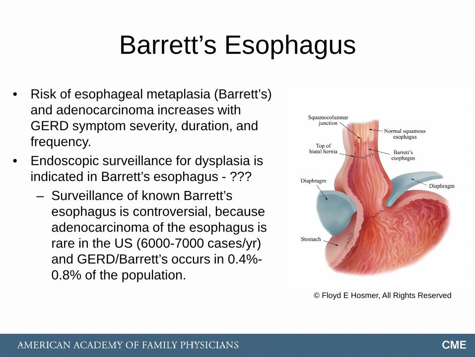
• Risk of esophageal metaplasia (Barrett’s) and adenocarcinoma increases with GERD symptom severity, duration, and frequency.
• Endoscopic surveillance for dysplasia is indicated in Barrett’s esophagus - ??? – Surveillance of known Barrett’s
esophagus is controversial, because adenocarcinoma of the esophagus is rare in the US (6000-7000 cases/yr) and GERD/Barrett’s occurs in 0.4%-0.8% of the population.
© Floyd E Hosmer, All Rights Reserved

AGA Position Statement on Screening for Barrett’s Esophagus 2011
• Whom to screen? – Long-standing (> 5 years) heartburn symptoms – Long-standing (> 5 years) need for medication
• Recommend against screening the general population
• Support is stronger for screening those patients with multiple risk factors.

Risk Factors for Barrett’s Esophagus
• Men – Screening women for Barrett’s is like screening men for
breast cancer. • Caucasian • Age > 50 years • Hiatal hernia • Increasing BMI • Abdominal fat distribution (abdominal obesity)

2008 ACG Guidelines for
Surveillance of Barrett’s Esophagus EGD
Barrett’s Esophagus
No dysplasia • Second EGD with biopsies within
year to confirm there is no dysplasia
• If both EGDs with biopsies (−) for dysplasia, repeat EGD with biopsy q 3 years.
Low-grade dysplasia (LGD) • Review by expert pathologist to
rule out HGD. • Repeat EGD with biopsies
within 6 months to reassess for dysplasia.
• If (−) for dysplasia on repeat EGD, yearly EGD with biopsy recommended until 2 years with EGDs showing no dysplasia.
High-grade dysplasia (HGD) • Expert pathologist confirm HGD. • Mucosal irregularity – remove
with endoscopic mucosal resection (EMR).
• EGD with biopsies repeated in 3 months to look for HGD and small cancers.
• Possible interventions for HGD: espohagectomy, EMR, photodynamic therapy, radiofrequency ablation, ablation using cryotherapy.
At the present time, only specialized intestinal metaplasia of the esophagus is classified as Barrett's esophagus. Currently, it is recommended that only patients with this diagnosis undergo periodic cancer surveillance.

GERD Surgical Treatment
• Who – Complications of reflux
• Non-responding esophagitis • Stricture • Barrett’s metaplasia
– Inability to tolerate medications – including non-compliance
– Persistent “reflux symptoms” despite acid suppression, ie, chronic reflux with recalcitrant symptoms (SOR A)
– Asthma

GERD Surgery
• Laparoscopic anti-reflux surgery – Treatment of choice
at many centers – Less perioperative
M&M – Shortens the
postoperative stay – Needs experienced
surgeon
• Complications – rate – Splenic injury – Esophageal or
gastric perforation – Dysphagia – Inability to belch or
vomit – Vagal denervation

GERD Follow-up and Surveillance
• If symptoms remain unchanged in a patient with a prior normal EGD, repeating EGD is not recommended (SOR C).
• Chronic reflux has been suspected to play a major role in the development of Barrett’s esophagus, yet it is unknown if outcomes can be improved through surveillance and medical treatment (SOR C).
• Anti-secretory therapy has not been shown to reduce the need for recurrent dilation from esophageal stricture formation (SOR A).

Inflammatory Disorders Esophagitis
Disorder Offending “Agents” Pill-induced Doxycycline, NSAIDs, steroids Infective* • Viral • Fungal
HSV, CMV Candida
Corrosive Alkalis or acids Eosinophilic: pronounced eosinophilic infiltration
Allergic or idiopathic; Tx – steroids, diet, anti-allergy medications
*Mostly in immunosuppressed patients

3. Which of the following statements regarding esophageal tumors is true?
A. 90% are cancer. B. Adenocarcinoma is the most common
histologic type. C. They are more common in women. D. The incidence of squamous cell carcinomas
is increasing.

3. Which of the following statements regarding esophageal tumors is true?
A. 90% are cancer. B. Adenocarcinoma is the most common
histologic type. C. They are more common in women. D. The incidence of squamous cell carcinomas
is increasing. 31%
21%
48%
0%

Esophageal Tumors • 90% are cancer.
– Much more common in males; 10% 5-yr survival rate overall (treatment improving)
– Dx – endoscopy and radiography • Squamous cell carcinoma
– Most common, declining incidence – Predominant esophageal cancer in
African Americans – More common with heavy alcohol and tobacco use
• Adenocarcinomas – Arise from columnar epithelium in cardia or from
Barrett’s – Recall that the lower esophagus is lined by
specialized intestinal epithelium. – GERD is a risk factor.
Source: Wikimedia

Diseases of the Stomach
Acid Peptic Disorders of the Stomach and Duodenum Infections
Motor Disorders Cancer

Acid Peptic Disorders Stomach and Duodenum
• Common Problem – 5%-10% of population will have PUD in their lifetimes; 50%
recurrence in 5 years – DU/GU 4:1
• 90% DU in duodenal bulb • GU most common on lesser curve
Mortality Rates
Ulcer Type Men Women
Duodenal Ulcer 1/100,000 0.5/100,000
Gastric Ulcer 1.5/100,000 1.2/100,000

4. Which of the following is a clear predisposing or exacerbating factor for acid peptic disorders?
A. Caffeine B. Stress C. Tobacco D. Chili peppers

4. Which of the following is a clear predisposing or exacerbating factor for acid peptic disorders?
A. Caffeine B. Stress C. Tobacco D. Chili peppers 4%
8%
11%
78%

Why Do Acid Peptic Disorders Develop?
• Current theory – PUD is an imbalance between protective and aggressive
factors.
Protective factors Aggressive factors • Surface epithelial cells with
mucus and bicarbonate secretions
• Apical surface membrane of gastric mucosal cells
• Prostaglandins E1 and E2
• Production of gastric acid • NSAIDs • Corticosteroids • Smoking • Alcohol consumption • ? Psychological stress • Probably not diet

Predisposing Factors
• H. pylori infection • NSAIDs
– Double the annual chance of complicated PUD from 1%-2% to 3%-4%
• Worse with alcohol • Longer-acting
NSAIDs are worse. – Dose, duration
important variables
• Milk: May slow healing of DU
• Caffeine: No clear evidence of worsening
• Peppers: No slowing of DU healing
• Alcohol: Worse with NSAIDs; unclear otherwise
• Tobacco: Much higher rates of ulcer and slower healing
• Stress: Remains controversial

Diagnosis PUD
• History – Persistent pain relieved by
food and antacids • Pain in upper abdomen
or back • Hematemesis, melena,
or hematochezia • Cannot usually separate
GU from DU by history • On exam
– Mid-epigastric tenderness
• Laboratory – Limited usefulness, except
H. pylori tests – Consider serum gastrin
(especially if recurrent ulcer disease)
– Hematocrit – Stool guaiac
• Endoscopy (SOR A) – 90% sensitivity and
specificity

Diagnostic Tests for Helicobacter pylori Test Usefulness Sens
(%) Specif (%)
Invasive Endoscopy with biopsy
Diagnostic strategy of choice in children with persistent or severe upper abdominal symptoms
Histology Sensitivity reduced by PPIs, antibiotics, bismuth-containing compounds.
> 95 100
Urease activity Test of choice when endoscopy indicated; rapid results (20 min); (−) results may need confirmation by histology or other test; sensitivity reduced by PPIs, antibiotics, bismuth-containing compounds, and active bleeding.
93-97 > 95
Culture Technically demanding; only use for resistant organism or refractory disease.
70-80 100

Diagnostic Tests for Helicobacter pylori Test Usefulness Sens
(%) Specif (%)
Noninvasive ✓ serology for immunoglobulin G
Sensitivity and specificity vary widely; assist with initial diagnosis; not as helpful in following patients due to prolonged presence of antibody after eradication.
85 79
Urea breath test Reliable test for cure – can document eradication as early as 4 weeks post-treatment; requires separate appointments; sensitivity reduced by PPIs, antibiotics, and bismuth-containing compounds.
> 95 100
H. pylori stool antigen
Detects active infection; test for cure seven days after therapy is accurate; sensitivity reduced by PPIs, antibiotics, and bismuth-containing compounds.
93-97 > 95

PUD and H. Pylori Disorder H. pylori (+) Associated
H. pylori gastritis
Treatment
Duodenal ulcer
90% 70% Eradication of infection markedly decreases recurrences of DU.
A 1-2 week course of H. pylori eradication therapy is an effective treatment for H. pylori (+) PUD.
Gastric ulcer
70% 60%-80%

Who Should Be Tested for H. Pylori?
• All newly diagnosed (by radiography or endoscopy) complicated or non-complicated DU or GU
• Hx of PUD receiving maintenance anti-secretory therapy or documented past ulcer
• Mucosa-associated lymphoid tissue lymphoma (MALT) • Some decision and cost-benefit analysis support non-endoscopic
diagnostic testing in patients < 55 with symptoms of ulcer-like dyspepsia and no alarm symptoms. – Deciding on what test to use in which situation relies upon
whether a patient requires evaluation with upper endoscopy and an understanding of the strengths, weaknesses, and costs of the individual test.
ACG Guideline on the Management of H. pylori infection. Am J Gastroenterol. 2007;102:1808.

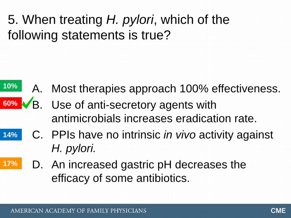
5. When treating H. pylori, which of the following statements is true?
A. Most therapies approach 100% effectiveness. B. Use of anti-secretory agents with
antimicrobials increases eradication rate. C. PPIs have no intrinsic in vivo activity against
H. pylori. D. An increased gastric pH decreases the
efficacy of some antibiotics.
5. When treating H. pylori, which of the following statements is true?
A. Most therapies approach 100% effectiveness. B. Use of anti-secretory agents with
antimicrobials increases eradication rate. C. PPIs have no intrinsic in vivo activity against
H. pylori. D. An increased gastric pH decreases the
efficacy of some antibiotics. 17%
10%
60%
14%

Specific H. Pylori Therapy Treatment Dosing/Comments PPI, amoxicillin 1g, clarithromycin 500 mg
• All BID for 7-14 days • First line (up to date – 1A recommendation)
Bismuth 525 mg, metronidazole 500 mg, tetracycline 500 mg
• All QID with PPI BID for 7-14 days • First line or retreatment
PPI, amoxicillin 1 g, metronidazole 500 mg
• All BID for 14 days • First line in macrolide allergic or retreatment
PPI, levofloxacin 250-500 mg, amoxicillin 1 g
• All BID for 14 days • Rescue for two prior treatments
PPI, rifabutin 150 mg, amoxicillin 1 g
• All BID for 14 days • Rescue
PPI plus amoxicillin 1 g • PPI BID, amoxicillin TID for 14 days • Rescue

6. Regarding anti-secretory therapy for PUD, which of the following is true?
A. H2 blockers lead to faster healing than proton pump inhibitors.
B. Therapy is usually longer for duodenal ulcers versus gastric ulcers.
C. The addition of sucralfate to the anti-secretory medications hastens healing.
D. It is the mainstay of therapy in uninfected patients.
65%
2%
15%
18%

Specific Ulcer Treatment Anti-secretory
• Anti-secretory therapy – Mainstay of therapy in uninfected patients
• PPI, H2 blocker – Appropriate for maintenance therapy in selected
cases – Usually 4-6 weeks for DU – Generally longer for GU – 12 weeks – PPIs lead to faster healing than H2 blockers.
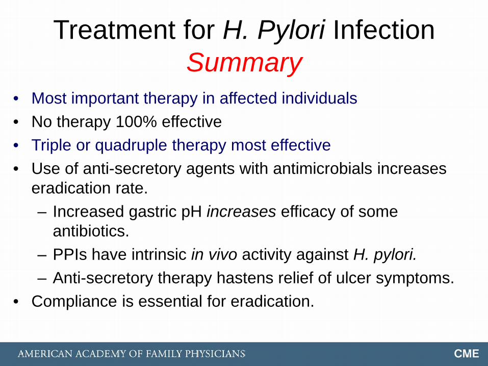
Treatment for H. Pylori Infection Summary
• Most important therapy in affected individuals • No therapy 100% effective • Triple or quadruple therapy most effective • Use of anti-secretory agents with antimicrobials increases
eradication rate. – Increased gastric pH increases efficacy of some
antibiotics. – PPIs have intrinsic in vivo activity against H. pylori. – Anti-secretory therapy hastens relief of ulcer symptoms.
• Compliance is essential for eradication.

7. In considering NSAID-induced ulcers, which of the following is true regarding prevention?
A. H2 blockers are superior to PPIs in preventing ulcers.
B. H. pylori should not be treated if present. C. PPIs superior to misoprostol (200 mg QID) in
preventing ulcer relapses. D. Sucralfate is contraindicated in preventing
NSAID-induced ulcers.

7. In considering NSAID-induced ulcers, which of the following is true regarding prevention?
A. H2 blockers are superior to PPIs in preventing ulcers.
B. H. pylori should not be treated if present. C. PPIs superior to misoprostol (200 mg QID) in
preventing ulcer relapses. D. Sucralfate is contraindicated in preventing
NSAID-induced ulcers. 6%
4%
3%
86%

NSAID Ulcers
• Risk factors – Prior adverse GI event (ulcer, hemorrhage) – Age > 60 – High-dose NSAID (> twice normal) – Glucocorticoid use – Anticoagulant use
• Risk for NSAID-induced GI toxicity 9% at 6 months with multiple risk factors present
• In naïve NSAID users, H. pylori is significant risk factor for complicated ulcer disease – screening may be indicated.

NSAID Users: Medical Treatment of Peptic Ulcer Practice Guidelines
• Treatment of NSAID ulcers – D/C NSAIDs – Treat H. pylori if present. – PPIs superior to H2 receptor antagonists
• Prevention of NSAID ulcers – PPIs superior to H2 blockers in preventing ulcers – PPIs superior to misoprostol (200 mg QID) in preventing
ulcer relapses – Treat H. pylori if present. NEJM. 1998;338:727.
Primary Care. September, 2001:28(3):487-503. Am J Gastrolenterol. 1998;93:2037.

Treatment Non−H. Pylori PUD
• Withdrawal of potential offending or contributing agents – NSAIDs, cigarettes, excess ETOH
• No firm dietary recommendations – avoid foods that precipitate dyspepsia.
• Address psychosocial issues and comorbidities – no firm evidence, but may have deleterious health consequences.

Controversy: Treatment of H. Pylori in Non-ulcer Dyspepsia
• Efficacy of treatment is controversial. • Recent review of RCTs: Eradication provides small
but significant benefit for dyspeptic symptoms.* • Eradication may be cost-effective intervention for
non-ulcer dyspepsia. • And … H. pylori appears to have a net suppressive
effect on acid production, so treating may make GERD worse.
*Practice Guideline. Moayyedi P, Soo S, et al. Eradication of Helicobacter pylori for non-ulcer dyspepsia. The Cochrane Database of Systematic Reviews. 2005. http://www.cochrane.org/cochrane/revabstr/ab002096.htm

An Approach to Dyspepsia Fails Fails
Fails Fails
Information from American College
Gastroenterology, 2005
H. Pylori prevalence < 10%
Trial of PPl
Test and treat for H. pylori.
Consider upper endoscopy.
Dyspepsia
H. pylori prevalence ≥ 10%
Test and treat for H. pylori.
Trial of PPl
Consider upper Endoscopy.
Upper endoscopy
> 55 years old or presence of alarm symptoms, family or personal Hx GI CA or PUD, wt. loss, GI bleeding, anemia, or dysphagia
55 years old No alarm features

8. Risk factors for ulcer complications include all of the following except:
A. Previous history of complications B. Prior refractory or protracted course C. Small ulcers (< 2 cm) D. Deformed ulcer bed or dense fibrosis

8. Risk factors for ulcer complications include all of the following except:
A. Previous history of complications B. Prior refractory or protracted course C. Small ulcers (< 2 cm) D. Deformed ulcer bed or dense fibrosis 3%
4%
2%
91%

Risk Factors for Ulcer Complications
• Previous history of complications • Prior refractory or protracted course • Big ulcers (> 2 cm) • Deformed ulcer bed or dense fibrosis
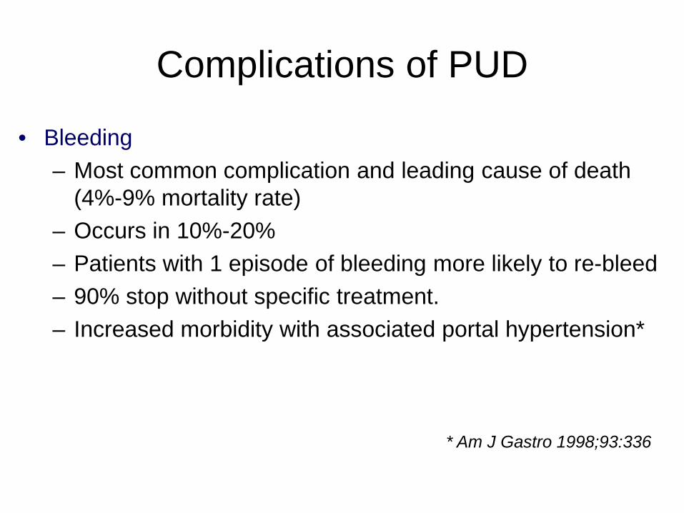
Complications of PUD
• Bleeding – Most common complication and leading cause of death
(4%-9% mortality rate) – Occurs in 10%-20% – Patients with 1 episode of bleeding more likely to re-bleed – 90% stop without specific treatment. – Increased morbidity with associated portal hypertension*
* Am J Gastro 1998;93:336

Complications of PUD
• Gastric outlet obstruction – Usually mechanical obstruction due to edema or scar – Most due to DU – Rare: 2% of ulcer patients
• Perforation and penetration – 2% of ulcers perforate. – Average duration of Sx prior to perforation: 5 years
• NOTE: Complicated ulcer disease less likely to involve H. pylori

PUD Surgical Treatment/Management
• Dramatically declined over past two decades • Indications
– Hemorrhage not responsive to medical therapy – Gastric outlet obstruction not reversed by medical
treatment – Perforation – Malignancy

SORT: Key Recommendations for Practice Ables AZ, Simon I, and Melton ER. Update on Helicobacter pylori Treatment. Am Fam Physician.
2007;75:351-8.
Clinical recommendation Evidence rating
Comment
A “test-and-treat” strategy is recommended in patients with symptoms of dyspepsia.
A Test-and-treat strategy reduces endoscopies and use of anti-secretory medications.
Helicobacter pylori eradication therapy is recommended to prevent recurrence and re-bleeding in patients with peptic ulcer.
A It is unnecessary to continue anti-secretory maintenance therapy in patients after H. pylori eradication.
Short-course drug therapy is an option for H. pylori eradication in adult patients.
C Eradication rates using short-course therapy are similar to those of traditional treatment with the potential for greater compliance.
The urea breath test is the most reliable noninvasive diagnostic test in children with suspected H. pylori infection.
C Urea breath test is more reliable in children older than six years; monoclonal antibody−based stool antigen is an alternative.

Gastric Dysmotility

Gastric Dysmotility Slow or Delayed Emptying
• Etiology – Mechanical or outlet obstruction
• PUD, bezoar, etc. – Functional obstruction (gastroparesis)
• Drugs – opiates, anticholinergics, beta and Ca channel blockers
• Diabetes, Parkinson’s, hypothyroidism, hypoparathyroidism
• Pregnancy • Post-vagotomy
Gastric Dysmotility Slow or Delayed Emptying
• Diagnosis – Nausea, vomiting, dysphagia, post-prandial
abdominal pain, GERD – Tests: Scintigraphy, electrogastrogram (evaluate
gastric myoelectrical utility), ultrasonography • Treatment
– Remove causes – Low-fat diet; avoid large meals – Metoclopramide, erythromycin, prokinetics

Gastric Dysmotility Rapid Gastric Emptying
• Dumping Syndrome – Most commonly seen post-operatively from
gastric surgery or a truncal vagotomy • Symptoms
– 15-30 minutes after eating – nausea, non-productive vomiting, sweating, flushing, abdominal cramping, diarrhea
• Treatment – 6-8 small, low-CHO meals/day; avoid excessive
liquids; use of opiates and anticholinergic drugs; fiber products; possibly surgery

9. A 65-yo male smoker complains of dyspepsia, weight loss, early satiety, and occasional nausea and vomiting. Which one of the following would be the initial diagnostic method of choice?
A. Upper GI endoscopy B. CT of the upper abdomen C. Single-contrast upper GI barium
swallow D. Endoscopic ultrasonography

9. A 65-yo male smoker complains of dyspepsia, weight loss, early satiety, and occasional nausea and vomiting. Which one of the following would be the initial diagnostic method of choice?
A. Upper GI endoscopy B. CT of the upper abdomen C. Single-contrast upper GI barium
swallow D. Endoscopic ultrasonography 2%
75%
12%
11%

Cancer of the Stomach • One of the most common internal malignancies in the world
– 95% are adenocarcinomas. • Chronic GERD is the leading cause of esophageal
adenocarcinoma (68%-90%). – Only 10%-20% of US GI tumors – probably because of
lower rates of H. pylori in US, due to cleaner food and water
• 2x as common in ♂ as in ♀ • 2x as common in African Americans and Hispanics as
in Caucasians • Dx: Endoscopic biopsy in patients with upper GI symptoms or
high-risk or double-contrast barium swallow • Tx: Surgical excision; 5-yr survival rate < 10%

• Fourth leading cause of cancer-related death; second only to CRC as cause of GI cancer-related death – Higher incidence: ♂ and African Americans – Risk factors: Smoking, chronic
pancreatitis, diabetes, hereditary predisposition
• History/PE – Abd pain, weight loss,
jaundice, pancreatitis – Jaundice, abdominal mass,
ascites
Pancreatic Cancer

Pancreatic Cancer
• Diagnosis – U/S, EUS CT, MRI ERCP, FNA, CA19-9 – All sensitive and specific
• Treatment – Surgical resection only potential curable treatment
• Prognosis – 5-yr survival – Node (−) 25%-30% – Node (+) 10%

Answers
1. C 2. C 3. A 4. C 5. B 6. D 7. C 8. C 9. A

Supplementary Slides

H. Pylori Resistance
• Metronidazole 22%-39% • Clarithromycin 11% • Amoxicillin, tetracycline Rare • Bismuth None