Embed Size (px)
Citation preview

Review of the Diseases of the Upper GI Tract
David G Weismiller, MD, ScM, FAAFP
Attending Physician – Rural Health Group, INC.
Disclosure StatementIt is the policy of the AAFP that all individuals in a position to control content disclose any relationships with commercial interests upon nomination/invitation of participation. Disclosure documents are reviewed for potential conflicts of interest. If conflicts are identified, they are resolved prior to confirmation of participation. Only participants who have no conflict of interest or who agree to an identified resolution process prior to their participation were involved in this CME activity.
All individuals in a position to control content for this session have indicated they have no relevant financial relationships to disclose.
Learning Objectives 1. Determine the approach to the patient with dyspepsia.2. Discuss the common disorders of the esophagus:
motility and GERD.3. Describe the diagnosis and treatment of PUD and
Helicobacter pylori.

Esophageal Disorders
Disorders of motility Gastro-esophageal Reflux Disease
Inflammatory Diseases Tumors of the Esophagus

Symptoms From the EsophagusHistory
• Swallowing difficulties – dysphagia• Pain – heartburn, odynophagia, chest pain• Regurgitation – effortless appearance of
gastric or esophageal contents in the oral cavity
Esophageal Motility Disorders
• Achalasia• Spasm
– Diffuse– Localized – “nutcracker” esophagus
• Scleroderma

1. Which of the following is an indicated treatment for achalasia?
A. Beta blockersB. Alpha blockersC. Calcium channel blockersD. H2 blockers

1. Which of the following is an indicated treatment for achalasia?
A. Beta blockersB. Alpha blockersC. Calcium channel blockersD. H2 blockers12%
3%
7%
78%
1

Motility DisordersDisorder Clinical Diagnosis Treatment
Achalasia(Absence of peristaltic progression)
Dysphagia –solids and liquids, increased risk of SCC
Barium swallow,manometry
Long-acting nitrates, Ca channel blockers, pneumatic dilatation of LES
Diffuse Esophageal Spasm(Spastic Motor Disorder)
Heartburn, chest pain, or dysphagia; often swallow-induced –always exclude CAD.
Barium swallow Long-acting nitrates, Ca channel blockers
Scleroderma Esophagus(90% of patients with scleroderma have esophagus involved.)
None to severe reflux; often with strictures, motility abnormalities
Barium swallow, manometry
Manage reflux; treat esophagitis with H2 blockers, PPIs, prokinetic drugs.

GERDSymptoms
• Typical Symptoms: heartburn, acid regurgitation• Atypical Symptoms: wheezing, hoarseness, atypical
chest pain• Diagnosis usually based on history and physical, and
trial of empiric therapy

2. Which of the following diagnostic tests is recommended in the patient with GERD refractory to maximum PPI therapy?
A. Esophageal manometryB. 24 hour pH monitoringC. Barium radiologyD. Urea Breath Test

2. Which of the following diagnostic tests is recommended in the patient with GERD refractory to maximum PPI therapy?
A. Esophageal manometryB. 24 hour pH monitoringC. Barium radiologyD. Urea Breath Test33%
15%
42%
10%
1

GERD Diagnosis
• No gold standard; EGD is to assess complications (SOR: A).– Erosive esophagitis– Stricture– Barrett’s esophagus– Cancer
• EGD lacks adequate sensitivity in determining pathologic reflux (SOR B)

Diagnostic Testing• Esophageal manometry is recommended in the pre-operative
evaluation, e.g. Nissen fundoplication (SOR: C)• Esophageal manometry is NOT recommended in the diagnosis of
GERD• 24-hour pH monitoring is recommended in patients refractory to
PPI therapy or when the diagnosis is in question (SOR: C)• 24-hour pH monitoring is NOT recommended in the routine diagnosis
of GERD and is NOT required in the presence of Barrett’s esophagus (SOR: B)
• Barium radiology: Limited usefulness; not recommended (SOR: A)Am J Gastroenterol 2013; 108:308 – 328

Diagnostic Testing• Screening for Helicobacter pylori infection is NOT
recommended (SOR: C)• Eradication of H. pylori is not routinely required as part of
antireflux therapy• FDA concluded that there was insufficient evidence to
recommend testing of all patients on long-term PPI therapy• Controversial: European recommendation in favor of
screening all patients for H. pyloriAm J Gastroenterol 2013; 108:308–328.
Gut. 2007;56:772- 81.

GERD Diagnosis
• An empiric trial of acid suppression therapy for 4-8 weeks can identify patients with GERD who do not have alarm symptoms (SOR: A)
• Alarm symptoms – Black or bloody stools– Choking– Chronic cough– Dysphagia– Early satiety– Hematemesis– Hoarseness– Iron deficiency anemia– Odynophagia– Weight loss
• Obtain upper endoscopy in patients with alarm symptoms or those at high risk for complications (SOR: B)
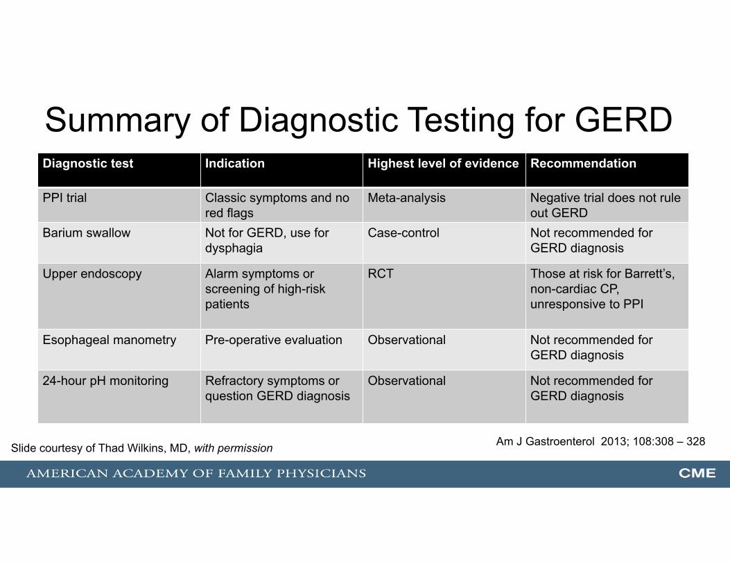
Summary of Diagnostic Testing for GERDDiagnostic test Indication Highest level of evidence Recommendation
PPI trial Classic symptoms and no red flags
Meta-analysis Negative trial does not rule out GERD
Barium swallow Not for GERD, use for dysphagia
Case-control Not recommended for GERD diagnosis
Upper endoscopy Alarm symptoms or screening of high-risk patients
RCT Those at risk for Barrett’s, non-cardiac CP, unresponsive to PPI
Esophageal manometry Pre-operative evaluation Observational Not recommended for GERD diagnosis
24-hour pH monitoring Refractory symptoms or question GERD diagnosis
Observational Not recommended for GERD diagnosis
Am J Gastroenterol 2013; 108:308 – 328Slide courtesy of Thad Wilkins, MD, with permission

Treatment• H2RA less effective than PPIs (less expensive than
PPIs)• PPIs are more effective for relieving heartburn than
H2RA or prokinetic agents (SOR: A)• Different PPIs have similar efficacy at standard doses
(SOR: A)• Long-term PPI use may increase fracture risk, but
inconsistent evidence regarding hip fracture (SOR: C)

On-demand Maintenance Therapy Versus Continuous PPI Therapy for GERD
Systematic review16 studies n = 14,142
*Statistically significant difference in favor of on-demand PPI.†Outcome is symptom relief.‡Statistically significant difference in favor of continuous PPI.§Outcome is endoscopic remission.
Aliment Pharmacol Ther. 2007;26:195-204.
Comparisons Severity of GERD
Number of trials (n)
Willingness to continue
with treatment
On-demand PPI vs continuous
PPI
Nonerosive 1 (622) 93% vs 88%*
Nonerosive or mildly erosive
1 (176) 75% vs 86%†
Uninvestigated 1 (1292) 52% vs 83%‡
Erosive1 (477) 58% vs 81%‡§
Slide courtesy of Thad Wilkins, MD, with permission

3. In discussing the initiation of a patient on a proton pump inhibitor (PPI), which of the following is a potential risk of therapy that should be reviewed with the patient?
A. Long-term PPI use may increase risk for community acquired pneumonia
B. Increased risk of hypermagnesemia C. Increased risk of Vitamin B12 deficiency D. Increased risk of iron deficiency anemia

3. In discussing the initiation of a patient on a proton pump inhibitor (PPI), which of the following is a potential risk of therapy that should be reviewed with the patient?
A. Long-term PPI use may increase risk for community acquired pneumonia
B. Increased risk of hypermagnesemia C. Increased risk of Vitamin B12 deficiency D. Increased risk of iron deficiency anemia14%
35%
4%
47%
1
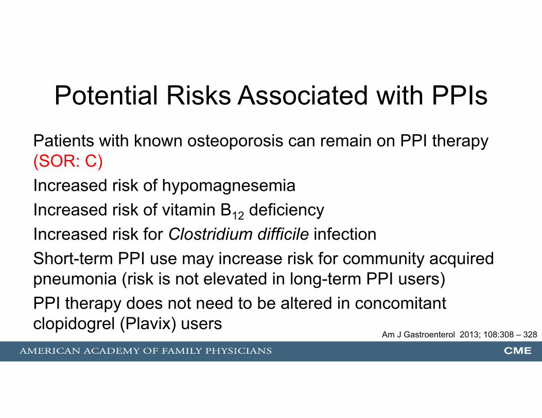
Potential Risks Associated with PPIsPatients with known osteoporosis can remain on PPI therapy (SOR: C)Increased risk of hypomagnesemiaIncreased risk of vitamin B12 deficiencyIncreased risk for Clostridium difficile infectionShort-term PPI use may increase risk for community acquired pneumonia (risk is not elevated in long-term PPI users)PPI therapy does not need to be altered in concomitant clopidogrel (Plavix) users
Am J Gastroenterol 2013; 108:308 – 328
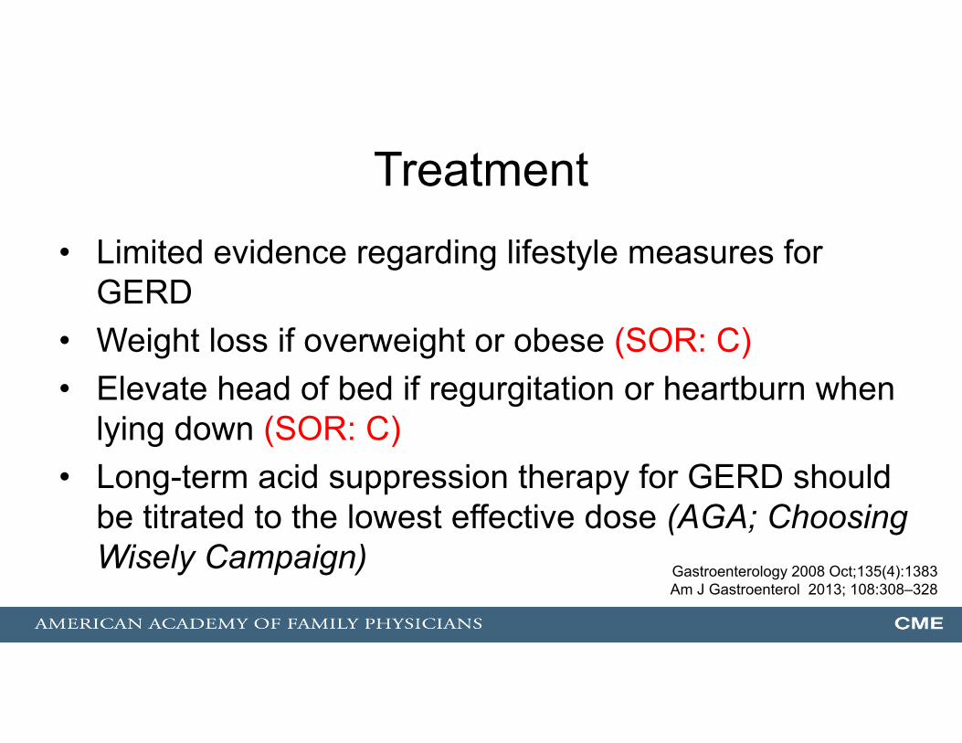
Treatment • Limited evidence regarding lifestyle measures for
GERD• Weight loss if overweight or obese (SOR: C) • Elevate head of bed if regurgitation or heartburn when
lying down (SOR: C) • Long-term acid suppression therapy for GERD should
be titrated to the lowest effective dose (AGA; Choosing Wisely Campaign) Gastroenterology 2008 Oct;135(4):1383
Am J Gastroenterol 2013; 108:308–328

Treatment Guidelines
•• Dietary modifications• Lifestyle modification (SOR C)• Trial of patient-directed therapy with antacids or
OTC H2 antagonists
Step 1Mild symptoms
•• Continue lifestyle/dietary modification.• H2 antagonists (SOR A)• Proton pump inhibitor (PPI)* (SOR A) • Pro-motility agent (SOR A)• 8-12 weeks of therapy
Step 2Non-respondersNon-erosive disease
• Continue with measures.• GI workup (+/−) endoscopy• High-dose H2 antagonists• Higher dose PPI
Step 3Severe symptomsErosive disease
* PPI should be taken 30-60 minutes prior to a meal (the first meal of the day) to optimize effectiveness (SOR: B).
American College of Gastroenterology – 2005 Volume 135, Issue 4; 1383-1391.e5, October 2008

4. Risk factors for esophageal intestinal metaplasia (Barrett’s Esophagus) include which one of the following characteristics?
A. Female sexB. African American race C. Tobacco D. Hiatal hernia

4. Risk factors for esophageal intestinal metaplasia (Barrett’s Esophagus) include which one of the following characteristics?
A. Female sexB. African American race C. Tobacco D. Hiatal hernia 15%
0%
1%
84% 1

Barrett’s Esophagus• Risk of intestinal metaplasia (Barrett’s) and
adenocarcinoma increases with GERD symptom severity, duration, and frequency.
• Endoscopic surveillance for dysplasia is indicated in Barrett’s esophagus – ???
– Surveillance of known Barrett’s esophagus is controversial, because adenocarcinoma of the esophagus is rare in the US (6000-7000 cases/yr) and GERD/Barrett’s occurs in 0.4%-0.8% of the population
– Risk of developing esophageal adenocarcinoma in patients with Barrett’s esophagus is less than 1%
© Floyd E Hosmer, All Rights Reserved
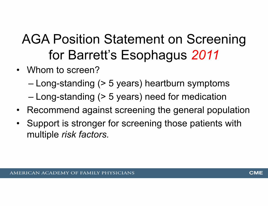
AGA Position Statement on Screening for Barrett’s Esophagus 2011
• Whom to screen?– Long-standing (> 5 years) heartburn symptoms– Long-standing (> 5 years) need for medication
• Recommend against screening the general population• Support is stronger for screening those patients with
multiple risk factors.

Risk Factors for Barrett’s Esophagus• Men
– Screening women for Barrett’s is like screening men for breast cancer.
• Caucasian• Age > 50 years• Hiatal hernia• Increasing BMI• Abdominal fat distribution (abdominal obesity)

2008 ACG Guidelines for Surveillance of Barrett’s Esophagus
EGD
Barrett’s Esophagus
No dysplasia• Second EGD with biopsies within
year to confirm there is no dysplasia• If both EGDs with biopsies (−) for
dysplasia, repeat EGD with biopsy q 3 years.
Low-grade dysplasia (LGD)• Review by expert pathologist to rule
out HGD.• Repeat EGD with biopsies within
6 months to reassess for dysplasia.• If (−) for dysplasia on repeat EGD,
yearly EGD with biopsy recommended until 2 years with EGDs showing no dysplasia.
High-grade dysplasia (HGD)• Expert pathologist confirm HGD.• Mucosal irregularity – remove with
endoscopic mucosal resection (EMR).• EGD with biopsies repeated in
3 months to look for HGD and small cancers.
• Possible interventions for HGD: espohagectomy, EMR, photodynamic therapy, radiofrequency ablation, ablation using cryotherapy.At the present time, only specialized intestinal metaplasia of the esophagus is
classified as Barrett's esophagus. Currently, it is recommended that only patients with this diagnosis undergo periodic cancer surveillance.

Medical Versus Surgical Treatment
• Medical treatment as effective as surgery (SOR:B)
• Surgery • Laparoscopic fundoplication increases quality of life in
patients with GERD but many patients still require medication after surgery (SOR: B)
• Endoscopic Procedures• May reduce symptoms in patients with GERD

Summary of GERD Treatment Recommendations Recommendation SORObtain upper endoscopy in patients with alarm symptoms or those at high risk for complications BStrong evidence supports association of GERD and esophageal adenocarcinoma with Barrett esophagus as precursor lesion
A
Chronic reflux has been suspected to play a major role in the development of Barrett’s esophagus, yet it is unknown if outcomes can be improved through surveillance and medical treatment
C
PPIs are more effective for relieving heartburn in short term than H2RA or prokinetic agents ADifferent PPIs have similar efficacy at standard doses APatients with known osteoporosis can remain on PPI therapy CConcomitant use of PPIs and clopidogrel appears safe BIf symptoms remain unchanged in a patient with a prior normal EGD, repeating EGD is notrecommended
C
Anti-secretory therapy has not been shown to reduce the need for recurrent dilation from esophageal stricture formation
A
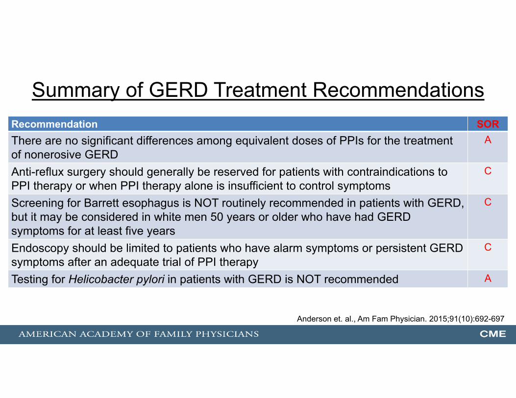
Summary of GERD Treatment Recommendations Recommendation SORThere are no significant differences among equivalent doses of PPIs for the treatment of nonerosive GERD
A
Anti-reflux surgery should generally be reserved for patients with contraindications to PPI therapy or when PPI therapy alone is insufficient to control symptoms
C
Screening for Barrett esophagus is NOT routinely recommended in patients with GERD, but it may be considered in white men 50 years or older who have had GERD symptoms for at least five years
C
Endoscopy should be limited to patients who have alarm symptoms or persistent GERD symptoms after an adequate trial of PPI therapy
C
Testing for Helicobacter pylori in patients with GERD is NOT recommended A
Anderson et. al., Am Fam Physician. 2015;91(10):692-697

Inflammatory DisordersEsophagitis
Disorder Offending “Agents”Pill-induced Doxycycline, NSAIDs, steroidsInfective*• Viral• Fungal
HSV, CMVCandida
Corrosive Alkalis or acidsEosinophilic: pronounced eosinophilic infiltration
Allergic or idiopathic; Tx – steroids, diet, anti-allergy medications
*Mostly in immunosuppressed patients

Esophageal Tumors• 90% are cancer
– Much more common in males; 10% 5-yr survival rate overall (treatment improving)– Dx – endoscopy and radiography
• Squamous cell carcinoma– Most common, declining incidence– Predominant esophageal cancer in African Americans– More common with heavy alcohol and tobacco use
• Adenocarcinomas– Arise from columnar epithelium in cardia or from Barrett’s– Recall that the lower esophagus is lined by specialized intestinal epithelium.– GERD is a risk factor
Source: Wikimedia

Diseases of the Stomach
Acid Peptic Disorders of the Stomach and Duodenum Infections
Motor Disorders Cancer

5. A 49 yo female presents with a 4-week history of epigastric pain. She reports the pain gets a bit better when she eats but worse within an hour of eating. She has been using an over-the-counter liquid antacid that she reports decreases her symptoms. She denies weight loss, hematemesis, melena, or hematochezia. With the exception of midepigastric tenderness, her exam is unremarkable. Her only medication is periodic acetaminophen, which she uses for headaches. Which of the following diagnostic tests would you recommend in your evaluation?
A. Serum gastrin
B. Helicobacter pylori serology
C. Esophagogastroduodenoscopy
D. Urea Breath Test

5. A 49 yo female presents with a 4-week history of epigastric pain. She reports the pain gets a bit better when she eats but worse within an hour of eating. She has been using an over-the-counter liquid antacid that she reports decreases her symptoms. She denies weight loss, hematemesis, melena, or hematochezia. With the exception of midepigastric tenderness, her exam is unremarkable. Her only medication is periodic acetaminophen, which she uses for headaches. Which of the following diagnostic tests would you recommend in your evaluation?
A. Serum gastrin
B. Helicobacter pylori serology
C. Esophagogastroduodenoscopy
D. Urea Breath Test 30%
8%
27%
36% 1

Acid Peptic DisordersStomach and Duodenum
• Common Problem – 5%-10% of population will have PUD in their lifetimes;
50% recurrence in 5 years– DU/GU 4:1
• 90% DU in duodenal bulb• GU most common on lesser curve
Mortality RatesUlcer Type Men Women
Duodenal Ulcer 1/100,000 0.5/100,000
Gastric Ulcer 1.5/100,000 1.2/100,000

Why Do Acid Peptic Disorders Develop?
• Current theory– PUD is an imbalance between protective and aggressive
factors.Protective factors Aggressive factors
• Surface epithelial cells with mucus and bicarbonate secretions
• Apical surface membrane of gastric mucosal cells
• Prostaglandins E1 and E2
• Production of gastric acid• NSAIDs• Corticosteroids• Smoking• Alcohol consumption• ? Psychological stress• Probably not diet
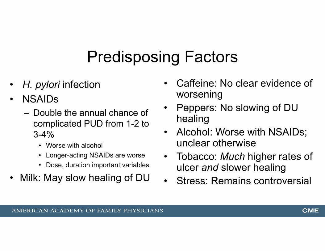
Predisposing Factors• H. pylori infection• NSAIDs
– Double the annual chance of complicated PUD from 1-2 to 3-4%
• Worse with alcohol• Longer-acting NSAIDs are worse• Dose, duration important variables
• Milk: May slow healing of DU
• Caffeine: No clear evidence of worsening
• Peppers: No slowing of DU healing
• Alcohol: Worse with NSAIDs; unclear otherwise
• Tobacco: Much higher rates of ulcer and slower healing
• Stress: Remains controversial
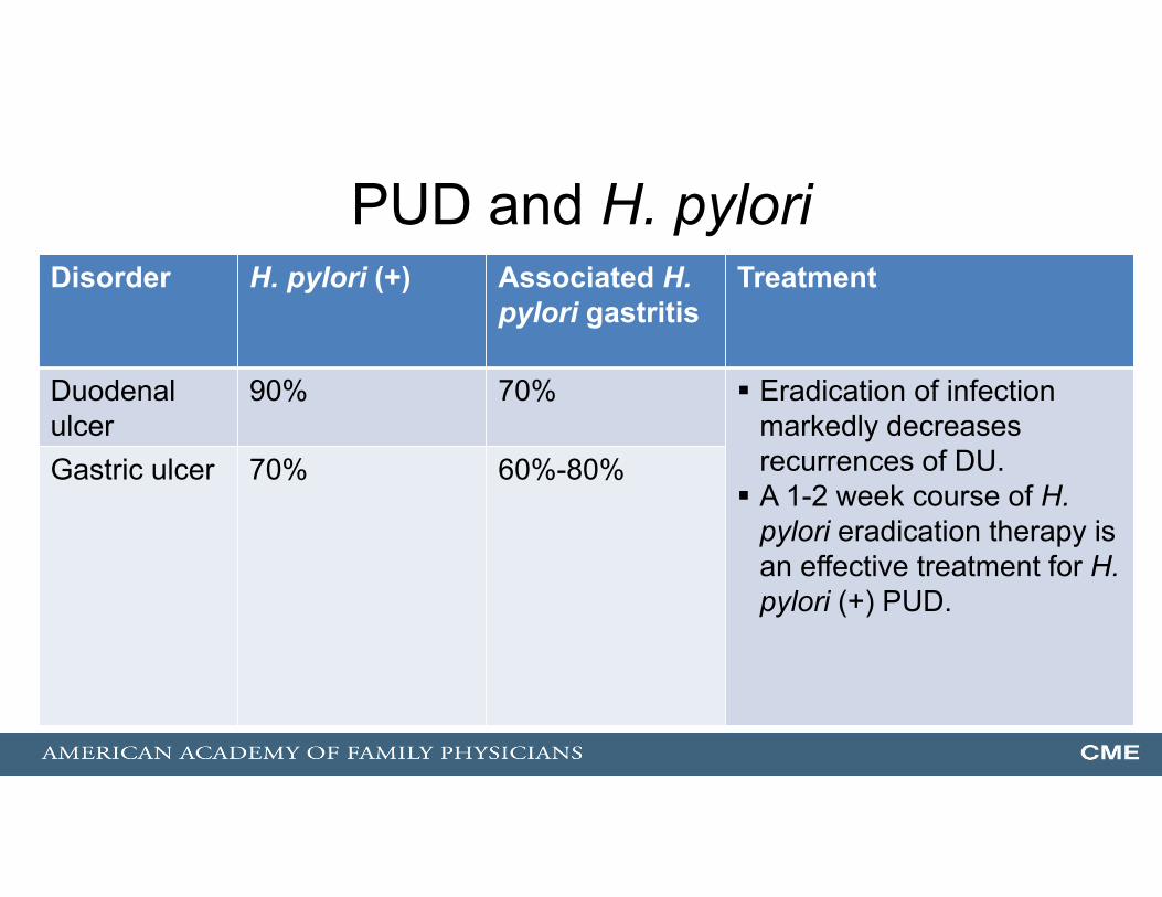
PUD and H. pylori Disorder H. pylori (+) Associated H.
pylori gastritisTreatment
Duodenal ulcer
90% 70% Eradication of infection markedly decreases recurrences of DU. A 1-2 week course of H.
pylori eradication therapy is an effective treatment for H. pylori (+) PUD.
Gastric ulcer 70% 60%-80%
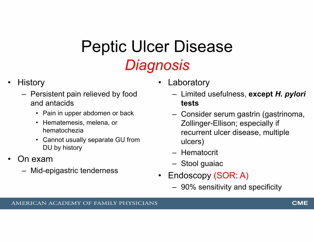
Peptic Ulcer DiseaseDiagnosis
• History– Persistent pain relieved by food
and antacids• Pain in upper abdomen or back• Hematemesis, melena, or
hematochezia• Cannot usually separate GU from
DU by history
• On exam– Mid-epigastric tenderness
• Laboratory– Limited usefulness, except H. pylori
tests– Consider serum gastrin (gastrinoma,
Zollinger-Ellison; especially if recurrent ulcer disease, multiple ulcers)
– Hematocrit– Stool guaiac
• Endoscopy (SOR: A)– 90% sensitivity and specificity
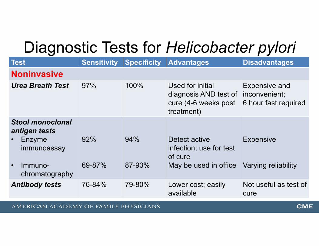
Diagnostic Tests for Helicobacter pyloriTest Sensitivity Specificity Advantages DisadvantagesNoninvasiveUrea Breath Test 97% 100% Used for initial
diagnosis AND test of cure (4-6 weeks post treatment)
Expensive and inconvenient; 6 hour fast required
Stool monoclonal antigen tests• Enzyme
immunoassay
• Immuno-chromatography
92%
69-87%
94%
87-93%
Detect active infection; use for test of cureMay be used in office
Expensive
Varying reliability
Antibody tests 76-84% 79-80% Lower cost; easilyavailable
Not useful as test of cure

Diagnostic Tests for Helicobacter pyloriTest Usefulness Sens (%) Specif (%)
Invasive Endoscopy with biopsy
Diagnostic strategy of choice in children with persistent or severe upper abdominal symptoms
Histology Sensitivity reduced by PPIs, antibiotics, bismuth-containing compounds.
> 95 100
Urease activity Test of choice when endoscopy indicated; rapid results (20 min); (−) results may need confirmation by histology or other test; sensitivity reduced by PPIs, antibiotics, bismuth-containing compounds, and active bleeding.
93-97 > 95
Culture Technically demanding; only use for resistant organism or refractory disease.
70-80 100

SummaryDiagnostic H. pylori Testing
• In patients who do not require endoscopic evaluation for evaluation of new onset dyspepsia (< age 55 and no alarm symptoms), initial diagnosis of H. pylori should be made with a test for active infection (stool antigen or urea breath test).
o Serology, as it cannot differentiate between past or current infection and has a low positive predictive value in much of the United States, is not recommended in patients with a low pretest probability.
• Endoscopic biopsy reserved for patients who are undergoing a diagnostic endoscopy and are found to have an ulcer and for those who require endoscopy to follow up a gastric ulcer or for the diagnosis or follow-up of suspected MALT lymphoma.
o Biopsy urease testing can be performed in patients not taking antibiotics or a proton pump inhibitor when histopathology is not required.
• Confirmation of eradication [because of the availability of accurate, relatively inexpensive, noninvasive tests (stool and breath tests) and because of increased resistance to antibiotic therapy,] at least four weeks after treatment (Grade 2B)
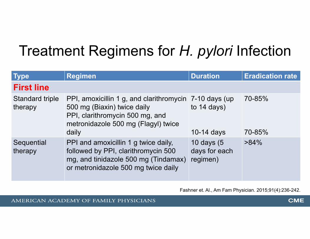
Treatment Regimens for H. pylori InfectionType Regimen Duration Eradication rateFirst lineStandard triple therapy
PPI, amoxicillin 1 g, and clarithromycin 500 mg (Biaxin) twice dailyPPI, clarithromycin 500 mg, and metronidazole 500 mg (Flagyl) twice daily
7-10 days (up to 14 days)
10-14 days
70-85%
70-85%Sequential therapy
PPI and amoxicillin 1 g twice daily, followed by PPI, clarithromycin 500 mg, and tinidazole 500 mg (Tindamax) or metronidazole 500 mg twice daily
10 days (5 days for each regimen)
>84%
Fashner et. Al., Am Fam Physician. 2015;91(4):236-242.

Treatment Regimens for H. pylori InfectionType Regimen Duration Eradication rateSecond lineNon-bismuth-based quadruple therapy (concomitant therapy)
PPI, amoxicillin 1 g, clarithromycin 500 mg, and tinidazole 500 mg or metronidazole 500 mg twice daily
10 days 90%
Bismuth-based quadruple therapy
Bismuth subsalicylate 525 mg or subcitrate 300 mg, metronidazole 250 mg, and tetracycline 500 mg, four times daily; and PPI twice daily
10-14 days 75-90%
Levofloxacin-based triple therapy
PPI and amoxicillin 1 g twice daily, and levofloxacin 500 mg (Levaquin) once daily
10 days -----
Fashner et. Al., Am Fam Physician. 2015;91(4):236-242.

Specific Ulcer TreatmentAnti-secretory
• Anti-secretory therapy– Mainstay of therapy in uninfected patients
• PPI, H2 blocker – Appropriate for maintenance therapy in selected cases– Usually 4-6 weeks for DU– Generally longer for GU – 12 weeks– PPIs lead to faster healing than H2 blockers.
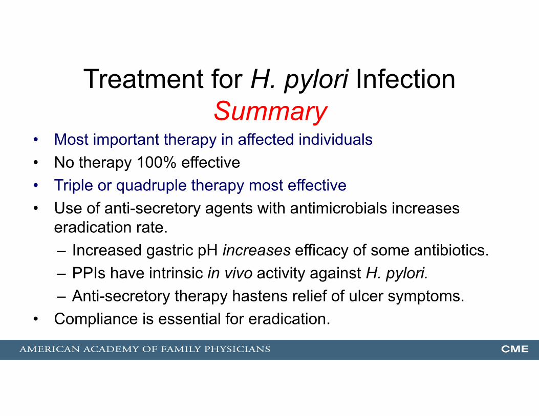
Treatment for H. pylori InfectionSummary
• Most important therapy in affected individuals• No therapy 100% effective• Triple or quadruple therapy most effective• Use of anti-secretory agents with antimicrobials increases
eradication rate.– Increased gastric pH increases efficacy of some antibiotics.– PPIs have intrinsic in vivo activity against H. pylori.– Anti-secretory therapy hastens relief of ulcer symptoms.
• Compliance is essential for eradication.

6. In considering NSAID-induced ulcers, which of the following statements is true?
A. Peptic ulcers are less common in patients taking NSAIDs who are H. pylori (+) compared with those who are (-)
B. Eradicating H. pylori in NSAID users reduces the likelihood of peptic ulcer by about one-half
C. The use of a maintenance PPI is less effective than H. pylori eradication therapy for preventing NSAID-related ulcers
D. Patients who will be on long-term NSAID therapy should be empirically treated for H. pylori
3%
2%
61%
33%1

NSAID Ulcers• Risk factors
– Prior adverse GI event (ulcer, hemorrhage) – Age > 60 (Older age) – High-dose NSAID (> twice normal) – Use of
• Glucocorticoid use• Anticoagulant use• Aspirin
• Risk for NSAID-induced GI toxicity 9% at 6 months with multiple risk factors present
• In naïve NSAID users, H. pylori is significant risk factor for complicated ulcer disease – screening may be indicated.

Recommendations for Prevention of NSAID-Related Ulcer Complications
Cardiovascular Risk Gastrointestinal Risk ACG Recommendation (2009)Low Low (No risk factors) NSAID
Moderate (1 or 2 risk factors)
NSAID plus PPI or misoprostol
High (> 2 risk factors) Alternative therapy IF POSSOBLE, or COX-2 inhibitor plus PPI or misoprostol
High Low Naproxen plus PPI or misoprostolModerate Naproxen plus PPI or misoprostolHigh Avoid NSAID and COX-2 inhibitor; alternative
therapy
Lanza FL, Chan FK, Quigley EM; Practice Parameters Committee of the American College of Gastroenterology. Guidelines for prevention of NSAID-related ulcer complications. Am J Gastroenterol. 2009;104(3):728-738.

NSAID Users: Medical Treatment of Peptic Ulcer Practice Guidelines
• Treatment of NSAID ulcers– D/C NSAIDs– PPIs superior to H2 receptor antagonists
• The ACG guideline recommends that patients who will be on long-term NSAID therapy be tested for H. pylori infection and eradication therapy given if (+)
NEJM. 1998;338:727.Primary Care. September, 2001:28(3):487-503.
Am J Gastrolenterol. 2009;104(3) :728-738

Conclusion• For patients who have multiple risk factors for
NSAID-related gastroduodenal toxicity – FDA approved options include:
• COX-2 selective inhibitor or • Nonselective NSAID in combination with a proton pump
inhibitor (PPI) or misoprostol. High dose H2 receptor antagonists are a reasonable alternative to a PPI or misoprostol• Approved doses of these drugs: Misoprostol (200 mcg QID), Lansoprazole
(15 or 30 mg daily), and esomeprazole (20 or 40 mg daily).

TreatmentNon−H. pylori PUD
• Withdrawal of potential offending or contributing agents– NSAIDs, cigarettes, excess ETOH
• No firm dietary recommendations – avoid foods that precipitate dyspepsia.
• Address psychosocial issues and comorbidities – no firm evidence, but may have deleterious health consequences.

Controversy: Treatment ofH. pylori in Non-ulcer Dyspepsia
• Efficacy of treatment is controversial. • Recent review of RCTs: Eradication provides small but
significant benefit for dyspeptic symptoms.*• Eradication may be cost-effective intervention for non-ulcer
dyspepsia.• And … H. pylori appears to have a net suppressive effect on
acid production, so treating may make GERD worse.
*Practice Guideline. Moayyedi P, Soo S, et al. Eradication of Helicobacter pylori for non-ulcer dyspepsia. The Cochrane Database of Systematic Reviews. 2005. http://www.cochrane.org/cochrane/revabstr/ab002096.htm

An Approach to Dyspepsia
Fails Fails
Fails Fails
H. Pyloriprevalence < 10%
Trial of PPl
Test and treatfor H. pylori.
Consider upperendoscopy.
Dyspepsia
H. pyloriprevalence ≥ 10%
Test and treat forH. pylori.
Trial of PPl
Consider upperEndoscopy.
Upper endoscopy
> 55 years old or presence of alarm symptoms, family or personal Hx GI CA or PUD, wt. loss, GI bleeding, anemia, or dysphagia
55 years oldNo alarm features

Risk Factors for Ulcer Complications
• Previous history of complications• Prior refractory or protracted course • Big ulcers (> 2 cm)• Deformed ulcer bed or dense fibrosis

Complications of PUD• Bleeding
– Most common complication and leading cause of death (4%-9% mortality rate)
– Occurs in 10%-20%– Patients with 1 episode of bleeding more likely to re-bleed– 90% stop without specific treatment.– Increased morbidity with associated portal hypertension*
* Am J Gastro 1998;93:336

Complications of PUD• Gastric outlet obstruction
– Usually mechanical obstruction due to edema or scar– Most due to DU– Rare: 2% of ulcer patients
• Perforation and penetration– 2% of ulcers perforate.– Average duration of Sx prior to perforation: 5 years
• NOTE: Complicated ulcer disease less likely to involve H. pylori

PUDSurgical Treatment/Management
• Dramatically declined over past two decades• Indications
– Hemorrhage not responsive to medical therapy– Gastric outlet obstruction not reversed by medical
treatment– Perforation– Malignancy
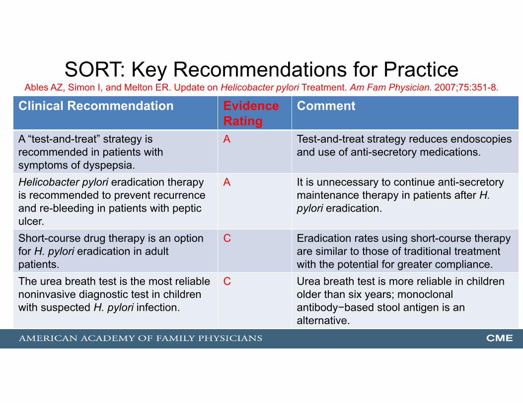
SORT: Key Recommendations for PracticeAbles AZ, Simon I, and Melton ER. Update on Helicobacter pylori Treatment. Am Fam Physician. 2007;75:351-8.
Clinical Recommendation Evidence Rating
Comment
A “test-and-treat” strategy is recommended in patients with symptoms of dyspepsia.
A Test-and-treat strategy reduces endoscopiesand use of anti-secretory medications.
Helicobacter pylori eradication therapy is recommended to prevent recurrence and re-bleeding in patients with peptic ulcer.
A It is unnecessary to continue anti-secretorymaintenance therapy in patients after H. pylori eradication.
Short-course drug therapy is an option for H. pylori eradication in adult patients.
C Eradication rates using short-course therapyare similar to those of traditional treatmentwith the potential for greater compliance.
The urea breath test is the most reliable noninvasive diagnostic test in children with suspected H. pylori infection.
C Urea breath test is more reliable in childrenolder than six years; monoclonal antibody−based stool antigen is an alternative.

SORT: Key Recommendations for PracticeFashner et. al. Diagnosis and Treatment of PUD and H. pylori Infection. Am Fam Physician. 2015;91(4):236-242
Clinical Recommendation Evidence Rating
Use the test-and-treat strategy for patients with dyspepsia who are <55 years and have no alarm symptoms for gastric cancer. Use endoscopy for all other patients.
A
Confirm eradication of H. pylori after therapy in patients with H. pylori-associated ulcer, continued dyspeptic symptoms, mucosa-associated lymphoid tissue lymphoma, and resection of gastric cancer.
C
Non–bismuth-based quadruple therapy (10 days of a proton pump inhibitor, amoxicillin 1 g, clarithromycin 500 mg [Biaxin], and metronidazole 500 mg [Flagyl] or tinidazole 500 mg [Tindamax] twice daily) has the highest success rate in eradicating H. pylori, although other regimens may also be used.
A
For patients at low risk of gastrointestinal complications, nonsteroidal anti-inflammatory drugs may be used, whereas cotherapy with a proton pump inhibitor or misoprostol (Cytotec) is recommended for patients with moderate risk of ulcer, and they should be avoided in those with a high risk of ulcer.
C

Gastric Dysmotility

Gastric DysmotilitySlow or Delayed Emptying
• Etiology– Mechanical or outlet obstruction
• PUD, bezoar, etc.– Functional obstruction (gastroparesis)
• Drugs – opiates, anticholinergics, beta and Ca channel blockers
• Diabetes, Parkinson’s, hypothyroidism, hypoparathyroidism• Pregnancy• Post-vagotomy
Trichobezoar being extracted through a gastrotomy

Gastric DysmotilitySlow or Delayed Emptying
• Diagnosis – Nausea, vomiting, dysphagia, post-prandial abdominal
pain, GERD– Tests: Scintigraphy, electrogastrogram (evaluate gastric
myoelectrical utility), ultrasonography• Treatment
– Remove causes– Low-fat diet; avoid large meals– Metoclopramide, erythromycin, prokinetics

Gastric DysmotilityRapid Gastric Emptying
• Dumping Syndrome – Most commonly seen post-operatively from gastric surgery or a
truncal vagotomy• Symptoms
– 15-30 minutes after eating – nausea, non-productive vomiting, sweating, flushing, abdominal cramping, diarrhea
• Treatment– 6-8 small, low-CHO meals/day; avoid excessive liquids; use of
opiates and anticholinergic drugs; fiber products; possibly surgery

7. A 65-yo male smoker complains of dyspepsia, weight loss, early satiety, and occasional nausea and vomiting. Which one of the following would be the initial diagnostic method of choice?
A. Upper GI endoscopy
B. CT of the upper abdomen
C. Single-contrast upper GI barium swallow
D. Endoscopic ultrasonography

7. A 65-yo male smoker complains of dyspepsia, weight loss, early satiety, and occasional nausea and vomiting. Which one of the following would be the initial diagnostic method of choice?
A. Upper GI endoscopy
B. CT of the upper abdomen
C. Single-contrast upper GI barium swallow
D. Endoscopic ultrasonography3%
71%
14%
12%
1
Cancer of the Stomach • One of the most common internal malignancies in the world
– 95% are adenocarcinomas.• Chronic GERD is the leading cause of esophageal adenocarcinoma (68%-90%).
– Only 10%-20% of US GI tumors – probably because of lower rates of H. pylori in US, due to cleaner food and water
• H. pylori can cause chronic active gastritis and atrophic gastritis, early steps in the carcinogenesis sequence
• 2x as common in ♂ as in ♀• 2x as common in African Americans and Hispanics as in Caucasians
• Dx: Endoscopic biopsy in patients with upper GI symptoms or high-risk or double-contrast barium swallow
• Tx: Surgical excision; 5-yr survival rate < 10%

Pancreatic Cancer• Fourth leading cause of cancer-related death; second only to
CRC as cause of GI cancer-related death– Higher incidence: ♂ and African Americans– Risk factors: Smoking, chronic pancreatitis, diabetes,
hereditary predisposition• History/PE
– Abd pain, weight loss, jaundice, pancreatitis– Jaundice, abdominal mass, ascites

Pancreatic Cancer• Diagnosis
– U/S, EUS CT, MRI ERCP, FNA, CA19-9– All sensitive and specific
• Treatment– Surgical resection only potential curable treatment
• Prognosis – 5-year survival – Node (−) 25%-30%– Node (+) 10%

Thank you!

Answers1. C2. B3. C4. D5. D6. B7. A

Supplementary Slides

GERD: Incidence and Prevalence• Peak prevalence at ages 30-60 years, more common
in women• Prevalence 10-20% in the Western world (lower
prevalence in Asia)• Most common GI-related diagnosis in the U.S.• 14% of U.S. population has frequent GERD symptoms
Arch Intern Med 2001 Jan 8;161(1):45Am J Gastroenterol 2006 Sep;101(9):2128Am J Gastroenterol 2013; 108:308 – 328

H. pylori Resistance
• Metronidazole 22%-39%• Clarithromycin 11%• Amoxicillin, tetracycline Rare• Bismuth None