Embed Size (px)
Citation preview
1
Enterovirus 71 in Malaysia: A decade later
Yoke-Fun CHAN PhD, I-Ching SAM MRCPath, Kai-Li WEE BSc, Sazaly ABUBAKAR PhD
Tropical Infectious Diseases Research and Education Centre, Department of Medical Microbiology, Faculty of Medicine, University Malaya, Kuala Lumpur, Malaysia
Abstract
In the last decade, Malaysia has experienced several hand, foot and mouth disease (HFMD) epidemics, complicated by fatalities due to severe neurological involvement. Enterovirus 71 (EV-71) has been implicated as the major causative agent for these epidemics. EV-71 infection is a global public health problem with pandemic potential. In many parts of Asia-Pacifi c, the virus has emerged as one of the most deadly virus infections amongst young children. The virus is highly transmissible through faecal-oral route and respiratory droplets. A recent rise in neurological complications and deaths suggests that the viruses currently circulating may be more virulent. The major risk factor associated with more severe EV-71 infection is young age and poor cellular immunity. Rapid laboratory diagnosis and molecular surveillance is important to closely monitor the emergence of new EV-71 subgenotypes. Since vaccine and anti-virals for EV-71 are not available, control and prevention strategies remain the only ways to combat the infection.
Neurology Asia 2011; 16(1) : 1 – 15
Address correspondence to: Yoke-Fun Chan, Department of Medical Microbiology, Faculty of Medicine, University Malaya, 50603 Kuala Lumpur, Malaysia. Tel: 60379676677, Fax: 60379675752, Email: [email protected]; [email protected]
INTRODUCTION
Since its fi rst isolation in California in 1969, enterovirus 71 (EV-71) has caused sporadic outbreaks of hand, foot and mouth disease (HFMD), as well as serious neurological diseases such as meningitis, encephalitis, and acute fl accid paralysis (AFP). In the last 15 years, these epidemics have increased in number and involved large numbers of children in Asia, including Malaysia. In this review, we will discuss the epidemiology, clinical manifestations, risk factors, diagnosis, and management of EV-71 infection, with particular reference to work in Malaysia, and our diagnostic virology laboratory in University Malaya Medical Centre (UMMC), in Kuala Lumpur, Malaysia.
VIROLOGY OF EV-71
EV-71 belongs to the Picornaviridae family. Picornaviruses are small, about 28 nm in size, non-enveloped, and consist of icosahedral particles with a positive-stranded RNA genome. Based on nucleotide sequences, physico-chemical properties, serological relatedness and pathogenicity in man or mice, EV-71 is classifi ed in the genus enterovirus along with polioviruses, coxsackievirus A (CV-A) and coxsackievirus B (CV-B), echoviruses, EV-70, and bovine
enteroviruses.1-2 EV-71 belongs to the species Human enterovirus A (HEV-A), which also includes other important human pathogens such as coxsackieviruses A2-A8, A10, A14, and A16.3-4 The EV-71 genome encodes a long polyprotein with a single open reading frame followed by a poly A tract. The single polyprotein is fl anked by untranslated regions at both the 5’ and 3’ ends, and are cleaved into different structural (VP1-VP4) proteins, which form the physical structure of the virus, and non-structural proteins (2A-2C, 3A-3D), which include enzymes important for host interaction and replication (Figure 1).
GLOBAL EPIDEMIOLOGY OF ENTEROVIRUS 71
Global prevalence of EV-71
After the fi rst outbreak in California in 1969, EV-71 was subsequently associated with several other outbreaks, in Bulgaria5, Japan6, Hungary7, Australia8-9, China10, Malaysia11, Taiwan12-13, Hong Kong14, Korea15-16, Vietnam17, Macao18, Brunei19, and, more recently, China20 and Singapore in 2008. European countries such as Germany21, Norway22, United Kingdom23, Austria24, Netherlands25, France26 and Hungary27 have also reported EV-71 cases. EV-71 infection is therefore a global health problem with pandemic potential.
REVIEW ARTICLE
Neurology Asia March 2011
2
Global increase of EV-71 activity
There has been increased EV-71 activity in the Asia Pacifi c region since 1997.28 We compared surveillance data from USA, Japan, Singapore and Taiwan. In the USA, more than 50% of the 1,520 HEV-A reported were CV-A16 (40.5%) and EV-71 (17.8%).29 In the 1970s and 1980s, CV-A16 was most frequently isolated, but was then replaced by EV-71 after the 1990s.29 In Japan, enterovirus surveillance from 1982-2010 showed that of the 28,882 HEV-A isolates, 24.4% were CV-A16 and 17.5% were EV-71.30 Of 4,181 HEV-A isolates reported in Taiwan between 1998-2005, the most common enteroviruses were EV-71 (40.9%) and CV-A16 (40.7%).31 Surveillance from Singapore from 2001-2007 also revealed similar trends, with CV-A16 (40.8%) and EV-71 (31.4%) as the main enteroviruses.32
ENTEROVIRUS 71 IN MALAYSIA
Outbreaks of fatal hand, foot and mouth disease in Malaysia
In mid-1997, an outbreak of acute viral infection in Sarawak, Malaysia caused 31 deaths among young children aged between fi ve months to six years.11, 33 In most cases, the children died within hours of admission to hospitals due to acute congestive heart failure and cardiovascular collapse, which was suggestive of acute viral myocarditis. Occurrence of the fatal infections in the midst of simultaneous outbreaks of EV71-associated HFMD raised the possibility that EV71, or another enterovirus, was associated with the deaths. Based on the clinical presentation resembling myocarditis, group B coxsackieviruses
Figure 1. EV-71 genome organization. The genome organization and function of the EV-71 genes is similar to other enteroviruses.4
(CV-B) were initially thought to be the causative agent. However, CV-B was not detected in these fatal cases. Instead, a novel adenovirus was isolated from some of the fatal cases, in addition to EV-71.34-35 A few months following the outbreak in Sarawak, fatal brainstem encephalomyelitis was reported in at least four young children in Peninsular Malaysia.36 In all cases, EV-71 was isolated and identifi ed from central nervous system tissue samples.11 In 2000, another fatal HFMD outbreak hit Malaysia. In addition to EV-71, echovirus 7 was also identifi ed as a possible causative agent for three cases of acute encephalomyelitis.37 Large outbreaks with fatalities continued to be seen in 3-yearly cycles in Sarawak in 2003, 200638 and 2008/2009.39
EV-71 in Malaysia before 1997
No data is available on EV-71 or HFMD prior to 1997. Laboratory records at UMMC showed that samples were received from patients with HFMD between 1982 to 1997, but no virus isolates are available. A seroprevalence study on archived sera will help to show if EV-71 was present in Malaysia before 1997.
EV-71 in Malaysia after 1997
After the 1997 EV-71 fatal outbreak in Malaysia, our laboratory began confi rmation of EV-71. From 1997 to 2008, 1,098 samples from patients suspected with enterovirus infection were received in the virology laboratory in UMMC, and of these, 202 (18.6%) samples were culture-confi rmed positive for EV-71, particularly during the outbreak years of 1997, 2000, and 2006. No
3
EV-71 was isolated in 2002 and 2004. From 2000 onwards, the laboratory also began confi rmation of CV-A16 and since then, CV-A16 has been identifi ed in 48 patients. Of these, 33.3% (16/48) and 37.5% (18/48) were reported in 2000 and 2002, respectively, while no CV-A16 was isolated in 2001 and 2003. The number of patients with confirmed CV-A16 infection was relatively small which suggests that it is uncommon for CV-A16 to cause more severe forms of HFMD that warrant hospitalization, at least in UMMC. In Sarawak, EV-71 was seen during the outbreak years, while CV-A16 was seen in both outbreak and inter-outbreak periods.40
The average incidence rate (per 100,000 population) for 2000-2008 was 25.0 (range, 1.5-60.6). Data from Ministry of Health Malaysia (MOH)41 showed that the incidence rate of HFMD was as high as 56.1 and 60.6 in 2007 and 2008, respectively. This may be because HFMD was declared a notifi able disease in 2006. Nevertheless, it is highly likely that the disease remains underreported. The average mortality rate (per 100,000 population) for 2000-2009 was 0.01 (range 0-0.3). No deaths were reported in 2002, 2004, 2007 and 2009.
EV-71 genotype distribution
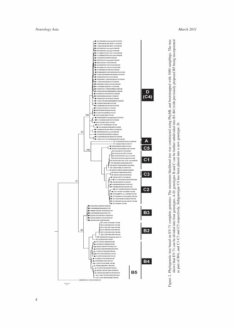
Generally, EV-71 is divided into four broad genotypes: A, B, C and D (previously designated as C4), based on analysis of complete genome sequences of many strains.42 Genotyping and phylogenetic analysis provides useful information on the evolution and epidemiology of EV-71, such as confi rming outbreaks and demonstrating geographical spread. Genotype A contains the prototype virus BrCr. Genotype B is further divided into B1-B4 (B4 was recently proposed to be merged with B5 based on its complete genome relatedness) while genotype C is further divided into C1-C3 and C5 (Figure 2). Van Sanden et al. (2009)25 reported the earliest EV-71 isolate, from 1963. Recent bioinformatics analysis showed that EV-71 probably evolved as early as 1942.43 Based on molecular epidemiological studies performed using publicly available EV-71 VP1 or VP4 gene sequences, the distribution of different EV-71 in different parts of the world was mapped (Figure 3). Our review of the published literature shows that only several EV71 subgenotypes - B4 (previously B5), C1, C2, C5 and D are currently circulating in the world. While all these genotypes are found in Asia, only genotype D, subgenotype C1 and C2
are circulating in Europe (Figure 3). Subgenotype B1 was last seen in 1986, subgenotype B2 in 1994, while subgenotypes B3 and C3 only circulated briefl y during 1997-1998 and 1997-2000, respectively. Of note, genotype A has been reported to have reemerged in China during the HFMD outbreaks in 2008 and 2009; however these isolates may not have evolved together with other subgenotypes because of its high similarity with the prototype BrCr.44 In Malaysia, subgenotypes C1, C2, B3 and B4 have been reported, but recently, predominantly, subgenotype B4 (B5) is circulating in both Peninsular Malaysia and Sarawak.
Seasonal distribution and cyclical EV71 outbreaks?
Seasonal distribution has been observed for enteroviruses in temperate countries between June to October, the summer and early fall months.4 HFMD surveillance in Singapore from 2002-2009 showed that annual peaks occurred between March to May.45 Similarly, sentinel surveillance for enteroviruses from 1998-2005 in Sarawak also showed the occurrence of HFMD around March to June.40 An 8-year epidemiologic study in Taiwan (1998-2005) showed that more severe HFMD occurred around April-June.31 Limited surveillance data is available for Peninsular Malaysia. Based on the small number of 202 laboratory confi rmed cases seen in UMMC in Kuala Lumpur, EV-71 was most frequently isolated between June to October. This hospital data refl ects more severe cases and increased reporting due to public awareness in seeking treatment, in contrast to the active surveillance of HFMD cases from sentinel clinics in Sarawak and Singapore. Some sites have reported cyclical patterns of EV-71 outbreaks, which we did not see with our limited hospital cases in Kuala Lumpur. In Sarawak, EV-71 HFMD outbreaks occurred in a cyclical pattern of approximately every two to three years.40 The Infectious Disease Surveillance Center in Japan showed a similar cyclical trend, with predominantly EV-71 isolation in 1983, 1986, 1990, 1993, 1997, 2000, 2003, 2006 and 2010.30
TRANSMISSION OF EV-71
Humans are the only known reservoir for EV-71. The faecal-oral route is the commonest mode of transmission, although respiratory droplet spread may also play a role.4 EV-71 shedding in the
Neurology Asia March 2011
4
Figu
re 2
. Ph
ylog
enet
ic t
ree
base
d on
EV
-71
com
plet
e ge
nom
es. T
he m
axim
um l
ikel
ihoo
d tr
ee w
as c
onst
ruct
ed u
sing
Phy
ML
and
boo
tstr
appe
d w
ith 1
000
sam
plin
gs. T
he t
ree
show
s th
at E
V-7
1 ca
n be
div
ided
into
fou
r ge
noty
pes,
A-D
; gen
otyp
es B
and
C c
an b
e fu
rthe
r su
bdiv
ided
into
B1-
B4
(with
pre
viou
sly
prop
osed
B5
bein
g in
corp
orat
ed
as p
art
of B
4), a
nd C
1-C
3 an
d C
5 re
spec
tivel
y. S
ubge
noty
pe C
4 ha
s be
en p
lace
d in
to a
new
gen
otyp
e, D
.
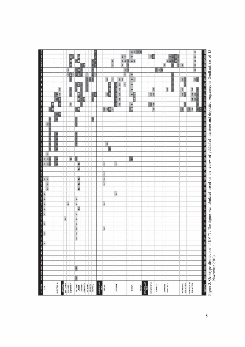
5
Figu
re 3
. G
enot
ype
dist
ribu
tion
of E
V-7
1. T
he fi
gur
e w
as t
abul
ated
bas
ed o
n th
e re
view
of
publ
ishe
d lit
erat
ure
and
depo
site
d se
quen
ces
in G
enB
ank
(as
of 1
5 N
ovem
ber
2010
).
Neurology Asia March 2011
6
stool was longer than in the throat46, and may be for up to 42 days post-infection.47 During the EV-71 outbreak in Taiwan in 2001-2002, the principal transmission was between children in childcare facilities. However, the transmission rate to household contacts was as high as 52%, especially to siblings and cousins.48 Adults are rarely infected, as most adults already have previous immunity. However, infected adults may excrete the virus without signs and symptoms, and transmit virus to susceptible children. EV-71 has been isolated from worldwide HFMD outbreaks every year, suggesting the continuous circulation of the virus in the population. The persistence of enteroviruses has been well documented in the environment, such as in sewage and water systems4, and in spring water and environmental water in Taiwan.49-50 EV-71 may also survive in the environment for at least three days at tropical room temperature.51 The persistence of EV-71 in the environment may provide a continuing source of potential exposure for susceptible populations. A very low rate of enterovirus infections amongst blood donors has been documented, suggesting that blood components are unlikely to be an important route of transmission.52 Perinatal transmission of enterovirus infections are common, however in EV-71 only one case has been documented.4,53
CLINICAL MANIFESTATIONS OF EV-71 INFECTION
Clinical manifestations of EV-71 range from asymptomatic to involvement of upper respiratory tract, gastrointestinal tract, central nervous system and cardiovascular system. Up to 71% of EV-71 infections in children may be asymptomatic, and may serve as a reservoir for transmission.54 EV-71 is associated with HFMD, meningoencephalitis, poliomyelitis-like paralysis and aseptic meningitis (non-paralytic poliomyelitis). Chang et al.
(2004)55 classifi ed symptomatic EV-71 infection into four stages.
Hand, foot and mouth disease and herpangina
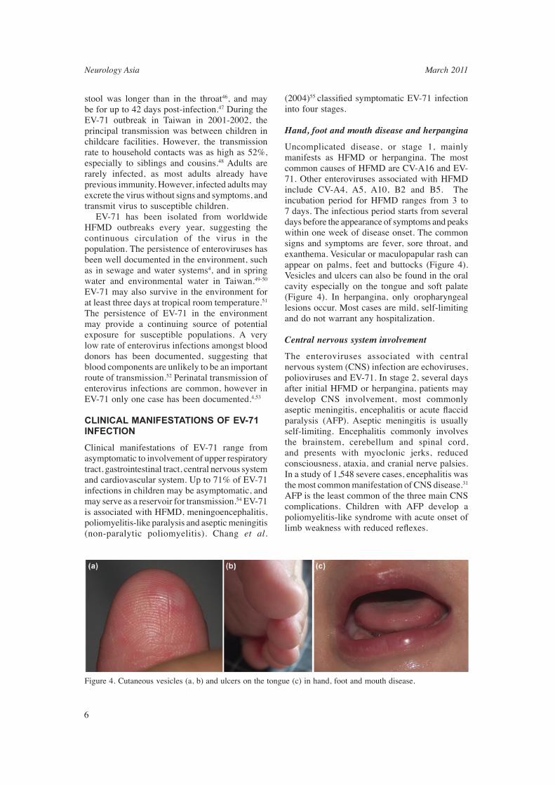
Uncomplicated disease, or stage 1, mainly manifests as HFMD or herpangina. The most common causes of HFMD are CV-A16 and EV-71. Other enteroviruses associated with HFMD include CV-A4, A5, A10, B2 and B5. The incubation period for HFMD ranges from 3 to 7 days. The infectious period starts from several days before the appearance of symptoms and peaks within one week of disease onset. The common signs and symptoms are fever, sore throat, and exanthema. Vesicular or maculopapular rash can appear on palms, feet and buttocks (Figure 4). Vesicles and ulcers can also be found in the oral cavity especially on the tongue and soft palate (Figure 4). In herpangina, only oropharyngeal lesions occur. Most cases are mild, self-limiting and do not warrant any hospitalization.
Central nervous system involvement
The enteroviruses associated with central nervous system (CNS) infection are echoviruses, polioviruses and EV-71. In stage 2, several days after initial HFMD or herpangina, patients may develop CNS involvement, most commonly aseptic meningitis, encephalitis or acute fl accid paralysis (AFP). Aseptic meningitis is usually self-limiting. Encephalitis commonly involves the brainstem, cerebellum and spinal cord, and presents with myoclonic jerks, reduced consciousness, ataxia, and cranial nerve palsies. In a study of 1,548 severe cases, encephalitis was the most common manifestation of CNS disease.31 AFP is the least common of the three main CNS complications. Children with AFP develop a poliomyelitis-like syndrome with acute onset of limb weakness with reduced refl exes.
Figure 4. Cutaneous vesicles (a, b) and ulcers on the tongue (c) in hand, foot and mouth disease.
7
Cardiopulmonary involvement
Between several hours to two days following CNS involvement, stage 3 may occur, involving dysregulation of autonomic nervous system, and reduced myocardial function with progressive hypotension or pulmonary edema.55 The onset of this stage may lead to death within hours. Extensive infl ammation is always seen in the brainstem and spinal cord, but only occasionally in the lungs or myocardium. It is hypothesized that the cardiopulmonary failure is neurogenic in origin, due to EV-71 damage of the vasomotor centers in the medulla.36
Long-term morbidity
Stage 4, the convalescence stage, is characterized by the long-term neurological sequelae of CNS involvement, which includes limb weakness, poorer cognitive ability, motor deficits and neurodevelopmental delay.13,56 Children recovered from EV-71 neurological illness may also have attention defi cit or hyperactivity problems in schools.57
RISK FACTORS FOR SEVERE EV-71
Genetic variation of EV-71
One of the possible reasons that EV-71 continues to emerge as a more virulent virus is the presence of many subgenotypes within the virus. Ooi et al. (2007)58 showed that subgenotype C1 and B5 were more likely to cause CNS infection compared to subgenotype B4. In Taiwan, outbreaks with CNS involvement due to subgenotypes C2, B4 and B5 were often preceded by shifts in the predominant circulating subgenotype of EV-71.59-60 Recombination in enteroviruses involves the breaking of viral nucleic acid before it is joined to another segment from a related virus. The resulting genetic variation may be benefi cial as it may allow immune escape, or it may alter viral virulence. The possible emergence of these different subgenotypes through inter-typic recombination with other HEV-A has been shown.61 It is now known that at least genotype D and subgenotype B3 are recombinants of EV-71 with mosaic sequences of CV-A16.61-
62 Interestingly, experiments performed in suckling mice showed that the non-recombinant subgenotype B4 and recombinant subgenotypes B3 had different neurovirulence.63 Sequencing studies have shown that different clinical outcomes of EV-71 infection are associated
with mutations at 5’UTR64-65, VP166, 2B64, 2C64, 3A67, 3C64,68, 3D65, and 3’UTR68 regions. However, there have not yet been any consistent fi ndings of specifi c mutations that increase virulence that may be used in a clinically relevant manner.
Co-infection
The possible role of co-infection of other infectious agents in increasing virulence cannot be excluded. During the 1997 HFMD outbreak in Malaysia, adenovirus was also isolated11,34, while in 2000, echovirus 7 was isolated in severe cases.37 Nonetheless, it has also been recently reported that dual infection with other viruses (such as other enteroviruses or adenovirus) in Sarawak did not increase the risk of neurological complications.58 It is likely that co-infection of EV-71 with other HEV-A provides opportunity for recombination.
Host factors
Age
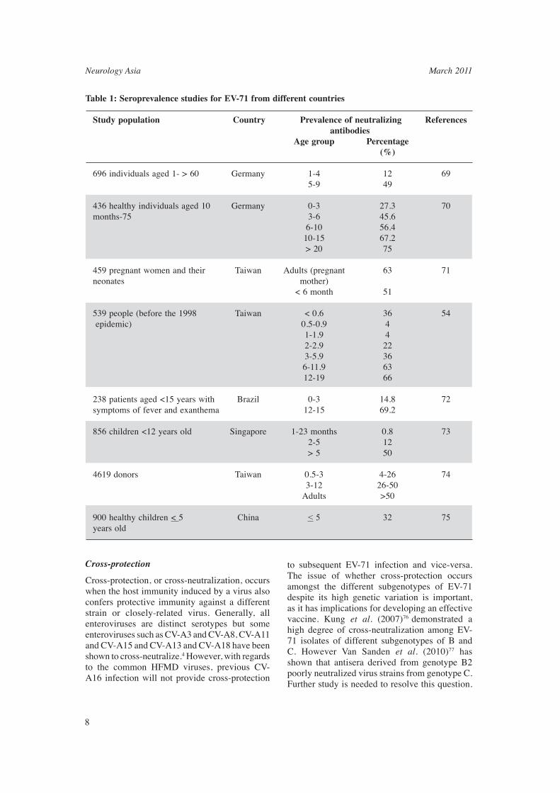
Based on our laboratory records of confi rmed HFMD cases from 1997-2007, the ages of children infected with EV-71 and CV-A16 were tabulated. Of the 145 confi rmed EV-71 cases from 1997-2007, 9% were aged < 1, 65.5% were aged 1-3, 18.6% were aged 4-6 and 5.5% were aged 7-12. Of the 32 confi rmed CV-A16 cases, 9.4% were aged <1, 71.9% were aged 1-3 and 18.8% were aged 4-6. Many studies have showed that children below four years are the most susceptible to infection, and are summarized in Table 1. In Singapore, <1% of children of 6-23 months of age have neutralizing antibodies against EV-71, but by the age of fi ve, the rate was >50%.73 In Taiwan, seroprevalence of EV-71 in children aged 0.5-2 years was only 4%, but in children above six years and adults, seroprevalence was 57-67%.54 In Brazil, about 85% of patients aged 0-3 years had no neutralizing antibodies against EV-71, but by the age of 12-15, >70% had neutralizing antibodies.72 Another recent Taiwan study has shown that maternal neutralizing antibodies are present in about 50% of neonates, but are undetectable by 6 months of age.71 Taking together all these fi ndings, children between 6 months and 3 years are at highest risk of acquiring EV-71 infection due to lack of protective immunity. In addition, Chang et al. (2002)54 also showed that more severe and fatal EV-71 cases occurred in this age group.
Neurology Asia March 2011
8
Study population Country Prevalence of neutralizing References antibodies Age group Percentage (%) 696 individuals aged 1- > 60 Germany 1-4 12 69 5-9 49
436 healthy individuals aged 10 Germany 0-3 27.3 70months-75 3-6 45.6 6-10 56.4 10-15 67.2 > 20 75
459 pregnant women and their Taiwan Adults (pregnant 63 71neonates mother) < 6 month 51
539 people (before the 1998 Taiwan < 0.6 36 54 epidemic) 0.5-0.9 4 1-1.9 4 2-2.9 22 3-5.9 36 6-11.9 63 12-19 66
238 patients aged <15 years with Brazil 0-3 14.8 72symptoms of fever and exanthema 12-15 69.2
856 children <12 years old Singapore 1-23 months 0.8 73 2-5 12 > 5 50 4619 donors Taiwan 0.5-3 4-26 74 3-12 26-50 Adults >50
900 healthy children < 5 China < 5 32 75years old
Table 1: Seroprevalence studies for EV-71 from different countries
Cross-protection
Cross-protection, or cross-neutralization, occurs when the host immunity induced by a virus also confers protective immunity against a different strain or closely-related virus. Generally, all enteroviruses are distinct serotypes but some enteroviruses such as CV-A3 and CV-A8, CV-A11 and CV-A15 and CV-A13 and CV-A18 have been shown to cross-neutralize.4 However, with regards to the common HFMD viruses, previous CV-A16 infection will not provide cross-protection
to subsequent EV-71 infection and vice-versa. The issue of whether cross-protection occurs amongst the different subgenotypes of EV-71 despite its high genetic variation is important, as it has implications for developing an effective vaccine. Kung et al. (2007)76 demonstrated a high degree of cross-neutralization among EV-71 isolates of different subgenotypes of B and C. However Van Sanden et al. (2010)77 has shown that antisera derived from genotype B2 poorly neutralized virus strains from genotype C. Further study is needed to resolve this question.
9
Antigenic variation in subgenotype B5 viruses did not fully protect the Taiwan population that had prior exposure to genotypes C and B4 and resulted in the Taiwan 2008 outbreak, the largest outbreak since 1998.60
Genetic factors
Genetic susceptibility to EV-71 infection has not been studied in detail. HLA-33 was strongly associated with EV-71 infection, while HLA-A2 was found more frequently in EV-71 cardiopulmonary failure patients.78 As HLA-33 is an HLA type commonly seen in 17-35% of Asian populations, including Taiwan and Malaysia, and seen in <1% of white populations, it was suggested that this may explain why EV-71 outbreaks occur more frequently in Asian countries.78 Glucose 6-phosphate defi ciency has also been shown to enhance EV-71 infection in vitro, but remains to be determined in human subjects.79
CLINICAL AND LABORATORY BIOMARKERS
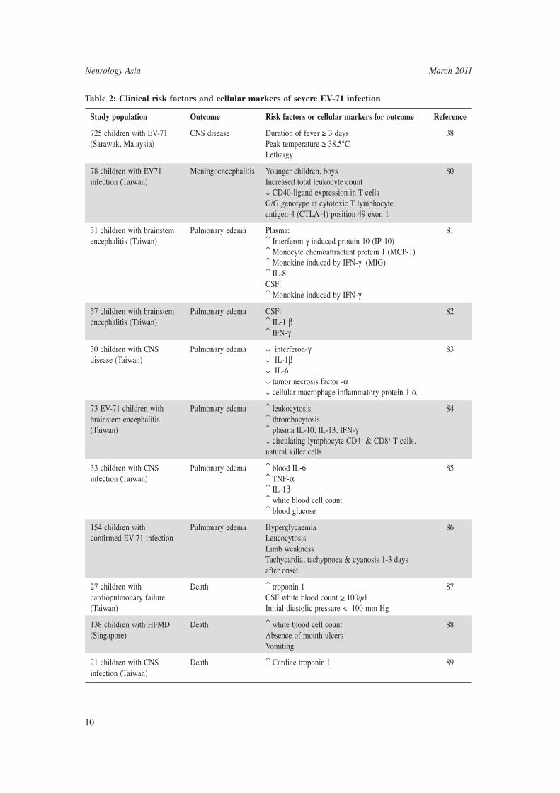
Reported clinical and laboratory markers of severe EV-71 infection are shown in Table 2. Increase of leukocyte counts in blood and CSF were noted in patients with pulmonary edema and encephalitis (Table 2). Generally, a poor cellular immunity response has been correlated with complicated EV-71 infection. Some studies have also reported the increase or decrease of cellular markers such as interleukins, however, most involved small number of patients with preliminary data and no conclusive fi ndings were noted. Cardiac troponin 1 has been shown to be an early indicator of fatal EV-71 infection.87,89 Understanding these risk factors will aid in the management of HFMD patients by allowing identifi cation of those at risk of progression to severe neurological disease.
LABORATORY DIAGNOSIS
During a HFMD outbreak, rapid diagnosis is important to identify the causative agent and implement proper control measures. The method of choice must be able to differentiate between EV-71 and CV-A16, because the latter usually causes uncomplicated HFMD. In addition, it is also imperative to closely monitor the emergence of new subgenotypes which may predispose to neurological complications and deaths.
Choice of specimens
For diagnosis of EV-71, clinical specimens can
include throat swabs, ulcer swabs, vesicle swabs/fl uid, cerebrospinal fl uid, rectal swabs, stool and brain tissues. Blood is generally not useful, as enteroviruses may only be detected in blood at very early stages of infection.90 Generally, enteroviruses can be recovered from these sites during the fi rst few days of illness, and from rectal swabs and stool for many weeks afterwards.4 In our laboratory, EV-71 and CV-A16 were most frequently isolated from vesicle swabs (47/147, 32%) followed by oral swabs (12/55, 21.8%), throat swabs (86/434, 19.8%), stool (34/180, 18.9%), rectal swabs (89/519, 17.2%), ulcer swabs (7/47, 14.9%) and cerebrospinal fl uid (7/151, 4.7%). These fi ndings were very similar to the evaluation performed in Sarawak and Taiwan.91-92 As enterovirus infections are often asymptomatic, the isolation of enteroviruses from non-sterile sites such as throat and stool should be interpreted in the context of full clinical information, especially in the event of unusual or severe clinical disease. Isolation of enteroviruses from sterile sites such as CSF is always signifi cant, but may be diffi cult to achieve due to lower virus loads.
Laboratory methods
The gold standard of EV-71 diagnosis is virus isolation using mammalian tissue cultures and subsequent confi rmation with EV-71 specifi c monoclonal antibody. Mammalian cell cultures such as Vero (African green monkey kidney), human RD (rhabdomyosarcoma) cells, HeLa (cervical adenocarcinoma) cells and BGMK (buffalo green monkey kidney) support the growth of many enteroviruses.4 Cytopathic effects can be observed as early as day 2. However, virus isolation is usually technically demanding, tedious, takes days to weeks, and has poor sensitivity. Detection of viral RNA by reverse-transcriptase PCR (RT-PCR) is increasingly becoming the method of choice, as it is rapid and sensitive. Careful interpretation of PCR results is necessary for non-sterile sites, as residual RNA and live viruses can be shed in throat and stool for weeks.47 As genotyping is now considered an essential epidemiological tool, the right choice of genes for RT-PCR and genotyping is crucial since EV-71 frequently recombines and has high genetic variation. 5’UTR is frequently used for detection of enteroviruses while VP1 is used for genotyping. Recently, 3D has also been proposed for genotyping, as this gene is commonly involved in recombination in enteroviruses.42,93
Commercial IgM capture ELISA is available
Neurology Asia March 2011
10
Table 2: Clinical risk factors and cellular markers of severe EV-71 infection
Study population Outcome Risk factors or cellular markers for outcome Reference
725 children with EV-71 CNS disease Duration of fever ≥ 3 days 38(Sarawak, Malaysia) Peak temperature ≥ 38.5°C Lethargy
78 children with EV71 Meningoencephalitis Younger children, boys 80infection (Taiwan) Increased total leukocyte count ↓ CD40-ligand expression in T cells G/G genotype at cytotoxic T lymphocyte antigen-4 (CTLA-4) position 49 exon 1
31 children with brainstem Pulmonary edema Plasma: 81encephalitis (Taiwan) ↑ Interferon-γ induced protein 10 (IP-10) ↑ Monocyte chemoattractant protein 1 (MCP-1) ↑ Monokine induced by IFN-γ (MIG) ↑ IL-8 CSF: ↑ Monokine induced by IFN-γ
57 children with brainstem Pulmonary edema CSF: 82encephalitis (Taiwan) ↑ IL-1 β ↑ IFN-γ
30 children with CNS Pulmonary edema ↓ interferon-γ 83disease (Taiwan) ↓ IL-1β ↓ IL-6 ↓ tumor necrosis factor -α ↓ cellular macrophage infl ammatory protein-1 α
73 EV-71 children with Pulmonary edema ↑ leukocytosis 84brainstem encephalitis ↑ thrombocytosis(Taiwan) ↑ plasma IL-10, IL-13, IFN-γ ↓ circulating lymphocyte CD4+ & CD8+ T cells, natural killer cells
33 children with CNS Pulmonary edema ↑ blood IL-6 85infection (Taiwan) ↑ TNF-α ↑ IL-1β ↑ white blood cell count ↑ blood glucose
154 children with Pulmonary edema Hyperglycaemia 86confi rmed EV-71 infection Leucocytosis Limb weakness Tachycardia, tachypnoea & cyanosis 1-3 days after onset
27 children with Death ↑ troponin 1 87cardiopulmonary failure CSF white blood count > 100/μl(Taiwan) Initial diastolic pressure < 100 mm Hg
138 children with HFMD Death ↑ white blood cell count 88(Singapore) Absence of mouth ulcers Vomiting
21 children with CNS Death ↑ Cardiac troponin I 89infection (Taiwan)
11
for diagnosis of EV-71.94 This would be useful as most diagnostic laboratories have the capability to perform ELISA, but not virus isolation or PCR. Neutralization assays may be used to detect EV-71 antibodies, but are laborious and used only in research settings.
TREATMENT AND PREVENTION
Management
The majority of EV-71 cases are mild, do not require hospitalization, and can be treated symptomatically. However, the challenge, particularly during an outbreak situation with large numbers of infected children, is to identify the small numbers who are at risk of severe neurological disease. A prospective study in Sarawak identified and validated three risk factors for neurological involvement: fever for >3 days, peak temperature >38.5°C, and lethargy.38 The stage management approach by Chang et al. (2004)55 has improved the outcome of EV-71 infection in Taiwan. Management of EV-71 infection is mainly supportive, as there are still no effective anti-virals. Patients in stage 2 who demonstrate CNS disease should be hospitalized, and closely monitored. Intravenous immunoglobulin (IVIG) has been used at this stage to reduce acute mortality in Taiwan and Malaysia.38,95 The mechanism of effect of IVIG is not known, but IVIG reduces plasma levels of cytokines which are raised in patients with EV-71-associated pulmonary edema.95 Patients in stage 3, with cardiopulmonary failure, require the necessary intensive care support, and may also benefi t from milrinone, which has inotropic and vasodilating properties to increase cardiac output.96 For Stage 4 patients, rehabilitation is required for limb weakness/atrophy and dysphagia, while those with diaphragm dysfunction, apnea or central hypoventilation may require long-term respiratory support.
Developments in anti-virals and vaccines
EV-71 infection is often self-limiting and does not require any medication. However, in the case of neurological complications, anti-viral drugs would be benefi cial to improve the outcomes. Many new developments in anti-viral therapy are on-going. Interferons, antibodies, capsid inhibitors, enviroxime-like compounds targeting the 3A and 3C protease inhibitors are potential anti-picornaviral compounds.97 The capsid inhibitor pleconaril is one of the most promising enteroviral
drugs, and is currently being tested in phase 2 clinical trials for viral meningitis. However, its use has not been demonstrated for EV-71. No anti-viral drug is currently available for EV-71. Nevertheless, there are numerous studies showing in vitro effects of many compounds against EV-71. Ribavirin, a nucleoside analogue has been shown to reduce viral load in cell culture and in animals.98 Lactoferrin has been shown to prevent entry of virus in the host cell by binding to EV-71 capsid VP1, protecting cells against EV-71 infection and mice against EV-71 lethal challenge.99-100 Other compounds reported with anti-EV-71 activity include the Raf-1 inhibitor, GW5074101, capsid-binding, pyridyl imidazolidinones102-103 and the interferon inducer, aloe emodin.104 In addition, the therapeutic use of small-interfering RNA targeting the capsid and non-structural genes of EV-71 showed promising viral inhibition activity.105 A vaccine is highly desirable for EV-71. The conventional formalin-inactivated EV-71106 and attenuated EV-71 strain107-108 are potential candidates for EV-71 vaccines. Other newer vaccines include oral vaccine with transgenic tomato109 and milk of transgenic mice100, virus-like particles110, VP1 subunit vaccine106,111-112 and synthetic peptides targeting VP1.113
Prevention and control strategies for EV-71
Hand-washing and environmental cleaning
The lack of proper hygiene and close contact between children facilitates faecal-oral spread. Simple handwashing with soap will remove the virus from hands. The virus is resistant to many disinfectants such as alcohol and phenolic disinfectants. The effi cacy of Virkon S, a peroxide-based disinfectant has been tested and a minimum 1% Virkon is suffi cient to kill the virus.51 Diluted chlorine (bleach) has also been recommended for cleaning surfaces and toys in nurseries during outbreaks.
Public health control measures
Adequate surveillance systems should be in place to enable early detection and control of outbreaks. In Malaysia, the statutory notifi cation of HFMD has been in enforcement since 12 October 2006, and HFMD guidelines were released by the Ministry of Health Malaysia in 2007.114 As previously shown, most of the transmission of EV-71 occurs in childcare settings or between household members.48,54,73 Under the guidelines,
Neurology Asia March 2011
12
when two or more cases occur in the same childcare facility, the premise is advised to close for ten days from the date of onset of the last case, and must be thoroughly disinfected before reopening. Infected children should always have minimal contact with other people, particularly other children in the kindergarten or household. Public education, particularly during outbreaks, will also help to alert the public to adopt preventive measures. These include washing hands before preparing food, after going to the toilet, and after handling stool-soiled clothes or diapers, and covering the nose and mouth when coughing or sneezing.
CONCLUSION
EV-71 is an emerging virus with pandemic potential to cause severe neurological disease. Although large outbreaks are mainly seen in Asia, EV-71 disease is also increasingly found in Europe. In the absence of effective anti-virals, a vaccine offers the best strategy for control. However, many questions have not been addressed in vaccine development such as the target vaccination group and possibility of an universal EV-71 vaccine for all subgenotypes. In Malaysia, seroprevalence studies are needed to understand the baseline population immunity, the role of cross-protective antibodies across the different subgenotypes, and reasons for the cyclical outbreaks every 2-3 years. Molecular surveillance at a national level will also help in understanding the viral genetic evolution that leads to recurring outbreaks, and contribute to development of vaccine and anti-virals. Meanwhile, improvement of surveillance and proper risk communication to the public will help keep the highly contagious and potentially life-threatening infection at bay.
ACKNOWLEDGEMENT
This study is funded by University Malaya High Impact Research Grant (J-00000-73564) and University Malaya Research Grant (RG002-09AFR).
REFERENCES
1. Poyry T, Kinnunen L, Hyypia T, et al. Genetic and phylogenetic clustering of enteroviruses. J Gen Virol 1996;77: 1699-717.
2. Hyypia T, Hovi T, Knowles NJ, Stanway G. Classifi cation of enteroviruses based on molecular and biological properties. J Gen Virol 1997; 78:1-11.
3. Institute for Animal Health, UK. Human enterovirus A. Available from http://www.picornaviridae.com/enterovirus/hev-a/hev-a.htm.
4. Melnick J . Enteroviruses : pol iovi ruses , coxsackieviruses, echoviruses, and newer enteroviruses. In: Fields BN KD, Howley PM, eds: Fields Virology. Philadelphia: Lippincott-Raven Publishers, 1996:655-705.
5. Chumakov M, Voroshilova M, Shindarov L, et al. Enterovirus 71 isolated from cases of epidemic poliomyelitis-like disease in Bulgaria. Arch Virol 1979; 60:329-40.
6. Hagiwara A, Tagaya I, Yoneyama T. Epidemic of hand, foot and mouth disease associated with enterovirus 71 infection. Intervirology 1978; 9:60-3.
7. Nagy G, Takatsy S, Kukan E, Mihaly I, Domok I. Virological diagnosis of enterovirus type 71 infections: experiences gained during an epidemic of acute CNS diseases in Hungary in 1978. Arch Virol 1982; 71:217-27.
8. Gilbert GL, Dickson KE, Waters MJ, Kennett ML, Land SA, Sneddon M. Outbreak of enterovirus 71 infection in Victoria, Australia, with a high incidence of neurologic involvement. Pediatr Infect Dis J 1988; 7:484-8.
9. McMinn P, Stratov I, Nagarajan L, Davis S. Neurological manifestations of enterovirus 71 infection in children during an outbreak of hand, foot, and mouth disease in Western Australia. Clin Infect Dis 2001; 32:236-42.
10. Zheng ZM, He PJ, Caueffi eld D, et al. Enterovirus 71 isolated from China is serologically similar to the prototype E71 BrCr strain but differs in the 5’-noncoding region. J Med Virol 1995; 47:161-7.
11. AbuBakar S, Chee HY, Al-Kobaisi MF, Xiaoshan J, Chua KB, Lam SK. Identifi cation of enterovirus 71 isolates from an outbreak of hand, foot and mouth disease (HFMD) with fatal cases of encephalomyelitis in Malaysia. Virus Res 1999; 61:1-9.
12. Wu TN, Tsai SF, Li SF, et al. Sentinel surveillance for enterovirus 71, Taiwan, 1998. Emerg Infect Dis 1999; 5:458-60.
13. Huang CC, Liu CC, Chang YC, Chen CY, Wang ST, Yeh TF. Neurologic complications in children with enterovirus 71 infection. N Engl J Med 1999; 341:936-42.
14. Ng DK, Law AK, Cherk SW, Mak KL. First fatal case of enterovirus 71 infection in Hong Kong. Hong Kong Med J 2001; 7:193-6.
15. Jee YM, Cheon DS, Kim K, et al. Genetic analysis of the VP1 region of human enterovirus 71 strains isolated in Korea during 2000. Arch Virol 2003; 148:1735-46.
16. Ryu WS, Kang B, Hong J, Hwang S, Kim J, Cheon DS. Clinical and etiological characteristics of enterovirus 71-related diseases during a recent 2-year period in Korea. J Clin Microbiol 2010; 48:2490-4.
17. ProMED-mail. Enterovirus, childhood deaths - Vietnam: suspected. ProMED-mail 2003. 2003;08 Apr:20030408.0853.
18. ProMED-mail. Enterovirus 71 encephalitis, fatal - China (Macao). ProMED-mail 2004. 2004;15 Apr:20040415.1027.
19. AbuBakar S, Sam IC, Yusof J, et al. Enterovirus 71 outbreak, Brunei. Emerg Infect Dis 2009; 15:79-82.
20. Yang F, Ren L, Xiong Z, et al. Enterovirus 71 outbreak
13
in the People’s Republic of China in 2008. J Clin Microbiol 2009; 47:2351-2.
21. Kehle J, Roth B, Metzger C, Pfi tzner A, Enders G. Molecular characterization of an enterovirus 71 causing neurological disease in Germany. J Neurovirol 2003; 9:126-8.
22. Witso E, Palacios G, Ronningen KS, et al. Asymptomatic circulation of HEV71 in Norway. Virus Res 2007; 123:19-29.
23. Bible JM, Iturriza-Gomara M, Megson B, et al. Molecular epidemiology of human enterovirus 71 in the United Kingdom from 1998 to 2006. J Clin Microbiol 2008; 46:3192-200.
24. Huemer HP, Ortner B, Huang CW, et al. Isolating Asian enterovirus 71 subgenogroup C4 in two Austrian clinical samples from 2004. Euro Surveill 2008;13.
25. Van der Sanden S, Koopmans M, Uslu G, Van der Avoort H. Epidemiology of enterovirus 71 in the Netherlands, 1963 to 2008. J Clin Microbiol 2009; 47:2826-33.
26. Mirand A, Schuffenecker I, Henquell C, et al. Phylogenetic evidence for a recent spread of two populations of human enterovirus 71 in European countries. J Gen Virol 2010; 91:2263-77.
27. Kapusinszky B, Szomor KN, Farkas A, Takacs M, Berencsi G. Detection of non-polio enteroviruses in Hungary 2000-2008 and molecular epidemiology of enterovirus 71, coxsackievirus A16, and echovirus 30. Virus Genes 2010; 40:163-73.
28. Cardosa MJ, Perera D, Brown BA, et al. Molecular epidemiology of human enterovirus 71 strains and recent outbreaks in the Asia-Pacifi c region: comparative analysis of the VP1 and VP4 genes. Emerg Infect Dis 2003; 9:461-8.
29. Khetsuriani N, LaMonte-Fowlkes A, Oberste S, Pallansch M. Enterovirus surveillance - United States, 1970-2005. MMWR Morb Mortal Wkly Rep 2006; 55:1-20.
30. Infectious Disease Surveillance Center. Isolation and detection report of viruses by year, 1982-2008: enterovirus 1 & enterovirus 2. Infectious agent surveillance report 2008; Available from http://idsc.nih.go.jp/iasr/index.html.
31. Chen KT, Chang HL, Wang ST, Cheng YT, Yang JY. Epidemiologic features of hand-foot-mouth disease and herpangina caused by enterovirus 71 in Taiwan, 1998-2005. Pediatrics 2007; 120:244-52.
32. Ang LW, Koh BK, Chan KP, Chua LT, James L, Goh KT. Epidemiology and control of hand, foot and mouth disease in Singapore, 2001-2007. Ann Acad Med Singapore 2009; 38:106-12.
33. Abubakar S, Chee HY, Shafee N, Chua KB, Lam SK. Molecular detection of enteroviruses from an outbreak of hand, foot and mouth disease in Malaysia in 1997. Scand J Infect Dis 1999; 31:331-5.
34. Cardosa MJ, Krishnan S, Tio PH, Perera D, Wong SC. Isolation of subgenus B adenovirus during a fatal outbreak of enterovirus 71-associated hand, foot, and mouth disease in Sibu, Sarawak. Lancet 1999; 354:987-91.
35. AbuBakar S, Shafee N, Chee HY. Adenovirus in EV71-associated hand, foot, and mouth disease. Lancet 2000; 355:146.
36. Lum LCS, Wong KT, Lam SK, et al. Fatal enterovirus 71 encephalomyelitis. J Pediatr 1998; 133:795-8.
37. Lum LCS, Chua KB, McMinn PC, et al. Echovirus 7 associated encephalomyelitis. J Clin Virol 2002; 23:153-60.
38. Ooi MH, Wong SC, Mohan A, et al. Identifi cation and validation of clinical predictors for the risk
of neurological involvement in children with hand, foot, and mouth disease in Sarawak. BMC Infect
Dis 2009; 9:3. 39. Solomon T, Lewthwaite P, Perera D, Cardosa MJ,
McMinn P, Ooi MH. Virology, epidemiology, pathogenesis, and control of enterovirus 71. Lancet Infect Dis 2010; 10;778-90.
40. Podin Y, Gias EL, Ong F, et al. Sentinel surveillance for human enterovirus 71 in Sarawak, Malaysia: lessons from the fi rst 7 years. BMC Public Health 2006; 6:180.
41. Ministry of Health, Malaysia. Incidence and mortality rate of HFMD in Malaysia. Health Facts 2000-2009; Available from http://www.moh.gov.my/v/stats_si.
42. Chan YF, Sam IC, AbuBakar S. Phylogenetic designation of enterovirus 71 genotypes and subgenotypes using complete genome sequences. Infect Genet Evol 2010; 10:404-12.
43. Tee KK, Lam TT, Chan YF, et al. Evolutionary genetics of human enterovirus 71: origin, population dynamics, natural selection, and seasonal periodicity of the VP1 gene. J Virol 2010; 84:3339-50.
44. Yu H, Chen W, Chang H, et al. Genetic analysis of the VP1 region of enterovirus 71 reveals the emergence of genotype A in central China in 2008. Virus Genes 2010; 41:1-4.
45. Ministry Of Health. Air/droplets-borne diseases - Hand, foot and mouth disease. Weekly Infectious Disease Bulletin Singapore 2002-2009; Available from http://www.moh.gov.sg/mohcorp/statisticsweeklybulletins.aspx.
46. Chung PW, Huang YC, Chang LY, Lin TY, Ning HC. Duration of enterovirus shedding in stool. J Microbiol Immunol Infect 2001; 34:167-70.
47. Han J, Ma XJ, Wan JF, et al. Long persistence of EV71 specifi c nucleotides in respiratory and feces samples of the patients with hand-foot-mouth disease after recovery. BMC Infect Dis 2010; 10:178.
48. Chang LY, Tsao KC, Hsia SH, et al. Transmission and clinical features of enterovirus 71 infections in household contacts in Taiwan. JAMA 2004; 291:222-7.
49. Hsu BM, Chen CH, Wan MT. Prevalence of enteroviruses in hot spring recreation areas of Taiwan. FEMS Immunol Med Microbiol 2008; 52:253-9.
50. Chen CH, Hsu BM, Wan MT. Detection of enteroviruses within brackish water from the damshui river watershed, Taiwan. J Environ Eng-Asce 2008; 134:486-92.
51. Chan YF, AbuBakar S. Virucidal activity of Virkon S on enterovirus 71. Med J Malaysia 2005; 60:246-8.
52. Welch J, Maclaran K, Jordan T, Simmonds P. Frequency, viral loads, and serotype identifi cation of enterovirus infections in Scottish blood donors. Transfusion 2003; 43:1060-6.
53. Nishikii Y, Nakatomi A, Doi T, Oka S, Moriuchi H. Favorable outcome in a case of perinatal enterovirus 71 infection. Pediatr Infect Dis J 2002; 21:886-7.
Neurology Asia March 2011
14
54. Chang LY, King CC, Hsu KH, et al. Risk factors of enterovirus 71 infection and associated hand, foot, and mouth disease/herpangina in children during an epidemic in Taiwan. Pediatrics 2002; 109:88.
55. Chang LY, Hsia SH, Wu CT, et al. Outcome of enterovirus 71 infections with or without stage-based management: 1998 to 2002. Pediatr Infect Dis J 2004; 23:327-32.
56. Chang LY, Huang LM, Gau SS, et al. Neurodevelopment and cognition in children after enterovirus 71 infection. N Engl J Med 2007; 356:1226-34.
57. Gau SS, Chang LY, Huang LM, Fan TY, Wu YY, Lin TY. Attention-defi cit/hyperactivity-related symptoms among children with enterovirus 71 infection of the central nervous system. Pediatrics 2008; 122:452-8.
58. Ooi MH, Wong SC, Podin Y, et al. Human enterovirus 71 disease in Sarawak, Malaysia: a prospective clinical, virological, and molecular epidemiological study. Clin Infect Dis 2007; 44:646-56.
59. Wang JR, Tuan YC, Tsai HP, Yan JJ, Liu CC, Su IJ. Change of major genotype of enterovirus 71 in outbreaks of hand-foot-and-mouth disease in Taiwan between 1998 and 2000. J Clin Microbiol 2002; 40:10-5.
60. Huang SW, Hsu YW, Smith DJ, et al. Reemergence of enterovirus 71 in 2008 in Taiwan: dynamics of genetic and antigenic evolution from 1998 to 2008. J Clin Microbiol 2009; 47:3653-62.
61. Yoke-Fun C, AbuBakar S. Phylogenetic evidence for inter-typic recombination in the emergence of human enterovirus 71 subgenotypes. BMC Microbiol 2006; 6:74.
62. Chan YF, AbuBakar S. Recombinant human enterovirus 71 in hand, foot and mouth disease patients. Emerg Infect Dis 2004; 10:1468-70.
63. Chan YF, AbuBakar S. Human enterovirus 71 subgenotype B3 lacks coxsackievirus A16-like neurovirulence in mice infection. Virol J 2005; 2:74.
64. Yan JJ, Su IJ, Chen PF, Liu CC, Yu CK, Wang JR. Complete genome analysis of enterovirus 71 isolated from an outbreak in Taiwan and rapid identifi cation of enterovirus 71 and coxsackievirus A16 by RT-PCR. J Med Virol 2001; 65:331-9.
65. Chang GH, Lin L, Luo YJ, et al. Sequence analysis of six enterovirus 71 strains with different virulences in humans. Virus Res 2010; 151:66-73.
66. McMinn P, Lindsay K, Perera D, Chan HM, Chan KP, Cardosa MJ. Phylogenetic analysis of enterovirus 71 strains isolated during linked epidemics in Malaysia, Singapore, and Western Australia. J Virol 2001; 75:7732-8.
67. Singh S, Poh CL, Chow VT. Complete sequence analyses of enterovirus 71 strains from fatal and non-fatal cases of the hand, foot and mouth disease outbreak in Singapore (2000). Microbiol Immunol 2002; 46:801-8.
68. Shih SR, Ho MS, Lin KH, et al. Genetic analysis of enterovirus 71 isolated from fatal and non-fatal cases of hand, foot and mouth disease during an epidemic in Taiwan, 1998. Virus Res 2000; 68:127-36.
69. Rabenau HF, Richter M, Doerr HW. Hand, foot and mouth disease: seroprevalence of coxsackie A16 and enterovirus 71 in Germany. Med Microbiol Immunol
2010; 199:45-51. 70. Diedrich S, Weinbrecht A, Schreier E. Seroprevalence
and molecular epidemiology of enterovirus 71 in Germany. Arch Virol 2009; 154:1139-42.
71. Luo ST, Chiang PS, Chao AS, et al. Enterovirus 71 maternal antibodies in infants, Taiwan. Emerg Infect Dis 2009; 15:581-4.
72. Gomes Mde L, de Castro CM, Oliveira MJ, da Silva EE. Neutralizing antibodies to enterovirus 71 in Belem, Brazil. Mem Inst Oswaldo Cruz 2002; 97:47-9.
73. Ooi EE, Phoon MC, Ishak B, Chan SH. Seroepidemiology of human enterovirus 71, Singapore. Emerg Infect Dis 2002; 8:995-7.
74. Ho M. Enterovirus 71: the virus, its infections and outbreaks. J Microbiol Immunol Infect 2000; 33:205-16.
75. Zhu Z, Zhu S, Guo X, et al. Retrospective seroepidemiology indicated that human enterovirus 71 and coxsackievirus A16 circulated wildly in central and southern China before large-scale outbreaks from 2008. Virol J 2010; 7:300.
76. Kung SH, Wang SF, Huang CW, Hsu CC, Liu HF, Yang JY. Genetic and antigenic analyses of enterovirus 71 isolates in Taiwan during 1998-2005. Clin Microbiol Infect 2007; 13:782-7.
77. Van der Sanden S, Van der Avoort H, Lemey P, Uslu G, Koopmans M. Evolutionary trajectory of the VP1 gene of human enterovirus 71 genogroup B and C viruses. J Gen Virol 2010; 91:1949-58.
78. Chang LY, Chang IS, Chen WJ, et al. HLA-A33 is associated with susceptibility to enterovirus 71 infection. Pediatrics 2008; 122:1271-6.
79. Ho HY, Cheng ML, Weng SF, et al. Glucose-6-phosphate dehydrogenase deficiency enhances enterovirus 71 infection. J Gen Virol 2008; 89:2080-9.
80. Yang KD, Yang MY, Li CC, et al. Altered cellular but not humoral reactions in children with complicated enterovirus 71 infections in Taiwan. J Infect Dis 2001; 183:850-6.
81. Wang SM, Lei HY, Yu CK, Wang JR, Su IJ, Liu CC. Acute chemokine response in the blood and cerebrospinal fl uid of children with enterovirus 71-associated brainstem encephalitis. J Infect Dis 2008; 198:1002-6.
82. Wang SM, Lei HY, Su LY, et al. Cerebrospinal fl uid cytokines in enterovirus 71 brain stem encephalitis and echovirus meningitis infections of varying severity. Clin Microbiol Infect 2007; 13:677-82.
83. Chang LY, Hsiung CA, Lu CY, et al. Status of cellular rather than humoral immunity is correlated with clinical outcome of enterovirus 71. Pediatr Res 2006; 60:466-71.
84. Wang SM, Lei HY, Huang KJ, et al. Pathogenesis of enterovirus 71 brainstem encephalitis in pediatric patients: roles of cytokines and cellular immune activation in patients with pulmonary edema. J Infect Dis 2003; 188:564-70.
85. Lin TY, Chang LY, Huang YC, Hsu KH, Chiu CH, Yang KD. Different proinfl ammatory reactions in fatal and non-fatal enterovirus 71 infections: implications for early recognition and therapy. Acta Paediatr 2002; 91:632-5.
15
86. Chang LY, Lin TY, Hsu KH, et al. Clinical features and risk factors of pulmonary oedema after enterovirus-71-related hand, foot, and mouth disease. Lancet 1999; 354:1682-6.
87. Hsia SH, Wu CT, Chang JJ, et al. Predictors of unfavorable outcomes in enterovirus 71-related cardiopulmonary failure in children. Pediatr Infect Dis J 2005; 24:331-4.
88. Chong CY, Chan KP, Shah VA, et al. Hand, foot and mouth disease in Singapore: a comparison of fatal and non-fatal cases. Acta Paediatr 2003; 92:1163-9.
89. Huang YF, Chiu PC, Chen CC, et al. Cardiac troponin 1: a reliable marker and early myocardial involvement with meningoencephalitis after fatal enterovirus-71 infection. J Infect 2003; 46:238-43.
90. Melnick JL, Proctor RO, Ocampo AR, Diwan AR, Ben-Porath E. Free and bound virus in serum after administration of oral poliovirus vaccine. Am J Epidemiol 1966; 84:329-42.
91. Ooi MH, Solomon T, Podin Y, et al. Evaluation of different clinical sample types in diagnosis of human enterovirus 71-associated hand-foot-and-mouth disease. J Clin Microbiol 2007; 45:1858-66.
92. Wang JR, Tsai HP, Chen PF, et al. An outbreak of enterovirus 71 infection in Taiwan, 1998. II. Laboratory diagnosis and genetic analysis. J Clin Virol 2000; 17:91-9.
93. Bessaud M, Jegouic S, Joffret ML, et al. Characterization of the genome of human enteroviruses: design of generic primers for amplifi cation and sequencing of different regions of the viral genome. J Virol Methods 2008; 149:277-84.
94. Xu F, Yan Q, Wang H, et al. Performance of detecting IgM antibodies against enterovirus 71 for early diagnosis. Plos One 2010; 5:11388.
95. Wang SM, Lei HY, Huang MC, et al. Modulation of cytokine production by intravenous immunoglobulin in patients with enterovirus 71-associated brainstem encephalitis. J Clin Virol 2006;37:47-52.
96. Wang SM, Ho TS, Shen CF, Liu CC. Enterovirus 71, one virus and many stories. Pediatr Neonatol 2008;49:113-5.
97. Rotbart HA. Treatment of picornavirus infections. Antiviral Res 2002;53:83-98.
98. Li ZH, Li CM, Ling P, et al. Ribavirin reduces mortality in enterovirus 71-infected mice by decreasing viral replication. J Infect Dis 2008;197:854-7.
99. Weng TY, Chen LC, Shyu HW, et al. Lactoferrin inhibits enterovirus 71 infection by binding to VP1 protein and host cells. Antiviral Res 2005;67:31-7.
100. Chen HL, Wang LC, Chang CH, et al. Recombinant porcine lactoferrin expressed in the milk of transgenic mice protects neonatal mice from a lethal challenge with enterovirus type 71. Vaccine 2008; 26:891-8.
101. Arita M, Wakita T, Shimizu H. Characterization of pharmacologically active compounds that inhibit poliovirus and enterovirus 71 infectivity. J Gen Virol 2008; 89:2518-30.
102. Shia KS, Li WT, Chang CM, et al. Design, synthesis, and structure-activity relationship of pyridyl imidazolidinones: a novel class of potent and selective human enterovirus 71 inhibitors. J Med Chem 2002; 45:1644-55.
103. Chen TC, Liu SC, Huang PN, Chang HY, Chern JH, Shih SR. Antiviral activity of pyridyl imidazolidinones against enterovirus 71 variants. J Biomed Sci 2008; 15:291-300.
104. Lin CW, Wu CF, Hsiao NW, et al. Aloe-emodin is an interferon-inducing agent with antiviral activity against Japanese encephalitis virus and enterovirus 71. Int J Antimicrob Agents 2008; 32:355-9.
105. Tan EL, Tan TM, Tak Kwong Chow V, Poh CL. Inhibition of enterovirus 71 in virus-infected mice by RNA interference. Mol Ther 2007; 15:1931-8.
106. Wu CN, Lin YC, Fann C, Liao NS, Shih SR, Ho MS. Protection against lethal enterovirus 71 infection in newborn mice by passive immunization with subunit VP1 vaccines and inactivated virus. Vaccine 2001; 20:895-904.
107. Lin YC, Wu CN, Shih SR, Ho MS. Characterization of a Vero cell-adapted virulent strain of enterovirus 71 suitable for use as a vaccine candidate. Vaccine 2002; 20:2485-93.
108. Wu TC, Wang YF, Lee YP, et al. Immunity to avirulent enterovirus 71 and coxsackie A16 virus protects against enterovirus 71 infection in mice. J Virol 2007; 81:10310-5.
109. Chen HF, Chang MH, Chiang BL, Jeng ST. Oral immunization of mice using transgenic tomato fruit expressing VP1 protein from enterovirus 71. Vaccine 2006; 24:2944-51.
110. Chung YC, Ho MS, Wu JC, et al. Immunization with virus-like particles of enterovirus 71 elicits potent immune responses and protects mice against lethal challenge. Vaccine 2008; 26:1855-62.
111. Chiu CH, Chu C, He CC, Lin TY. Protection of neonatal mice from lethal enterovirus 71 infection by maternal immunization with attenuated Salmonella enterica serovar Typhimurium expressing VP1 of enterovirus 71. Microbes Infect 2006; 8:1671-8.
112. Tung WS, Bakar SA, Sekawi Z, Rosli R. DNA vaccine constructs against enterovirus 71 elicit immune response in mice. Genet Vaccines Ther 2007; 5:6.
113. Foo DG, Alonso S, Chow VT, Poh CL. Passive protection against lethal enterovirus 71 infection in newborn mice by neutralizing antibodies elicited by a synthetic peptide. Microbes Infect 2007; 9:1299-306.
114. Ministry of Health Malaysia. Hand foot and mouth disease (HFMD) guidelines 2007.