Embed Size (px)
Citation preview

HAND FOOT AND MOUTH DISEASE (HFMD)
GUIDELINES
2
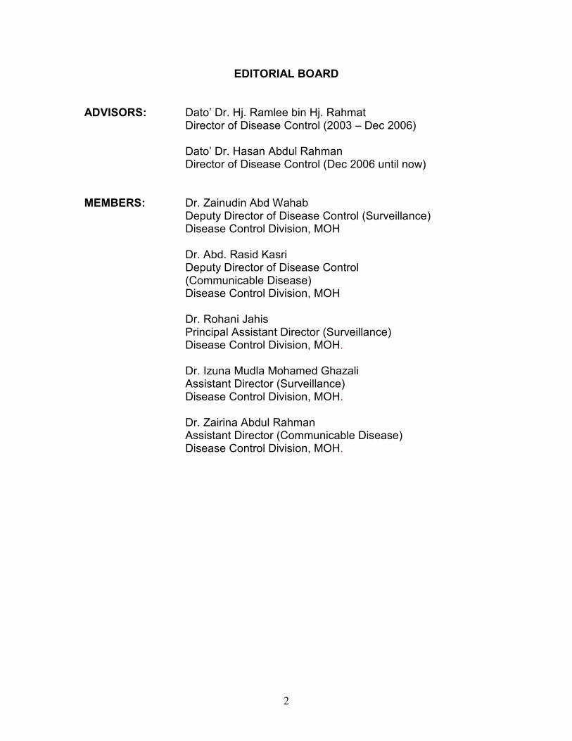
EDITORIAL BOARD
ADVISORS: Dato’ Dr. Hj. Ramlee bin Hj. Rahmat
Director of Disease Control (2003 – Dec 2006)
Dato’ Dr. Hasan Abdul Rahman Director of Disease Control (Dec 2006 until now)
MEMBERS: Dr. Zainudin Abd Wahab Deputy Director of Disease Control (Surveillance)
Disease Control Division, MOH
Dr. Abd. Rasid Kasri Deputy Director of Disease Control
(Communicable Disease) Disease Control Division, MOH
Dr. Rohani Jahis Principal Assistant Director (Surveillance) Disease Control Division, MOH. Dr. Izuna Mudla Mohamed Ghazali Assistant Director (Surveillance) Disease Control Division, MOH.
3
TABLE OF CONTENTS
1. INTRODUCTION.............................................................................................. 4
2. THE DISEASE ................................................................................................. 4
2.1 TRANSMISSION.............................................................................................. 4 2.2 CLINICAL FEATURES ...................................................................................... 4 2.3 MAGNITUDE OF THE DISEASE .......................................................................... 6
3. CLINICAL MANAGEMENT OF HFMD ............................................................ 7
3.1 SYMPTOMATIC TREATMENT ............................................................................ 7 3.2 HOSPITALISATION. .................................................................................... 7 3.2.1 Criteria for admission ........................................................................... 7 3.2.2 Infection control .................................................................................. 7
3.3 PATIENTS WITH NEUROLOGIC COMPLICATION .............................................. 8 3.4. ADVICE GIVEN UPON PATIENT’S DISCHARGE ............................................ 9
4. SURVEILLANCE ........................................................................................... 10
3.1 OBJECTIVE OF SURVEILLANCE ...................................................................... 10 3.2 CASE DEFINITION......................................................................................... 10 3.2.1 Clinical case definition........................................................................ 10 3.2.2 Laboratory criteria .............................................................................. 11 3.2.3 Case classification ............................................................................. 11
3.3 TYPES OF SURVEILLANCE ............................................................................. 11 3.3.1 Clinical surveillance............................................................................ 11 3.2.2 Laboratory surveillance ...................................................................... 12
3.4 Flow of surveillance data…………………………………………………... .. 14
5. MANAGEMENT OF SPORADIC CASES...................................................... 16
5.1DEFINITIONS OF SPORADIC CASE: .................................................................. 16 5.2THE PUBLIC HEALTH MEASURES ..................................................................... 16
6. MANAGEMENT OF OUTBREAK.................................................................. 18
6.1 DEFINITIONS OF OUTBREAK .......................................................................... 18 6.2 IDENTIFICATION OF OUTBREAKS .................................................................... 18 6.3 RISK FACTORS FOR OUTBREAKS ................................................................... 18 6.4 MANAGEMENT OF OUTBREAKS ...................................................................... 18 6.5 DOCUMENTATION AND REVIEW...................................................................... 20
7. REFERENCES............................................................................................. 216

4
1. INTRODUCTION
Hand foot and mouth disease (HFMD) is typically a benign and common illness among children and infants characterized by rapidly ulcerating vesicles in the mouth and lesions, usually vesicular, on the hands and feet.1 HFMD is an endemic disease in Malaysia. HFMD has become an important public health disease due to its tendency to cause large outbreaks and deaths among children and infants. The purpose of the guidelines is to help health personnels and health authorities, at any levels;
� To update current knowledge about HFMD
� To detect and control epidemic of HFMD as early as possible
� To strengthen the capacity for emergency response to epidemics of HFMD
� To improve the outcome of HFMD cases through effective case management.
2. THE DISEASE
Hand foot and mouth disease (HFMD) is caused by systemic infections with human enteroviruses. Human enteroviruses comprise one genus in the family Picornaviridae, which also contains the genera rhinovirus, cardiovirus, aphthovirus, hepatovirus and parechovirus. The members of the enterovirus genus that infect humans include the polioviruses, the Coxsackie virus groups A and B, the echoviruses and the enteroviruses 68-71.2
2.1 Transmission
Hand foot and mouth disease (HFMD) is moderately contagious. Infection is spread from person to person by direct contact with nose and throat discharges, saliva, fluid from blisters, or the stool of infected persons. A person is most contagious during the first week of the illness.
2.2 Clinical Features
HFMD is characterised by vesicular eruptions on the hands, wrists, feet and within the mouth. Lesions on the hands are almost always present, but oral lesions are present in 90% of patients and can occasionally be the only manifestation of the disease. The oral vesicles are often on the palate, tongue, and buccal mucosa and may range from a few isolated lesions to a marked stomatitis.
In addition patients may suffer fever, malaise, conjunctival injection, headache, and abdominal pain and occasionally diarrhea. The lesions on the feet and hands are flaccid, grayish vesicles, most often on the sides of the fingers, instep and
5
toes. If the disease is confined to the oral cavity it is almost indistinguishable from primary herpetic gingivostomatitis.3
Hand foot and mouth disease may be related to coxsackieviruses A16, A5, A9, A10, B2, B5 and enterovirus 71. Coxsackievirus A16 (Cox A16) is a frequently encountered pathogen in cases of HFMD and its clinical course is usually uneventful, with full recovery.
Fatal cases of Cox A16 infection are rare. Literature search revealed only three fatal cases of Cox A16 since 1963. All the cases were infants. The first case was reported by Wright et al in 1963 involving a 10 month-old girl with respiratory infection.4 The second case involved a 7 month-old boy with grunting and tongue ulcers.5 The third case was reported by Wang et al involving a 15 month-old boy who presented with hand foot and mouth disease complicated with myocarditis and intractable shock.6
HFMD caused by enterovirus 71 (EV71) can be more severe and may be complicated with meningitis, encephalitis and neurogenic pulmonary edema. In Taiwan, based on 1998 epidemic, the significant difference in the clinical features of Coxsackievirus A16 infection and EV 71 infection was that in EV71 the fever
was usually higher than 39°C and longer than 3 days.7
Enterovirus 71 infection can be classified into 4 stages:
Stage 1 – Hand foot and mouth disease (HFMD), oral ulcers and vesicular rash appearing on the hands, feet, knees and / or buttocks; or herpangina including oral ulceration over anterior tonsillar pillars, the soft palate, buccal mucosa or uvula.
Stage 2 – CNS involvement - aseptic meningitis with headache, irritability or myoclonic jerk and CSF pleocytosis (> 5 x 10⁶ leucocytes / litre) but without altered level of consciousness and focal signs, or encephalitis with altered level of consciousness plus CSF pleocytosis or poliomyelitis like syndrome with acute limb weakness and decreased reflex and muscle strength, or encephalomyelitis with the occurrence of both encephalitis and poliomyelitis like syndrome.
Stage 3 – Cardiopulmonary failure, pulmonary edema or hemorrhage with decreased ejection fraction of left ventricle as assessed by echocardiography necessitating inotropic agent support.

6
Stage 4 – Convalescence - is defined as recovery from cardiopulmonary failure.
2.3 Magnitude of the disease
Hand foot and mouth disease (HFMD) generally is a common and self limiting disease among children. EV 71 infection is the more important public health concern. In temperate countries most HFMD cases occur in the summer and fall and the incubation period is 4 – 6 days.
EV 71 was first isolated from a child with aseptic meningitis in California in 1969, and by 1974, the virus had been described as a new serotype of genus Enterovirus. In the years following the initial isolation of EV71, outbreaks occurred in USA, Australia, Sweden and Japan. In 1975 EV71 gained global attention when it was responsible for an outbreak in Bulgaria that resulted in 705 cases of poliomyelitis-like disease and the deaths of 44 people; 93% of the poliomyelitis-like disease cases occurred in children under the age of five. A similar outbreak occurred in Hungary in 1978, which also involved many cases of poliomyelitis-like disease and 47 deaths. Since this time outbreaks of EV71 have continued to occur throughout the world.
More recently there has been an increase in EV71 activity in the Asia Pacific region with several epidemics of HFMD being reported, including multiple cases associated with brain stem encephalitis and pulmonary edema. The first such epidemic occurred in Sarawak (Malaysian Borneo) in 1997 followed by smaller outbreaks in peninsular Malaysia and Japan. In 1998 outbreaks continued in Singapore and Taiwan.
The outbreak that occurred in Taiwan is the largest recorded, with greater than 100,000 cases of HFMD. Of these 400 children were admitted to hospital with central nervous system (CNS) involvement and 78 died of brainstem encephalitis with neurogenic pulmonary oedema.8 In 1999, a large HFMD outbreak occurred in Perth, western Australia and in 2000, EV71 was the cause of epidemics in Korea, Japan, Singapore, Taiwan and peninsular Malaysia, resulting in a range of clinical presentations including HFMD, aseptic meningitis, encephalitis and poliomyelitis like disease. Another outbreak occurred in Sarawak in 2003 which coincide with the SARS outbreak in the region and the public health measures put into place during this time evidently served to control transmission of enteroviruses as well.9

7
3. CLINICAL MANAGEMENT OF HFMD
HFMD is usually a mild and self limiting. In general, most cases of HFMD do not require admission but can be managed as outpatients. Most fatal HFMD cases were due to enterovirus infection.
3.1 Symptomatic treatment
Mild HFMD cases only need symptomatic treatment. Treatment of fever and relief of symptoms, adequate hydration and rest are important. Parents and care takers should be educated on hygiene and measures that they should take to prevent transmission to other children.
3.2 Hospitalisation.
3.2.1 Criteria for admission
• When the child is unable to tolerate oral feeds and there is a need for intravenous hydration;
• When the child is clinically very ill or toxic-looking
• When some other more serious disease cannot be excluded
• When there is persistent hyperpyrexia (e.g >38ºC) for >48 hours;
• When there is a suspicion of neurological complications, e.g increased lethargy, myoclonus, increased drowsiness, change in sensorium and/or seizures;
• When there is a suspicion of cardiac complications (myocarditis), e.g low blood pressure, low pulse volume, heart rhythm abnormalities, murmurs, gallop rhythm, displaced apex beat;
• When parents are unable to cope with child’s illness; and
• When there is inadequate family or social support in looking after the child at home.
3.2.2 Infection control
• Proper hygiene including mandatory hand washing after contact with patient, appropriate cleanliness during diaper changes is imperative
• Personal items such as spoons, cups and utensils should not be shared and should be properly washed with detergent after use;
• The use of gowns may act as a useful protection for health personnel looking after these patients; and
• Patients with HFMD should be isolated and the usual isolation procedures followed for infection control.

8
3.3 Patients with neurologic complication
Huang et al reviewed HFMD cases in Taiwan in 1998 and they found that the mean age of patients with neurological manifestations was 2.5 (range, 3 months to 8.2 years10. Higher incidence occurs in the younger age group. The neurologic disorders usually began 2 to 5 days after the onset of skin or mucosa lesion or fever.
Wu JM et al carried out a prospective study among infants confirmed to have enterovirus 71 and found that11;
• EV71 infection can lead to severe neurologic complications and acute pulmonary edema with or without hemorrhage
• All patients with EV71 infection and pulmonary edema had brainstem lesions
• Viral myocarditis is not the direct cause of acute PE and cardiopulmonary failure in EV71 Infection
• The excessive central sympathetic activation resulting in vasoconstriction was not the major cause of PE or hemorrhage
Based on the findings, they suggested that;
• Vasodilators should be used with caution since pulmonary edema in EV71 infection is not resulting from excessive vasoconstriction alone
• Cardiopulmonary function usually returns to nearly normal within days but the neurogic sequelae are severe and usually permanent. Therefore the use of extracorporeal membrane oxygenation support or a heart assist device should be weighed carefully
• Direct intracardiac hemodynamic monitoring may provide additional information to guide critical care

9
3.4. ADVICE GIVEN UPON PATIENT’S DISCHARGE
Parents and guardians should be advised upon patients discharge on complications that may occur; a statement as shown below can be given:
“Your child has been diagnosed to have hand-foot-mouth disease. This disease is normally not dangerous but in the light of recent events, we advise that you bring back your child to this hospital if he / she has any of the following symptoms:
• High fever.
• Lethargy and weakness.
• Refusing feeds and passing less urine.
• Rapid breathing.
• Vomiting.
• Drowsiness or irritably.
• Fits.”
10
4. SURVEILLANCE
The actual burden of HFMD in this country is not known. HFMD surveillance in Malaysia started after the Sarawak outbreak in 1997 with sentinel surveillance. Both private and government clinics were included as sentinel sites for each district. The number of sentinel sites for each district depends on the assumed prevalence of HFMD in the district / state.
HFMD notification is administratively required and started since October 2005. However death of HFMD and admitted cases to the ward of children aged 10 years old and below is mandatory for notification. However due to frequent outbreaks and fatality in Sarawak, Sarawak State has required all HFMD cases in the state to be notified.
The notification of HFMD came into enforcement on 12 October 2006 under the PU.A 374 / 2006 (appendix 1).
3.1 Objective of surveillance
i. To detect impending outbreak.
ii. To monitor circulating infectious agent.
iii. To estimate the magnitude of HFMD in the population at risk.
iv. To estimate the magnitude of the HFMD complication in the population at risk.
3.2 Case definition
3.2.1 Clinical case definition
3.2.1.1 Case of HFMD:
Any child with:
a. mouth / tongue ulcer and
b. maculopapular rashes and / or vesicles on palms and soles
c. with OR without history of fever
3.2.1.2 HFMD myocarditis:
2(i) Suspected HFMD myocarditis
Asymptomatic child with unexplained tachycardia to fulminant congestive cardiac failure.
With history of dirrhoea or URTI.

11
Accompanied by fever, malaise, tachypnoea, tachycardia (above and beyond expected for age and fever) or dehydration.
Pallor and shock in the absence of or obvious pulmonary sign or in the presence of crepitations (no murmurs) and / or a clinically enlarged heart.
2(i) Confirmed HFMD myocarditis.
The above 2(i) with virologic confirmation of enteroviruses.
3.2.1.3 HFMD meningoencephalitis / AFP
3(i) Suspected HFMD meningoencephalitis
Evidence of aseptic meningitis.
Evidence of aseptic encephalitis.
Evidence of AFP.
3(ii) Confirmed HFMD meningoencephalitis / AFP.
The above 3(i) with virologic confirmation of enteroviruses
3.2.2 Laboratory criteria
Any case that has the clinical symptoms and positive for viruses (coxsackieviruses (Cox) A16, A5, A9, A10, B2, B5 and enterovirus (EV) 71) which could cause HFMD, isolated or detected from stool or vesicle fluid or mouth ulcer or saliva.
3.2.3 Case classification
Suspected: A case that meets the clinical case definition.
Confirmed: A suspected case in which laboratory investigation confirms the presence of virus OR when cases are epidemiologically linked to a laboratory confirmed case.
3.3 Types of surveillance
3.3.1 Clinical surveillance
It is mandatory to notify HFMD under the National Notification of Infectious Diseases (list of notifiable diseases). Notification must be done within 24 hours of diagnosis via telephone, then it should be followed by notification form.
12
All HFMD cases detected must be entered to CDCIS. If states have other methods of data storing, it must be shared with MOH.
Laboratory confirmation is NOT required for notification.
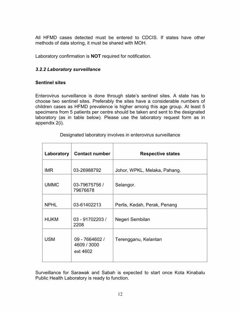
3.2.2 Laboratory surveillance
Sentinel sites
Enterovirus surveillance is done through state’s sentinel sites. A state has to choose two sentinel sites. Preferably the sites have a considerable numbers of children cases as HFMD prevalence is higher among this age group. At least 5 specimens from 5 patients per centre should be taken and sent to the designated laboratory (as in table below). Please use the laboratory request form as in appendix 2(i).
Designated laboratory involves in enterovirus surveillance
Laboratory
Contact number
Respective states
IMR
03-26988792
Johor, WPKL, Melaka, Pahang.
UMMC
03-79675756 / 79676678
Selangor.
NPHL
03-61402213
Perlis, Kedah, Perak, Penang
HUKM
03 - 91702203 / 2208
Negeri Sembilan
USM
09 - 7664602 / 4609 / 3000
ext 4602
Terengganu, Kelantan
Surveillance for Sarawak and Sabah is expected to start once Kota Kinabalu Public Health Laboratory is ready to function.

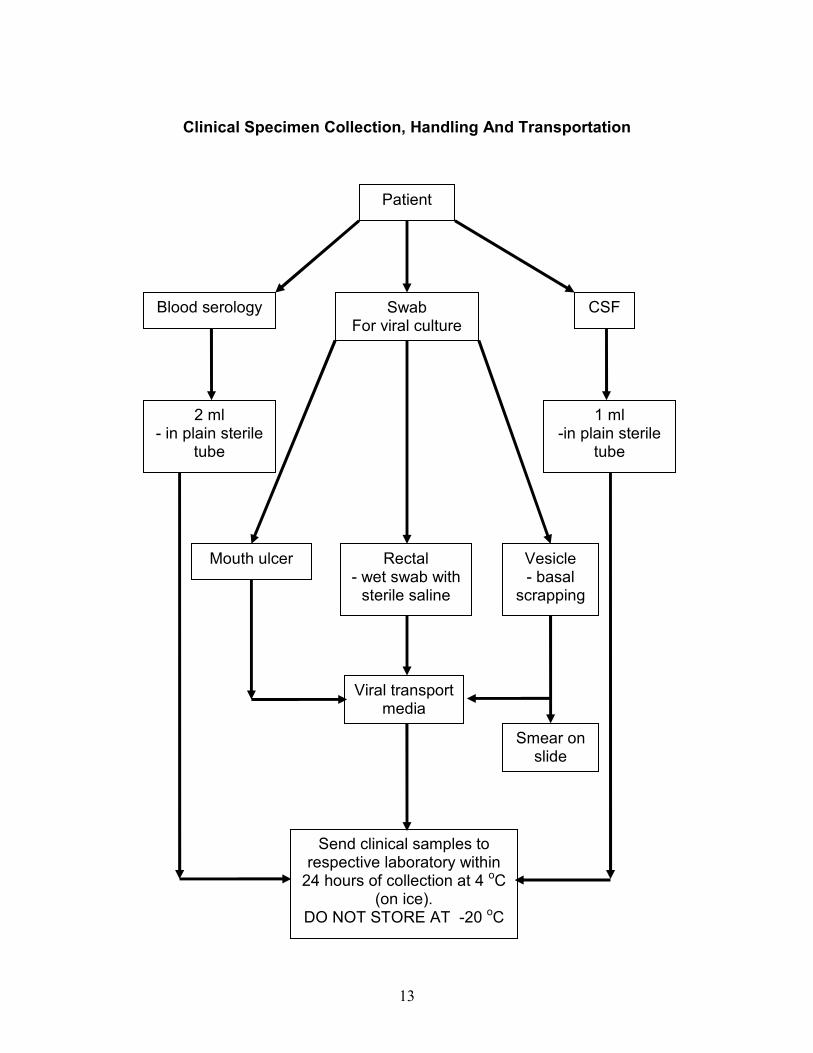
13
Clinical Specimen Collection, Handling And Transportation
Patient
Swab For viral culture
Blood serology CSF
2 ml - in plain sterile
tube
1 ml -in plain sterile
tube
Mouth ulcer Rectal - wet swab with sterile saline
Vesicle - basal
scrapping
Viral transport media
Smear on slide
Send clinical samples to respective laboratory within 24 hours of collection at 4 oC
(on ice). DO NOT STORE AT -20 oC

14
3.4 Flow of surveillance data
Notification data should be entered in CDCIS as and when there is HFMD case. Other sources of data must be shared with Disease Control Division. Data must have the variables listed in appendix 1.
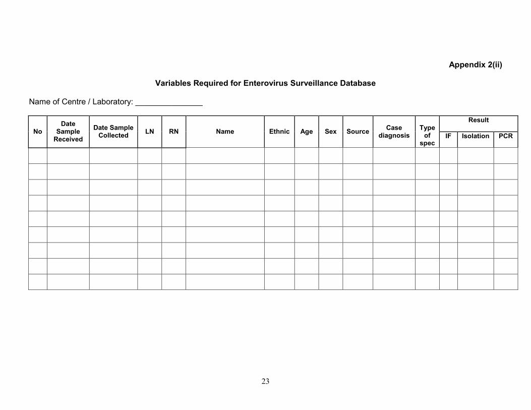
Laboratory results for enterovirus surveillance must be shared with respective states and Disease Control Division using the suggested format in appendix 2(ii).

15
Flow of Information Management
Yes
Suspected HFMD Case
CDCIS
Case fulfilled criteria
Investigate & Register
Lab-Confirmed Cases
LIS
Positive EV71
Investigate
Clinics Laboratories
Cluster of cases ?
Finish
Preventive and control activities
Report
No

16
5. MANAGEMENT OF SPORADIC CASES
5.1 Definitions of sporadic case:
A single case of HFMD in the absence of previous known close contact with another case.
5.2 The public health measures
5.2.1 Case Investigation
Case investigation is required following the criteria below:
a. every notified HFMD case that fulfilled any one of the criteria below:
i. case is admitted to hospital ii. case died. iii. case aged 6 years and below and goes to any pre-schools or nurseries.
The first two criteria indicate severity of illness and it is a proxy of possible EV71 infection. Meanwhile the third criterion is important in preventing spread of illness among the pre-school children (high risk group) and reducing public anxiety.
b. every case with positive EV71 laboratory result. Investigation should be carried out to determine the course, source and severity of infection. Enquiries shall be made into;
1. Particulars of the person affected
2. Clinical signs and symptoms with date of onset. In addition, enquiries shall be elicited for associated complications e.g. seizures, weakness of limbs, staring episodes, drowsiness, profound lethargy and refusal to play.
Enterovirus infections that are not associated with complications may not have cutaneous manifestations.
3. Duration of illness
4. Type of treatment sought, including details of hospitalization and reason for admission
5. History of travel especially to outbreak area in the past two weeks
6. Contacts with similar illness in an institution (child care centre or kindergarten), family or neighbourhood where relevant.
7. If newborn, antenatal history including maternal history of febrile illness, mode of delivery

17
5.2.2 Incidence management Following steps should be taken to prevent further spread of disease / pathogen:
1. Ensure that the infected children remain away from the institution for at least ten days after onset of symptoms and must be certified free from infection by a registered medical practitioner prior to returning to school.
2. Active case detection shall be carried out among contacts in the institution and family, and all those with illness shall be referred for treatment at the nearest health clinics.
3. Swabs from oral ulcers and vesicular lesions on the hands and feet shall be collected for virus isolation at identified laboratories (IMR or NPHL). If these ulcers and vesicles are no longer present, stool specimens shall be sent. Always consult pathologist for specimens taking, handling and transportation.
4. Health education of supervisors of institutions on the disease, mode of transmission, importance of good personal hygiene and isolation of infected children shall be carried out.
5. Principals, teachers and supervisors shall be alerted to look out for children with fever, rash / blisters on palms and soles and to isolate them immediately. Screening before coming to class is recommended (please refer Buku Panduan Latihan Taska dan Prasekolah).
6. If there are two or case in the premise, advice the premise to be closed to cut disease transmission. It should be closed for two incubation period (10 days) from the date of onset of the last case.
7. Supervision on disinfection procedures shall be given to the premise operators. Articles such as toys contaminated by infected cases are disinfected with 0.5% sodium hypochlorite solutions (please refer Buku Panduan Latihan Taska dan Prasekolah).

18
6. MANAGEMENT OF OUTBREAK
6.1 Definitions of outbreak
The occurrence of two or more cases in the same locality within the incubation period. Organisation based outbreak – two or more cases with onset in two weeks interval in a group which makes epidemiological sense.
6.2 Identification of outbreaks
Surveillance data on HFMD cases should be reviewed on continuous basis to identify cases and detects outbreaks.
6.3 Risk factors for outbreaks
Risk factors for outbreaks are not completely understood. A combination of conditions (environment, host and organism) is necessary for an epidemic to occur. However large HFMD outbreaks have been associated with EV71 infection.
6.4 Management of outbreaks
When a suspected outbreak is reported, it is important to determine rapidly whether a true outbreak is occurring based on the definition above. Similar steps as in sporadic case management should be carried out together with following measures;
1. Ensure that the infected children remain away from the institution for at least ten days after onset of symptoms and must be certified free from infection by a registered medical practitioner prior to returning to school.
2. An assessment of the number of cases and susceptible population involved shall be made, and the overall attack rate and age-specific attack rate computed.
3. Active case detection shall be carried out among contacts in the institution and family, and all those with illness shall be referred for treatment at the nearest health clinics.
4. Where a case is observed or suspected to have unusual signs and symptoms e.g. seizures, weakness of limbs, profound lethargy, refusal to play, the case shall be immediately referred to hospital.
5. Swabs from oral ulcers and vesicular lesions on the hands and feet shall be collected for virus isolation at identified laboratories (IMR or NPHL). If these ulcers and vesicles are no longer present, stool specimens shall be sent. Always consult pathologist for specimens taking, handling and transportation.

19
6. Principals, teachers and supervisors shall be alerted to look out for children with fever, rash / blisters on palms and soles and to isolate them immediately. Screening before coming to class is recommended (please refer Buku Panduan Latihan Taska dan Prasekolah).
7. Health education of supervisors of institutions on the disease, mode of transmission, importance of good personal hygiene and isolation of infected children shall be carried out.
8. Close the premise closed for two incubation period (10 days) from the date of onset of the last case. Supervision on disinfection procedures shall be given to the premise operators.
In addition the investigating team shall also look into environmental sanitation and hygiene to ensure that:
a. Articles such as toys contaminated by infected cases are disinfected with 0.5% sodium hypochlorite solutions (please refer Buku Panduan Latihan Taska dan Prasekolah).
b. A high standard of food and personal hygiene is maintained by the institution involved which include proper waste and diapers disposal.
c. The approved enrolment of Department of Social Welfare (JKM), Ministry of Education, Department of National Integration and other relevant parties, where applicable, and shall notify the relevant authorities if regulations not complied.
If the outbreak occurs in primary schools;
i. Ensure that the infected students remain away from the institution for at least ten days.
ii. Health education to the students on the disease, mode of transmission, importance of good personal hygiene.
iii. Principals, teachers and supervisors shall be look for children with fever, rash / blisters on palms and soles and to isolate them immediately. Screening before coming to class is recommended (please refer Buku Panduan Latihan Taska dan Prasekolah).
iv. If closure is necessary, just closed the affected class. Closure of the whole school is unnecessary as HFMD in older children is usually very mild and so far no complication has been documented from this agegroup.

20
6.5 Documentation and review
Disease activity and emergency response should be adequately documented so that further outbreaks can be handled more effectively. A summary report should be prepared within a month from the last date of case; and distributed to all the stakeholders for feedback. Format of report, dissemination and tabling of report is discussed in more details in Infectious Disease Outbreak Rapid Response Manual.12

21
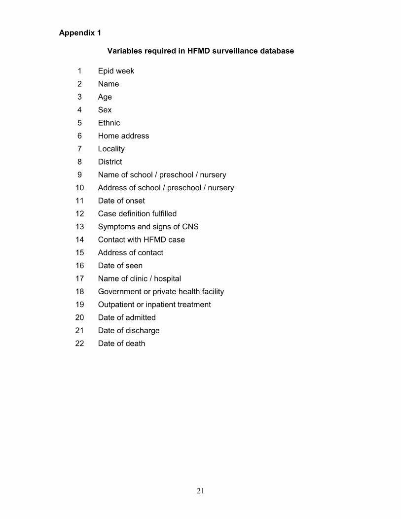
Appendix 1
Variables required in HFMD surveillance database
1 Epid week
2 Name
3 Age
4 Sex
5 Ethnic
6 Home address
7 Locality
8 District
9 Name of school / preschool / nursery
10 Address of school / preschool / nursery
11 Date of onset
12 Case definition fulfilled
13 Symptoms and signs of CNS
14 Contact with HFMD case
15 Address of contact
16 Date of seen
17 Name of clinic / hospital
18 Government or private health facility
19 Outpatient or inpatient treatment
20 Date of admitted
21 Date of discharge
22 Date of death

22
No. Rujukan Makmal
NATIONAL ENTEROVIRUS SURVEILLANCE
DIAGNOSTIC REQUEST FORM
A. MAKLUMAT PESAKIT
Negeri : Daerah :
Hospital (wad) / Klinik Kesihatan No. R/N :
Nama Pesakit :
No. K/P : Umur : Jantina : L / P
Bangsa : Tarikh lahir :
B. MAKLUMAT KLINIKAL
Gejala (Simptom) Ada / Tiada (Tandakan √) Tarikh mula
Demam > 38 0C
Ulser di mulut & tekak
Maculopapular rashes dan / Vesikel pada tapak
tangan dan tapak kaki
Tanda dan gejala URTI
C. SPESIMEN KLINIKAL
Spesimen Tarikh diambil Tarikh penghantaran
Rectal swab / / / /
Mouth ulcer / / / /
Vesicle swab / / / /
Stool / / / /
D. MAKLUMAT PEMOHON
Nama dan Cop Pegawai :
Tandatangan :
No. telefon :
No. fax :
e-mail :
E. MAKMAL (Untuk Kegunaan Makmal)
Keadaan spesimen : Tarikh terima spesimen :
Spesimen Jenis ujian Keputusan ujian Komen
Rectal swab
Mouth ulcer
Vesicle swab
Stool
Nama dan tandatangan Pegawai Makmal :
Jawatan Pegawai Makmal dan Cop Makmal :
Tarikh :
Nota * : All samples except blood / serum and stool must be in the viral transport media (VTM) and
send immediately in ice pack to HUKM Microbiological Laboratory.
Appendix 2(i)
23
Appendix 2(ii)
Variables Required for Enterovirus Surveillance Database
Name of Centre / Laboratory: _______________
Result
No Date
Sample Received
Date Sample Collected
LN RN Name Ethnic Age Sex Source
Case
diagnosis
Type of
spec IF Isolation PCR
24
BKP / S / HFMD (H) Ver 2 / 2007
BORANG SIASATAN PESAKIT DI WAD
PENYAKIT TANGAN, KAKI DAN MULUT (HFMD)
BAHAGIAN KAWALAN PENYAKIT, KEMENTERIAN KESIHATAN MALAYSIA
BAHAGIAN 1
Tarikh masuk wad: Masa:
Tarikh discaj: Masa:
Tarikh kematian Masa:
A. PERIHAL PESAKIT
1. Nama pesakit: 2. Tarikh lahir:
3. Nama ibu: 4. No K/P ibu:
5. Alamat rumah: 6. No telefon: (rumah)
(H/P)
Kedudukan rumah (GPS): X : _________________ Y : __________________
7. Jantina: Lelaki 8. Kumpulan etnik
Perempuan Melayu Kadazan/Dusun
Cina Murut
9. Kewarganegaraan: Malaysia India Bajau
Warga asing Asli semenanjung Melanau
Bidayuh Iban
Lain-lain pribumi Lain-lain (nyatakan)
Sabah/ Sarawak
10. Menghadiri sekolah / taska
Ya
Taska / Nurseri
Prasekolah
Sekolah
Nyatakan nama: ________________________________
Tidak
B. MAKLUMAT KLINIKAL
1. Tarikh mula sakit (onset)
2. Tanda dan gejala: Ya Tidak
Riwayat demam
25
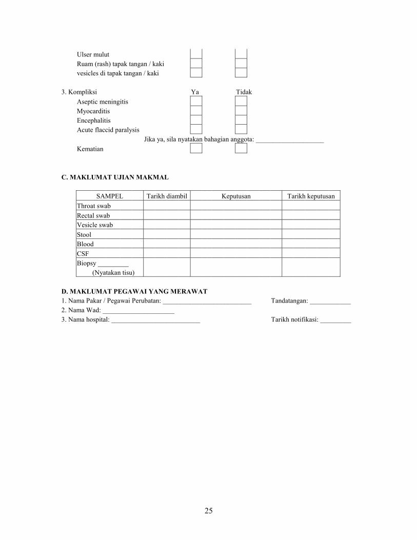
Ulser mulut
Ruam (rash) tapak tangan / kaki
vesicles di tapak tangan / kaki
3. Kompliksi Ya Tidak
Aseptic meningitis
Myocarditis
Encephalitis
Acute flaccid paralysis
Jika ya, sila nyatakan bahagian anggota: ____________________
Kematian
C. MAKLUMAT UJIAN MAKMAL
SAMPEL Tarikh diambil Keputusan Tarikh keputusan
Throat swab
Rectal swab
Vesicle swab
Stool
Blood
CSF
Biopsy _________
(Nyatakan tisu)
D. MAKLUMAT PEGAWAI YANG MERAWAT
1. Nama Pakar / Pegawai Perubatan: __________________________ Tandatangan: ____________
2. Nama Wad: _____________________
3. Nama hospital: __________________________ Tarikh notifikasi: _________

26
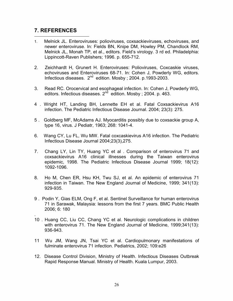
7. REFERENCES
1. Melnick JL. Enteroviruses: polioviruses, coxsackieviruses, echoviruses, and newer enteroviruse. In: Fields BN, Knipe DM, Howley PM, Chandlock RM, Melnick JL, Monah TP, et al., editors. Field’s virology. 3 rd ed. Philadelphia: Lippincott-Raven Publishers; 1996. p. 655-712.
2. Zeichhardt H, Grunert H. Enteroviruses: Polioviruses, Coxcaskie viruses,
echoviruses and Enteroviruses 68-71. In: Cohen J, Powderly WG, editors. Infectious diseases. 2nd edition. Mosby ; 2004. p.1993-2003.
3. Read RC. Orocervical and esophageal infection. In: Cohen J, Powderly WG,
editors. Infectious diseases. 2nd edition. Mosby ; 2004. p. 463. 4 . Wright HT, Landing BH, Lennette EH et al. Fatal Coxsackievirus A16
infection. The Pediatric Infectious Disease Journal. 2004; 23(3): 275. 5 . Goldberg MF, McAdams AJ. Myocarditis possibly due to coxsackie group A,
type 16, virus. J Pediatr, 1963; 268: 1041-4. 6. Wang CY, Lu FL, Wu MW. Fatal coxcaskievirus A16 infection. The Pediatric
Infectious Disease Journal 2004;23(3),275. 7. Chang LY, Lin TY, Huang YC et al . Comparison of enterovirus 71 and
coxsackievirus A16 clinical illnesses during the Taiwan enterovirus epidemic, 1998. The Pediatric Infectious Disease Journal 1999; 18(12): 1092-1096.
8. Ho M, Chen ER, Hsu KH, Twu SJ, et al. An epidemic of enterovirus 71
infection in Taiwan. The New England Journal of Medicine, 1999; 341(13): 929-935.
9 . Podin Y, Gias ELM, Ong F, et al. Sentinel Surveillance for human enterovirus
71 in Sarawak, Malaysia: lessons from the first 7 years. BMC Public Health 2006; 6: 180
. 10 . Huang CC, Liu CC, Chang YC et al. Neurologic complications in children
with enterovirus 71. The New England Journal of Medicine, 1999;341(13): 936-943.
11 Wu JM, Wang JN, Tsai YC et al. Cardiopulmonary manifestations of
fulminate enterovirus 71 infection. Pediatrics, 2002; 109:e26 12. Disease Control Division, Ministry of Health. Infectious Diseases Outbreak
Rapid Response Manual. Ministry of Health. Kuala Lumpur, 2003.
27
CONTRIBUTORS State Epidemiologists 2006 – 2007
1. Pulau Pinang Dr. Saraswathi Bina Rai
2. Kedah Dr. Uma Salamah Abdul Kadir
3. Perlis Dr. Zulhizzam Hj Abdullah
4. Perak Dr. Asiah Ayob
Dr. Zamri Md Ali
5. Selangor Dr. Prema Rajendran
Dr. B. Venugopalan
6. WPKL Dr. Norbizura Abdul Hamid
7. N. Sembilan Dr. Zulfa Vety Dol Bakri
8. Melaka Dr. Hashimah Hasan
9. Johor Dr. Shaharom Nor Azian Che Mat Din
Dr. Fatimah Othman
10. Pahang Dr. Shaari Ngadiman
Dr. Adibah Hani Harun
11. Terengganu Dr. Anita Surani Abdul Shukor
Dr. Balkis Ab Karim
12. Kelantan Dr. Hani Mat Hussin
13. Sarawak Dr. Ooi Choo Huck
Dr. Kamaliah Mohd Nor
Dr. Rohani Mat Bah
14. Sabah Dr. Jenarun Jelip
Dr. Kristina Rundi
15. Bahagian Kawalan Dr. Husnina Ibrahim
Penyakit Dr. Nazarudin Safian
Dr. Fadzilah Kamaludin
Dr. Wan Mansor Hamzah
Dr. Mohd Hanif Zailani
16 Laboratory Dr. Chua Kaw Bing
HAND FOOT AND MOUTH DISEASE (HFMD)
GUIDELINES

2
EDITORIAL BOARD
ADVISORS: Dato’ Dr. Hj. Ramlee bin Hj. Rahmat
Director of Disease Control (2003 – Dec 2006)
Dato’ Dr. Hasan Abdul Rahman Director of Disease Control (Dec 2006 until now)
MEMBERS: Dr. Zainudin Abd Wahab Deputy Director of Disease Control (Surveillance)
Disease Control Division, MOH
Dr. Abd. Rasid Kasri Deputy Director of Disease Control
(Communicable Disease) Disease Control Division, MOH
Dr. Rohani Jahis Principal Assistant Director (Surveillance) Disease Control Division, MOH. Dr. Izuna Mudla Mohamed Ghazali Assistant Director (Surveillance) Disease Control Division, MOH.

3
TABLE OF CONTENTS
1. INTRODUCTION.............................................................................................. 4
2. THE DISEASE ................................................................................................. 4
2.1 TRANSMISSION.............................................................................................. 4 2.2 CLINICAL FEATURES ...................................................................................... 4 2.3 MAGNITUDE OF THE DISEASE .......................................................................... 6
3. CLINICAL MANAGEMENT OF HFMD ............................................................ 7
3.1 SYMPTOMATIC TREATMENT ............................................................................ 7 3.2 HOSPITALISATION. .................................................................................... 7 3.2.1 Criteria for admission ........................................................................... 7 3.2.2 Infection control .................................................................................. 7
3.3 PATIENTS WITH NEUROLOGIC COMPLICATION .............................................. 8 3.4. ADVICE GIVEN UPON PATIENT’S DISCHARGE ............................................ 9
4. SURVEILLANCE ........................................................................................... 10
3.1 OBJECTIVE OF SURVEILLANCE ...................................................................... 10 3.2 CASE DEFINITION......................................................................................... 10 3.2.1 Clinical case definition........................................................................ 10 3.2.2 Laboratory criteria .............................................................................. 11 3.2.3 Case classification ............................................................................. 11
3.3 TYPES OF SURVEILLANCE ............................................................................. 11 3.3.1 Clinical surveillance............................................................................ 11 3.2.2 Laboratory surveillance ...................................................................... 12
3.4 Flow of surveillance data…………………………………………………... .. 14
5. MANAGEMENT OF SPORADIC CASES...................................................... 16
5.1DEFINITIONS OF SPORADIC CASE: .................................................................. 16 5.2THE PUBLIC HEALTH MEASURES ..................................................................... 16
6. MANAGEMENT OF OUTBREAK.................................................................. 18
6.1 DEFINITIONS OF OUTBREAK .......................................................................... 18 6.2 IDENTIFICATION OF OUTBREAKS .................................................................... 18 6.3 RISK FACTORS FOR OUTBREAKS ................................................................... 18 6.4 MANAGEMENT OF OUTBREAKS ...................................................................... 18 6.5 DOCUMENTATION AND REVIEW...................................................................... 20
7. REFERENCES............................................................................................. 216
4
1. INTRODUCTION
Hand foot and mouth disease (HFMD) is typically a benign and common illness among children and infants characterized by rapidly ulcerating vesicles in the mouth and lesions, usually vesicular, on the hands and feet.1 HFMD is an endemic disease in Malaysia. HFMD has become an important public health disease due to its tendency to cause large outbreaks and deaths among children and infants. The purpose of the guidelines is to help health personnels and health authorities, at any levels;
� To update current knowledge about HFMD
� To detect and control epidemic of HFMD as early as possible
� To strengthen the capacity for emergency response to epidemics of HFMD
� To improve the outcome of HFMD cases through effective case management.
2. THE DISEASE
Hand foot and mouth disease (HFMD) is caused by systemic infections with human enteroviruses. Human enteroviruses comprise one genus in the family Picornaviridae, which also contains the genera rhinovirus, cardiovirus, aphthovirus, hepatovirus and parechovirus. The members of the enterovirus genus that infect humans include the polioviruses, the Coxsackie virus groups A and B, the echoviruses and the enteroviruses 68-71.2
2.1 Transmission
Hand foot and mouth disease (HFMD) is moderately contagious. Infection is spread from person to person by direct contact with nose and throat discharges, saliva, fluid from blisters, or the stool of infected persons. A person is most contagious during the first week of the illness.
2.2 Clinical Features
HFMD is characterised by vesicular eruptions on the hands, wrists, feet and within the mouth. Lesions on the hands are almost always present, but oral lesions are present in 90% of patients and can occasionally be the only manifestation of the disease. The oral vesicles are often on the palate, tongue, and buccal mucosa and may range from a few isolated lesions to a marked stomatitis.
In addition patients may suffer fever, malaise, conjunctival injection, headache, and abdominal pain and occasionally diarrhea. The lesions on the feet and hands are flaccid, grayish vesicles, most often on the sides of the fingers, instep and

5
toes. If the disease is confined to the oral cavity it is almost indistinguishable from primary herpetic gingivostomatitis.3
Hand foot and mouth disease may be related to coxsackieviruses A16, A5, A9, A10, B2, B5 and enterovirus 71. Coxsackievirus A16 (Cox A16) is a frequently encountered pathogen in cases of HFMD and its clinical course is usually uneventful, with full recovery.
Fatal cases of Cox A16 infection are rare. Literature search revealed only three fatal cases of Cox A16 since 1963. All the cases were infants. The first case was reported by Wright et al in 1963 involving a 10 month-old girl with respiratory infection.4 The second case involved a 7 month-old boy with grunting and tongue ulcers.5 The third case was reported by Wang et al involving a 15 month-old boy who presented with hand foot and mouth disease complicated with myocarditis and intractable shock.6
HFMD caused by enterovirus 71 (EV71) can be more severe and may be complicated with meningitis, encephalitis and neurogenic pulmonary edema. In Taiwan, based on 1998 epidemic, the significant difference in the clinical features of Coxsackievirus A16 infection and EV 71 infection was that in EV71 the fever
was usually higher than 39°C and longer than 3 days.7
Enterovirus 71 infection can be classified into 4 stages:
Stage 1 – Hand foot and mouth disease (HFMD), oral ulcers and vesicular rash appearing on the hands, feet, knees and / or buttocks; or herpangina including oral ulceration over anterior tonsillar pillars, the soft palate, buccal mucosa or uvula.
Stage 2 – CNS involvement - aseptic meningitis with headache, irritability or myoclonic jerk and CSF pleocytosis (> 5 x 10⁶ leucocytes / litre) but without altered level of consciousness and focal signs, or encephalitis with altered level of consciousness plus CSF pleocytosis or poliomyelitis like syndrome with acute limb weakness and decreased reflex and muscle strength, or encephalomyelitis with the occurrence of both encephalitis and poliomyelitis like syndrome.
Stage 3 – Cardiopulmonary failure, pulmonary edema or hemorrhage with decreased ejection fraction of left ventricle as assessed by echocardiography necessitating inotropic agent support.

6
Stage 4 – Convalescence - is defined as recovery from cardiopulmonary failure.
2.3 Magnitude of the disease
Hand foot and mouth disease (HFMD) generally is a common and self limiting disease among children. EV 71 infection is the more important public health concern. In temperate countries most HFMD cases occur in the summer and fall and the incubation period is 4 – 6 days.
EV 71 was first isolated from a child with aseptic meningitis in California in 1969, and by 1974, the virus had been described as a new serotype of genus Enterovirus. In the years following the initial isolation of EV71, outbreaks occurred in USA, Australia, Sweden and Japan. In 1975 EV71 gained global attention when it was responsible for an outbreak in Bulgaria that resulted in 705 cases of poliomyelitis-like disease and the deaths of 44 people; 93% of the poliomyelitis-like disease cases occurred in children under the age of five. A similar outbreak occurred in Hungary in 1978, which also involved many cases of poliomyelitis-like disease and 47 deaths. Since this time outbreaks of EV71 have continued to occur throughout the world.
More recently there has been an increase in EV71 activity in the Asia Pacific region with several epidemics of HFMD being reported, including multiple cases associated with brain stem encephalitis and pulmonary edema. The first such epidemic occurred in Sarawak (Malaysian Borneo) in 1997 followed by smaller outbreaks in peninsular Malaysia and Japan. In 1998 outbreaks continued in Singapore and Taiwan.
The outbreak that occurred in Taiwan is the largest recorded, with greater than 100,000 cases of HFMD. Of these 400 children were admitted to hospital with central nervous system (CNS) involvement and 78 died of brainstem encephalitis with neurogenic pulmonary oedema.8 In 1999, a large HFMD outbreak occurred in Perth, western Australia and in 2000, EV71 was the cause of epidemics in Korea, Japan, Singapore, Taiwan and peninsular Malaysia, resulting in a range of clinical presentations including HFMD, aseptic meningitis, encephalitis and poliomyelitis like disease. Another outbreak occurred in Sarawak in 2003 which coincide with the SARS outbreak in the region and the public health measures put into place during this time evidently served to control transmission of enteroviruses as well.9

7
3. CLINICAL MANAGEMENT OF HFMD
HFMD is usually a mild and self limiting. In general, most cases of HFMD do not require admission but can be managed as outpatients. Most fatal HFMD cases were due to enterovirus infection.
3.1 Symptomatic treatment
Mild HFMD cases only need symptomatic treatment. Treatment of fever and relief of symptoms, adequate hydration and rest are important. Parents and care takers should be educated on hygiene and measures that they should take to prevent transmission to other children.
3.2 Hospitalisation.
3.2.1 Criteria for admission
• When the child is unable to tolerate oral feeds and there is a need for intravenous hydration;
• When the child is clinically very ill or toxic-looking
• When some other more serious disease cannot be excluded
• When there is persistent hyperpyrexia (e.g >38ºC) for >48 hours;
• When there is a suspicion of neurological complications, e.g increased lethargy, myoclonus, increased drowsiness, change in sensorium and/or seizures;
• When there is a suspicion of cardiac complications (myocarditis), e.g low blood pressure, low pulse volume, heart rhythm abnormalities, murmurs, gallop rhythm, displaced apex beat;
• When parents are unable to cope with child’s illness; and
• When there is inadequate family or social support in looking after the child at home.
3.2.2 Infection control
• Proper hygiene including mandatory hand washing after contact with patient, appropriate cleanliness during diaper changes is imperative
• Personal items such as spoons, cups and utensils should not be shared and should be properly washed with detergent after use;
• The use of gowns may act as a useful protection for health personnel looking after these patients; and
• Patients with HFMD should be isolated and the usual isolation procedures followed for infection control.

8
3.3 Patients with neurologic complication
Huang et al reviewed HFMD cases in Taiwan in 1998 and they found that the mean age of patients with neurological manifestations was 2.5 (range, 3 months to 8.2 years10. Higher incidence occurs in the younger age group. The neurologic disorders usually began 2 to 5 days after the onset of skin or mucosa lesion or fever.
Wu JM et al carried out a prospective study among infants confirmed to have enterovirus 71 and found that11;
• EV71 infection can lead to severe neurologic complications and acute pulmonary edema with or without hemorrhage
• All patients with EV71 infection and pulmonary edema had brainstem lesions
• Viral myocarditis is not the direct cause of acute PE and cardiopulmonary failure in EV71 Infection
• The excessive central sympathetic activation resulting in vasoconstriction was not the major cause of PE or hemorrhage
Based on the findings, they suggested that;
• Vasodilators should be used with caution since pulmonary edema in EV71 infection is not resulting from excessive vasoconstriction alone
• Cardiopulmonary function usually returns to nearly normal within days but the neurogic sequelae are severe and usually permanent. Therefore the use of extracorporeal membrane oxygenation support or a heart assist device should be weighed carefully
• Direct intracardiac hemodynamic monitoring may provide additional information to guide critical care

9
3.4. ADVICE GIVEN UPON PATIENT’S DISCHARGE
Parents and guardians should be advised upon patients discharge on complications that may occur; a statement as shown below can be given:
“Your child has been diagnosed to have hand-foot-mouth disease. This disease is normally not dangerous but in the light of recent events, we advise that you bring back your child to this hospital if he / she has any of the following symptoms:
• High fever.
• Lethargy and weakness.
• Refusing feeds and passing less urine.
• Rapid breathing.
• Vomiting.
• Drowsiness or irritably.
• Fits.”

10
4. SURVEILLANCE
The actual burden of HFMD in this country is not known. HFMD surveillance in Malaysia started after the Sarawak outbreak in 1997 with sentinel surveillance. Both private and government clinics were included as sentinel sites for each district. The number of sentinel sites for each district depends on the assumed prevalence of HFMD in the district / state.
HFMD notification is administratively required and started since October 2005. However death of HFMD and admitted cases to the ward of children aged 10 years old and below is mandatory for notification. However due to frequent outbreaks and fatality in Sarawak, Sarawak State has required all HFMD cases in the state to be notified.
The notification of HFMD came into enforcement on 12 October 2006 under the PU.A 374 / 2006 (appendix 1).
3.1 Objective of surveillance
i. To detect impending outbreak.
ii. To monitor circulating infectious agent.
iii. To estimate the magnitude of HFMD in the population at risk.
iv. To estimate the magnitude of the HFMD complication in the population at risk.
3.2 Case definition
3.2.1 Clinical case definition
3.2.1.1 Case of HFMD:
Any child with:
a. mouth / tongue ulcer and
b. maculopapular rashes and / or vesicles on palms and soles
c. with OR without history of fever
3.2.1.2 HFMD myocarditis:
2(i) Suspected HFMD myocarditis
Asymptomatic child with unexplained tachycardia to fulminant congestive cardiac failure.
With history of dirrhoea or URTI.

11
Accompanied by fever, malaise, tachypnoea, tachycardia (above and beyond expected for age and fever) or dehydration.
Pallor and shock in the absence of or obvious pulmonary sign or in the presence of crepitations (no murmurs) and / or a clinically enlarged heart.
2(i) Confirmed HFMD myocarditis.
The above 2(i) with virologic confirmation of enteroviruses.
3.2.1.3 HFMD meningoencephalitis / AFP
3(i) Suspected HFMD meningoencephalitis
Evidence of aseptic meningitis.
Evidence of aseptic encephalitis.
Evidence of AFP.
3(ii) Confirmed HFMD meningoencephalitis / AFP.
The above 3(i) with virologic confirmation of enteroviruses
3.2.2 Laboratory criteria
Any case that has the clinical symptoms and positive for viruses (coxsackieviruses (Cox) A16, A5, A9, A10, B2, B5 and enterovirus (EV) 71) which could cause HFMD, isolated or detected from stool or vesicle fluid or mouth ulcer or saliva.
3.2.3 Case classification
Suspected: A case that meets the clinical case definition.
Confirmed: A suspected case in which laboratory investigation confirms the presence of virus OR when cases are epidemiologically linked to a laboratory confirmed case.
3.3 Types of surveillance
3.3.1 Clinical surveillance
It is mandatory to notify HFMD under the National Notification of Infectious Diseases (list of notifiable diseases). Notification must be done within 24 hours of diagnosis via telephone, then it should be followed by notification form.

12
All HFMD cases detected must be entered to CDCIS. If states have other methods of data storing, it must be shared with MOH.
Laboratory confirmation is NOT required for notification.
3.2.2 Laboratory surveillance
Sentinel sites
Enterovirus surveillance is done through state’s sentinel sites. A state has to choose two sentinel sites. Preferably the sites have a considerable numbers of children cases as HFMD prevalence is higher among this age group. At least 5 specimens from 5 patients per centre should be taken and sent to the designated laboratory (as in table below). Please use the laboratory request form as in appendix 2(i).
Designated laboratory involves in enterovirus surveillance
Laboratory
Contact number
Respective states
IMR
03-26988792
Johor, WPKL, Melaka, Terengganu, Pahang.
UMMC
03-79675756 / 79676678
Selangor.
NPHL
03-61402213
Perlis, Kedah, Perak, Penang
HUKM
03 - 91702203 / 2208
Negeri Sembilan
USM
09 - 7664602 / 4609 / 3000
ext 4602
Kelantan
Surveillance for Sarawak and Sabah is expected to start once Kota Kinabalu Public Health Laboratory is ready to function.
13
Clinical Specimen Collection, Handling And Transportation
Patient
Swab For viral culture
Blood serology CSF
2 ml - in plain sterile
tube
1 ml -in plain sterile
tube
Mouth ulcer Rectal - wet swab with sterile saline
Vesicle - basal
scrapping
Viral transport media
Smear on slide
Send clinical samples to respective laboratory within 24 hours of collection at 4 oC
(on ice). DO NOT STORE AT -20 oC
14
3.4 Flow of surveillance data
Notification data should be entered in CDCIS as and when there is HFMD case. Other sources of data must be shared with Disease Control Division. Data must have the variables listed in appendix 1.
Laboratory results for enterovirus surveillance must be shared with respective states and Disease Control Division using the suggested format in appendix 2(ii).

15
Flow of Information Management
Yes
Suspected HFMD Case
CDCIS
Case fulfilled criteria
Investigate & Register
Lab-Confirmed Cases
LIS
Positive EV71
Investigate
Clinics Laboratories
Cluster of cases ?
Finish
Preventive and control activities
Report
No

16
5. MANAGEMENT OF SPORADIC CASES
5.1 Definitions of sporadic case:
A single case of HFMD in the absence of previous known close contact with another case.
5.2 The public health measures
5.2.1 Case Investigation
Case investigation is required following the criteria below:
a. every notified HFMD case that fulfilled any one of the criteria below:
i. case is admitted to hospital ii. case died. iii. case aged 6 years and below and goes to any pre-schools or nurseries.
The first two criteria indicate severity of illness and it is a proxy of possible EV71 infection. Meanwhile the third criterion is important in preventing spread of illness among the pre-school children (high risk group) and reducing public anxiety.
b. every case with positive EV71 laboratory result. Investigation should be carried out to determine the course, source and severity of infection. Enquiries shall be made into;
1. Particulars of the person affected
2. Clinical signs and symptoms with date of onset. In addition, enquiries shall be elicited for associated complications e.g. seizures, weakness of limbs, staring episodes, drowsiness, profound lethargy and refusal to play.
Enterovirus infections that are not associated with complications may not have cutaneous manifestations.
3. Duration of illness
4. Type of treatment sought, including details of hospitalization and reason for admission
5. History of travel especially to outbreak area in the past two weeks
6. Contacts with similar illness in an institution (child care centre or kindergarten), family or neighbourhood where relevant.
7. If newborn, antenatal history including maternal history of febrile illness, mode of delivery

17
5.2.2 Incidence management Following steps should be taken to prevent further spread of disease / pathogen:
1. Ensure that the infected children remain away from the institution for at least ten days after onset of symptoms and must be certified free from infection by a registered medical practitioner prior to returning to school.
2. Active case detection shall be carried out among contacts in the institution and family, and all those with illness shall be referred for treatment at the nearest health clinics.
3. Swabs from oral ulcers and vesicular lesions on the hands and feet shall be collected for virus isolation at identified laboratories (IMR or NPHL). If these ulcers and vesicles are no longer present, stool specimens shall be sent. Always consult pathologist for specimens taking, handling and transportation.
4. Health education of supervisors of institutions on the disease, mode of transmission, importance of good personal hygiene and isolation of infected children shall be carried out.
5. Principals, teachers and supervisors shall be alerted to look out for children with fever, rash / blisters on palms and soles and to isolate them immediately. Screening before coming to class is recommended (please refer Buku Panduan Latihan Taska dan Prasekolah).
6. If there are two or case in the premise, advice the premise to be closed to cut disease transmission. It should be closed for two incubation period (10 days) from the date of onset of the last case.
7. Supervision on disinfection procedures shall be given to the premise operators. Articles such as toys contaminated by infected cases are disinfected with 0.5% sodium hypochlorite solutions (please refer Buku Panduan Latihan Taska dan Prasekolah).
18
6. MANAGEMENT OF OUTBREAK
6.1 Definitions of outbreak
The occurrence of two or more cases in the same locality within the incubation period. Organisation based outbreak – two or more cases with onset in two weeks interval in a group which makes epidemiological sense.
6.2 Identification of outbreaks
Surveillance data on HFMD cases should be reviewed on continuous basis to identify cases and detects outbreaks.
6.3 Risk factors for outbreaks
Risk factors for outbreaks are not completely understood. A combination of conditions (environment, host and organism) is necessary for an epidemic to occur. However large HFMD outbreaks have been associated with EV71 infection.
6.4 Management of outbreaks
When a suspected outbreak is reported, it is important to determine rapidly whether a true outbreak is occurring based on the definition above. Similar steps as in sporadic case management should be carried out together with following measures;
1. Ensure that the infected children remain away from the institution for at least ten days after onset of symptoms and must be certified free from infection by a registered medical practitioner prior to returning to school.
2. An assessment of the number of cases and susceptible population involved shall be made, and the overall attack rate and age-specific attack rate computed.
3. Active case detection shall be carried out among contacts in the institution and family, and all those with illness shall be referred for treatment at the nearest health clinics.
4. Where a case is observed or suspected to have unusual signs and symptoms e.g. seizures, weakness of limbs, profound lethargy, refusal to play, the case shall be immediately referred to hospital.
5. Swabs from oral ulcers and vesicular lesions on the hands and feet shall be collected for virus isolation at identified laboratories (IMR or NPHL). If these ulcers and vesicles are no longer present, stool specimens shall be sent. Always consult pathologist for specimens taking, handling and transportation.
19
6. Principals, teachers and supervisors shall be alerted to look out for children with fever, rash / blisters on palms and soles and to isolate them immediately. Screening before coming to class is recommended (please refer Buku Panduan Latihan Taska dan Prasekolah).
7. Health education of supervisors of institutions on the disease, mode of transmission, importance of good personal hygiene and isolation of infected children shall be carried out.
8. Close the premise closed for two incubation period (10 days) from the date of onset of the last case. Supervision on disinfection procedures shall be given to the premise operators.
In addition the investigating team shall also look into environmental sanitation and hygiene to ensure that:
a. Articles such as toys contaminated by infected cases are disinfected with 0.5% sodium hypochlorite solutions (please refer Buku Panduan Latihan Taska dan Prasekolah).
b. A high standard of food and personal hygiene is maintained by the institution involved which include proper waste and diapers disposal.
c. The approved enrolment of Department of Social Welfare (JKM), Ministry of Education, Department of National Integration and other relevant parties, where applicable, and shall notify the relevant authorities if regulations not complied.
If the outbreak occurs in primary schools;
i. Ensure that the infected students remain away from the institution for at least ten days.
ii. Health education to the students on the disease, mode of transmission, importance of good personal hygiene.
iii. Principals, teachers and supervisors shall be look for children with fever, rash / blisters on palms and soles and to isolate them immediately. Screening before coming to class is recommended (please refer Buku Panduan Latihan Taska dan Prasekolah).
iv. If closure is necessary, just closed the affected class. Closure of the whole school is unnecessary as HFMD in older children is usually very mild and so far no complication has been documented from this agegroup.

20
6.5 Documentation and review
Disease activity and emergency response should be adequately documented so that further outbreaks can be handled more effectively. A summary report should be prepared within a month from the last date of case; and distributed to all the stakeholders for feedback. Format of report, dissemination and tabling of report is discussed in more details in Infectious Disease Outbreak Rapid Response Manual.12
21
Appendix 1
Variables required in HFMD surveillance database
1 Epid week
2 Name
3 Age
4 Sex
5 Ethnic
6 Home address
7 Locality
8 District
9 Name of school / preschool / nursery
10 Address of school / preschool / nursery
11 Date of onset
12 Case definition fulfilled
13 Symptoms and signs of CNS
14 Contact with HFMD case
15 Address of contact
16 Date of seen
17 Name of clinic / hospital
18 Government or private health facility
19 Outpatient or inpatient treatment
20 Date of admitted
21 Date of discharge
22 Date of death

22
No. Rujukan Makmal
NATIONAL ENTEROVIRUS SURVEILLANCE
DIAGNOSTIC REQUEST FORM
A. MAKLUMAT PESAKIT
Negeri : Daerah :
Hospital (wad) / Klinik Kesihatan No. R/N :
Nama Pesakit :
No. K/P : Umur : Jantina : L / P
Bangsa : Tarikh lahir :
B. MAKLUMAT KLINIKAL
Gejala (Simptom) Ada / Tiada (Tandakan √) Tarikh mula
Demam > 38 0C
Ulser di mulut & tekak
Maculopapular rashes dan / Vesikel pada tapak
tangan dan tapak kaki
Tanda dan gejala URTI
C. SPESIMEN KLINIKAL
Spesimen Tarikh diambil Tarikh penghantaran
Rectal swab / / / /
Mouth ulcer / / / /
Vesicle swab / / / /
Stool / / / /
D. MAKLUMAT PEMOHON
Nama dan Cop Pegawai :
Tandatangan :
No. telefon :
No. fax :
e-mail :
E. MAKMAL (Untuk Kegunaan Makmal)
Keadaan spesimen : Tarikh terima spesimen :
Spesimen Jenis ujian Keputusan ujian Komen
Rectal swab
Mouth ulcer
Vesicle swab
Stool
Nama dan tandatangan Pegawai Makmal :
Jawatan Pegawai Makmal dan Cop Makmal :
Tarikh :
Nota * : All samples except blood / serum and stool must be in the viral transport media (VTM) and
send immediately in ice pack to HUKM Microbiological Laboratory.
Appendix 2(i)

23
Appendix 2(ii)
Variables Required for Enterovirus Surveillance Database
Name of Centre / Laboratory: _______________
Result
No Date
Sample Received
Date Sample Collected
LN RN Name Ethnic Age Sex Source
Case
diagnosis
Type of
spec IF Isolation PCR

24
BKP / S / HFMD (H) Ver 2 / 2007
BORANG SIASATAN PESAKIT DI WAD
PENYAKIT TANGAN, KAKI DAN MULUT (HFMD)
BAHAGIAN KAWALAN PENYAKIT, KEMENTERIAN KESIHATAN MALAYSIA
BAHAGIAN 1
Tarikh masuk wad: Masa:
Tarikh discaj: Masa:
Tarikh kematian Masa:
A. PERIHAL PESAKIT
1. Nama pesakit: 2. Tarikh lahir:
3. Nama ibu: 4. No K/P ibu:
5. Alamat rumah: 6. No telefon: (rumah)
(H/P)
Kedudukan rumah (GPS): X : _________________ Y : __________________
7. Jantina: Lelaki 8. Kumpulan etnik
Perempuan Melayu Kadazan/Dusun
Cina Murut
9. Kewarganegaraan: Malaysia India Bajau
Warga asing Asli semenanjung Melanau
Bidayuh Iban
Lain-lain pribumi Lain-lain (nyatakan)
Sabah/ Sarawak
10. Menghadiri sekolah / taska
Ya
Taska / Nurseri
Prasekolah
Sekolah
Nyatakan nama: ________________________________
Tidak
B. MAKLUMAT KLINIKAL
1. Tarikh mula sakit (onset)
2. Tanda dan gejala: Ya Tidak
Riwayat demam

25
Ulser mulut
Ruam (rash) tapak tangan / kaki
vesicles di tapak tangan / kaki
3. Kompliksi Ya Tidak
Aseptic meningitis
Myocarditis
Encephalitis
Acute flaccid paralysis
Jika ya, sila nyatakan bahagian anggota: ____________________
Kematian
C. MAKLUMAT UJIAN MAKMAL
SAMPEL Tarikh diambil Keputusan Tarikh keputusan
Throat swab
Rectal swab
Vesicle swab
Stool
Blood
CSF
Biopsy _________
(Nyatakan tisu)
D. MAKLUMAT PEGAWAI YANG MERAWAT
1. Nama Pakar / Pegawai Perubatan: __________________________ Tandatangan: ____________
2. Nama Wad: _____________________
3. Nama hospital: __________________________ Tarikh notifikasi: _________
26
7. REFERENCES
1. Melnick JL. Enteroviruses: polioviruses, coxsackieviruses, echoviruses, and newer enteroviruse. In: Fields BN, Knipe DM, Howley PM, Chandlock RM, Melnick JL, Monah TP, et al., editors. Field’s virology. 3 rd ed. Philadelphia: Lippincott-Raven Publishers; 1996. p. 655-712.
2. Zeichhardt H, Grunert H. Enteroviruses: Polioviruses, Coxcaskie viruses,
echoviruses and Enteroviruses 68-71. In: Cohen J, Powderly WG, editors. Infectious diseases. 2nd edition. Mosby ; 2004. p.1993-2003.
3. Read RC. Orocervical and esophageal infection. In: Cohen J, Powderly WG,
editors. Infectious diseases. 2nd edition. Mosby ; 2004. p. 463. 4 . Wright HT, Landing BH, Lennette EH et al. Fatal Coxsackievirus A16
infection. The Pediatric Infectious Disease Journal. 2004; 23(3): 275. 5 . Goldberg MF, McAdams AJ. Myocarditis possibly due to coxsackie group A,
type 16, virus. J Pediatr, 1963; 268: 1041-4. 6. Wang CY, Lu FL, Wu MW. Fatal coxcaskievirus A16 infection. The Pediatric
Infectious Disease Journal 2004;23(3),275. 7. Chang LY, Lin TY, Huang YC et al . Comparison of enterovirus 71 and
coxsackievirus A16 clinical illnesses during the Taiwan enterovirus epidemic, 1998. The Pediatric Infectious Disease Journal 1999; 18(12): 1092-1096.
8. Ho M, Chen ER, Hsu KH, Twu SJ, et al. An epidemic of enterovirus 71
infection in Taiwan. The New England Journal of Medicine, 1999; 341(13): 929-935.
9 . Podin Y, Gias ELM, Ong F, et al. Sentinel Surveillance for human enterovirus
71 in Sarawak, Malaysia: lessons from the first 7 years. BMC Public Health 2006; 6: 180
. 10 . Huang CC, Liu CC, Chang YC et al. Neurologic complications in children
with enterovirus 71. The New England Journal of Medicine, 1999;341(13): 936-943.
11 Wu JM, Wang JN, Tsai YC et al. Cardiopulmonary manifestations of
fulminate enterovirus 71 infection. Pediatrics, 2002; 109:e26 12. Disease Control Division, Ministry of Health. Infectious Diseases Outbreak
Rapid Response Manual. Ministry of Health. Kuala Lumpur, 2003.

27
CONTRIBUTORS State Epidemiologists 2006 – 2007
1. Pulau Pinang Dr. Saraswathi Bina Rai
2. Kedah Dr. Uma Salamah Abdul Kadir
3. Perlis Dr. Zulhizzam Hj Abdullah
4. Perak Dr. Asiah Ayob
Dr. Zamri Md Ali
5. Selangor Dr. Prema Rajendran
Dr. B. Venugopalan
6. WPKL Dr. Norbizura Abdul Hamid
7. N. Sembilan Dr. Zulfa Vety Dol Bakri
8. Melaka Dr. Hashimah Hasan
9. Johor Dr. Shaharom Nor Azian Che Mat Din
Dr. Fatimah Othman
10. Pahang Dr. Shaari Ngadiman
Dr. Adibah Hani Harun
11. Terengganu Dr. Anita Surani Abdul Shukor
Dr. Balkis Ab Karim
12. Kelantan Dr. Hani Mat Hussin
13. Sarawak Dr. Ooi Choo Huck
Dr. Kamaliah Mohd Nor
Dr. Rohani Mat Bah
14. Sabah Dr. Jenarun Jelip
Dr. Kristina Rundi
15. Bahagian Kawalan Dr. Husnina Ibrahim
Penyakit Dr. Nazarudin Safian
Dr. Fadzilah Kamaludin
Dr. Wan Mansor Hamzah
Dr. Mohd Hanif Zailani
16 Laboratory Dr. Chua Kaw Bing

HAND FOOT AND MOUTH DISEASE (HFMD)
GUIDELINES
2
EDITORIAL BOARD
ADVISORS: Dato’ Dr. Hj. Ramlee bin Hj. Rahmat
Director of Disease Control (2003 – Dec 2006)
Dato’ Dr. Hasan Abdul Rahman Director of Disease Control (Dec 2006 until now)
MEMBERS: Dr. Zainudin Abd Wahab Deputy Director of Disease Control (Surveillance)
Disease Control Division, MOH
Dr. Abd. Rasid Kasri Deputy Director of Disease Control
(Communicable Disease) Disease Control Division, MOH
Dr. Rohani Jahis Principal Assistant Director (Surveillance) Disease Control Division, MOH. Dr. Izuna Mudla Mohamed Ghazali Assistant Director (Surveillance) Disease Control Division, MOH. Dr. Zairina Abdul Rahman Assistant Director (Communicable Disease) Disease Control Division, MOH.

3
TABLE OF CONTENTS
1. INTRODUCTION.............................................................................................. 4
2. THE DISEASE ................................................................................................. 4
2.1 TRANSMISSION.............................................................................................. 4 2.2 CLINICAL FEATURES ...................................................................................... 4 2.3 MAGNITUDE OF THE DISEASE .......................................................................... 6
3. CLINICAL MANAGEMENT OF HFMD ............................................................ 7
3.1 SYMPTOMATIC TREATMENT ............................................................................ 7 3.2 HOSPITALISATION. .................................................................................... 7 3.2.1 Criteria for admission ........................................................................... 7 3.2.2 Infection control .................................................................................. 7
3.3 PATIENTS WITH NEUROLOGIC COMPLICATION .............................................. 8 3.4. ADVICE GIVEN UPON PATIENT’S DISCHARGE ............................................ 9
4. SURVEILLANCE ........................................................................................... 10
3.1 OBJECTIVE OF SURVEILLANCE ...................................................................... 10 3.2 CASE DEFINITION......................................................................................... 10 3.2.1 Clinical case definition........................................................................ 10 3.2.2 Laboratory criteria .............................................................................. 11 3.2.3 Case classification ............................................................................. 11
3.3 TYPES OF SURVEILLANCE ............................................................................. 11 3.3.1 Clinical surveillance............................................................................ 11 3.2.2 Laboratory surveillance ...................................................................... 12
3.4 Flow of surveillance data…………………………………………………... .. 14
5. MANAGEMENT OF SPORADIC CASES...................................................... 16
5.1DEFINITIONS OF SPORADIC CASE: .................................................................. 16 5.2THE PUBLIC HEALTH MEASURES ..................................................................... 16
6. MANAGEMENT OF OUTBREAK.................................................................. 18
6.1 DEFINITIONS OF OUTBREAK .......................................................................... 18 6.2 IDENTIFICATION OF OUTBREAKS .................................................................... 18 6.3 RISK FACTORS FOR OUTBREAKS ................................................................... 18 6.4 MANAGEMENT OF OUTBREAKS ...................................................................... 18 6.5 DOCUMENTATION AND REVIEW...................................................................... 20
7. REFERENCES............................................................................................. 216

4
1. INTRODUCTION
Hand foot and mouth disease (HFMD) is typically a benign and common illness among children and infants characterized by rapidly ulcerating vesicles in the mouth and lesions, usually vesicular, on the hands and feet.1 HFMD is an endemic disease in Malaysia. HFMD has become an important public health disease due to its tendency to cause large outbreaks and deaths among children and infants. The purpose of the guidelines is to help health personnels and health authorities, at any levels;
� To update current knowledge about HFMD
� To detect and control epidemic of HFMD as early as possible
� To strengthen the capacity for emergency response to epidemics of HFMD
� To improve the outcome of HFMD cases through effective case management.
2. THE DISEASE
Hand foot and mouth disease (HFMD) is caused by systemic infections with human enteroviruses. Human enteroviruses comprise one genus in the family Picornaviridae, which also contains the genera rhinovirus, cardiovirus, aphthovirus, hepatovirus and parechovirus. The members of the enterovirus genus that infect humans include the polioviruses, the Coxsackie virus groups A and B, the echoviruses and the enteroviruses 68-71.2
2.1 Transmission
Hand foot and mouth disease (HFMD) is moderately contagious. Infection is spread from person to person by direct contact with nose and throat discharges, saliva, fluid from blisters, or the stool of infected persons. A person is most contagious during the first week of the illness.
2.2 Clinical Features
HFMD is characterised by vesicular eruptions on the hands, wrists, feet and within the mouth. Lesions on the hands are almost always present, but oral lesions are present in 90% of patients and can occasionally be the only manifestation of the disease. The oral vesicles are often on the palate, tongue, and buccal mucosa and may range from a few isolated lesions to a marked stomatitis.
In addition patients may suffer fever, malaise, conjunctival injection, headache, and abdominal pain and occasionally diarrhea. The lesions on the feet and hands are flaccid, grayish vesicles, most often on the sides of the fingers, instep and

5
toes. If the disease is confined to the oral cavity it is almost indistinguishable from primary herpetic gingivostomatitis.3
Hand foot and mouth disease may be related to coxsackieviruses A16, A5, A9, A10, B2, B5 and enterovirus 71. Coxsackievirus A16 (Cox A16) is a frequently encountered pathogen in cases of HFMD and its clinical course is usually uneventful, with full recovery.
Fatal cases of Cox A16 infection are rare. Literature search revealed only three fatal cases of Cox A16 since 1963. All the cases were infants. The first case was reported by Wright et al in 1963 involving a 10 month-old girl with respiratory infection.4 The second case involved a 7 month-old boy with grunting and tongue ulcers.5 The third case was reported by Wang et al involving a 15 month-old boy who presented with hand foot and mouth disease complicated with myocarditis and intractable shock.6
HFMD caused by enterovirus 71 (EV71) can be more severe and may be complicated with meningitis, encephalitis and neurogenic pulmonary edema. In Taiwan, based on 1998 epidemic, the significant difference in the clinical features of Coxsackievirus A16 infection and EV 71 infection was that in EV71 the fever
was usually higher than 39°C and longer than 3 days.7
Enterovirus 71 infection can be classified into 4 stages:
Stage 1 – Hand foot and mouth disease (HFMD), oral ulcers and vesicular rash appearing on the hands, feet, knees and / or buttocks; or herpangina including oral ulceration over anterior tonsillar pillars, the soft palate, buccal mucosa or uvula.
Stage 2 – CNS involvement - aseptic meningitis with headache, irritability or myoclonic jerk and CSF pleocytosis (> 5 x 10⁶ leucocytes / litre) but without altered level of consciousness and focal signs, or encephalitis with altered level of consciousness plus CSF pleocytosis or poliomyelitis like syndrome with acute limb weakness and decreased reflex and muscle strength, or encephalomyelitis with the occurrence of both encephalitis and poliomyelitis like syndrome.
Stage 3 – Cardiopulmonary failure, pulmonary edema or hemorrhage with decreased ejection fraction of left ventricle as assessed by echocardiography necessitating inotropic agent support.

6
Stage 4 – Convalescence - is defined as recovery from cardiopulmonary failure.
2.3 Magnitude of the disease
Hand foot and mouth disease (HFMD) generally is a common and self limiting disease among children. EV 71 infection is the more important public health concern. In temperate countries most HFMD cases occur in the summer and fall and the incubation period is 4 – 6 days.
EV 71 was first isolated from a child with aseptic meningitis in California in 1969, and by 1974, the virus had been described as a new serotype of genus Enterovirus. In the years following the initial isolation of EV71, outbreaks occurred in USA, Australia, Sweden and Japan. In 1975 EV71 gained global attention when it was responsible for an outbreak in Bulgaria that resulted in 705 cases of poliomyelitis-like disease and the deaths of 44 people; 93% of the poliomyelitis-like disease cases occurred in children under the age of five. A similar outbreak occurred in Hungary in 1978, which also involved many cases of poliomyelitis-like disease and 47 deaths. Since this time outbreaks of EV71 have continued to occur throughout the world.
More recently there has been an increase in EV71 activity in the Asia Pacific region with several epidemics of HFMD being reported, including multiple cases associated with brain stem encephalitis and pulmonary edema. The first such epidemic occurred in Sarawak (Malaysian Borneo) in 1997 followed by smaller outbreaks in peninsular Malaysia and Japan. In 1998 outbreaks continued in Singapore and Taiwan.
The outbreak that occurred in Taiwan is the largest recorded, with greater than 100,000 cases of HFMD. Of these 400 children were admitted to hospital with central nervous system (CNS) involvement and 78 died of brainstem encephalitis with neurogenic pulmonary oedema.8 In 1999, a large HFMD outbreak occurred in Perth, western Australia and in 2000, EV71 was the cause of epidemics in Korea, Japan, Singapore, Taiwan and peninsular Malaysia, resulting in a range of clinical presentations including HFMD, aseptic meningitis, encephalitis and poliomyelitis like disease. Another outbreak occurred in Sarawak in 2003 which coincide with the SARS outbreak in the region and the public health measures put into place during this time evidently served to control transmission of enteroviruses as well.9

7
3. CLINICAL MANAGEMENT OF HFMD
HFMD is usually a mild and self limiting. In general, most cases of HFMD do not require admission but can be managed as outpatients. Most fatal HFMD cases were due to enterovirus infection.
3.1 Symptomatic treatment
Mild HFMD cases only need symptomatic treatment. Treatment of fever and relief of symptoms, adequate hydration and rest are important. Parents and care takers should be educated on hygiene and measures that they should take to prevent transmission to other children.
3.2 Hospitalisation.
3.2.1 Criteria for admission
• When the child is unable to tolerate oral feeds and there is a need for intravenous hydration;
• When the child is clinically very ill or toxic-looking
• When some other more serious disease cannot be excluded
• When there is persistent hyperpyrexia (e.g >38ºC) for >48 hours;
• When there is a suspicion of neurological complications, e.g increased lethargy, myoclonus, increased drowsiness, change in sensorium and/or seizures;
• When there is a suspicion of cardiac complications (myocarditis), e.g low blood pressure, low pulse volume, heart rhythm abnormalities, murmurs, gallop rhythm, displaced apex beat;
• When parents are unable to cope with child’s illness; and
• When there is inadequate family or social support in looking after the child at home.
3.2.2 Infection control
• Proper hygiene including mandatory hand washing after contact with patient, appropriate cleanliness during diaper changes is imperative
• Personal items such as spoons, cups and utensils should not be shared and should be properly washed with detergent after use;
• The use of gowns may act as a useful protection for health personnel looking after these patients; and
• Patients with HFMD should be isolated and the usual isolation procedures followed for infection control.

8
3.3 Patients with neurologic complication
Huang et al reviewed HFMD cases in Taiwan in 1998 and they found that the mean age of patients with neurological manifestations was 2.5 (range, 3 months to 8.2 years10. Higher incidence occurs in the younger age group. The neurologic disorders usually began 2 to 5 days after the onset of skin or mucosa lesion or fever.
Wu JM et al carried out a prospective study among infants confirmed to have enterovirus 71 and found that11;
• EV71 infection can lead to severe neurologic complications and acute pulmonary edema with or without hemorrhage
• All patients with EV71 infection and pulmonary edema had brainstem lesions
• Viral myocarditis is not the direct cause of acute PE and cardiopulmonary failure in EV71 Infection
• The excessive central sympathetic activation resulting in vasoconstriction was not the major cause of PE or hemorrhage
Based on the findings, they suggested that;
• Vasodilators should be used with caution since pulmonary edema in EV71 infection is not resulting from excessive vasoconstriction alone
• Cardiopulmonary function usually returns to nearly normal within days but the neurogic sequelae are severe and usually permanent. Therefore the use of extracorporeal membrane oxygenation support or a heart assist device should be weighed carefully
• Direct intracardiac hemodynamic monitoring may provide additional information to guide critical care

9
3.4. ADVICE GIVEN UPON PATIENT’S DISCHARGE
Parents and guardians should be advised upon patients discharge on complications that may occur; a statement as shown below can be given:
“Your child has been diagnosed to have hand-foot-mouth disease. This disease is normally not dangerous but in the light of recent events, we advise that you bring back your child to this hospital if he / she has any of the following symptoms:
• High fever.
• Lethargy and weakness.
• Refusing feeds and passing less urine.
• Rapid breathing.
• Vomiting.
• Drowsiness or irritably.
• Fits.”
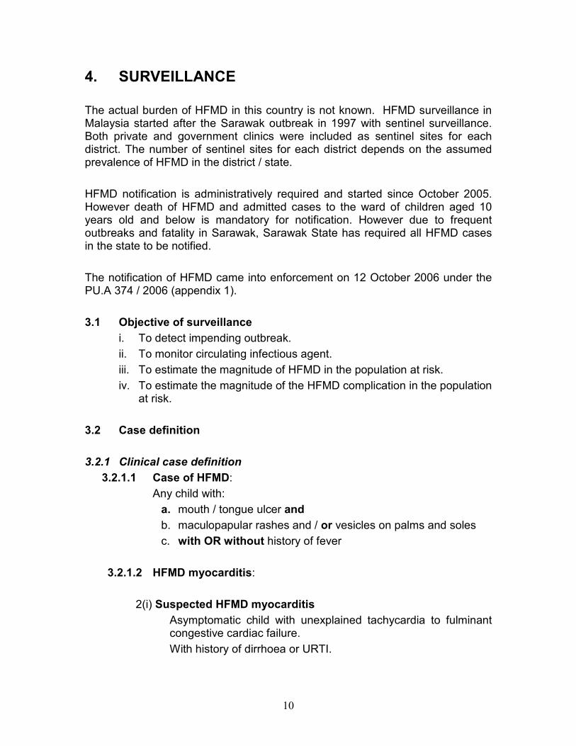
10
4. SURVEILLANCE
The actual burden of HFMD in this country is not known. HFMD surveillance in Malaysia started after the Sarawak outbreak in 1997 with sentinel surveillance. Both private and government clinics were included as sentinel sites for each district. The number of sentinel sites for each district depends on the assumed prevalence of HFMD in the district / state.
HFMD notification is administratively required and started since October 2005. However death of HFMD and admitted cases to the ward of children aged 10 years old and below is mandatory for notification. However due to frequent outbreaks and fatality in Sarawak, Sarawak State has required all HFMD cases in the state to be notified.
The notification of HFMD came into enforcement on 12 October 2006 under the PU.A 374 / 2006 (appendix 1).
3.1 Objective of surveillance
i. To detect impending outbreak.
ii. To monitor circulating infectious agent.
iii. To estimate the magnitude of HFMD in the population at risk.
iv. To estimate the magnitude of the HFMD complication in the population at risk.
3.2 Case definition
3.2.1 Clinical case definition
3.2.1.1 Case of HFMD:
Any child with:
a. mouth / tongue ulcer and
b. maculopapular rashes and / or vesicles on palms and soles
c. with OR without history of fever
3.2.1.2 HFMD myocarditis:
2(i) Suspected HFMD myocarditis
Asymptomatic child with unexplained tachycardia to fulminant congestive cardiac failure.
With history of dirrhoea or URTI.

11
Accompanied by fever, malaise, tachypnoea, tachycardia (above and beyond expected for age and fever) or dehydration.
Pallor and shock in the absence of or obvious pulmonary sign or in the presence of crepitations (no murmurs) and / or a clinically enlarged heart.
2(i) Confirmed HFMD myocarditis.
The above 2(i) with virologic confirmation of enteroviruses.
3.2.1.3 HFMD meningoencephalitis / AFP
3(i) Suspected HFMD meningoencephalitis
Evidence of aseptic meningitis.
Evidence of aseptic encephalitis.
Evidence of AFP.
3(ii) Confirmed HFMD meningoencephalitis / AFP.
The above 3(i) with virologic confirmation of enteroviruses
3.2.2 Laboratory criteria
Any case that has the clinical symptoms and positive for viruses (coxsackieviruses (Cox) A16, A5, A9, A10, B2, B5 and enterovirus (EV) 71) which could cause HFMD, isolated or detected from stool or vesicle fluid or mouth ulcer or saliva.
3.2.3 Case classification
Suspected: A case that meets the clinical case definition.
Confirmed: A suspected case in which laboratory investigation confirms the presence of virus OR when cases are epidemiologically linked to a laboratory confirmed case.
3.3 Types of surveillance
3.3.1 Clinical surveillance
It is mandatory to notify HFMD under the National Notification of Infectious Diseases (list of notifiable diseases). Notification must be done within 24 hours of diagnosis via telephone, then it should be followed by notification form.

12
All HFMD cases detected must be entered to CDCIS. If states have other methods of data storing, it must be shared with MOH.
Laboratory confirmation is NOT required for notification.
3.2.2 Laboratory surveillance
Sentinel sites
Enterovirus surveillance is done through state’s sentinel sites. A state has to choose two sentinel sites. Preferably the sites have a considerable numbers of children cases as HFMD prevalence is higher among this age group. At least 5 specimens from 5 patients per centre should be taken and sent to the designated laboratory (as in table below). Please use the laboratory request form as in appendix 2(i).
Designated laboratory involves in enterovirus surveillance
Laboratory
Contact number
Respective states
IMR
03-26988792
Johor, WPKL, Melaka, Terengganu, Pahang.
UMMC
03-79675756 / 79676678
Selangor.
NPHL
03-61402213
Perlis, Kedah, Perak, Penang
‘HUKM
03 - 91702203 / 2208
Negeri Sembilan
USM
09 - 7664602 / 4609 / 3000
ext 4602
Kelantan
Surveillance for Sarawak and Sabah is expected to start once Kota Kinabalu Public Health Laboratory is ready to function.

13
Clinical Specimen Collection, Handling And Transportation
Patient
Swab For viral culture
Blood serology CSF
2 ml - in plain sterile
tube
1 ml -in plain sterile
tube
Mouth ulcer Rectal - wet swab with sterile saline
Vesicle - basal
scrapping
Viral transport media
Smear on slide
Send clinical samples to respective laboratory within 24 hours of collection at 4 oC
(on ice). DO NOT STORE AT -20 oC
14
3.4 Flow of surveillance data
Notification data should be entered in CDCIS as and when there is HFMD case. Other sources of data must be shared with Disease Control Division. Data must have the variables listed in appendix 1.
Laboratory results for enterovirus surveillance must be shared with respective states and Disease Control Division using the suggested format in appendix 2(ii).

15
Flow of Information Management
Yes
Suspected HFMD Case
CDCIS
Case fulfilled criteria
Investigate & Register
Lab-Confirmed Cases
LIS
Positive EV71
Investigate
Clinics Laboratories
Cluster of cases ?
Finish
Preventive and control activities
Report
No

16
5. MANAGEMENT OF SPORADIC CASES
5.1 Definitions of sporadic case:
A single case of HFMD in the absence of previous known close contact with another case.
5.2 The public health measures
5.2.1 Case Investigation
Case investigation is required following the criteria below:
a. every notified HFMD case that fulfilled any one of the criteria below:
i. case is admitted to hospital ii. case died. iii. case aged 6 years and below and goes to any pre-schools or nurseries.
The first two criteria indicate severity of illness and it is a proxy of possible EV71 infection. Meanwhile the third criterion is important in preventing spread of illness among the pre-school children (high risk group) and reducing public anxiety.
b. every case with positive EV71 laboratory result. Investigation should be carried out to determine the course, source and severity of infection. Enquiries shall be made into;
1. Particulars of the person affected
2. Clinical signs and symptoms with date of onset. In addition, enquiries shall be elicited for associated complications e.g. seizures, weakness of limbs, staring episodes, drowsiness, profound lethargy and refusal to play.
Enterovirus infections that are not associated with complications may not have cutaneous manifestations.
3. Duration of illness
4. Type of treatment sought, including details of hospitalization and reason for admission
5. History of travel especially to outbreak area in the past two weeks
6. Contacts with similar illness in an institution (child care centre or kindergarten), family or neighbourhood where relevant.
7. If newborn, antenatal history including maternal history of febrile illness, mode of delivery
17
5.2.2 Incidence management Following steps should be taken to prevent further spread of disease / pathogen:
1. Ensure that the infected children remain away from the institution for at least ten days after onset of symptoms and must be certified free from infection by a registered medical practitioner prior to returning to school.
2. Active case detection shall be carried out among contacts in the institution and family, and all those with illness shall be referred for treatment at the nearest health clinics.
3. Swabs from oral ulcers and vesicular lesions on the hands and feet shall be collected for virus isolation at identified laboratories (IMR or NPHL). If these ulcers and vesicles are no longer present, stool specimens shall be sent. Always consult pathologist for specimens taking, handling and transportation.
4. Health education of supervisors of institutions on the disease, mode of transmission, importance of good personal hygiene and isolation of infected children shall be carried out.
5. Principals, teachers and supervisors shall be alerted to look out for children with fever, rash / blisters on palms and soles and to isolate them immediately. Screening before coming to class is recommended (please refer Buku Panduan Latihan Taska dan Prasekolah).
6. If there are two or case in the premise, advice the premise to be closed to cut disease transmission. It should be closed for two incubation period (10 days) from the date of onset of the last case.
7. Supervision on disinfection procedures shall be given to the premise operators. Articles such as toys contaminated by infected cases are disinfected with 0.5% sodium hypochlorite solutions (please refer Buku Panduan Latihan Taska dan Prasekolah).

18
6. MANAGEMENT OF OUTBREAK
6.1 Definitions of outbreak
The occurrence of two or more cases in the same locality within the incubation period. Organisation based outbreak – two or more cases with onset in two weeks interval in a group which makes epidemiological sense.
6.2 Identification of outbreaks
Surveillance data on HFMD cases should be reviewed on continuous basis to identify cases and detects outbreaks.
6.3 Risk factors for outbreaks
Risk factors for outbreaks are not completely understood. A combination of conditions (environment, host and organism) is necessary for an epidemic to occur. However large HFMD outbreaks have been associated with EV71 infection.
6.4 Management of outbreaks
When a suspected outbreak is reported, it is important to determine rapidly whether a true outbreak is occurring based on the definition above. Similar steps as in sporadic case management should be carried out together with following measures;
1. Ensure that the infected children remain away from the institution for at least ten days after onset of symptoms and must be certified free from infection by a registered medical practitioner prior to returning to school.
2. An assessment of the number of cases and susceptible population involved shall be made, and the overall attack rate and age-specific attack rate computed.
3. Active case detection shall be carried out among contacts in the institution and family, and all those with illness shall be referred for treatment at the nearest health clinics.
4. Where a case is observed or suspected to have unusual signs and symptoms e.g. seizures, weakness of limbs, profound lethargy, refusal to play, the case shall be immediately referred to hospital.
5. Swabs from oral ulcers and vesicular lesions on the hands and feet shall be collected for virus isolation at identified laboratories (IMR or NPHL). If these ulcers and vesicles are no longer present, stool specimens shall be sent. Always consult pathologist for specimens taking, handling and transportation.

19
6. Principals, teachers and supervisors shall be alerted to look out for children with fever, rash / blisters on palms and soles and to isolate them immediately. Screening before coming to class is recommended (please refer Buku Panduan Latihan Taska dan Prasekolah).
7. Health education of supervisors of institutions on the disease, mode of transmission, importance of good personal hygiene and isolation of infected children shall be carried out.
8. Close the premise closed for two incubation period (10 days) from the date of onset of the last case. Supervision on disinfection procedures shall be given to the premise operators.
In addition the investigating team shall also look into environmental sanitation and hygiene to ensure that:
a. Articles such as toys contaminated by infected cases are disinfected with 0.5% sodium hypochlorite solutions (please refer Buku Panduan Latihan Taska dan Prasekolah).
b. A high standard of food and personal hygiene is maintained by the institution involved which include proper waste and diapers disposal.
c. The approved enrolment of Department of Social Welfare (JKM), Ministry of Education, Department of National Integration and other relevant parties, where applicable, and shall notify the relevant authorities if regulations not complied.
If the outbreak occurs in primary schools;
i. Ensure that the infected students remain away from the institution for at least ten days.
ii. Health education to the students on the disease, mode of transmission, importance of good personal hygiene.
iii. Principals, teachers and supervisors shall be look for children with fever, rash / blisters on palms and soles and to isolate them immediately. Screening before coming to class is recommended (please refer Buku Panduan Latihan Taska dan Prasekolah).
iv. If closure is necessary, just closed the affected class. Closure of the whole school is unnecessary as HFMD in older children is usually very mild and so far no complication has been documented from this agegroup.

20
6.5 Documentation and review
Disease activity and emergency response should be adequately documented so that further outbreaks can be handled more effectively. A summary report should be prepared within a month from the last date of case; and distributed to all the stakeholders for feedback. Format of report, dissemination and tabling of report is discussed in more details in Infectious Disease Outbreak Rapid Response Manual.12

21
Appendix 1
Variables required in HFMD surveillance database
1 Epid week
2 Name
3 Age
4 Sex
5 Ethnic
6 Home address
7 Locality
8 District
9 Name of school / preschool / nursery
10 Address of school / preschool / nursery
11 Date of onset
12 Case definition fulfilled
13 Symptoms and signs of CNS
14 Contact with HFMD case
15 Address of contact
16 Date of seen
17 Name of clinic / hospital
18 Government or private health facility
19 Outpatient or inpatient treatment
20 Date of admitted
21 Date of discharge
22 Date of death

22
No. Rujukan Makmal
NATIONAL ENTEROVIRUS SURVEILLANCE
DIAGNOSTIC REQUEST FORM
A. MAKLUMAT PESAKIT
Negeri : Daerah :
Hospital (wad) / Klinik Kesihatan No. R/N :
Nama Pesakit :
No. K/P : Umur : Jantina : L / P
Bangsa : Tarikh lahir :
B. MAKLUMAT KLINIKAL
Gejala (Simptom) Ada / Tiada (Tandakan √) Tarikh mula
Demam > 38 0C
Ulser di mulut & tekak
Maculopapular rashes dan / Vesikel pada tapak
tangan dan tapak kaki
Tanda dan gejala URTI
C. SPESIMEN KLINIKAL
Spesimen Tarikh diambil Tarikh penghantaran
Rectal swab / / / /
Mouth ulcer / / / /
Vesicle swab / / / /
Stool / / / /
D. MAKLUMAT PEMOHON
Nama dan Cop Pegawai :
Tandatangan :
No. telefon :
No. fax :
e-mail :
E. MAKMAL (Untuk Kegunaan Makmal)
Keadaan spesimen : Tarikh terima spesimen :
Spesimen Jenis ujian Keputusan ujian Komen
Rectal swab
Mouth ulcer
Vesicle swab
Stool
Nama dan tandatangan Pegawai Makmal :
Jawatan Pegawai Makmal dan Cop Makmal :
Tarikh :
Nota * : All samples except blood / serum and stool must be in the viral transport media (VTM) and
send immediately in ice pack to HUKM Microbiological Laboratory.
Appendix 2(i)

23
Appendix 2(ii)
Variables Required for Enterovirus Surveillance Database
Name of Centre / Laboratory: _______________
Result
No Date
Sample Received
Date Sample Collected
LN RN Name Ethnic Age Sex Source
Case
diagnosis
Type of
spec IF Isolation PCR

24
BKP / S / HFMD (H) Ver 2 / 2007
BORANG SIASATAN PESAKIT DI WAD
PENYAKIT TANGAN, KAKI DAN MULUT (HFMD)
BAHAGIAN KAWALAN PENYAKIT, KEMENTERIAN KESIHATAN MALAYSIA
BAHAGIAN 1
Tarikh masuk wad: Masa:
Tarikh discaj: Masa:
Tarikh kematian Masa:
A. PERIHAL PESAKIT
1. Nama pesakit: 2. Tarikh lahir:
3. Nama ibu: 4. No K/P ibu:
5. Alamat rumah: 6. No telefon: (rumah)
(H/P)
Kedudukan rumah (GPS): X : _________________ Y : __________________
7. Jantina: Lelaki 8. Kumpulan etnik
Perempuan Melayu Kadazan/Dusun
Cina Murut
9. Kewarganegaraan: Malaysia India Bajau
Warga asing Asli semenanjung Melanau
Bidayuh Iban
Lain-lain pribumi Lain-lain (nyatakan)
Sabah/ Sarawak
10. Menghadiri sekolah / taska
Ya
Taska / Nurseri
Prasekolah
Sekolah
Nyatakan nama: ________________________________
Tidak
B. MAKLUMAT KLINIKAL
1. Tarikh mula sakit (onset)
2. Tanda dan gejala: Ya Tidak
Riwayat demam

25
Ulser mulut
Ruam (rash) tapak tangan / kaki
vesicles di tapak tangan / kaki
3. Kompliksi Ya Tidak
Aseptic meningitis
Myocarditis
Encephalitis
Acute flaccid paralysis
Jika ya, sila nyatakan bahagian anggota: ____________________
Kematian
C. MAKLUMAT UJIAN MAKMAL
SAMPEL Tarikh diambil Keputusan Tarikh keputusan
Throat swab
Rectal swab
Vesicle swab
Stool
Blood
CSF
Biopsy _________
(Nyatakan tisu)
D. MAKLUMAT PEGAWAI YANG MERAWAT
1. Nama Pakar / Pegawai Perubatan: __________________________ Tandatangan: ____________
2. Nama Wad: _____________________
3. Nama hospital: __________________________ Tarikh notifikasi: _________

26
7. REFERENCES
1. Melnick JL. Enteroviruses: polioviruses, coxsackieviruses, echoviruses, and newer enteroviruse. In: Fields BN, Knipe DM, Howley PM, Chandlock RM, Melnick JL, Monah TP, et al., editors. Field’s virology. 3 rd ed. Philadelphia: Lippincott-Raven Publishers; 1996. p. 655-712.
2. Zeichhardt H, Grunert H. Enteroviruses: Polioviruses, Coxcaskie viruses,
echoviruses and Enteroviruses 68-71. In: Cohen J, Powderly WG, editors. Infectious diseases. 2nd edition. Mosby ; 2004. p.1993-2003.
3. Read RC. Orocervical and esophageal infection. In: Cohen J, Powderly WG,
editors. Infectious diseases. 2nd edition. Mosby ; 2004. p. 463. 4 . Wright HT, Landing BH, Lennette EH et al. Fatal Coxsackievirus A16
infection. The Pediatric Infectious Disease Journal. 2004; 23(3): 275. 5 . Goldberg MF, McAdams AJ. Myocarditis possibly due to coxsackie group A,
type 16, virus. J Pediatr, 1963; 268: 1041-4. 6. Wang CY, Lu FL, Wu MW. Fatal coxcaskievirus A16 infection. The Pediatric
Infectious Disease Journal 2004;23(3),275. 7. Chang LY, Lin TY, Huang YC et al . Comparison of enterovirus 71 and
coxsackievirus A16 clinical illnesses during the Taiwan enterovirus epidemic, 1998. The Pediatric Infectious Disease Journal 1999; 18(12): 1092-1096.
8. Ho M, Chen ER, Hsu KH, Twu SJ, et al. An epidemic of enterovirus 71
infection in Taiwan. The New England Journal of Medicine, 1999; 341(13): 929-935.
9 . Podin Y, Gias ELM, Ong F, et al. Sentinel Surveillance for human enterovirus
71 in Sarawak, Malaysia: lessons from the first 7 years. BMC Public Health 2006; 6: 180
. 10 . Huang CC, Liu CC, Chang YC et al. Neurologic complications in children
with enterovirus 71. The New England Journal of Medicine, 1999;341(13): 936-943.
11 Wu JM, Wang JN, Tsai YC et al. Cardiopulmonary manifestations of
fulminate enterovirus 71 infection. Pediatrics, 2002; 109:e26 12. Disease Control Division, Ministry of Health. Infectious Diseases Outbreak
Rapid Response Manual. Ministry of Health. Kuala Lumpur, 2003.
27
CONTRIBUTORS State Epidemiologists 2006 – 2007
1. Pulau Pinang Dr. Saraswathi Bina Rai
2. Kedah Dr. Uma Salamah Abdul Kadir
3. Perlis Dr. Zulhizzam Hj Abdullah
4. Perak Dr. Asiah Ayob
Dr. Zamri Md Ali
5. Selangor Dr. Prema Rajendran
Dr. B. Venugopalan
6. WPKL Dr. Norbizura Abdul Hamid
7. N. Sembilan Dr. Zulfa Vety Dol Bakri
8. Melaka Dr. Hashimah Hasan
9. Johor Dr. Shaharom Nor Azian Che Mat Din
Dr. Fatimah Othman
10. Pahang Dr. Shaari Ngadiman
Dr. Adibah Hani Harun
11. Terengganu Dr. Anita Surani Abdul Shukor
Dr. Balkis Ab Karim
12. Kelantan Dr. Hani Mat Hussin
13. Sarawak Dr. Ooi Choo Huck
Dr. Kamaliah Mohd Nor
Dr. Rohani Mat Bah
14. Sabah Dr. Jenarun Jelip
Dr. Kristina Rundi
15. Bahagian Kawalan Dr. Husnina Ibrahim
Penyakit Dr. Nazarudin Safian
Dr. Fadzilah Kamaludin
Dr. Wan Mansor Hamzah
Dr. Mohd Hanif Zailani
16 Laboratory Dr. Chua Kaw Bing

HAND FOOT AND MOUTH DISEASE (HFMD)
GUIDELINES
2
EDITORIAL BOARD
ADVISORS: Dato’ Dr. Hj. Ramlee bin Hj. Rahmat
Director of Disease Control (2003 – Dec 2006)
Dato’ Dr. Hasan Abdul Rahman Director of Disease Control (Dec 2006 until now)
MEMBERS: Dr. Zainudin Abd Wahab Deputy Director of Disease Control (Surveillance)
Disease Control Division, MOH
Dr. Abd. Rasid Kasri Deputy Director of Disease Control
(Communicable Disease) Disease Control Division, MOH
Dr. Rohani Jahis Principal Assistant Director (Surveillance) Disease Control Division, MOH. Dr. Izuna Mudla Mohamed Ghazali Assistant Director (Surveillance) Disease Control Division, MOH. Dr. Zairina Abdul Rahman Assistant Director (Communicable Disease) Disease Control Division, MOH.

3
TABLE OF CONTENTS
1. INTRODUCTION.............................................................................................. 4
2. THE DISEASE ................................................................................................. 4
2.1 TRANSMISSION.............................................................................................. 4 2.2 CLINICAL FEATURES ...................................................................................... 4 2.3 MAGNITUDE OF THE DISEASE .......................................................................... 6
3. CLINICAL MANAGEMENT OF HFMD ............................................................ 7
3.1 SYMPTOMATIC TREATMENT ............................................................................ 7 3.2 HOSPITALISATION. .................................................................................... 7 3.2.1 Criteria for admission ........................................................................... 7 3.2.2 Infection control .................................................................................. 7
3.3 PATIENTS WITH NEUROLOGIC COMPLICATION .............................................. 8 3.4. ADVICE GIVEN UPON PATIENT’S DISCHARGE ............................................ 9
4. SURVEILLANCE ........................................................................................... 10
3.1 OBJECTIVE OF SURVEILLANCE ...................................................................... 10 3.2 CASE DEFINITION......................................................................................... 10 3.2.1 Clinical case definition........................................................................ 10 3.2.2 Laboratory criteria .............................................................................. 11 3.2.3 Case classification ............................................................................. 11
3.3 TYPES OF SURVEILLANCE ............................................................................. 11 3.3.1 Clinical surveillance............................................................................ 11 3.2.2 Laboratory surveillance ...................................................................... 12
3.4 Flow of surveillance data…………………………………………………... .. 14
5. MANAGEMENT OF SPORADIC CASES...................................................... 16
5.1DEFINITIONS OF SPORADIC CASE: .................................................................. 16 5.2THE PUBLIC HEALTH MEASURES ..................................................................... 16
6. MANAGEMENT OF OUTBREAK.................................................................. 18
6.1 DEFINITIONS OF OUTBREAK .......................................................................... 18 6.2 IDENTIFICATION OF OUTBREAKS .................................................................... 18 6.3 RISK FACTORS FOR OUTBREAKS ................................................................... 18 6.4 MANAGEMENT OF OUTBREAKS ...................................................................... 18 6.5 DOCUMENTATION AND REVIEW...................................................................... 20
7. REFERENCES............................................................................................. 216

4
1. INTRODUCTION
Hand foot and mouth disease (HFMD) is typically a benign and common illness among children and infants characterized by rapidly ulcerating vesicles in the mouth and lesions, usually vesicular, on the hands and feet.1 HFMD is an endemic disease in Malaysia. HFMD has become an important public health disease due to its tendency to cause large outbreaks and deaths among children and infants. The purpose of the guidelines is to help health personnels and health authorities, at any levels;
� To update current knowledge about HFMD
� To detect and control epidemic of HFMD as early as possible
� To strengthen the capacity for emergency response to epidemics of HFMD
� To improve the outcome of HFMD cases through effective case management.
2. THE DISEASE
Hand foot and mouth disease (HFMD) is caused by systemic infections with human enteroviruses. Human enteroviruses comprise one genus in the family Picornaviridae, which also contains the genera rhinovirus, cardiovirus, aphthovirus, hepatovirus and parechovirus. The members of the enterovirus genus that infect humans include the polioviruses, the Coxsackie virus groups A and B, the echoviruses and the enteroviruses 68-71.2
2.1 Transmission
Hand foot and mouth disease (HFMD) is moderately contagious. Infection is spread from person to person by direct contact with nose and throat discharges, saliva, fluid from blisters, or the stool of infected persons. A person is most contagious during the first week of the illness.
2.2 Clinical Features
HFMD is characterised by vesicular eruptions on the hands, wrists, feet and within the mouth. Lesions on the hands are almost always present, but oral lesions are present in 90% of patients and can occasionally be the only manifestation of the disease. The oral vesicles are often on the palate, tongue, and buccal mucosa and may range from a few isolated lesions to a marked stomatitis.
In addition patients may suffer fever, malaise, conjunctival injection, headache, and abdominal pain and occasionally diarrhea. The lesions on the feet and hands are flaccid, grayish vesicles, most often on the sides of the fingers, instep and

5
toes. If the disease is confined to the oral cavity it is almost indistinguishable from primary herpetic gingivostomatitis.3
Hand foot and mouth disease may be related to coxsackieviruses A16, A5, A9, A10, B2, B5 and enterovirus 71. Coxsackievirus A16 (Cox A16) is a frequently encountered pathogen in cases of HFMD and its clinical course is usually uneventful, with full recovery.
Fatal cases of Cox A16 infection are rare. Literature search revealed only three fatal cases of Cox A16 since 1963. All the cases were infants. The first case was reported by Wright et al in 1963 involving a 10 month-old girl with respiratory infection.4 The second case involved a 7 month-old boy with grunting and tongue ulcers.5 The third case was reported by Wang et al involving a 15 month-old boy who presented with hand foot and mouth disease complicated with myocarditis and intractable shock.6
HFMD caused by enterovirus 71 (EV71) can be more severe and may be complicated with meningitis, encephalitis and neurogenic pulmonary edema. In Taiwan, based on 1998 epidemic, the significant difference in the clinical features of Coxsackievirus A16 infection and EV 71 infection was that in EV71 the fever
was usually higher than 39°C and longer than 3 days.7
Enterovirus 71 infection can be classified into 4 stages:
Stage 1 – Hand foot and mouth disease (HFMD), oral ulcers and vesicular rash appearing on the hands, feet, knees and / or buttocks; or herpangina including oral ulceration over anterior tonsillar pillars, the soft palate, buccal mucosa or uvula.
Stage 2 – CNS involvement - aseptic meningitis with headache, irritability or myoclonic jerk and CSF pleocytosis (> 5 x 10⁶ leucocytes / litre) but without altered level of consciousness and focal signs, or encephalitis with altered level of consciousness plus CSF pleocytosis or poliomyelitis like syndrome with acute limb weakness and decreased reflex and muscle strength, or encephalomyelitis with the occurrence of both encephalitis and poliomyelitis like syndrome.
Stage 3 – Cardiopulmonary failure, pulmonary edema or hemorrhage with decreased ejection fraction of left ventricle as assessed by echocardiography necessitating inotropic agent support.

6
Stage 4 – Convalescence - is defined as recovery from cardiopulmonary failure.
2.3 Magnitude of the disease
Hand foot and mouth disease (HFMD) generally is a common and self limiting disease among children. EV 71 infection is the more important public health concern. In temperate countries most HFMD cases occur in the summer and fall and the incubation period is 4 – 6 days.
EV 71 was first isolated from a child with aseptic meningitis in California in 1969, and by 1974, the virus had been described as a new serotype of genus Enterovirus. In the years following the initial isolation of EV71, outbreaks occurred in USA, Australia, Sweden and Japan. In 1975 EV71 gained global attention when it was responsible for an outbreak in Bulgaria that resulted in 705 cases of poliomyelitis-like disease and the deaths of 44 people; 93% of the poliomyelitis-like disease cases occurred in children under the age of five. A similar outbreak occurred in Hungary in 1978, which also involved many cases of poliomyelitis-like disease and 47 deaths. Since this time outbreaks of EV71 have continued to occur throughout the world.
More recently there has been an increase in EV71 activity in the Asia Pacific region with several epidemics of HFMD being reported, including multiple cases associated with brain stem encephalitis and pulmonary edema. The first such epidemic occurred in Sarawak (Malaysian Borneo) in 1997 followed by smaller outbreaks in peninsular Malaysia and Japan. In 1998 outbreaks continued in Singapore and Taiwan.
The outbreak that occurred in Taiwan is the largest recorded, with greater than 100,000 cases of HFMD. Of these 400 children were admitted to hospital with central nervous system (CNS) involvement and 78 died of brainstem encephalitis with neurogenic pulmonary oedema.8 In 1999, a large HFMD outbreak occurred in Perth, western Australia and in 2000, EV71 was the cause of epidemics in Korea, Japan, Singapore, Taiwan and peninsular Malaysia, resulting in a range of clinical presentations including HFMD, aseptic meningitis, encephalitis and poliomyelitis like disease. Another outbreak occurred in Sarawak in 2003 which coincide with the SARS outbreak in the region and the public health measures put into place during this time evidently served to control transmission of enteroviruses as well.9

7
3. CLINICAL MANAGEMENT OF HFMD
HFMD is usually a mild and self limiting. In general, most cases of HFMD do not require admission but can be managed as outpatients. Most fatal HFMD cases were due to enterovirus infection.
3.1 Symptomatic treatment
Mild HFMD cases only need symptomatic treatment. Treatment of fever and relief of symptoms, adequate hydration and rest are important. Parents and care takers should be educated on hygiene and measures that they should take to prevent transmission to other children.
3.2 Hospitalisation.
3.2.1 Criteria for admission
• When the child is unable to tolerate oral feeds and there is a need for intravenous hydration;
• When the child is clinically very ill or toxic-looking
• When some other more serious disease cannot be excluded
• When there is persistent hyperpyrexia (e.g >38ºC) for >48 hours;
• When there is a suspicion of neurological complications, e.g increased lethargy, myoclonus, increased drowsiness, change in sensorium and/or seizures;
• When there is a suspicion of cardiac complications (myocarditis), e.g low blood pressure, low pulse volume, heart rhythm abnormalities, murmurs, gallop rhythm, displaced apex beat;
• When parents are unable to cope with child’s illness; and
• When there is inadequate family or social support in looking after the child at home.
3.2.2 Infection control
• Proper hygiene including mandatory hand washing after contact with patient, appropriate cleanliness during diaper changes is imperative
• Personal items such as spoons, cups and utensils should not be shared and should be properly washed with detergent after use;
• The use of gowns may act as a useful protection for health personnel looking after these patients; and
• Patients with HFMD should be isolated and the usual isolation procedures followed for infection control.

8
3.3 Patients with neurologic complication
Huang et al reviewed HFMD cases in Taiwan in 1998 and they found that the mean age of patients with neurological manifestations was 2.5 (range, 3 months to 8.2 years10. Higher incidence occurs in the younger age group. The neurologic disorders usually began 2 to 5 days after the onset of skin or mucosa lesion or fever.
Wu JM et al carried out a prospective study among infants confirmed to have enterovirus 71 and found that11;
• EV71 infection can lead to severe neurologic complications and acute pulmonary edema with or without hemorrhage
• All patients with EV71 infection and pulmonary edema had brainstem lesions
• Viral myocarditis is not the direct cause of acute PE and cardiopulmonary failure in EV71 Infection
• The excessive central sympathetic activation resulting in vasoconstriction was not the major cause of PE or hemorrhage
Based on the findings, they suggested that;
• Vasodilators should be used with caution since pulmonary edema in EV71 infection is not resulting from excessive vasoconstriction alone
• Cardiopulmonary function usually returns to nearly normal within days but the neurogic sequelae are severe and usually permanent. Therefore the use of extracorporeal membrane oxygenation support or a heart assist device should be weighed carefully
• Direct intracardiac hemodynamic monitoring may provide additional information to guide critical care

9
3.4. ADVICE GIVEN UPON PATIENT’S DISCHARGE
Parents and guardians should be advised upon patients discharge on complications that may occur; a statement as shown below can be given:
“Your child has been diagnosed to have hand-foot-mouth disease. This disease is normally not dangerous but in the light of recent events, we advise that you bring back your child to this hospital if he / she has any of the following symptoms:
• High fever.
• Lethargy and weakness.
• Refusing feeds and passing less urine.
• Rapid breathing.
• Vomiting.
• Drowsiness or irritably.
• Fits.”

10
4. SURVEILLANCE
The actual burden of HFMD in this country is not known. HFMD surveillance in Malaysia started after the Sarawak outbreak in 1997 with sentinel surveillance. Both private and government clinics were included as sentinel sites for each district. The number of sentinel sites for each district depends on the assumed prevalence of HFMD in the district / state.
HFMD notification is administratively required and started since October 2005. However death of HFMD and admitted cases to the ward of children aged 10 years old and below is mandatory for notification. However due to frequent outbreaks and fatality in Sarawak, Sarawak State has required all HFMD cases in the state to be notified.
The notification of HFMD came into enforcement on 12 October 2006 under the PU.A 374 / 2006 (appendix 1).
3.1 Objective of surveillance
i. To detect impending outbreak.
ii. To monitor circulating infectious agent.
iii. To estimate the magnitude of HFMD in the population at risk.
iv. To estimate the magnitude of the HFMD complication in the population at risk.
3.2 Case definition
3.2.1 Clinical case definition
3.2.1.1 Case of HFMD:
Any child with:
a. mouth / tongue ulcer and
b. maculopapular rashes and / or vesicles on palms and soles
c. with OR without history of fever
3.2.1.2 HFMD myocarditis:
2(i) Suspected HFMD myocarditis
Asymptomatic child with unexplained tachycardia to fulminant congestive cardiac failure.
With history of dirrhoea or URTI.
11
Accompanied by fever, malaise, tachypnoea, tachycardia (above and beyond expected for age and fever) or dehydration.
Pallor and shock in the absence of or obvious pulmonary sign or in the presence of crepitations (no murmurs) and / or a clinically enlarged heart.
2(i) Confirmed HFMD myocarditis.
The above 2(i) with virologic confirmation of enteroviruses.
3.2.1.3 HFMD meningoencephalitis / AFP
3(i) Suspected HFMD meningoencephalitis
Evidence of aseptic meningitis.
Evidence of aseptic encephalitis.
Evidence of AFP.
3(ii) Confirmed HFMD meningoencephalitis / AFP.
The above 3(i) with virologic confirmation of enteroviruses
3.2.2 Laboratory criteria
Any case that has the clinical symptoms and positive for viruses (coxsackieviruses (Cox) A16, A5, A9, A10, B2, B5 and enterovirus (EV) 71) which could cause HFMD, isolated or detected from stool or vesicle fluid or mouth ulcer or saliva.
3.2.3 Case classification
Suspected: A case that meets the clinical case definition.
Confirmed: A suspected case in which laboratory investigation confirms the presence of virus OR when cases are epidemiologically linked to a laboratory confirmed case.
3.3 Types of surveillance
3.3.1 Clinical surveillance
It is mandatory to notify HFMD under the National Notification of Infectious Diseases (list of notifiable diseases). Notification must be done within 24 hours of diagnosis via telephone, then it should be followed by notification form.

12
All HFMD cases detected must be entered to CDCIS. If states have other methods of data storing, it must be shared with MOH.
Laboratory confirmation is NOT required for notification.
3.2.2 Laboratory surveillance
Sentinel sites
Enterovirus surveillance is done through state’s sentinel sites. A state has to choose two sentinel sites. Preferably the sites have a considerable numbers of children cases as HFMD prevalence is higher among this age group. At least 5 specimens from 5 patients per centre should be taken and sent to the designated laboratory (as in table below). Please use the laboratory request form as in appendix 2(i).
Designated laboratory involves in enterovirus surveillance
Laboratory
Contact number
Respective states
IMR
03-26988792
Johor, WPKL, Melaka, Terengganu, Pahang.
UMMC
03-79675756 / 79676678
Selangor.
NPHL
03-61402213
Perlis, Kedah, Perak, Penang
‘HUKM
03 - 91702203 / 2208
Negeri Sembilan
USM
09 - 7664602 / 4609 / 3000
ext 4602
Kelantan
Surveillance for Sarawak and Sabah is expected to start once Kota Kinabalu Public Health Laboratory is ready to function.

13
Clinical Specimen Collection, Handling And Transportation
Patient
Swab For viral culture
Blood serology CSF
2 ml - in plain sterile
tube
1 ml -in plain sterile
tube
Mouth ulcer Rectal - wet swab with sterile saline
Vesicle - basal
scrapping
Viral transport media
Smear on slide
Send clinical samples to respective laboratory within 24 hours of collection at 4 oC
(on ice). DO NOT STORE AT -20 oC

14
3.4 Flow of surveillance data
Notification data should be entered in CDCIS as and when there is HFMD case. Other sources of data must be shared with Disease Control Division. Data must have the variables listed in appendix 1.
Laboratory results for enterovirus surveillance must be shared with respective states and Disease Control Division using the suggested format in appendix 2(ii).

15
Flow of Information Management
Yes
Suspected HFMD Case
CDCIS
Case fulfilled criteria
Investigate & Register
Lab-Confirmed Cases
LIS
Positive EV71
Investigate
Clinics Laboratories
Cluster of cases ?
Finish
Preventive and control activities
Report
No
16
5. MANAGEMENT OF SPORADIC CASES
5.1 Definitions of sporadic case:
A single case of HFMD in the absence of previous known close contact with another case.
5.2 The public health measures
5.2.1 Case Investigation
Case investigation is required following the criteria below:
a. every notified HFMD case that fulfilled any one of the criteria below:
i. case is admitted to hospital ii. case died. iii. case aged 6 years and below and goes to any pre-schools or nurseries.
The first two criteria indicate severity of illness and it is a proxy of possible EV71 infection. Meanwhile the third criterion is important in preventing spread of illness among the pre-school children (high risk group) and reducing public anxiety.
b. every case with positive EV71 laboratory result. Investigation should be carried out to determine the course, source and severity of infection. Enquiries shall be made into;
1. Particulars of the person affected
2. Clinical signs and symptoms with date of onset. In addition, enquiries shall be elicited for associated complications e.g. seizures, weakness of limbs, staring episodes, drowsiness, profound lethargy and refusal to play.
Enterovirus infections that are not associated with complications may not have cutaneous manifestations.
3. Duration of illness
4. Type of treatment sought, including details of hospitalization and reason for admission
5. History of travel especially to outbreak area in the past two weeks
6. Contacts with similar illness in an institution (child care centre or kindergarten), family or neighbourhood where relevant.
7. If newborn, antenatal history including maternal history of febrile illness, mode of delivery

17
5.2.2 Incidence management Following steps should be taken to prevent further spread of disease / pathogen:
1. Ensure that the infected children remain away from the institution for at least ten days after onset of symptoms and must be certified free from infection by a registered medical practitioner prior to returning to school.
2. Active case detection shall be carried out among contacts in the institution and family, and all those with illness shall be referred for treatment at the nearest health clinics.
3. Swabs from oral ulcers and vesicular lesions on the hands and feet shall be collected for virus isolation at identified laboratories (IMR or NPHL). If these ulcers and vesicles are no longer present, stool specimens shall be sent. Always consult pathologist for specimens taking, handling and transportation.
4. Health education of supervisors of institutions on the disease, mode of transmission, importance of good personal hygiene and isolation of infected children shall be carried out.
5. Principals, teachers and supervisors shall be alerted to look out for children with fever, rash / blisters on palms and soles and to isolate them immediately. Screening before coming to class is recommended (please refer Buku Panduan Latihan Taska dan Prasekolah).
6. If there are two or case in the premise, advice the premise to be closed to cut disease transmission. It should be closed for two incubation period (10 days) from the date of onset of the last case.
7. Supervision on disinfection procedures shall be given to the premise operators. Articles such as toys contaminated by infected cases are disinfected with 0.5% sodium hypochlorite solutions (please refer Buku Panduan Latihan Taska dan Prasekolah).

18
6. MANAGEMENT OF OUTBREAK
6.1 Definitions of outbreak
The occurrence of two or more cases in the same locality within the incubation period. Organisation based outbreak – two or more cases with onset in two weeks interval in a group which makes epidemiological sense.
6.2 Identification of outbreaks
Surveillance data on HFMD cases should be reviewed on continuous basis to identify cases and detects outbreaks.
6.3 Risk factors for outbreaks
Risk factors for outbreaks are not completely understood. A combination of conditions (environment, host and organism) is necessary for an epidemic to occur. However large HFMD outbreaks have been associated with EV71 infection.
6.4 Management of outbreaks
When a suspected outbreak is reported, it is important to determine rapidly whether a true outbreak is occurring based on the definition above. Similar steps as in sporadic case management should be carried out together with following measures;
1. Ensure that the infected children remain away from the institution for at least ten days after onset of symptoms and must be certified free from infection by a registered medical practitioner prior to returning to school.
2. An assessment of the number of cases and susceptible population involved shall be made, and the overall attack rate and age-specific attack rate computed.
3. Active case detection shall be carried out among contacts in the institution and family, and all those with illness shall be referred for treatment at the nearest health clinics.
4. Where a case is observed or suspected to have unusual signs and symptoms e.g. seizures, weakness of limbs, profound lethargy, refusal to play, the case shall be immediately referred to hospital.
5. Swabs from oral ulcers and vesicular lesions on the hands and feet shall be collected for virus isolation at identified laboratories (IMR or NPHL). If these ulcers and vesicles are no longer present, stool specimens shall be sent. Always consult pathologist for specimens taking, handling and transportation.

19
6. Principals, teachers and supervisors shall be alerted to look out for children with fever, rash / blisters on palms and soles and to isolate them immediately. Screening before coming to class is recommended (please refer Buku Panduan Latihan Taska dan Prasekolah).
7. Health education of supervisors of institutions on the disease, mode of transmission, importance of good personal hygiene and isolation of infected children shall be carried out.
8. Close the premise closed for two incubation period (10 days) from the date of onset of the last case. Supervision on disinfection procedures shall be given to the premise operators.
In addition the investigating team shall also look into environmental sanitation and hygiene to ensure that:
a. Articles such as toys contaminated by infected cases are disinfected with 0.5% sodium hypochlorite solutions (please refer Buku Panduan Latihan Taska dan Prasekolah).
b. A high standard of food and personal hygiene is maintained by the institution involved which include proper waste and diapers disposal.
c. The approved enrolment of Department of Social Welfare (JKM), Ministry of Education, Department of National Integration and other relevant parties, where applicable, and shall notify the relevant authorities if regulations not complied.
If the outbreak occurs in primary schools;
i. Ensure that the infected students remain away from the institution for at least ten days.
ii. Health education to the students on the disease, mode of transmission, importance of good personal hygiene.
iii. Principals, teachers and supervisors shall be look for children with fever, rash / blisters on palms and soles and to isolate them immediately. Screening before coming to class is recommended (please refer Buku Panduan Latihan Taska dan Prasekolah).
iv. If closure is necessary, just closed the affected class. Closure of the whole school is unnecessary as HFMD in older children is usually very mild and so far no complication has been documented from this agegroup.

20
6.5 Documentation and review
Disease activity and emergency response should be adequately documented so that further outbreaks can be handled more effectively. A summary report should be prepared within a month from the last date of case; and distributed to all the stakeholders for feedback. Format of report, dissemination and tabling of report is discussed in more details in Infectious Disease Outbreak Rapid Response Manual.12

21
Appendix 1
Variables required in HFMD surveillance database
1 Epid week
2 Name
3 Age
4 Sex
5 Ethnic
6 Home address
7 Locality
8 District
9 Name of school / preschool / nursery
10 Address of school / preschool / nursery
11 Date of onset
12 Case definition fulfilled
13 Symptoms and signs of CNS
14 Contact with HFMD case
15 Address of contact
16 Date of seen
17 Name of clinic / hospital
18 Government or private health facility
19 Outpatient or inpatient treatment
20 Date of admitted
21 Date of discharge
22 Date of death

22
No. Rujukan Makmal
NATIONAL ENTEROVIRUS SURVEILLANCE
DIAGNOSTIC REQUEST FORM
A. MAKLUMAT PESAKIT
Negeri : Daerah :
Hospital (wad) / Klinik Kesihatan No. R/N :
Nama Pesakit :
No. K/P : Umur : Jantina : L / P
Bangsa : Tarikh lahir :
B. MAKLUMAT KLINIKAL
Gejala (Simptom) Ada / Tiada (Tandakan √) Tarikh mula
Demam > 38 0C
Ulser di mulut & tekak
Maculopapular rashes dan / Vesikel pada tapak
tangan dan tapak kaki
Tanda dan gejala URTI
C. SPESIMEN KLINIKAL
Spesimen Tarikh diambil Tarikh penghantaran
Rectal swab / / / /
Mouth ulcer / / / /
Vesicle swab / / / /
Stool / / / /
D. MAKLUMAT PEMOHON
Nama dan Cop Pegawai :
Tandatangan :
No. telefon :
No. fax :
e-mail :
E. MAKMAL (Untuk Kegunaan Makmal)
Keadaan spesimen : Tarikh terima spesimen :
Spesimen Jenis ujian Keputusan ujian Komen
Rectal swab
Mouth ulcer
Vesicle swab
Stool
Nama dan tandatangan Pegawai Makmal :
Jawatan Pegawai Makmal dan Cop Makmal :
Tarikh :
Nota * : All samples except blood / serum and stool must be in the viral transport media (VTM) and
send immediately in ice pack to HUKM Microbiological Laboratory.
Appendix 2(i)

23
Appendix 2(ii)
Variables Required for Enterovirus Surveillance Database
Name of Centre / Laboratory: _______________
Result
No Date
Sample Received
Date Sample Collected
LN RN Name Ethnic Age Sex Source
Case
diagnosis
Type of
spec IF Isolation PCR

24
BKP / S / HFMD (H) Ver 2 / 2007
BORANG SIASATAN PESAKIT DI WAD
PENYAKIT TANGAN, KAKI DAN MULUT (HFMD)
BAHAGIAN KAWALAN PENYAKIT, KEMENTERIAN KESIHATAN MALAYSIA
BAHAGIAN 1
Tarikh masuk wad: Masa:
Tarikh discaj: Masa:
Tarikh kematian Masa:
A. PERIHAL PESAKIT
1. Nama pesakit: 2. Tarikh lahir:
3. Nama ibu: 4. No K/P ibu:
5. Alamat rumah: 6. No telefon: (rumah)
(H/P)
Kedudukan rumah (GPS): X : _________________ Y : __________________
7. Jantina: Lelaki 8. Kumpulan etnik
Perempuan Melayu Kadazan/Dusun
Cina Murut
9. Kewarganegaraan: Malaysia India Bajau
Warga asing Asli semenanjung Melanau
Bidayuh Iban
Lain-lain pribumi Lain-lain (nyatakan)
Sabah/ Sarawak
10. Menghadiri sekolah / taska
Ya
Taska / Nurseri
Prasekolah
Sekolah
Nyatakan nama: ________________________________
Tidak
B. MAKLUMAT KLINIKAL
1. Tarikh mula sakit (onset)
2. Tanda dan gejala: Ya Tidak
Riwayat demam

25
Ulser mulut
Ruam (rash) tapak tangan / kaki
vesicles di tapak tangan / kaki
3. Kompliksi Ya Tidak
Aseptic meningitis
Myocarditis
Encephalitis
Acute flaccid paralysis
Jika ya, sila nyatakan bahagian anggota: ____________________
Kematian
C. MAKLUMAT UJIAN MAKMAL
SAMPEL Tarikh diambil Keputusan Tarikh keputusan
Throat swab
Rectal swab
Vesicle swab
Stool
Blood
CSF
Biopsy _________
(Nyatakan tisu)
D. MAKLUMAT PEGAWAI YANG MERAWAT
1. Nama Pakar / Pegawai Perubatan: __________________________ Tandatangan: ____________
2. Nama Wad: _____________________
3. Nama hospital: __________________________ Tarikh notifikasi: _________
26
7. REFERENCES
1. Melnick JL. Enteroviruses: polioviruses, coxsackieviruses, echoviruses, and newer enteroviruse. In: Fields BN, Knipe DM, Howley PM, Chandlock RM, Melnick JL, Monah TP, et al., editors. Field’s virology. 3 rd ed. Philadelphia: Lippincott-Raven Publishers; 1996. p. 655-712.
2. Zeichhardt H, Grunert H. Enteroviruses: Polioviruses, Coxcaskie viruses,
echoviruses and Enteroviruses 68-71. In: Cohen J, Powderly WG, editors. Infectious diseases. 2nd edition. Mosby ; 2004. p.1993-2003.
3. Read RC. Orocervical and esophageal infection. In: Cohen J, Powderly WG,
editors. Infectious diseases. 2nd edition. Mosby ; 2004. p. 463. 4 . Wright HT, Landing BH, Lennette EH et al. Fatal Coxsackievirus A16
infection. The Pediatric Infectious Disease Journal. 2004; 23(3): 275. 5 . Goldberg MF, McAdams AJ. Myocarditis possibly due to coxsackie group A,
type 16, virus. J Pediatr, 1963; 268: 1041-4. 6. Wang CY, Lu FL, Wu MW. Fatal coxcaskievirus A16 infection. The Pediatric
Infectious Disease Journal 2004;23(3),275. 7. Chang LY, Lin TY, Huang YC et al . Comparison of enterovirus 71 and
coxsackievirus A16 clinical illnesses during the Taiwan enterovirus epidemic, 1998. The Pediatric Infectious Disease Journal 1999; 18(12): 1092-1096.
8. Ho M, Chen ER, Hsu KH, Twu SJ, et al. An epidemic of enterovirus 71
infection in Taiwan. The New England Journal of Medicine, 1999; 341(13): 929-935.
9 . Podin Y, Gias ELM, Ong F, et al. Sentinel Surveillance for human enterovirus
71 in Sarawak, Malaysia: lessons from the first 7 years. BMC Public Health 2006; 6: 180
. 10 . Huang CC, Liu CC, Chang YC et al. Neurologic complications in children
with enterovirus 71. The New England Journal of Medicine, 1999;341(13): 936-943.
11 Wu JM, Wang JN, Tsai YC et al. Cardiopulmonary manifestations of
fulminate enterovirus 71 infection. Pediatrics, 2002; 109:e26 12. Disease Control Division, Ministry of Health. Infectious Diseases Outbreak
Rapid Response Manual. Ministry of Health. Kuala Lumpur, 2003.

27
CONTRIBUTORS State Epidemiologists 2006 – 2007
1. Pulau Pinang Dr. Saraswathi Bina Rai
2. Kedah Dr. Uma Salamah Abdul Kadir
3. Perlis Dr. Zulhizzam Hj Abdullah
4. Perak Dr. Asiah Ayob
Dr. Zamri Md Ali
5. Selangor Dr. Prema Rajendran
Dr. B. Venugopalan
6. WPKL Dr. Norbizura Abdul Hamid
7. N. Sembilan Dr. Zulfa Vety Dol Bakri
8. Melaka Dr. Hashimah Hasan
9. Johor Dr. Shaharom Nor Azian Che Mat Din
Dr. Fatimah Othman
10. Pahang Dr. Shaari Ngadiman
Dr. Adibah Hani Harun
11. Terengganu Dr. Anita Surani Abdul Shukor
Dr. Balkis Ab Karim
12. Kelantan Dr. Hani Mat Hussin
13. Sarawak Dr. Ooi Choo Huck
Dr. Kamaliah Mohd Nor
Dr. Rohani Mat Bah
14. Sabah Dr. Jenarun Jelip
Dr. Kristina Rundi
15. Bahagian Kawalan Dr. Husnina Ibrahim
Penyakit Dr. Nazarudin Safian
Dr. Fadzilah Kamaludin
Dr. Wan Mansor Hamzah
Dr. Mohd Hanif Zailani
16 Laboratory Dr. Chua Kaw Bing

HAND FOOT AND MOUTH DISEASE (HFMD)
GUIDELINES
2
EDITORIAL BOARD
ADVISORS: Dato’ Dr. Hj. Ramlee bin Hj. Rahmat
Director of Disease Control (2003 – Dec 2006)
Dato’ Dr. Hasan Abdul Rahman Director of Disease Control (Dec 2006 until now)
MEMBERS: Dr. Zainudin Abd Wahab Deputy Director of Disease Control (Surveillance)
Disease Control Division, MOH
Dr. Abd. Rasid Kasri Deputy Director of Disease Control
(Communicable Disease) Disease Control Division, MOH
Dr. Rohani Jahis Principal Assistant Director (Surveillance) Disease Control Division, MOH. Dr. Izuna Mudla Mohamed Ghazali Assistant Director (Surveillance) Disease Control Division, MOH. Dr. Zairina Abdul Rahman Assistant Director (Communicable Disease) Disease Control Division, MOH.

3
TABLE OF CONTENTS
1. INTRODUCTION.............................................................................................. 4
2. THE DISEASE ................................................................................................. 4
2.1 TRANSMISSION.............................................................................................. 4 2.2 CLINICAL FEATURES ...................................................................................... 4 2.3 MAGNITUDE OF THE DISEASE .......................................................................... 6
3. CLINICAL MANAGEMENT OF HFMD ............................................................ 7
3.1 SYMPTOMATIC TREATMENT ............................................................................ 7 3.2 HOSPITALISATION. .................................................................................... 7 3.2.1 Criteria for admission ........................................................................... 7 3.2.2 Infection control .................................................................................. 7
3.3 PATIENTS WITH NEUROLOGIC COMPLICATION .............................................. 8 3.4. ADVICE GIVEN UPON PATIENT’S DISCHARGE ............................................ 9
4. SURVEILLANCE ........................................................................................... 10
3.1 OBJECTIVE OF SURVEILLANCE ...................................................................... 10 3.2 CASE DEFINITION......................................................................................... 10 3.2.1 Clinical case definition........................................................................ 10 3.2.2 Laboratory criteria .............................................................................. 11 3.2.3 Case classification ............................................................................. 11
3.3 TYPES OF SURVEILLANCE ............................................................................. 11 3.3.1 Clinical surveillance............................................................................ 11 3.2.2 Laboratory surveillance ...................................................................... 12
3.4 Flow of surveillance data…………………………………………………... .. 14
5. MANAGEMENT OF SPORADIC CASES...................................................... 16
5.1DEFINITIONS OF SPORADIC CASE: .................................................................. 16 5.2THE PUBLIC HEALTH MEASURES ..................................................................... 16
6. MANAGEMENT OF OUTBREAK.................................................................. 18
6.1 DEFINITIONS OF OUTBREAK .......................................................................... 18 6.2 IDENTIFICATION OF OUTBREAKS .................................................................... 18 6.3 RISK FACTORS FOR OUTBREAKS ................................................................... 18 6.4 MANAGEMENT OF OUTBREAKS ...................................................................... 18 6.5 DOCUMENTATION AND REVIEW...................................................................... 20
7. REFERENCES............................................................................................. 216
4
1. INTRODUCTION
Hand foot and mouth disease (HFMD) is typically a benign and common illness among children and infants characterized by rapidly ulcerating vesicles in the mouth and lesions, usually vesicular, on the hands and feet.1 HFMD is an endemic disease in Malaysia. HFMD has become an important public health disease due to its tendency to cause large outbreaks and deaths among children and infants. The purpose of the guidelines is to help health personnels and health authorities, at any levels;
� To update current knowledge about HFMD
� To detect and control epidemic of HFMD as early as possible
� To strengthen the capacity for emergency response to epidemics of HFMD
� To improve the outcome of HFMD cases through effective case management.
2. THE DISEASE
Hand foot and mouth disease (HFMD) is caused by systemic infections with human enteroviruses. Human enteroviruses comprise one genus in the family Picornaviridae, which also contains the genera rhinovirus, cardiovirus, aphthovirus, hepatovirus and parechovirus. The members of the enterovirus genus that infect humans include the polioviruses, the Coxsackie virus groups A and B, the echoviruses and the enteroviruses 68-71.2
2.1 Transmission
Hand foot and mouth disease (HFMD) is moderately contagious. Infection is spread from person to person by direct contact with nose and throat discharges, saliva, fluid from blisters, or the stool of infected persons. A person is most contagious during the first week of the illness.
2.2 Clinical Features
HFMD is characterised by vesicular eruptions on the hands, wrists, feet and within the mouth. Lesions on the hands are almost always present, but oral lesions are present in 90% of patients and can occasionally be the only manifestation of the disease. The oral vesicles are often on the palate, tongue, and buccal mucosa and may range from a few isolated lesions to a marked stomatitis.
In addition patients may suffer fever, malaise, conjunctival injection, headache, and abdominal pain and occasionally diarrhea. The lesions on the feet and hands are flaccid, grayish vesicles, most often on the sides of the fingers, instep and

5
toes. If the disease is confined to the oral cavity it is almost indistinguishable from primary herpetic gingivostomatitis.3
Hand foot and mouth disease may be related to coxsackieviruses A16, A5, A9, A10, B2, B5 and enterovirus 71. Coxsackievirus A16 (Cox A16) is a frequently encountered pathogen in cases of HFMD and its clinical course is usually uneventful, with full recovery.
Fatal cases of Cox A16 infection are rare. Literature search revealed only three fatal cases of Cox A16 since 1963. All the cases were infants. The first case was reported by Wright et al in 1963 involving a 10 month-old girl with respiratory infection.4 The second case involved a 7 month-old boy with grunting and tongue ulcers.5 The third case was reported by Wang et al involving a 15 month-old boy who presented with hand foot and mouth disease complicated with myocarditis and intractable shock.6
HFMD caused by enterovirus 71 (EV71) can be more severe and may be complicated with meningitis, encephalitis and neurogenic pulmonary edema. In Taiwan, based on 1998 epidemic, the significant difference in the clinical features of Coxsackievirus A16 infection and EV 71 infection was that in EV71 the fever
was usually higher than 39°C and longer than 3 days.7
Enterovirus 71 infection can be classified into 4 stages:
Stage 1 – Hand foot and mouth disease (HFMD), oral ulcers and vesicular rash appearing on the hands, feet, knees and / or buttocks; or herpangina including oral ulceration over anterior tonsillar pillars, the soft palate, buccal mucosa or uvula.
Stage 2 – CNS involvement - aseptic meningitis with headache, irritability or myoclonic jerk and CSF pleocytosis (> 5 x 10⁶ leucocytes / litre) but without altered level of consciousness and focal signs, or encephalitis with altered level of consciousness plus CSF pleocytosis or poliomyelitis like syndrome with acute limb weakness and decreased reflex and muscle strength, or encephalomyelitis with the occurrence of both encephalitis and poliomyelitis like syndrome.
Stage 3 – Cardiopulmonary failure, pulmonary edema or hemorrhage with decreased ejection fraction of left ventricle as assessed by echocardiography necessitating inotropic agent support.
6
Stage 4 – Convalescence - is defined as recovery from cardiopulmonary failure.
2.3 Magnitude of the disease
Hand foot and mouth disease (HFMD) generally is a common and self limiting disease among children. EV 71 infection is the more important public health concern. In temperate countries most HFMD cases occur in the summer and fall and the incubation period is 4 – 6 days.
EV 71 was first isolated from a child with aseptic meningitis in California in 1969, and by 1974, the virus had been described as a new serotype of genus Enterovirus. In the years following the initial isolation of EV71, outbreaks occurred in USA, Australia, Sweden and Japan. In 1975 EV71 gained global attention when it was responsible for an outbreak in Bulgaria that resulted in 705 cases of poliomyelitis-like disease and the deaths of 44 people; 93% of the poliomyelitis-like disease cases occurred in children under the age of five. A similar outbreak occurred in Hungary in 1978, which also involved many cases of poliomyelitis-like disease and 47 deaths. Since this time outbreaks of EV71 have continued to occur throughout the world.
More recently there has been an increase in EV71 activity in the Asia Pacific region with several epidemics of HFMD being reported, including multiple cases associated with brain stem encephalitis and pulmonary edema. The first such epidemic occurred in Sarawak (Malaysian Borneo) in 1997 followed by smaller outbreaks in peninsular Malaysia and Japan. In 1998 outbreaks continued in Singapore and Taiwan.
The outbreak that occurred in Taiwan is the largest recorded, with greater than 100,000 cases of HFMD. Of these 400 children were admitted to hospital with central nervous system (CNS) involvement and 78 died of brainstem encephalitis with neurogenic pulmonary oedema.8 In 1999, a large HFMD outbreak occurred in Perth, western Australia and in 2000, EV71 was the cause of epidemics in Korea, Japan, Singapore, Taiwan and peninsular Malaysia, resulting in a range of clinical presentations including HFMD, aseptic meningitis, encephalitis and poliomyelitis like disease. Another outbreak occurred in Sarawak in 2003 which coincide with the SARS outbreak in the region and the public health measures put into place during this time evidently served to control transmission of enteroviruses as well.9

7
3. CLINICAL MANAGEMENT OF HFMD
HFMD is usually a mild and self limiting. In general, most cases of HFMD do not require admission but can be managed as outpatients. Most fatal HFMD cases were due to enterovirus infection.
3.1 Symptomatic treatment
Mild HFMD cases only need symptomatic treatment. Treatment of fever and relief of symptoms, adequate hydration and rest are important. Parents and care takers should be educated on hygiene and measures that they should take to prevent transmission to other children.
3.2 Hospitalisation.
3.2.1 Criteria for admission
• When the child is unable to tolerate oral feeds and there is a need for intravenous hydration;
• When the child is clinically very ill or toxic-looking
• When some other more serious disease cannot be excluded
• When there is persistent hyperpyrexia (e.g >38ºC) for >48 hours;
• When there is a suspicion of neurological complications, e.g increased lethargy, myoclonus, increased drowsiness, change in sensorium and/or seizures;
• When there is a suspicion of cardiac complications (myocarditis), e.g low blood pressure, low pulse volume, heart rhythm abnormalities, murmurs, gallop rhythm, displaced apex beat;
• When parents are unable to cope with child’s illness; and
• When there is inadequate family or social support in looking after the child at home.
3.2.2 Infection control
• Proper hygiene including mandatory hand washing after contact with patient, appropriate cleanliness during diaper changes is imperative
• Personal items such as spoons, cups and utensils should not be shared and should be properly washed with detergent after use;
• The use of gowns may act as a useful protection for health personnel looking after these patients; and
• Patients with HFMD should be isolated and the usual isolation procedures followed for infection control.
8
3.3 Patients with neurologic complication
Huang et al reviewed HFMD cases in Taiwan in 1998 and they found that the mean age of patients with neurological manifestations was 2.5 (range, 3 months to 8.2 years10. Higher incidence occurs in the younger age group. The neurologic disorders usually began 2 to 5 days after the onset of skin or mucosa lesion or fever.
Wu JM et al carried out a prospective study among infants confirmed to have enterovirus 71 and found that11;
• EV71 infection can lead to severe neurologic complications and acute pulmonary edema with or without hemorrhage
• All patients with EV71 infection and pulmonary edema had brainstem lesions
• Viral myocarditis is not the direct cause of acute PE and cardiopulmonary failure in EV71 Infection
• The excessive central sympathetic activation resulting in vasoconstriction was not the major cause of PE or hemorrhage
Based on the findings, they suggested that;
• Vasodilators should be used with caution since pulmonary edema in EV71 infection is not resulting from excessive vasoconstriction alone
• Cardiopulmonary function usually returns to nearly normal within days but the neurogic sequelae are severe and usually permanent. Therefore the use of extracorporeal membrane oxygenation support or a heart assist device should be weighed carefully
• Direct intracardiac hemodynamic monitoring may provide additional information to guide critical care
9
3.4. ADVICE GIVEN UPON PATIENT’S DISCHARGE
Parents and guardians should be advised upon patients discharge on complications that may occur; a statement as shown below can be given:
“Your child has been diagnosed to have hand-foot-mouth disease. This disease is normally not dangerous but in the light of recent events, we advise that you bring back your child to this hospital if he / she has any of the following symptoms:
• High fever.
• Lethargy and weakness.
• Refusing feeds and passing less urine.
• Rapid breathing.
• Vomiting.
• Drowsiness or irritably.
• Fits.”

10
4. SURVEILLANCE
The actual burden of HFMD in this country is not known. HFMD surveillance in Malaysia started after the Sarawak outbreak in 1997 with sentinel surveillance. Both private and government clinics were included as sentinel sites for each district. The number of sentinel sites for each district depends on the assumed prevalence of HFMD in the district / state.
HFMD notification is administratively required and started since October 2005. However death of HFMD and admitted cases to the ward of children aged 10 years old and below is mandatory for notification. However due to frequent outbreaks and fatality in Sarawak, Sarawak State has required all HFMD cases in the state to be notified.
The notification of HFMD came into enforcement on 12 October 2006 under the PU.A 374 / 2006 (appendix 1).
3.1 Objective of surveillance
i. To detect impending outbreak.
ii. To monitor circulating infectious agent.
iii. To estimate the magnitude of HFMD in the population at risk.
iv. To estimate the magnitude of the HFMD complication in the population at risk.
3.2 Case definition
3.2.1 Clinical case definition
3.2.1.1 Case of HFMD:
Any child with:
a. mouth / tongue ulcer and
b. maculopapular rashes and / or vesicles on palms and soles
c. with OR without history of fever
3.2.1.2 HFMD myocarditis:
2(i) Suspected HFMD myocarditis
Asymptomatic child with unexplained tachycardia to fulminant congestive cardiac failure.
With history of dirrhoea or URTI.
11
Accompanied by fever, malaise, tachypnoea, tachycardia (above and beyond expected for age and fever) or dehydration.
Pallor and shock in the absence of or obvious pulmonary sign or in the presence of crepitations (no murmurs) and / or a clinically enlarged heart.
2(i) Confirmed HFMD myocarditis.
The above 2(i) with virologic confirmation of enteroviruses.
3.2.1.3 HFMD meningoencephalitis / AFP
3(i) Suspected HFMD meningoencephalitis
Evidence of aseptic meningitis.
Evidence of aseptic encephalitis.
Evidence of AFP.
3(ii) Confirmed HFMD meningoencephalitis / AFP.
The above 3(i) with virologic confirmation of enteroviruses
3.2.2 Laboratory criteria
Any case that has the clinical symptoms and positive for viruses (coxsackieviruses (Cox) A16, A5, A9, A10, B2, B5 and enterovirus (EV) 71) which could cause HFMD, isolated or detected from stool or vesicle fluid or mouth ulcer or saliva.
3.2.3 Case classification
Suspected: A case that meets the clinical case definition.
Confirmed: A suspected case in which laboratory investigation confirms the presence of virus OR when cases are epidemiologically linked to a laboratory confirmed case.
3.3 Types of surveillance
3.3.1 Clinical surveillance
It is mandatory to notify HFMD under the National Notification of Infectious Diseases (list of notifiable diseases). Notification must be done within 24 hours of diagnosis via telephone, then it should be followed by notification form.
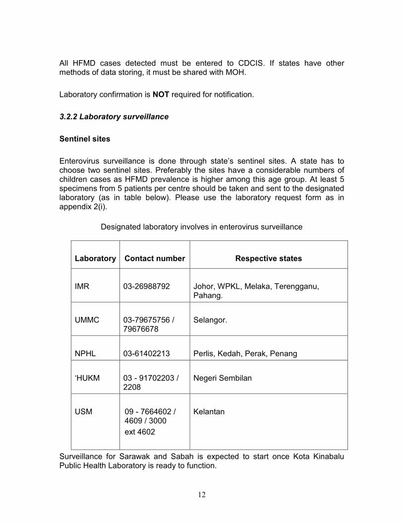
12
All HFMD cases detected must be entered to CDCIS. If states have other methods of data storing, it must be shared with MOH.
Laboratory confirmation is NOT required for notification.
3.2.2 Laboratory surveillance
Sentinel sites
Enterovirus surveillance is done through state’s sentinel sites. A state has to choose two sentinel sites. Preferably the sites have a considerable numbers of children cases as HFMD prevalence is higher among this age group. At least 5 specimens from 5 patients per centre should be taken and sent to the designated laboratory (as in table below). Please use the laboratory request form as in appendix 2(i).
Designated laboratory involves in enterovirus surveillance
Laboratory
Contact number
Respective states
IMR
03-26988792
Johor, WPKL, Melaka, Terengganu, Pahang.
UMMC
03-79675756 / 79676678
Selangor.
NPHL
03-61402213
Perlis, Kedah, Perak, Penang
‘HUKM
03 - 91702203 / 2208
Negeri Sembilan
USM
09 - 7664602 / 4609 / 3000
ext 4602
Kelantan
Surveillance for Sarawak and Sabah is expected to start once Kota Kinabalu Public Health Laboratory is ready to function.

13
Clinical Specimen Collection, Handling And Transportation
Patient
Swab For viral culture
Blood serology CSF
2 ml - in plain sterile
tube
1 ml -in plain sterile
tube
Mouth ulcer Rectal - wet swab with sterile saline
Vesicle - basal
scrapping
Viral transport media
Smear on slide
Send clinical samples to respective laboratory within 24 hours of collection at 4 oC
(on ice). DO NOT STORE AT -20 oC

14
3.4 Flow of surveillance data
Notification data should be entered in CDCIS as and when there is HFMD case. Other sources of data must be shared with Disease Control Division. Data must have the variables listed in appendix 1.
Laboratory results for enterovirus surveillance must be shared with respective states and Disease Control Division using the suggested format in appendix 2(ii).
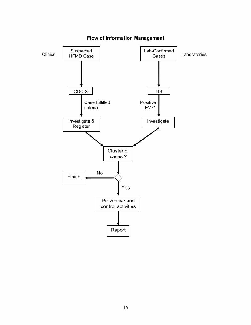
15
Flow of Information Management
Yes
Suspected HFMD Case
CDCIS
Case fulfilled criteria
Investigate & Register
Lab-Confirmed Cases
LIS
Positive EV71
Investigate
Clinics Laboratories
Cluster of cases ?
Finish
Preventive and control activities
Report
No

16
5. MANAGEMENT OF SPORADIC CASES
5.1 Definitions of sporadic case:
A single case of HFMD in the absence of previous known close contact with another case.
5.2 The public health measures
5.2.1 Case Investigation
Case investigation is required following the criteria below:
a. every notified HFMD case that fulfilled any one of the criteria below:
i. case is admitted to hospital ii. case died. iii. case aged 6 years and below and goes to any pre-schools or nurseries.
The first two criteria indicate severity of illness and it is a proxy of possible EV71 infection. Meanwhile the third criterion is important in preventing spread of illness among the pre-school children (high risk group) and reducing public anxiety.
b. every case with positive EV71 laboratory result. Investigation should be carried out to determine the course, source and severity of infection. Enquiries shall be made into;
1. Particulars of the person affected
2. Clinical signs and symptoms with date of onset. In addition, enquiries shall be elicited for associated complications e.g. seizures, weakness of limbs, staring episodes, drowsiness, profound lethargy and refusal to play.
Enterovirus infections that are not associated with complications may not have cutaneous manifestations.
3. Duration of illness
4. Type of treatment sought, including details of hospitalization and reason for admission
5. History of travel especially to outbreak area in the past two weeks
6. Contacts with similar illness in an institution (child care centre or kindergarten), family or neighbourhood where relevant.
7. If newborn, antenatal history including maternal history of febrile illness, mode of delivery

17
5.2.2 Incidence management Following steps should be taken to prevent further spread of disease / pathogen:
1. Ensure that the infected children remain away from the institution for at least ten days after onset of symptoms and must be certified free from infection by a registered medical practitioner prior to returning to school.
2. Active case detection shall be carried out among contacts in the institution and family, and all those with illness shall be referred for treatment at the nearest health clinics.
3. Swabs from oral ulcers and vesicular lesions on the hands and feet shall be collected for virus isolation at identified laboratories (IMR or NPHL). If these ulcers and vesicles are no longer present, stool specimens shall be sent. Always consult pathologist for specimens taking, handling and transportation.
4. Health education of supervisors of institutions on the disease, mode of transmission, importance of good personal hygiene and isolation of infected children shall be carried out.
5. Principals, teachers and supervisors shall be alerted to look out for children with fever, rash / blisters on palms and soles and to isolate them immediately. Screening before coming to class is recommended (please refer Buku Panduan Latihan Taska dan Prasekolah).
6. If there are two or case in the premise, advice the premise to be closed to cut disease transmission. It should be closed for two incubation period (10 days) from the date of onset of the last case.
7. Supervision on disinfection procedures shall be given to the premise operators. Articles such as toys contaminated by infected cases are disinfected with 0.5% sodium hypochlorite solutions (please refer Buku Panduan Latihan Taska dan Prasekolah).

18
6. MANAGEMENT OF OUTBREAK
6.1 Definitions of outbreak
The occurrence of two or more cases in the same locality within the incubation period. Organisation based outbreak – two or more cases with onset in two weeks interval in a group which makes epidemiological sense.
6.2 Identification of outbreaks
Surveillance data on HFMD cases should be reviewed on continuous basis to identify cases and detects outbreaks.
6.3 Risk factors for outbreaks
Risk factors for outbreaks are not completely understood. A combination of conditions (environment, host and organism) is necessary for an epidemic to occur. However large HFMD outbreaks have been associated with EV71 infection.
6.4 Management of outbreaks
When a suspected outbreak is reported, it is important to determine rapidly whether a true outbreak is occurring based on the definition above. Similar steps as in sporadic case management should be carried out together with following measures;
1. Ensure that the infected children remain away from the institution for at least ten days after onset of symptoms and must be certified free from infection by a registered medical practitioner prior to returning to school.
2. An assessment of the number of cases and susceptible population involved shall be made, and the overall attack rate and age-specific attack rate computed.
3. Active case detection shall be carried out among contacts in the institution and family, and all those with illness shall be referred for treatment at the nearest health clinics.
4. Where a case is observed or suspected to have unusual signs and symptoms e.g. seizures, weakness of limbs, profound lethargy, refusal to play, the case shall be immediately referred to hospital.
5. Swabs from oral ulcers and vesicular lesions on the hands and feet shall be collected for virus isolation at identified laboratories (IMR or NPHL). If these ulcers and vesicles are no longer present, stool specimens shall be sent. Always consult pathologist for specimens taking, handling and transportation.

19
6. Principals, teachers and supervisors shall be alerted to look out for children with fever, rash / blisters on palms and soles and to isolate them immediately. Screening before coming to class is recommended (please refer Buku Panduan Latihan Taska dan Prasekolah).
7. Health education of supervisors of institutions on the disease, mode of transmission, importance of good personal hygiene and isolation of infected children shall be carried out.
8. Close the premise closed for two incubation period (10 days) from the date of onset of the last case. Supervision on disinfection procedures shall be given to the premise operators.
In addition the investigating team shall also look into environmental sanitation and hygiene to ensure that:
a. Articles such as toys contaminated by infected cases are disinfected with 0.5% sodium hypochlorite solutions (please refer Buku Panduan Latihan Taska dan Prasekolah).
b. A high standard of food and personal hygiene is maintained by the institution involved which include proper waste and diapers disposal.
c. The approved enrolment of Department of Social Welfare (JKM), Ministry of Education, Department of National Integration and other relevant parties, where applicable, and shall notify the relevant authorities if regulations not complied.
If the outbreak occurs in primary schools;
i. Ensure that the infected students remain away from the institution for at least ten days.
ii. Health education to the students on the disease, mode of transmission, importance of good personal hygiene.
iii. Principals, teachers and supervisors shall be look for children with fever, rash / blisters on palms and soles and to isolate them immediately. Screening before coming to class is recommended (please refer Buku Panduan Latihan Taska dan Prasekolah).
iv. If closure is necessary, just closed the affected class. Closure of the whole school is unnecessary as HFMD in older children is usually very mild and so far no complication has been documented from this agegroup.

20
6.5 Documentation and review
Disease activity and emergency response should be adequately documented so that further outbreaks can be handled more effectively. A summary report should be prepared within a month from the last date of case; and distributed to all the stakeholders for feedback. Format of report, dissemination and tabling of report is discussed in more details in Infectious Disease Outbreak Rapid Response Manual.12
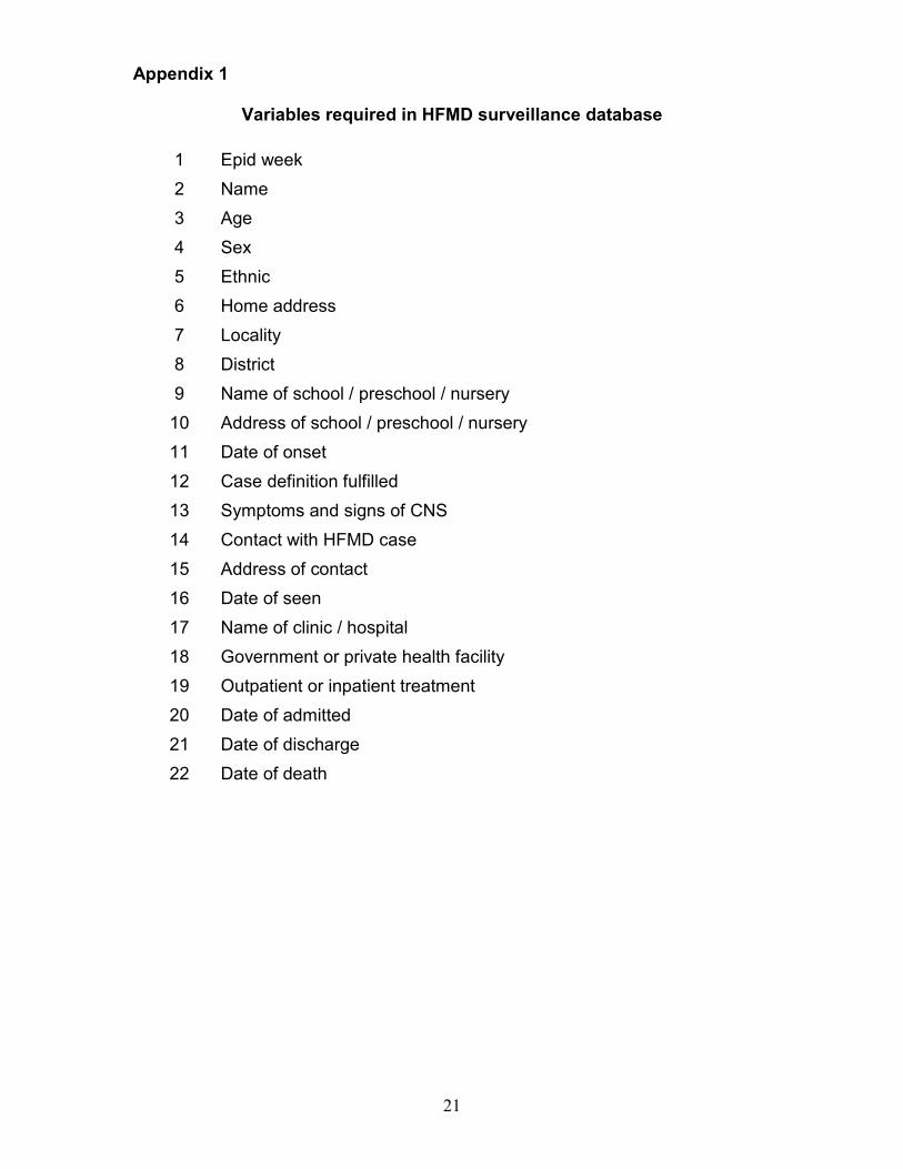
21
Appendix 1
Variables required in HFMD surveillance database
1 Epid week
2 Name
3 Age
4 Sex
5 Ethnic
6 Home address
7 Locality
8 District
9 Name of school / preschool / nursery
10 Address of school / preschool / nursery
11 Date of onset
12 Case definition fulfilled
13 Symptoms and signs of CNS
14 Contact with HFMD case
15 Address of contact
16 Date of seen
17 Name of clinic / hospital
18 Government or private health facility
19 Outpatient or inpatient treatment
20 Date of admitted
21 Date of discharge
22 Date of death
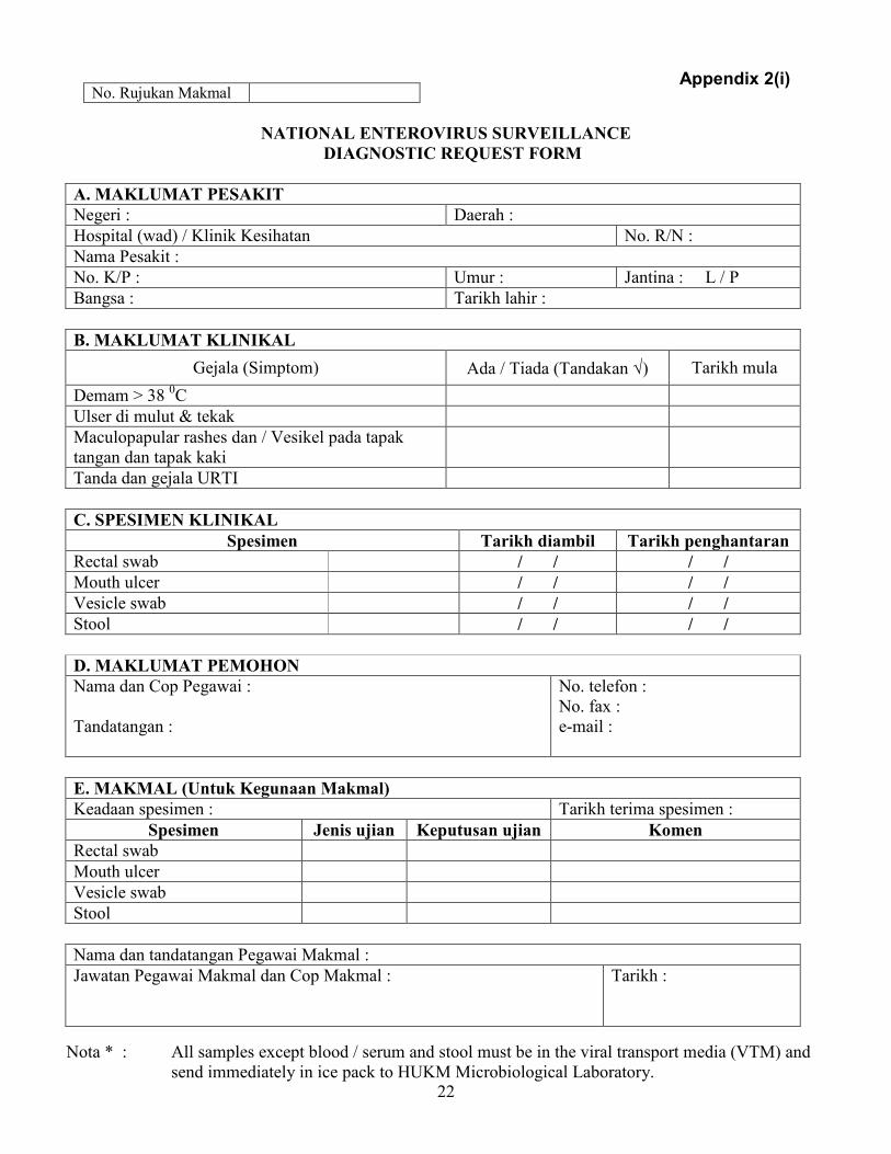
22
No. Rujukan Makmal
NATIONAL ENTEROVIRUS SURVEILLANCE
DIAGNOSTIC REQUEST FORM
A. MAKLUMAT PESAKIT
Negeri : Daerah :
Hospital (wad) / Klinik Kesihatan No. R/N :
Nama Pesakit :
No. K/P : Umur : Jantina : L / P
Bangsa : Tarikh lahir :
B. MAKLUMAT KLINIKAL
Gejala (Simptom) Ada / Tiada (Tandakan √) Tarikh mula
Demam > 38 0C
Ulser di mulut & tekak
Maculopapular rashes dan / Vesikel pada tapak
tangan dan tapak kaki
Tanda dan gejala URTI
C. SPESIMEN KLINIKAL
Spesimen Tarikh diambil Tarikh penghantaran
Rectal swab / / / /
Mouth ulcer / / / /
Vesicle swab / / / /
Stool / / / /
D. MAKLUMAT PEMOHON
Nama dan Cop Pegawai :
Tandatangan :
No. telefon :
No. fax :
e-mail :
E. MAKMAL (Untuk Kegunaan Makmal)
Keadaan spesimen : Tarikh terima spesimen :
Spesimen Jenis ujian Keputusan ujian Komen
Rectal swab
Mouth ulcer
Vesicle swab
Stool
Nama dan tandatangan Pegawai Makmal :
Jawatan Pegawai Makmal dan Cop Makmal :
Tarikh :
Nota * : All samples except blood / serum and stool must be in the viral transport media (VTM) and
send immediately in ice pack to HUKM Microbiological Laboratory.
Appendix 2(i)

23
Appendix 2(ii)
Variables Required for Enterovirus Surveillance Database
Name of Centre / Laboratory: _______________
Result
No Date
Sample Received
Date Sample Collected
LN RN Name Ethnic Age Sex Source
Case
diagnosis
Type of
spec IF Isolation PCR

24
BKP / S / HFMD (H) Ver 2 / 2007
BORANG SIASATAN PESAKIT DI WAD
PENYAKIT TANGAN, KAKI DAN MULUT (HFMD)
BAHAGIAN KAWALAN PENYAKIT, KEMENTERIAN KESIHATAN MALAYSIA
BAHAGIAN 1
Tarikh masuk wad: Masa:
Tarikh discaj: Masa:
Tarikh kematian Masa:
A. PERIHAL PESAKIT
1. Nama pesakit: 2. Tarikh lahir:
3. Nama ibu: 4. No K/P ibu:
5. Alamat rumah: 6. No telefon: (rumah)
(H/P)
Kedudukan rumah (GPS): X : _________________ Y : __________________
7. Jantina: Lelaki 8. Kumpulan etnik
Perempuan Melayu Kadazan/Dusun
Cina Murut
9. Kewarganegaraan: Malaysia India Bajau
Warga asing Asli semenanjung Melanau
Bidayuh Iban
Lain-lain pribumi Lain-lain (nyatakan)
Sabah/ Sarawak
10. Menghadiri sekolah / taska
Ya
Taska / Nurseri
Prasekolah
Sekolah
Nyatakan nama: ________________________________
Tidak
B. MAKLUMAT KLINIKAL
1. Tarikh mula sakit (onset)
2. Tanda dan gejala: Ya Tidak
Riwayat demam

25
Ulser mulut
Ruam (rash) tapak tangan / kaki
vesicles di tapak tangan / kaki
3. Kompliksi Ya Tidak
Aseptic meningitis
Myocarditis
Encephalitis
Acute flaccid paralysis
Jika ya, sila nyatakan bahagian anggota: ____________________
Kematian
C. MAKLUMAT UJIAN MAKMAL
SAMPEL Tarikh diambil Keputusan Tarikh keputusan
Throat swab
Rectal swab
Vesicle swab
Stool
Blood
CSF
Biopsy _________
(Nyatakan tisu)
D. MAKLUMAT PEGAWAI YANG MERAWAT
1. Nama Pakar / Pegawai Perubatan: __________________________ Tandatangan: ____________
2. Nama Wad: _____________________
3. Nama hospital: __________________________ Tarikh notifikasi: _________

26
7. REFERENCES
1. Melnick JL. Enteroviruses: polioviruses, coxsackieviruses, echoviruses, and newer enteroviruse. In: Fields BN, Knipe DM, Howley PM, Chandlock RM, Melnick JL, Monah TP, et al., editors. Field’s virology. 3 rd ed. Philadelphia: Lippincott-Raven Publishers; 1996. p. 655-712.
2. Zeichhardt H, Grunert H. Enteroviruses: Polioviruses, Coxcaskie viruses,
echoviruses and Enteroviruses 68-71. In: Cohen J, Powderly WG, editors. Infectious diseases. 2nd edition. Mosby ; 2004. p.1993-2003.
3. Read RC. Orocervical and esophageal infection. In: Cohen J, Powderly WG,
editors. Infectious diseases. 2nd edition. Mosby ; 2004. p. 463. 4 . Wright HT, Landing BH, Lennette EH et al. Fatal Coxsackievirus A16
infection. The Pediatric Infectious Disease Journal. 2004; 23(3): 275. 5 . Goldberg MF, McAdams AJ. Myocarditis possibly due to coxsackie group A,
type 16, virus. J Pediatr, 1963; 268: 1041-4. 6. Wang CY, Lu FL, Wu MW. Fatal coxcaskievirus A16 infection. The Pediatric
Infectious Disease Journal 2004;23(3),275. 7. Chang LY, Lin TY, Huang YC et al . Comparison of enterovirus 71 and
coxsackievirus A16 clinical illnesses during the Taiwan enterovirus epidemic, 1998. The Pediatric Infectious Disease Journal 1999; 18(12): 1092-1096.
8. Ho M, Chen ER, Hsu KH, Twu SJ, et al. An epidemic of enterovirus 71
infection in Taiwan. The New England Journal of Medicine, 1999; 341(13): 929-935.
9 . Podin Y, Gias ELM, Ong F, et al. Sentinel Surveillance for human enterovirus
71 in Sarawak, Malaysia: lessons from the first 7 years. BMC Public Health 2006; 6: 180
. 10 . Huang CC, Liu CC, Chang YC et al. Neurologic complications in children
with enterovirus 71. The New England Journal of Medicine, 1999;341(13): 936-943.
11 Wu JM, Wang JN, Tsai YC et al. Cardiopulmonary manifestations of
fulminate enterovirus 71 infection. Pediatrics, 2002; 109:e26 12. Disease Control Division, Ministry of Health. Infectious Diseases Outbreak
Rapid Response Manual. Ministry of Health. Kuala Lumpur, 2003.
27
CONTRIBUTORS State Epidemiologists 2006 – 2007
1. Pulau Pinang Dr. Saraswathi Bina Rai
2. Kedah Dr. Uma Salamah Abdul Kadir
3. Perlis Dr. Zulhizzam Hj Abdullah
4. Perak Dr. Asiah Ayob
Dr. Zamri Md Ali
5. Selangor Dr. Prema Rajendran
Dr. B. Venugopalan
6. WPKL Dr. Norbizura Abdul Hamid
7. N. Sembilan Dr. Zulfa Vety Dol Bakri
8. Melaka Dr. Hashimah Hasan
9. Johor Dr. Shaharom Nor Azian Che Mat Din
Dr. Fatimah Othman
10. Pahang Dr. Shaari Ngadiman
Dr. Adibah Hani Harun
11. Terengganu Dr. Anita Surani Abdul Shukor
Dr. Balkis Ab Karim
12. Kelantan Dr. Hani Mat Hussin
13. Sarawak Dr. Ooi Choo Huck
Dr. Kamaliah Mohd Nor
Dr. Rohani Mat Bah
14. Sabah Dr. Jenarun Jelip
Dr. Kristina Rundi
15. Bahagian Kawalan Dr. Rohani Jahis
Penyakit Dr. Husnina Ibrahim
Dr. Nazarudin Safian
Dr. Izuna Mudla Mohamad Ghazali
Dr. Zairina Abdul Rahman
16. Laboratory Dr. Chua Kaw Bing