Embed Size (px)
Citation preview
Hindawi Publishing CorporationInternational Journal of Cell BiologyVolume 2010, Article ID 758230, 9 pagesdoi:10.1155/2010/758230
Review Article
Conserved Molecular Mechanisms Underlying Homeostasis ofthe Golgi Complex
Cathal Wilson1 and Antonella Ragnini-Wilson1, 2
1 Department of Translational and Cellular Pharmacology, Consorzio Mario Negri Sud, Santa Maria Imbaro, 66030 Chieti, Italy2 Department of Biology, Tor Vergata University of Rome, 00133 Rome, Italy
Correspondence should be addressed to Cathal Wilson, [email protected]
Received 6 June 2010; Revised 30 July 2010; Accepted 19 August 2010
Academic Editor: Jerome Rattner
Copyright © 2010 C. Wilson and A. Ragnini-Wilson. This is an open access article distributed under the Creative CommonsAttribution License, which permits unrestricted use, distribution, and reproduction in any medium, provided the original work isproperly cited.
The Golgi complex performs a central function in the secretory pathway in the sorting and sequential processing of a large numberof proteins destined for other endomembrane organelles, the plasma membrane, or secretion from the cell, in addition to lipidmetabolism and signaling. The Golgi apparatus can be regarded as a self-organizing system that maintains a relatively stablemorphofunctional organization in the face of an enormous flux of lipids and proteins. A large number of the molecular playersthat operate in these processes have been identified, their functions and interactions defined, but there is still debate about manyaspects that regulate protein trafficking and, in particular, the maintenance of these highly dynamic structures and processes.Here, we consider how an evolutionarily conserved underlying mechanism based on retrograde trafficking that uses lipids, COPI,SNAREs, and tethers could maintain such a homeodynamic system.
1. Introduction
Despite the ancient origin of the Golgi and the differencesin its structure across species, there is a striking conservationof a number of molecular machineries and principles thatappear to operate in intra-Golgi trafficking. We soughtto use these observations as a starting point from whichto discuss how the maintenance of Golgi structure mightbe intrinsically related with the conservation of the basicmolecular machineries that regulate intra-Golgi trafficking.
In most organisms the Golgi apparatus is composed ofa series of flattened, membrane-bounded sacks (cisternae)arranged in a cis-to-trans fashion to form a stack. Thesestacks are laterally linked to form a ribbon-like membranesystem in mammalian cells [1] but this ribbon-like structuredoes not link the Golgi stacks in plants and Drosophila [2, 3].In the yeast Saccharomyces cerevisiae the Golgi compartmentsare not arranged as a stack at all but exist as separate scatteredcompartments in the cell [4, 5] while in some developmentalstages of Drosophila no stacks are present [3]. Yet, the basicfunctions of the Golgi in transport and sorting appear to beconserved across species, so neither the stacked structure nor
the ribbon can be considered as fundamental for the basicfunctions of the Golgi apparatus in transport and sorting ofsecretory cargo molecules.
In addition, it is possible to argue that ER-to-Golgitransport and the COPII complex, which is required forcargo selection and packaging at the ER [6], is not part ofthe self-organizing system per se. Many, but not all, Golgi-associated proteins recycle through the ER and are thenreexported back to the Golgi in a continuous cycle [7–9].Therefore ER-to-Golgi transport is required for constructinga Golgi, but only in the sense that it supplies some of theraw materials but is not part of the underlying mechanismthat maintains the peculiar membrane organization of theGolgi. Therefore, here we do not consider COPII, whichhas been extensively reviewed elsewhere [10, 11], as part ofthe mechanism that maintains the homeostasis of the Golgistructure.
The Golgi complex can be considered a self-organizingsystem [12] where a dynamic equilibrium is maintainedthrough multiple molecular interactions. Under steady stateconditions the Golgi appears as a stable structure that wasoriginally proposed to be a series of relatively stable cisternae
2 International Journal of Cell Biology
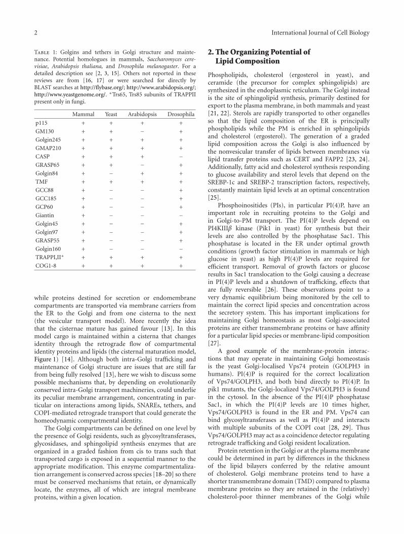
Table 1: Golgins and tethers in Golgi structure and mainte-nance. Potential homologues in mammals, Saccharomyces cere-visiae, Arabidopsis thaliana, and Drosophila melanogaster. For adetailed description see [2, 3, 15]. Others not reported in thesereviews are from [16, 17] or were searched for directly byBLAST searches at http://flybase.org/; http://www.arabidopsis.org/;http://www.yeastgenome.org/. ∗Trs65, Trs85 subunits of TRAPPIIpresent only in fungi.
Mammal Yeast Arabidopsis Drosophila
p115 + + + +
GM130 + + − +
Golgin245 + + + +
GMAP210 + + + +
CASP + + + −GRASP65 + + − +
Golgin84 + − + +
TMF + + + +
GCC88 + − − +
GCC185 + − − +
GCP60 + − − +
Giantin + − − −Golgin45 + − − +
Golgin97 + − − +
GRASP55 + − − +
Golgin160 + − − −TRAPPI,II∗ + + + +
COG1-8 + + + +
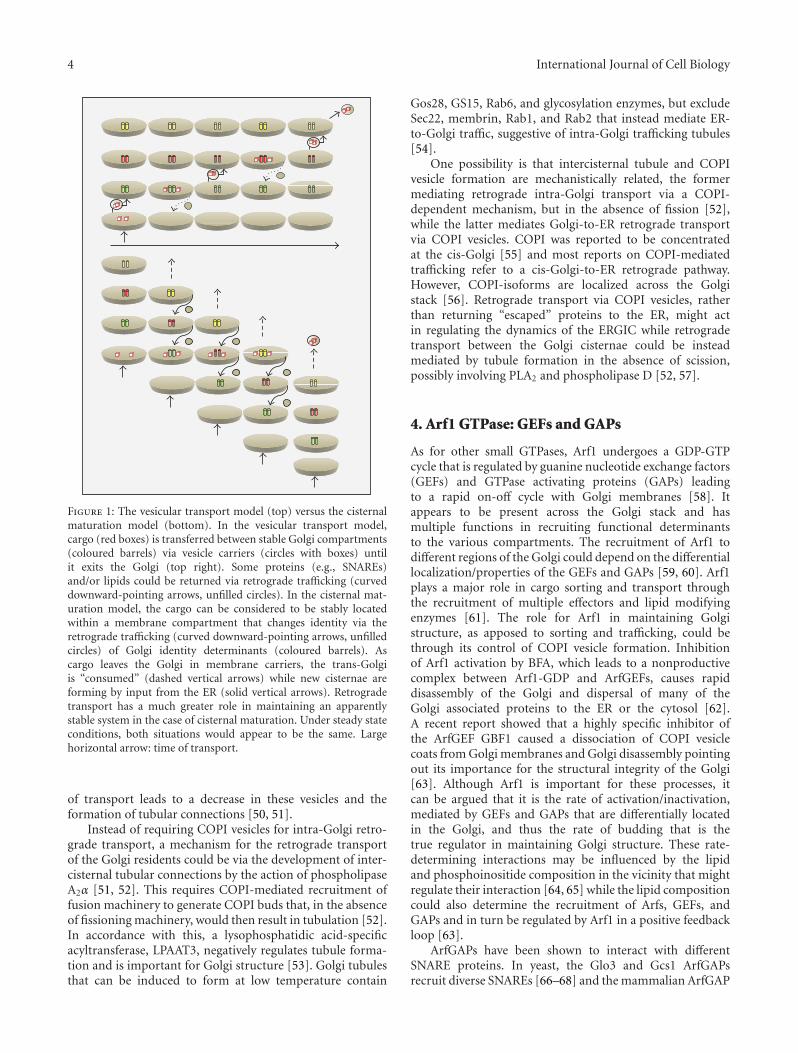
while proteins destined for secretion or endomembranecompartments are transported via membrane carriers fromthe ER to the Golgi and from one cisterna to the next(the vesicular transport model). More recently the ideathat the cisternae mature has gained favour [13]. In thismodel cargo is maintained within a cisterna that changesidentity through the retrograde flow of compartmentalidentity proteins and lipids (the cisternal maturation model,Figure 1) [14]. Although both intra-Golgi trafficking andmaintenance of Golgi structure are issues that are still farfrom being fully resolved [13], here we wish to discuss somepossible mechanisms that, by depending on evolutionarilyconserved intra-Golgi transport machineries, could underlieits peculiar membrane arrangement, concentrating in par-ticular on interactions among lipids, SNAREs, tethers, andCOPI-mediated retrograde transport that could generate thehomeodynamic compartmental identity.
The Golgi compartments can be defined on one level bythe presence of Golgi residents, such as glycosyltransferases,glycosidases, and sphingolipid synthesis enzymes that areorganized in a graded fashion from cis to trans such thattransported cargo is exposed in a sequential manner to theappropriate modification. This enzyme compartmentaliza-tion arrangement is conserved across species [18–20] so theremust be conserved mechanisms that retain, or dynamicallylocate, the enzymes, all of which are integral membraneproteins, within a given location.
2. The Organizing Potential ofLipid Composition
Phospholipids, cholesterol (ergosterol in yeast), andceramide (the precursor for complex sphingolipids) aresynthesized in the endoplasmic reticulum. The Golgi insteadis the site of sphingolipid synthesis, primarily destined forexport to the plasma membrane, in both mammals and yeast[21, 22]. Sterols are rapidly transported to other organellesso that the lipid composition of the ER is principallyphospholipids while the PM is enriched in sphingolipidsand cholesterol (ergosterol). The generation of a gradedlipid composition across the Golgi is also influenced bythe nonvesicular transfer of lipids between membranes vialipid transfer proteins such as CERT and FAPP2 [23, 24].Additionally, fatty acid and cholesterol synthesis respondingto glucose availability and sterol levels that depend on theSREBP-1c and SREBP-2 transcription factors, respectively,constantly maintain lipid levels at an optimal concentration[25].
Phosphoinositides (PIs), in particular PI(4)P, have animportant role in recruiting proteins to the Golgi andin Golgi-to-PM transport. The PI(4)P levels depend onPI4KIIIβ kinase (Pik1 in yeast) for synthesis but theirlevels are also controlled by the phosphatase Sac1. Thisphosphatase is located in the ER under optimal growthconditions (growth factor stimulation in mammals or highglucose in yeast) as high PI(4)P levels are required forefficient transport. Removal of growth factors or glucoseresults in Sac1 translocation to the Golgi causing a decreasein PI(4)P levels and a shutdown of trafficking, effects thatare fully reversible [26]. These observations point to avery dynamic equilibrium being monitored by the cell tomaintain the correct lipid species and concentration acrossthe secretory system. This has important implications formaintaining Golgi homeostasis as most Golgi-associatedproteins are either transmembrane proteins or have affinityfor a particular lipid species or membrane-lipid composition[27].
A good example of the membrane-protein interac-tions that may operate in maintaining Golgi homeostasisis the yeast Golgi-localised Vps74 protein (GOLPH3 inhumans). PI(4)P is required for the correct localizationof Vps74/GOLPH3, and both bind directly to PI(4)P. Inpik1 mutants, the Golgi-localized Vps74/GOLPH3 is foundin the cytosol. In the absence of the PI(4)P phosphataseSac1, in which the PI(4)P levels are 10 times higher,Vps74/GOLPH3 is found in the ER and PM. Vps74 canbind glycosyltransferases as well as PI(4)P and interactswith multiple subunits of the COPI coat [28, 29]. ThusVps74/GOLPH3 may act as a coincidence detector regulatingretrograde trafficking and Golgi resident localization.
Protein retention in the Golgi or at the plasma membranecould be determined in part by differences in the thicknessof the lipid bilayers conferred by the relative amountof cholesterol. Golgi membrane proteins tend to have ashorter transmembrane domain (TMD) compared to plasmamembrane proteins so they are retained in the (relatively)cholesterol-poor thinner membranes of the Golgi while
International Journal of Cell Biology 3
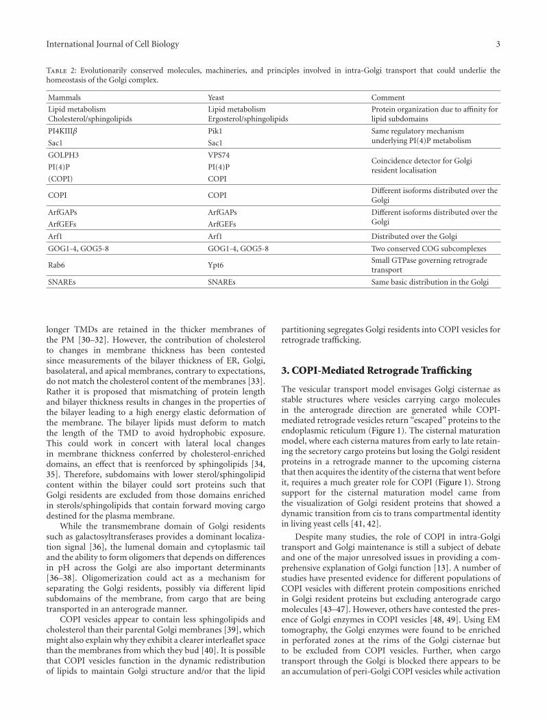
Table 2: Evolutionarily conserved molecules, machineries, and principles involved in intra-Golgi transport that could underlie thehomeostasis of the Golgi complex.
Mammals Yeast Comment
Lipid metabolismCholesterol/sphingolipids
Lipid metabolismErgosterol/sphingolipids
Protein organization due to affinity forlipid subdomains
PI4KIIIβ Pik1 Same regulatory mechanismunderlying PI(4)P metabolismSac1 Sac1
GOLPH3 VPS74Coincidence detector for Golgiresident localisationPI(4)P PI(4)P
(COPI) COPI
COPI COPIDifferent isoforms distributed over theGolgi
ArfGAPs ArfGAPs Different isoforms distributed over theGolgiArfGEFs ArfGEFs
Arf1 Arf1 Distributed over the Golgi
GOG1-4, GOG5-8 GOG1-4, GOG5-8 Two conserved COG subcomplexes
Rab6 Ypt6Small GTPase governing retrogradetransport
SNAREs SNAREs Same basic distribution in the Golgi
longer TMDs are retained in the thicker membranes ofthe PM [30–32]. However, the contribution of cholesterolto changes in membrane thickness has been contestedsince measurements of the bilayer thickness of ER, Golgi,basolateral, and apical membranes, contrary to expectations,do not match the cholesterol content of the membranes [33].Rather it is proposed that mismatching of protein lengthand bilayer thickness results in changes in the properties ofthe bilayer leading to a high energy elastic deformation ofthe membrane. The bilayer lipids must deform to matchthe length of the TMD to avoid hydrophobic exposure.This could work in concert with lateral local changesin membrane thickness conferred by cholesterol-enricheddomains, an effect that is reenforced by sphingolipids [34,35]. Therefore, subdomains with lower sterol/sphingolipidcontent within the bilayer could sort proteins such thatGolgi residents are excluded from those domains enrichedin sterols/sphingolipids that contain forward moving cargodestined for the plasma membrane.
While the transmembrane domain of Golgi residentssuch as galactosyltransferases provides a dominant localiza-tion signal [36], the lumenal domain and cytoplasmic tailand the ability to form oligomers that depends on differencesin pH across the Golgi are also important determinants[36–38]. Oligomerization could act as a mechanism forseparating the Golgi residents, possibly via different lipidsubdomains of the membrane, from cargo that are beingtransported in an anterograde manner.
COPI vesicles appear to contain less sphingolipids andcholesterol than their parental Golgi membranes [39], whichmight also explain why they exhibit a clearer interleaflet spacethan the membranes from which they bud [40]. It is possiblethat COPI vesicles function in the dynamic redistributionof lipids to maintain Golgi structure and/or that the lipid
partitioning segregates Golgi residents into COPI vesicles forretrograde trafficking.
3. COPI-Mediated Retrograde Trafficking
The vesicular transport model envisages Golgi cisternae asstable structures where vesicles carrying cargo moleculesin the anterograde direction are generated while COPI-mediated retrograde vesicles return “escaped” proteins to theendoplasmic reticulum (Figure 1). The cisternal maturationmodel, where each cisterna matures from early to late retain-ing the secretory cargo proteins but losing the Golgi residentproteins in a retrograde manner to the upcoming cisternathat then acquires the identity of the cisterna that went beforeit, requires a much greater role for COPI (Figure 1). Strongsupport for the cisternal maturation model came fromthe visualization of Golgi resident proteins that showed adynamic transition from cis to trans compartmental identityin living yeast cells [41, 42].
Despite many studies, the role of COPI in intra-Golgitransport and Golgi maintenance is still a subject of debateand one of the major unresolved issues in providing a com-prehensive explanation of Golgi function [13]. A number ofstudies have presented evidence for different populations ofCOPI vesicles with different protein compositions enrichedin Golgi resident proteins but excluding anterograde cargomolecules [43–47]. However, others have contested the pres-ence of Golgi enzymes in COPI vesicles [48, 49]. Using EMtomography, the Golgi enzymes were found to be enrichedin perforated zones at the rims of the Golgi cisternae butto be excluded from COPI vesicles. Further, when cargotransport through the Golgi is blocked there appears to bean accumulation of peri-Golgi COPI vesicles while activation
4 International Journal of Cell Biology
Figure 1: The vesicular transport model (top) versus the cisternalmaturation model (bottom). In the vesicular transport model,cargo (red boxes) is transferred between stable Golgi compartments(coloured barrels) via vesicle carriers (circles with boxes) untilit exits the Golgi (top right). Some proteins (e.g., SNAREs)and/or lipids could be returned via retrograde trafficking (curveddownward-pointing arrows, unfilled circles). In the cisternal mat-uration model, the cargo can be considered to be stably locatedwithin a membrane compartment that changes identity via theretrograde trafficking (curved downward-pointing arrows, unfilledcircles) of Golgi identity determinants (coloured barrels). Ascargo leaves the Golgi in membrane carriers, the trans-Golgiis “consumed” (dashed vertical arrows) while new cisternae areforming by input from the ER (solid vertical arrows). Retrogradetransport has a much greater role in maintaining an apparentlystable system in the case of cisternal maturation. Under steady stateconditions, both situations would appear to be the same. Largehorizontal arrow: time of transport.
of transport leads to a decrease in these vesicles and theformation of tubular connections [50, 51].
Instead of requiring COPI vesicles for intra-Golgi retro-grade transport, a mechanism for the retrograde transportof the Golgi residents could be via the development of inter-cisternal tubular connections by the action of phospholipaseA2α [51, 52]. This requires COPI-mediated recruitment offusion machinery to generate COPI buds that, in the absenceof fissioning machinery, would then result in tubulation [52].In accordance with this, a lysophosphatidic acid-specificacyltransferase, LPAAT3, negatively regulates tubule forma-tion and is important for Golgi structure [53]. Golgi tubulesthat can be induced to form at low temperature contain
Gos28, GS15, Rab6, and glycosylation enzymes, but excludeSec22, membrin, Rab1, and Rab2 that instead mediate ER-to-Golgi traffic, suggestive of intra-Golgi trafficking tubules[54].
One possibility is that intercisternal tubule and COPIvesicle formation are mechanistically related, the formermediating retrograde intra-Golgi transport via a COPI-dependent mechanism, but in the absence of fission [52],while the latter mediates Golgi-to-ER retrograde transportvia COPI vesicles. COPI was reported to be concentratedat the cis-Golgi [55] and most reports on COPI-mediatedtrafficking refer to a cis-Golgi-to-ER retrograde pathway.However, COPI-isoforms are localized across the Golgistack [56]. Retrograde transport via COPI vesicles, ratherthan returning “escaped” proteins to the ER, might actin regulating the dynamics of the ERGIC while retrogradetransport between the Golgi cisternae could be insteadmediated by tubule formation in the absence of scission,possibly involving PLA2 and phospholipase D [52, 57].
4. Arf1 GTPase: GEFs and GAPs
As for other small GTPases, Arf1 undergoes a GDP-GTPcycle that is regulated by guanine nucleotide exchange factors(GEFs) and GTPase activating proteins (GAPs) leadingto a rapid on-off cycle with Golgi membranes [58]. Itappears to be present across the Golgi stack and hasmultiple functions in recruiting functional determinantsto the various compartments. The recruitment of Arf1 todifferent regions of the Golgi could depend on the differentiallocalization/properties of the GEFs and GAPs [59, 60]. Arf1plays a major role in cargo sorting and transport throughthe recruitment of multiple effectors and lipid modifyingenzymes [61]. The role for Arf1 in maintaining Golgistructure, as apposed to sorting and trafficking, could bethrough its control of COPI vesicle formation. Inhibitionof Arf1 activation by BFA, which leads to a nonproductivecomplex between Arf1-GDP and ArfGEFs, causes rapiddisassembly of the Golgi and dispersal of many of theGolgi associated proteins to the ER or the cytosol [62].A recent report showed that a highly specific inhibitor ofthe ArfGEF GBF1 caused a dissociation of COPI vesiclecoats from Golgi membranes and Golgi disassembly pointingout its importance for the structural integrity of the Golgi[63]. Although Arf1 is important for these processes, itcan be argued that it is the rate of activation/inactivation,mediated by GEFs and GAPs that are differentially locatedin the Golgi, and thus the rate of budding that is thetrue regulator in maintaining Golgi structure. These rate-determining interactions may be influenced by the lipidand phosphoinositide composition in the vicinity that mightregulate their interaction [64, 65] while the lipid compositioncould also determine the recruitment of Arfs, GEFs, andGAPs and in turn be regulated by Arf1 in a positive feedbackloop [63].
ArfGAPs have been shown to interact with differentSNARE proteins. In yeast, the Glo3 and Gcs1 ArfGAPsrecruit diverse SNAREs [66–68] and the mammalian ArfGAP
International Journal of Cell Biology 5
C a r g o , m o d i fi e d c a r g o
T r a n s
M e d i a l
C i s
E R G I C
E R
R e t r o g r a d e t r a n s p o r t v i a i n t e r c i s t e r n a l t u b u l e
L i p i d s u b d o m a i n s
G o l g i e n z y m e s
C O P I v e s i c l e
S N A R E s
C O G
A r f 1 A r f - G E F
A r f - G A P
EndosomalGolgiinterface(TGN)
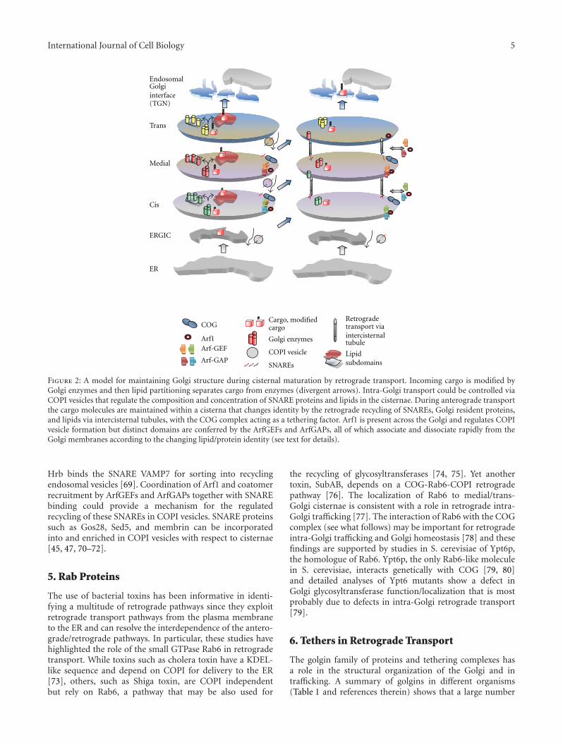
Figure 2: A model for maintaining Golgi structure during cisternal maturation by retrograde transport. Incoming cargo is modified byGolgi enzymes and then lipid partitioning separates cargo from enzymes (divergent arrows). Intra-Golgi transport could be controlled viaCOPI vesicles that regulate the composition and concentration of SNARE proteins and lipids in the cisternae. During anterograde transportthe cargo molecules are maintained within a cisterna that changes identity by the retrograde recycling of SNAREs, Golgi resident proteins,and lipids via intercisternal tubules, with the COG complex acting as a tethering factor. Arf1 is present across the Golgi and regulates COPIvesicle formation but distinct domains are conferred by the ArfGEFs and ArfGAPs, all of which associate and dissociate rapidly from theGolgi membranes according to the changing lipid/protein identity (see text for details).
Hrb binds the SNARE VAMP7 for sorting into recyclingendosomal vesicles [69]. Coordination of Arf1 and coatomerrecruitment by ArfGEFs and ArfGAPs together with SNAREbinding could provide a mechanism for the regulatedrecycling of these SNAREs in COPI vesicles. SNARE proteinssuch as Gos28, Sed5, and membrin can be incorporatedinto and enriched in COPI vesicles with respect to cisternae[45, 47, 70–72].
5. Rab Proteins
The use of bacterial toxins has been informative in identi-fying a multitude of retrograde pathways since they exploitretrograde transport pathways from the plasma membraneto the ER and can resolve the interdependence of the antero-grade/retrograde pathways. In particular, these studies havehighlighted the role of the small GTPase Rab6 in retrogradetransport. While toxins such as cholera toxin have a KDEL-like sequence and depend on COPI for delivery to the ER[73], others, such as Shiga toxin, are COPI independentbut rely on Rab6, a pathway that may be also used for
the recycling of glycosyltransferases [74, 75]. Yet anothertoxin, SubAB, depends on a COG-Rab6-COPI retrogradepathway [76]. The localization of Rab6 to medial/trans-Golgi cisternae is consistent with a role in retrograde intra-Golgi trafficking [77]. The interaction of Rab6 with the COGcomplex (see what follows) may be important for retrogradeintra-Golgi trafficking and Golgi homeostasis [78] and thesefindings are supported by studies in S. cerevisiae of Ypt6p,the homologue of Rab6. Ypt6p, the only Rab6-like moleculein S. cerevisiae, interacts genetically with COG [79, 80]and detailed analyses of Ypt6 mutants show a defect inGolgi glycosyltransferase function/localization that is mostprobably due to defects in intra-Golgi retrograde transport[79].
6. Tethers in Retrograde Transport
The golgin family of proteins and tethering complexes hasa role in the structural organization of the Golgi and intrafficking. A summary of golgins in different organisms(Table 1 and references therein) shows that a large number
6 International Journal of Cell Biology
of them are not present in yeast or plants, suggesting thatthey play a role in Golgi ribbon formation in mammals [81].The Golgi in Drosophila cells do not show ribbon formationbut two stacks are tightly apposed that may represent a“minimal” ribbon that could explain the presence of thesegolgins [3].
Within the present context, the COG (ConservedOligomeric Golgi) complex appears to be of particularrelevance for retrograde intra-Golgi trafficking. This highlyconserved peripheral membrane protein complex is pro-posed to act as a retrograde vesicle-tethering factor in intra-Golgi trafficking [15]. Downregulation of COG function inmammals and yeast results in the mislocalization of residentGolgi glycosyltransferases/glycosidases [82–84] and defectsin the recycling of Golgi proteins [85]. COG subunits showgenetic and physical interactions with intra-Golgi SNAREsand the COPI coat [86] and can bind to the t-SNARESyntaxin5a/Sed5p thus enhancing the stability of intra-GolgiSNARE complexes [87]. Immunogold electron microscopyshowed that the COG1 subunit is localized across the Golgistack on or close to the tips and rims of the Golgi’s cisternaeand in some cases on COPI containing vesicles [88].
7. SNAREs as Generators of Compartments
Soluble N-ethylmaleimide-sensitive factor attachment pro-tein receptor (SNARE) proteins are essential for the fusionof transport vesicles, or a donor membrane compartment,with an acceptor membrane. A complex of SNARE proteinsthat is localized in opposing membranes drives membranefusion. The four SNARE proteins that contribute to theformation of the complex direct different trafficking steps atdifferent locations in the cell. By and large, the combinationsof the different SNARE proteins at different locations withinthe main intracellular trafficking pathways are conservedbetween organisms [89]. Interestingly, computer simulationmodeling has suggested that this differential localizationof the SNAREs could contribute to the generation andmaintenance of stable nonidentical Golgi compartments. Adifferential affinity of a coat protein for one set of SNAREsover another could lead to the concentration of theseSNAREs and this, together with the selective fusion with theircognate SNAREs, was sufficient not only to generate non-identical Golgi compartments de novo but also to maintainthe steady state system of non-identical Golgi compartments[90, 91]. As mentioned above, this could be controlled viaCOPI vesicles and/or tubular connections.
8. Conclusions
The Golgi apparatus shows characteristics consistent withit being a self-organizing system. Such a system relies onmultiple interdependent interactions to maintain it in ahomeodynamic state. In relatively simple systems, such asthe cytoskeleton, it has been possible to describe the factorsthat regulate the self-organization [12]. However, in a systemas complicated as the Golgi it is difficult to arrive at asimple underlying molecular mechanism that is responsible
for its maintenance. However, a number of factors that havebeen described in relation to the functioning of the Golgicomplex can be considered as operating together to generateand maintain this system. The mechanisms described aboveare conserved across species and are therefore applicable indescribing the basic functioning of the Golgi (Table 2). Wespeculate that a combination of lipid partitioning, SNAREs,tethers, and retrograde trafficking that relies on COPI, couldbe sufficient to generate a compartmental system as seen inthe Golgi (Figure 2). Lipid input from the ER and endosomalcompartments and from the nonvesicular transfer of lipidsbetween membranes via lipid transfer proteins could setup the basic membrane platform that recruits various lipidbinding proteins that then organize the whole into a self-regulating homeostatic system by counterbalancing forwardtransport with the retrograde trafficking of proteins andlipids to generate differential protein and lipid compositionacross the Golgi cisternae. The concentration gradientof different lipids across the Golgi could determine thelocalization of Golgi residents that define the identity ofthe compartments. Further, this lipid sorting may be animportant factor not only in regulating Golgi structurebut also in providing a driving force for intra-Golgi cargosegregation and transport [92].
Abbreviations
BFA: Brefeldin AEM: Electron microscopyER: Endoplasmic reticulumGAP: GTPase-activating proteinGEF: Guanine nucleotide-exchange factorPM: Plasma membraneSNARE: Soluble N-ethylmaleimide-sensitive factor
attachment protein receptorTMD: Transmembrane domain.
Acknowledgments
Work in the authors’ lab is supported by Telethon Italia (C.Wilson and A. Ragnini-Wilson) and the Italian Associationfor Cancer Research (AIRC) (A. Ragnini-Wilson).
References
[1] P. Marra, L. Salvatore, A. Mironov Jr. et al., “The biogenesisof the Golgi ribbon: the roles of membrane input from the ERand of GM130,” Molecular Biology of the Cell, vol. 18, no. 5, pp.1595–1608, 2007.
[2] M. Latijnhouwers, C. Hawes, and C. Carvalho, “Holding itall together? Candidate proteins for the plant Golgi matrix,”Current Opinion in Plant Biology, vol. 8, no. 6, pp. 632–639,2005.
[3] V. Kondylis and C. Rabouille, “The Golgi apparatus: lessonsfrom Drosophila,” FEBS Letters, vol. 583, no. 23, pp. 3827–3838, 2009.
[4] D. Preuss, J. Mulholland, A. Franzusoff, N. Segev, and D.Botstein, “Characterization of the Saccharomyces Golgi com-plex through the cell cycle by immunoelectron microscopy,”Molecular Biology of the Cell, vol. 3, no. 7, pp. 789–803, 1992.
International Journal of Cell Biology 7
[5] A. Rambourg, C. L. Jackson, and Y. Clermont, “Three dimen-sional configuration of the secretory pathway and segregationof secretion granules in the yeast Saccharomyces cerevisiae,”The Journal of Cell Science, vol. 114, no. 12, pp. 2231–2239,2001.
[6] M. C. S. Lee, E. A. Miller, J. Goldberg, L. Orci, and R.Schekman, “Bi-directional protein transport between the ERand Golgi,” Annual Review of Cell and Developmental Biology,vol. 20, pp. 87–123, 2004.
[7] S. Wooding and H. R. B. Pelham, “The dynamics of Golgiprotein traffic visualized in living yeast cells,” MolecularBiology of the Cell, vol. 9, no. 9, pp. 2667–2680, 1998.
[8] S. Miles, H. McManus, K. E. Forsten, and B. Storrie, “Evidencethat the entire Golgi apparatus cycles in interphase HeLacells: sensitivity of Golgi matrix proteins to an ER exit block,”Journal of Cell Biology, vol. 155, no. 3, pp. 543–555, 2001.
[9] T. H. Ward, R. S. Polishchuk, S. Caplan, K. Hirschberg, andJ. Lippincott-Schwartz, “Maintenance of Golgi structure andfunction depends on the integrity of ER export,” Journal of CellBiology, vol. 155, no. 3, pp. 557–570, 2001.
[10] M. C. S. Lee and E. A. Miller, “Molecular mechanisms ofCOPII vesicle formation,” Seminars in Cell and DevelopmentalBiology, vol. 18, no. 4, pp. 424–434, 2007.
[11] K. Sato and A. Nakano, “Mechanisms of COPII vesicleformation and protein sorting,” FEBS Letters, vol. 581, no. 11,pp. 2076–2082, 2007.
[12] T. Misteli, “The concept of self-organization in cellulararchitecture,” Journal of Cell Biology, vol. 155, no. 2, pp. 181–185, 2001.
[13] S. Emr, B. S. Glick, A. D. Linstedt et al., “Journeys through theGolgi—taking stock in a new era,” Journal of Cell Biology, vol.187, no. 4, pp. 449–453, 2009.
[14] B. S. Glick, “Organization of the Golgi apparatus,” CurrentOpinion in Cell Biology, vol. 12, no. 4, pp. 450–456, 2000.
[15] D. Ungar, T. Oka, M. Krieger, and F. M. Hughson, “Retrogradetransport on the COG railway,” Trends in Cell Biology, vol. 16,no. 2, pp. 113–120, 2006.
[16] R. Cox, S. H. Chen, E. Yoo, and N. Segev, “Conservation of theTRAPPII-specific subunits of a Ypt/Rab exchanger complex,”BMC Evolutionary Biology, vol. 7, article 12, 2007.
[17] Y. Fridmann-Sirkis, S. Siniossoglou, and H. R. B. Pelham,“TMF is a golgin that binds Rab6 and influences Golgimorphology,” BMC Cell Biology, vol. 5, article 18, 2004.
[18] W. T. Brigance, C. Barlowe, and T. R. Graham, “Organizationof the yeast Golgi complex into at least four funtionallydistinct compartments,” Molecular Biology of the Cell, vol. 11,no. 1, pp. 171–182, 2000.
[19] S. Munro, “What can yeast tell us about N-linked glycosylationin the Golgi apparatus?” FEBS Letters, vol. 498, no. 2-3, pp.223–227, 2001.
[20] A. S. Opat, F. Houghton, and P. A. Gleeson, “Medial Golgibut not late golgi glycosyltransferases exist as high molecularweight complexes. Role of luminal domain in complex forma-tion and localization,” The Journal of Biological Chemistry, vol.275, no. 16, pp. 11836–11845, 2000.
[21] J. C. M. Holthuis, T. Pomorski, R. J. Raggers, H. Sprong, andG. Van Meer, “The organizing potential of sphingolipids inintracellular membrane transport,” Physiological Reviews, vol.81, no. 4, pp. 1689–1723, 2001.
[22] K. Funato, B. Vallee, and H. Riezman, “Biosynthesis and traf-ficking of sphingolipids in the yeast Saccharomyces cerevisiae,”Biochemistry, vol. 41, no. 51, pp. 15105–15114, 2002.
[23] G. D’Angelo, E. Polishchuk, G. D. Tullio et al., “Glycosph-ingolipid synthesis requires FAPP2 transfer of glucosylce-ramide,” Nature, vol. 449, no. 7158, pp. 62–67, 2007.
[24] D. Halter, S. Neumann, S. M. van Dijk et al., “Pre- and post-Golgi translocation of glucosylceramide in glycosphingolipidsynthesis,” Journal of Cell Biology, vol. 179, no. 1, pp. 101–115,2007.
[25] T. F. Osborne and P. J. Espenshade, “Evolutionary con-servation and adaptation in the mechanism that regulatesSREBP action: what a long, strange tRIP it’s been,” Genes andDevelopment, vol. 23, no. 22, pp. 2578–2591, 2009.
[26] P. Mayinger, “Regulation of Golgi function via phosphoinosi-tide lipids,” Seminars in Cell and Developmental Biology, vol.20, no. 7, pp. 793–800, 2009.
[27] M. A. De Matteis and A. Godi, “Protein-lipid interactions inmembrane trafficking at the Golgi complex,” Biochimica etBiophysica Acta, vol. 1666, no. 1-2, pp. 264–274, 2004.
[28] K. L. Scott, O. Kabbarah, M.-C. Liang et al., “G0LPH3modulates mTOR signalling and rapamydn sensitivity incancer,” Nature, vol. 459, no. 7250, pp. 1085–1090, 2009.
[29] L. Tu, W. C. S. Tai, L. Chen, and D. K. Banfield, “Signal-mediated dynamic retention of glycosyltransferases in theGolgi,” Science, vol. 321, no. 5887, pp. 404–407, 2008.
[30] M. S. Bretscher and S. Munro, “Cholesterol and the Golgiapparatus,” Science, vol. 261, no. 5126, pp. 1280–1281, 1993.
[31] D. K. Banfield, M. J. Lewis, C. Rabouille, G. Warren, andH. R. B. Pelham, “Localization of Sed5, a putative vesicletargeting molecule, to the cis- Golgi network involves bothits transmembrane and cytoplasmic domains,” Journal of CellBiology, vol. 127, no. 2, pp. 357–371, 1994.
[32] J. C. Rayner and H. R. B. Pelham, “Transmembrane domain-dependent sorting of proteins to the ER and plasma mem-brane in yeast,” The EMBO Journal, vol. 16, no. 8, pp. 1832–1841, 1997.
[33] K. Mitra, I. Ubarretxena-Belandia, T. Taguchi, G. Warren,and D. M. Engelman, “Modulation of the bilayer thickness ofexocytic pathway membranes by membrane proteins ratherthan cholesterol,” Proceedings of the National Academy ofSciences of the United States of America, vol. 101, no. 12, pp.4083–4088, 2004.
[34] J. A. Lundbæk, O. S. Andersen, T. Werge, and C. Nielsen,“Cholesterol-induced protein sorting: an analysis of energeticfeasibility,” Biophysical Journal, vol. 84, no. 3, pp. 2080–2089,2003.
[35] J. Aittoniemi, P. S. Niemela, M. T. Hyvonen, M. Karttunen, andI. Vattulainen, “Insight into the putative specific interactionsbetween cholesterol, sphingomyelin, and palmitoyl-oleoylphosphatidylcholine,” Biophysical Journal, vol. 92, no. 4, pp.1125–1137, 2007.
[36] A. S. Opat, C. van Vliet, and P. A. Gleeson, “Traffickingand localisation of resident Golgi glycosylation enzymes,”Biochimie, vol. 83, no. 8, pp. 763–773, 2001.
[37] E. Grabenhorst and H. S. Conradt, “The cytoplasmic, trans-membrane, and stem regions of glycosyltransferases specifytheir in vivo functional sublocalization and stability in theGolgi,” The Journal of Biological Chemistry, vol. 274, no. 51,pp. 36107–36116, 1999.
[38] C. Chen, J. Ma, A. Lazic, M. Backovic, and K. J. Colley,“Formation of insoluble oligomers correlates with ST6GalI stable localization in the Golgi,” The Journal of BiologicalChemistry, vol. 275, no. 18, pp. 13819–13826, 2000.
[39] B. Brugger, R. Sandhoff, S. Wegehingel et al., “Evidencefor segregation of sphingomyelin and cholesterol during
8 International Journal of Cell Biology
formation of COPI-coated vesicles,” Journal of Cell Biology,vol. 151, no. 3, pp. 507–517, 2000.
[40] L. Orci, R. Schekman, and A. Perrelet, “Interleaflet clear spaceis reduced in the membrane of COP I and COP II-coatedbuds/vesicles,” Proceedings of the National Academy of Sciencesof the United States of America, vol. 93, no. 17, pp. 8968–8970,1996.
[41] E. Losev, C. A. Reinke, J. Jellen, D. E. Strongin, B. J. Bevis,and B. S. Glick, “Golgi maturation visualized in living yeast,”Nature, vol. 441, no. 7096, pp. 1002–1006, 2006.
[42] K. Matsuura-Tokita, M. Takeuchi, A. Ichihara, K. Mikuriya,and A. Nakano, “Live imaging of yeast Golgi cisternalmaturation,” Nature, vol. 441, no. 7096, pp. 1007–1010, 2006.
[43] A. Gilchrist, C. E. Au, J. Hiding et al., “Quantitative ProteomicsAnalysis of the Secretory Pathway,” Cell, vol. 127, no. 6, pp.1265–1281, 2006.
[44] J. Lanoix, J. Ouwendijk, L. Chung-Chih et al., “GTP hydrolysisby arf-1 mediates sorting and concentration of Golgi residentenzymes into functional COP I vesicles,” The EMBO Journal,vol. 18, no. 18, pp. 4935–4948, 1999.
[45] J. Lanoix, J. Ouwendijk, A. Stark et al., “Sorting of Golgiresident proteins into different subpopulations of COPIvesicles: a role for ArfGAP1,” Journal of Cell Biology, vol. 155,no. 7, pp. 1199–1212, 2001.
[46] J. A. Martınez-Menarguez, R. Prekeris, V. M. J. Oorschot etal., “Peri-Golgi vesicles contain retrograde but not anterogradeproteins consistent with the cisternal progression model ofintra-Golgi transport,” Journal of Cell Biology, vol. 155, no. 7,pp. 1213–1224, 2001.
[47] J. Malsam, A. Satoh, L. Pelletier, and G. Warren, “Golgintethers define subpopulations of COPI vesicles,” Science, vol.307, no. 5712, pp. 1095–1098, 2005.
[48] H.-S. Kweon, G. V. Beznoussenko, M. Micaroni et al., “Golgienzymes are enriched in perforated zones of Golgi cisternaebut are depleted in COPI vesicles,” Molecular Biology of theCell, vol. 15, no. 10, pp. 4710–4724, 2004.
[49] L. Orci, M. Ravazzola, A. Volchuk et al., “Anterograde flowof cargo across the Golgi stack potentially mediated viabidirectional ”percolating” COPI vesicles,” Proceedings of theNational Academy of Sciences of the United States of America,vol. 97, no. 19, pp. 10400–10405, 2000.
[50] A. A. Mironov, G. V. Beznoussenko, R. S. Polishchuk, and A.Trucco, “Intra-Golgi transport: a way to a new paradigm?”Biochimica et Biophysica Acta, vol. 1744, no. 3, pp. 340–350,2005.
[51] A. Trucco, R. S. Polischuck, O. Martella et al., “Secretory traffictriggers the formation of tubular continuities across Golgisub-compartments,” Nature Cell Biology, vol. 6, no. 11, pp.1071–1081, 2004.
[52] E. San Pietro, M. Capestrano, E. V. Polishchuk et al., “GroupIV phospholipase A2α controls the formation of inter-cisternal continuities involved in intra-golgi transport,” PLoSBiology, vol. 7, no. 9, Article ID e1000194, 2009.
[53] J. A. Schmidt and W. J. Brown, “Lysophosphatidic acid acyl-transferase 3 regulates Golgi complex structure and function,”Journal of Cell Biology, vol. 186, no. 2, pp. 211–218, 2009.
[54] E. Martınez-Alonso, J. Ballesta, and J. A. Martınez-Menarguez,“Low-temperature-induced Golgi tubules are transient mem-branes enriched in molecules regulating intra-Golgi trans-port,” Traffic, vol. 8, no. 4, pp. 359–368, 2007.
[55] A. Oprins, R. Duden, T. E. Kreis, H. J. Geuze, and J. W.Slot, “β-COP localizes mainly to the cis-Golgi side in exocrinepancreas,” Journal of Cell Biology, vol. 121, no. 1, pp. 49–60,1993.
[56] J. Moelleken, J. Malsam, M. J. Betts et al., “Differentiallocalization of coatomer complex isoforms within the Golgiapparatus,” Proceedings of the National Academy of Sciences ofthe United States of America, vol. 104, no. 11, pp. 4425–4430,2007.
[57] J.-S. Yang, H. Gad, S. Y. Lee et al., “A role for phosphatidic acidin COPI vesicle fission yields insights into Golgi maintenance,”Nature Cell Biology, vol. 10, no. 10, pp. 1146–1153, 2008.
[58] C. D’Souza-Schorey and P. Chavrier, “ARF proteins: roles inmembrane traffic and beyond,” Nature Reviews Molecular CellBiology, vol. 7, no. 5, pp. 347–358, 2006.
[59] L. Kliouchnikov, J. Bigay, B. Mesmin et al., “Discrete deter-minants in ArfGAP2/3 conferring golgi localization andregulation by the COPI coat,” Molecular Biology of the Cell, vol.20, no. 3, pp. 859–869, 2009.
[60] X. Zhao, T. K. R. Lasell, and P. Melancon, “Localization oflarge ADP-ribosylation factor-guanine nucleotide exchangefactors to different golgi compartments: evidence for distinctfunctions in protein traffic,” Molecular Biology of the Cell, vol.13, no. 1, pp. 119–133, 2002.
[61] J. G. Donaldson, A. Honda, and R. Weigert, “Multiple activ-ities for Arf1 at the Golgi complex,” Biochimica et BiophysicaActa, vol. 1744, no. 3, pp. 364–373, 2005.
[62] R. Beck, M. Rawet, F. T. Wieland, and D. Cassel, “The COPIsystem: molecular mechanisms and function,” FEBS Letters,vol. 583, no. 2701, p. 2709, 2009.
[63] J. B. Saenz, W. J. Sun, J. W. Chang et al., “Golgicide A revealsessential roles for GBF1 in Golgi assembly and function,”Nature Chemical Biology, vol. 5, no. 3, pp. 157–165, 2009.
[64] E. Szafer, E. Pick, M. Rotman, S. Zuck, I. Huber, and D. Cassel,“Role of coatormer and phospholipids in GTPase-activatingprotein-dependent hydrolysis of GTP by ADP-ribosylationfactor-1,” The Journal of Biological Chemistry, vol. 275, no. 31,pp. 23615–23619, 2000.
[65] P. De Camilli, S. D. Emr, P. S. McPherson, and P. Novick,“Phosphoinositides as regulators in membrane traffic,” Sci-ence, vol. 271, no. 5255, pp. 1533–1539, 1996.
[66] U. Rein, U. Andag, R. Duden, H. D. Schmitt, and A. Spang,“ARF-GAP-mediated interaction between the ER-Golgi v-SNAREs and the COPI coat,” Journal of Cell Biology, vol. 157,no. 3, pp. 395–404, 2002.
[67] M. Robinson, P. P. Poon, C. Schindler et al., “The Gcs1 Arf-GAP mediates Snc1,2 v-SNAKE retrieval to the Golgi in yeast,”Molecular Biology of the Cell, vol. 17, no. 4, pp. 1845–1858,2006.
[68] C. Schindler and A. Spang, “Interaction of SNAREs withArfGAPs precedes recruitment of Sec18p/NSF,” MolecularBiology of the Cell, vol. 18, no. 8, pp. 2852–2863, 2007.
[69] P. R. Pryor, L. Jackson, S. R. Gray et al., “Molecular Basis forthe Sorting of the SNARE VAMP7 into Endocytic Clathrin-Coated Vesicles by the ArfGAP Hrb,” Cell, vol. 134, no. 5, pp.817–827, 2008.
[70] U. Rein, U. Andag, R. Duden, H. D. Schmitt, and A. Spang,“ARF-GAP-mediated interaction between the ER-Golgi v-SNAREs and the COPI coat,” Journal of Cell Biology, vol. 157,no. 3, pp. 395–404, 2002.
[71] A. Trucco, R. S. Polischuck, O. Martella et al., “Secretory traffictriggers the formation of tubular continuities across Golgisub-compartments,” Nature Cell Biology, vol. 6, no. 11, pp.1071–1081, 2004.
[72] A. Volchuk, M. Ravazzola, A. Perrelet et al., “CountercurrentDistribution of Two Distinct SNARE Complexes MediatingTransport within the Golgi Stack,” Molecular Biology of theCell, vol. 15, no. 4, pp. 1506–1518, 2004.
International Journal of Cell Biology 9
[73] I. V. Majoul, P. I. H. Bastiaens, and H.-D. Soling, “Transport ofan external Lys-Asp-Glu-Leu (KDEL) protein from the plasmamembrane to the endoplasmic reticulum: studies with choleratoxin in Vero cells,” Journal of Cell Biology, vol. 133, no. 4, pp.777–789, 1996.
[74] A. Girod, B. Storrie, J. C. Simpson et al., “Evidence for a COP-I-independent transport route from the Golgi complex to theendoplasmic reticulum,” Nature Cell Biology, vol. 1, no. 7, pp.423–430, 1999.
[75] J. White, L. Johannes, F. Mallard et al., “Rab6 coordinates anovel Golgi to ER retrograde transport pathway in live cells,”Journal of Cell Biology, vol. 147, no. 4, pp. 743–759, 1999.
[76] R. D. Smith, R. Willett, T. Kudlyk et al., “The COG complex,Rab6 and COPI define a novel golgi retrograde traffickingpathway that is exploited by SubAB toxin,” Traffic, vol. 10, no.10, pp. 1502–1517, 2009.
[77] C. Antony, C. Cibert, G. Geraud et al., “The small GTP-binding protein rab6p is distributed from medial Golgi to thetrans-Golgi network as determined by a confocal microscopicapproach,” Journal of Cell Science, vol. 103, no. 3, pp. 785–796,1992.
[78] Y. Sun, A. Shestakova, L. Hunt, S. Sehgal, V. Lupashin, and B.Storrie, “Rab6 regulates both ZW10/RINT-1- and conservedoligomeric Golgi complex-dependent Golgi trafficking andhomeostasis,” Molecular Biology of the Cell, vol. 18, no. 10, pp.4129–4142, 2007.
[79] J. R. C. Whyte and S. Munro, “The Sec34/35 Golgi TransportComplex Is Related to the Exocyst, Defining a Family ofComplexes Involved in Multiple Steps of Membrane Traffic,”Developmental Cell, vol. 1, no. 4, pp. 527–537, 2001.
[80] Z. Luo and D. Gallwitz, “Biochemical and genetic evidencefor the involvement of yeast Ypt6-GTPase in protein retrievalto different Golgi compartments,” The Journal of BiologicalChemistry, vol. 278, no. 2, pp. 791–799, 2003.
[81] M. A. De Matteis, A. A. Mironov, and G. V. Beznoussenko,“The Golgi ribbon and the function of the golgin,” in TheGolgi Apparatus, State of the Art 110 Years after Camillo Golgi’sDiscovery, A. A. Mironov and M. Pavelka, Eds., pp. 223–246,Springer, New York, NY, USA, 2008.
[82] P. Bruinsma, R. G. Spelbrink, and S. F. Nothwehr, “Retrogradetransport of the mannosyltransferase Och1p to the early Golgirequires a component of the COG transport complex,” TheJournal of Biological Chemistry, vol. 279, no. 38, pp. 39814–39823, 2004.
[83] A. Shestakova, S. Zolov, and V. Lupashin, “COG complex-mediated recycling of golgi glycosyltransferases is essential fornormal protein glycosylation,” Traffic, vol. 7, no. 2, pp. 191–204, 2006.
[84] R. D. Smith and V. V. Lupashin, “Role of the conservedoligomeric Golgi (COG) complex in protein glycosylation,”Carbohydrate Research, vol. 343, no. 12, pp. 2024–2031, 2008.
[85] R. Steet and S. Kornfeld, “COG-7-deficient human fibroblastsexhibit altered recycling of Golgi proteins,” Molecular Biologyof the Cell, vol. 17, no. 5, pp. 2312–2321, 2006.
[86] E. S. Suvorova, R. Duden, and V. V. Lupashin, “TheSec34/Sec35p complex, a Ypt1p effector required for retro-grade intra-Golgi trafficking, interacts with Golgi SNAREs andCOPI vesicle coat proteins,” Journal of Cell Biology, vol. 157,no. 4, pp. 631–643, 2002.
[87] A. Shestakova, E. Suvorova, O. Pavliv, G. Khaidakova, andV. Lupashin, “Interaction of the conserved oligomeric Golgicomplex with t-SNARE Syntaxin5a/Sed5 enhances intra-GolgiSNARE complex stability,” Journal of Cell Biology, vol. 179, no.6, pp. 1179–1192, 2007.
[88] E. Vasile, T. Oka, M. Ericsson, N. Nakamura, and M. Krieger,“IntraGolgi distribution of the Conserved Oligomeric Golgi(COG) complex,” Experimental Cell Research, vol. 312, no. 16,pp. 3132–3141, 2006.
[89] R. Jahn and R. H. Scheller, “SNAREs—engines for membranefusion,” Nature Reviews Molecular Cell Biology, vol. 7, no. 9,pp. 631–643, 2006.
[90] R. Heinrich and T. A. Rapoport, “Generation of nonidenticalcompartments in vesicular transport systems,” Journal of CellBiology, vol. 168, no. 2, pp. 271–280, 2005.
[91] H. Gong, D. Sengupta, A. D. Linstedt, and R. Schwartz,“Simulated de novo assembly of Golgi compartments byselective cargo capture during vesicle budding and targetedvesicle fusion,” Biophysical Journal, vol. 95, no. 4, pp. 1674–1688, 2008.
[92] G. H. Patterson, K. Hirschberg, R. S. Polishchuk, D. Gerlich,R. D. Phair, and J. Lippincott-Schwartz, “Transport throughthe Golgi Apparatus by Rapid Partitioning within a Two-PhaseMembrane System,” Cell, vol. 133, no. 6, pp. 1055–1067, 2008.