Embed Size (px)
Citation preview
Santa Clara UniversityScholar Commons
Bioengineering Senior Theses Student Scholarship
6-11-2014
Reverse Protein Engineering Of Firefly LuciferaseKahler BugtongSanta Clara Univeristy
Skyler HerczegSanta Clara Univeristy
Abraham MunozSanta Clara Univeristy
Alexandra ObataSanta Clara Univeristy
Follow this and additional works at: http://scholarcommons.scu.edu/bioe_senior
Part of the Biomedical Engineering and Bioengineering Commons
This Thesis is brought to you for free and open access by the Student Scholarship at Scholar Commons. It has been accepted for inclusion inBioengineering Senior Theses by an authorized administrator of Scholar Commons. For more information, please contact [email protected].
Recommended CitationBugtong, Kahler; Herczeg, Skyler; Munoz, Abraham; and Obata, Alexandra, "Reverse Protein Engineering Of Firefly Luciferase"(2014). Bioengineering Senior Theses. Paper 10.
ii
REVERSE PROTEIN ENGINEERING OF FIREFLY LUCIFERASE
by
Kahler Bugtong Skyler Herczeg
Abraham Muñoz &
Alexandra Obata
SENIOR DESIGN PROJECT REPORT
Submitted in partial fulfillment of the requirements
for the degree of
Bachelor of Science in Biomedical Engineering School of Engineering Santa Clara University
Santa Clara, California
June 11, 2014
iii
Acknowledgements
We would like to acknowledge and give thanks to the following people for assisting us with the development of our project, providing us with the necessary knowledge and informational resources, guidance within the laboratory, and the appropriate funding to pursue this type of research. Dr. Zhiwen (Jonathan) Zhang, Associate professor & project advisor Blake Williams, SCU bioengineering graduate student Elyse Shimomora, SCU bioengineering graduate student Santa Clara University School of Engineering, Project grant funding
iv
Abstract
Firefly luciferase is a bioluminescent protein commonly used as a bioluminescent tag in biological studies and applications. However, because the protein is fairly large in size, it is sometimes larger than the molecules it is intended to measure and is therefore not a sufficient tag in smaller applications. The active site of firely luciferase is also not well understood, making it difficult to engineer the protein without affecting its bioluminescent activity. In this paper, we discuss the experimental methods of Reverse Protein Engineering: a bioengineering technology that reduces the size of a protein while retaining its original function. This involves subcloning a core section of the protein, attaching a DNA library to the core to achieve a large pool of randomized peptide variants, and screening those variants for any bioluminescent properties. Successful conduction of this technique would achieve two goals: 1) create a peptide alternative to resolve the protein’s current size limitations and 2) confirm the importance of specific amino acids that might contribute to the active site’s activity. Our experiments show that Reverse Protein Engineering can be done to decrease the size of Firefly Luciferase (550 amino acids) to a much smaller peptidic version of the protein (less than 80 amino acids). However, to determine successful function of the peptide variants, more research in screening the peptides for bioluminescent activity needs to be done. In addition, Reverse Protein Engineering with a different range of amino acids within the core could further the chances of achieving a successful bioluminescent peptide variant of firefly luciferase.
1
Table of Contents
1. Certificate of Approval……………………………………………………………………….i 2. Title Page..……………………………………………………………………………………ii 3. Acknowledgements………………………………………………………………………….iii 4. Abstract………………………………………………………………………………………iv 5. Table of Contents……………………………………………………………………………1 6. List of Figures………………………………………………………………………………..3 7. List of Tables………………………………………………………………………………...4 8. Introduction and Background………………………………………………………………5
1.1. Reverse Protein Engineering ……………………………………………………...…5 1.1.1. GFP……………………………………………………………….…………...6
1.2. Bioluminescence ………………………………………………………………...…….6 1.3. Firefly Luciferase……………………………………………………….………………7
8.1.1. Background…………..………………………………………………….……7 8.1.2. Structure…………………………………………………………....…………8 8.1.3. Mechanism……………………………………………………..…………..…9 8.1.4. Catalytic Residues….……………………………………………………......9
1.4. Review of the Field……………………………………………………………………10 1.1.1. Promega NanoLuc®………………………………………………………..10
1.5. Statement of project goal, objective and results…………………………………..11 9. Cost Analysis…………………………………………………………………………….…12
1.1. Budget proposal……………………………………………………………………….12 1.2. Breakdown of Expenditures…………………………………………………….……13
10. System Integration, Test, & Results…………………………………………………..…14 1.1. Experimental Method…………………………………………………………..……..14
10.1.1. Determination of Core Sequences……………………………………..…14 10.1.2. Primer Design……………………………………………………………….14 10.1.3. Random Flanking Sequences………………………………………..……14
1.2. Results……………………………………………..…………………………………..15 1.3. Discussion……………………………………………………………………………..17
1.1.1. Analysis………………………………………………………………………17 1.1.2. Comparison to predictions ………………………………………………...18
11. Summary & Conclusion………………………………………………………………..….20 1.1. Summary & Conclusion………………………………………………………..……..20 1.2. Future Work………………………………………………………………………..….21 1.3. Reflection/ Lessons learned…………………………………………………..……..21
12. Engineering Standards …………………………………………………………..……….23 1.1. Ethics…………………………………………………………………………………...23 1.2. Environmental…………………………………………………………..……………..23 1.3. Health and Safety……………………………………………………………………..24 1.4. Manufacturability…………………………………………………………….………..24 1.5. Social ……………………………………………………………………..…………..25

2
13. Bibliography……………………………………………………………..……………….…26 14. Appendices…………………………………………………………………………………28
3
6. List of Figures
Figure 1………………………………………………………………………………………......6 Figure 2……………………………………………………………………………………...…...8 Figure 3……………………………………………………………………………………...…...9 Figure 4…………….………………………………………………………………………..….16 Figure A1……………………………………………………………………………………….28 Figure A2……………………………………………………………………………………….29

4
7. List of Tables
Table 1 ….…………………………………………………………………………………..….10 Table 2 ……….………………………………………………………………………………...12 Table 3 ……….………………………………………………………………………………...13
5
8. Introduction and Background
8.1. Reverse Protein Engineering
The technique of reverse protein engineering encompasses removing all non-essential
amino acids that do not contribute to the catalytic function of the protein. The remaining
essential amino acid sequence then has a random flanking sequence of 20 amino acids
added to each side of the core region. This creates a combinatorial library of millions of
different proteins all with the same functional core region. This library is then put
through a stress test to determine which sequences are functional. There are usually
less than 10 functional proteins produced from the library and each of them is then
tested to determine their attributes and sequence. It is from this data that the smallest
functional protein is determined. The result of this is decreased steric hindrance at the
molecular level which has the potential to increase the efficacy of the protein.
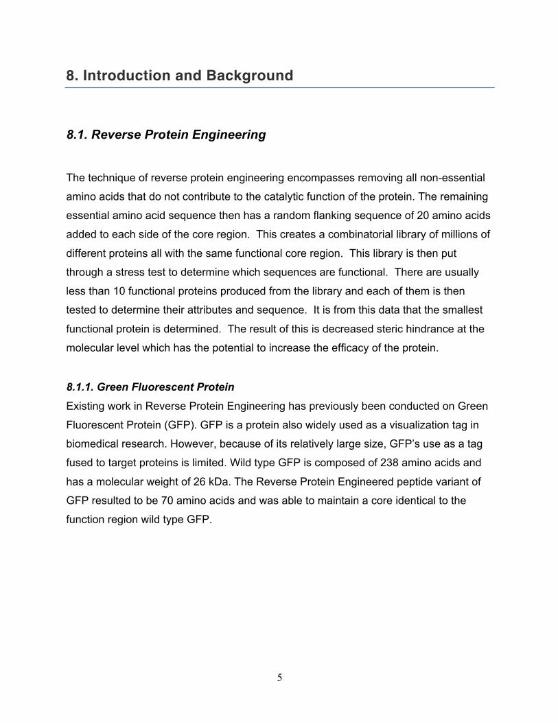
8.1.1. Green Fluorescent Protein
Existing work in Reverse Protein Engineering has previously been conducted on Green
Fluorescent Protein (GFP). GFP is a protein also widely used as a visualization tag in
biomedical research. However, because of its relatively large size, GFP’s use as a tag
fused to target proteins is limited. Wild type GFP is composed of 238 amino acids and
has a molecular weight of 26 kDa. The Reverse Protein Engineered peptide variant of
GFP resulted to be 70 amino acids and was able to maintain a core identical to the
function region wild type GFP.
6
Figure 1. GFP core. Full length wild type Aequorea GFP (left) and the core peptide region C48-T97
(right). Akido Umeda Dissertation
8.2. Bioluminescence
Bioluminescence is the emission of light by a living organism through a biochemical
reaction. Organisms that possess bioluminescent properties include fireflies, beetles,
glow worms, bacteria, fungi, and deep-sea fish. In nature, bioluminescence serves
organisms as a defense mechanism or device to lure prey. However, in the medical
field, it is widely used in biomedical applications as a means for visualizing biological
interactions on the molecular level. Some applications of bioluminescence in the
biomedical field include in vivo imaging, activity assays, microarrays, and biosensors.
Bioluminescent imaging is also deemed an important technology because of its
advantages over fluorescent imaging. Fluorescence is another type of light emission
7
commonly used in biological research and is caused by energy excitation in a molecule
from light. While both bioluminescence and fluorescence are widely used in scientific
applications, bioluminescence is often considered better than fluorescence because the
emission of light is intrinsically supplied by an enzymatic reaction, whereas fluorescence
requires another source of light in order to emit energy. In addition, because of its
enzymatic nature, bioluminescent reporters display an ultrasensitive detection capacity
and have a wider dynamic range compared to fluorescent reporters.
Fluorescent reporters, on the other hand, are susceptible to photo-bleaching, provide
low quantum yields and have greater protein stability in cell-based assays compared to
bioluminescent reporters, which make them less amenable for use as real-time
reporters. Cellular components also have auto-fluorescent properties, which increase
the non-specific background and decreases the sensitivity of fluorescent detection in
cell-based assays. Conversely, cellular components have no inherent bioluminescence,
allowing for greater sensitivity with bioluminescent assays. Thus, further research in
bioluminescence is worth investing in to advance current and future applications within
the biomedical field.
8.3. Firefly Luciferase
8.3.1. Background
Luciferase is a bioluminescent protein that glows green upon reacting with its substrate
luciferin. In current biological technologies, it is used to study a variety of biological
applications, such as in-vivo imaging, cell proliferation assays, protein folding and
secretion, and reporter gene assays. In nature, varying forms of luciferase exist in
different types of organisms, such as firefly, bacteria, and marine animals. For the
purposes of this project, firefly luciferase was studied because it is most commonly used
in biological applications and is more commercially available.
8
8.3.2. Structure
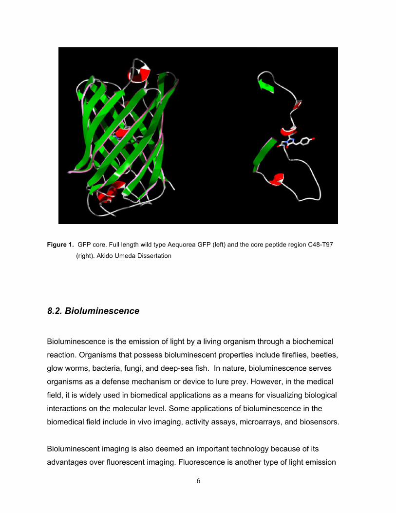
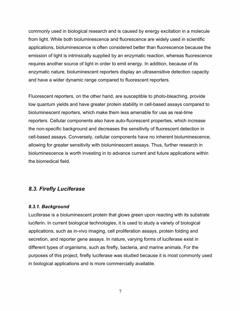
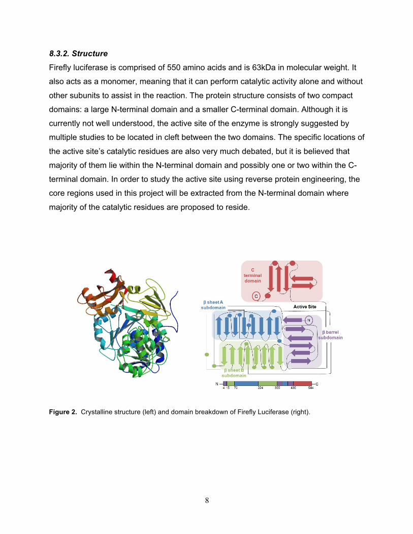
Firefly luciferase is comprised of 550 amino acids and is 63kDa in molecular weight. It
also acts as a monomer, meaning that it can perform catalytic activity alone and without
other subunits to assist in the reaction. The protein structure consists of two compact
domains: a large N-terminal domain and a smaller C-terminal domain. Although it is
currently not well understood, the active site of the enzyme is strongly suggested by
multiple studies to be located in cleft between the two domains. The specific locations of
the active site’s catalytic residues are also very much debated, but it is believed that
majority of them lie within the N-terminal domain and possibly one or two within the C-
terminal domain. In order to study the active site using reverse protein engineering, the
core regions used in this project will be extracted from the N-terminal domain where
majority of the catalytic residues are proposed to reside.
Figure 2. Crystalline structure (left) and domain breakdown of Firefly Luciferase (right).
9
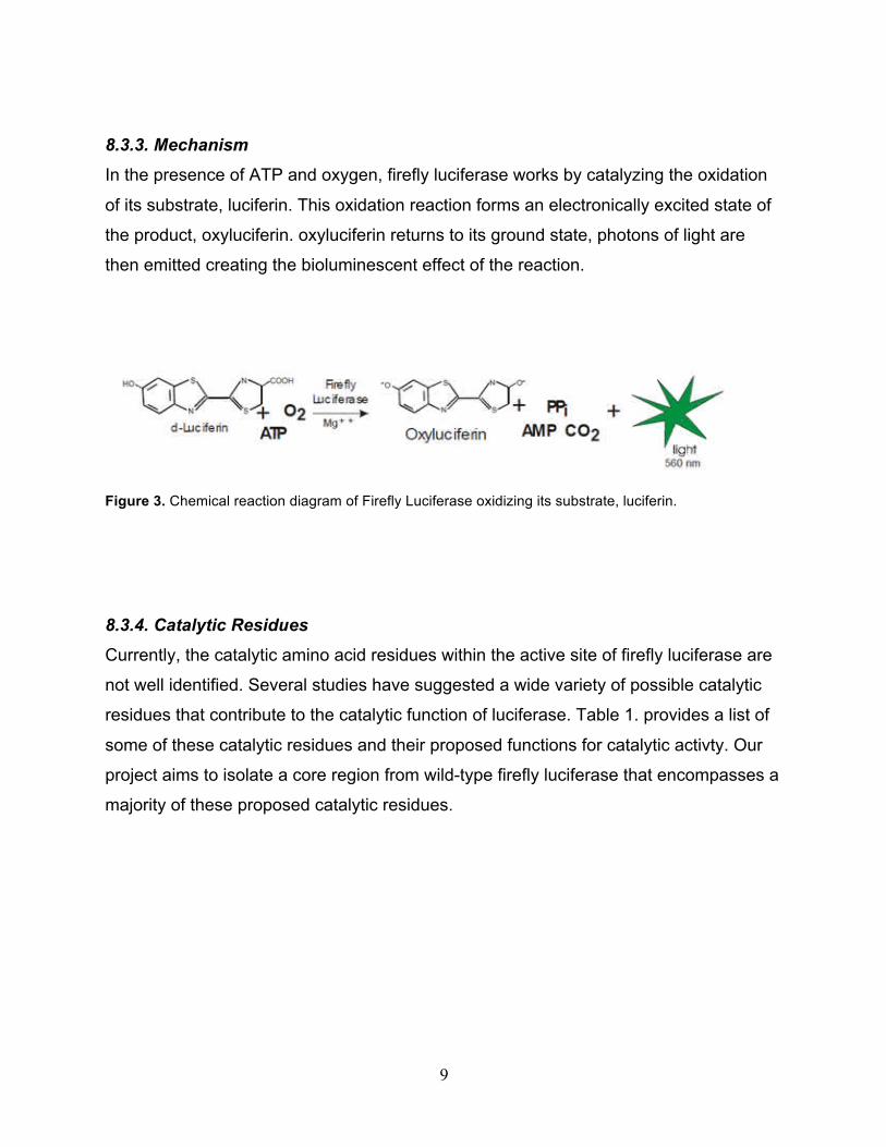
8.3.3. Mechanism
In the presence of ATP and oxygen, firefly luciferase works by catalyzing the oxidation
of its substrate, luciferin. This oxidation reaction forms an electronically excited state of
the product, oxyluciferin. oxyluciferin returns to its ground state, photons of light are
then emitted creating the bioluminescent effect of the reaction.
Figure 3. Chemical reaction diagram of Firefly Luciferase oxidizing its substrate, luciferin.
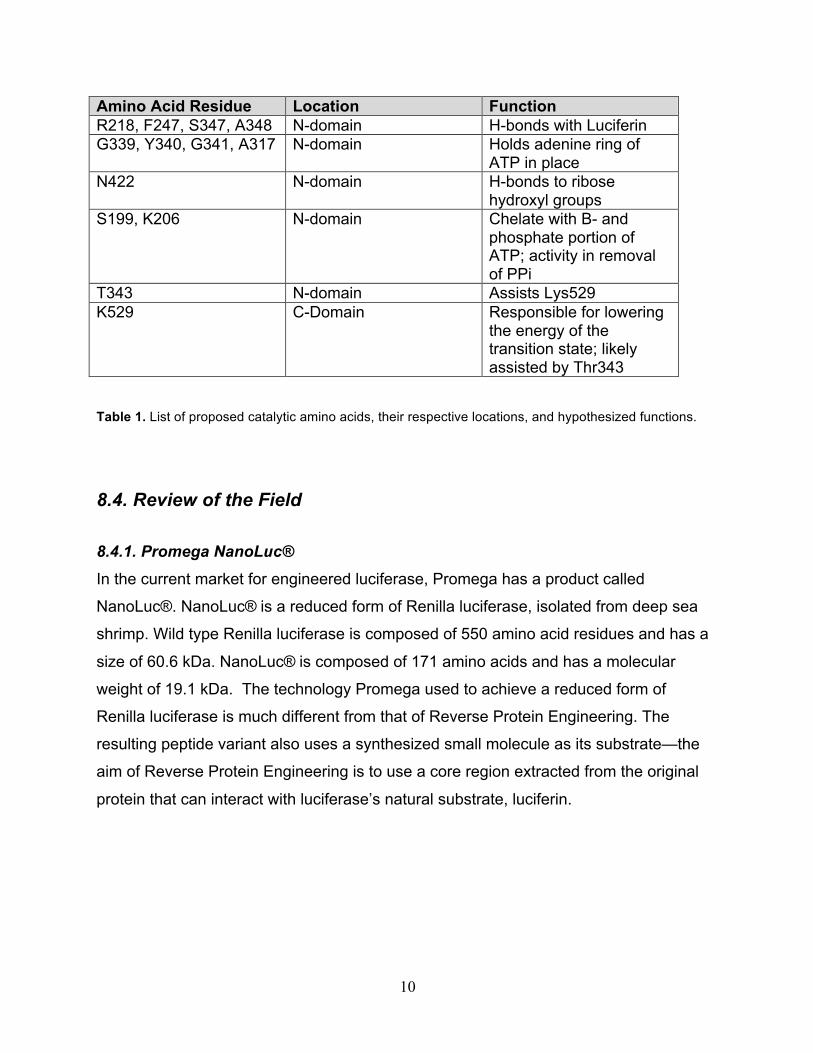
8.3.4. Catalytic Residues
Currently, the catalytic amino acid residues within the active site of firefly luciferase are
not well identified. Several studies have suggested a wide variety of possible catalytic
residues that contribute to the catalytic function of luciferase. Table 1. provides a list of
some of these catalytic residues and their proposed functions for catalytic activty. Our
project aims to isolate a core region from wild-type firefly luciferase that encompasses a
majority of these proposed catalytic residues.
10
Amino Acid Residue Location Function R218, F247, S347, A348 N-domain H-bonds with Luciferin G339, Y340, G341, A317 N-domain Holds adenine ring of
ATP in place N422 N-domain H-bonds to ribose
hydroxyl groups S199, K206 N-domain Chelate with B- and
phosphate portion of ATP; activity in removal of PPi
T343 N-domain Assists Lys529 K529 C-Domain Responsible for lowering
the energy of the transition state; likely assisted by Thr343
Table 1. List of proposed catalytic amino acids, their respective locations, and hypothesized functions.
8.4. Review of the Field 8.4.1. Promega NanoLuc®
In the current market for engineered luciferase, Promega has a product called
NanoLuc®. NanoLuc® is a reduced form of Renilla luciferase, isolated from deep sea
shrimp. Wild type Renilla luciferase is composed of 550 amino acid residues and has a
size of 60.6 kDa. NanoLuc® is composed of 171 amino acids and has a molecular
weight of 19.1 kDa. The technology Promega used to achieve a reduced form of
Renilla luciferase is much different from that of Reverse Protein Engineering. The
resulting peptide variant also uses a synthesized small molecule as its substrate—the
aim of Reverse Protein Engineering is to use a core region extracted from the original
protein that can interact with luciferase’s natural substrate, luciferin.

11
8.5 Statement of project goal, objective and results
The first goal our project hopes to achieve is to create a peptide alternative for wild-type
firefly luciferase to resolve protein’s current size limitations. The successful reduction in
size would allow the luciferase protein to be used as a tag molecule in small molecular
studies and applications that its wild-type form is currently unfitting for. Specifically this
smaller peptide form of luciferase could be used in very small microarrays, biosensors,
and in-vivo imaging.
The second goal of this project is to contribute to the characterization of luciferases
active site and identification of its catalytic residues. We do this by isolating a core
region that encompasses many of the currently proposed catalytic residues and
determining if that isolated region can create a functional peptide that glows. If a peptide
is successful, it would confirm that the included catalytic residues are important to
catalytic function.
The last goal of the project is to support the theory of a peptide world. This theory states
that all current proteins today existed in the past as peptides and that the world once
operated by peptide interactions alone before it became driven by protein interactions.
Successful results from this project can show that a wild-type protein can exist and
function as a peptide, thus supporting the theory of a previous peptide world. Again, the
method in which all of these goals will be achieved is by reverse protein engineering.
12
9. Cost Analysis
9.1 Budget Proposal
Advisor: Jonathan Zhang <[email protected]>
This study was previously conducted using Green Fluorescent Protein where it was
found that certain amino acids in the protein were unessential to the protein’s function.
When these unessential amino acids were removed the protein was less of a
hinderance to other proteins when used as a marker. This year we plan to take the next
step in this study by finding, and removing the nonessential amino acids in some
therapeutic bacterial proteins. The idea is that this will increase the efficiency of these
enzymes which has huge potential in the biomolecular industry.
This study is currently the only of its kind in the world as Professor Zhang is pioneering
this field. Increasing the efficiency of of an enzyme is essentially increasing the
efficiency of the entire bio-molecular world. These are the first steps towards opening
an entirely new door in the future of medicine as it has the potential to create faster
acting, more accurate, and more effective pharmaceutical drugs.
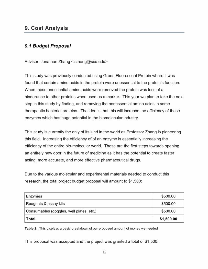
Due to the various molecular and experimental materials needed to conduct this
research, the total project budget proposal will amount to $1,500:
Enzymes $500.00
Reagents & assay kits $500.00
Consumables (goggles, well plates, etc.) $500.00
Total $1,500.00 Table 2. This displays a basic breakdown of our proposed amount of money we needed This proposal was accepted and the project was granted a total of $1,500.
13
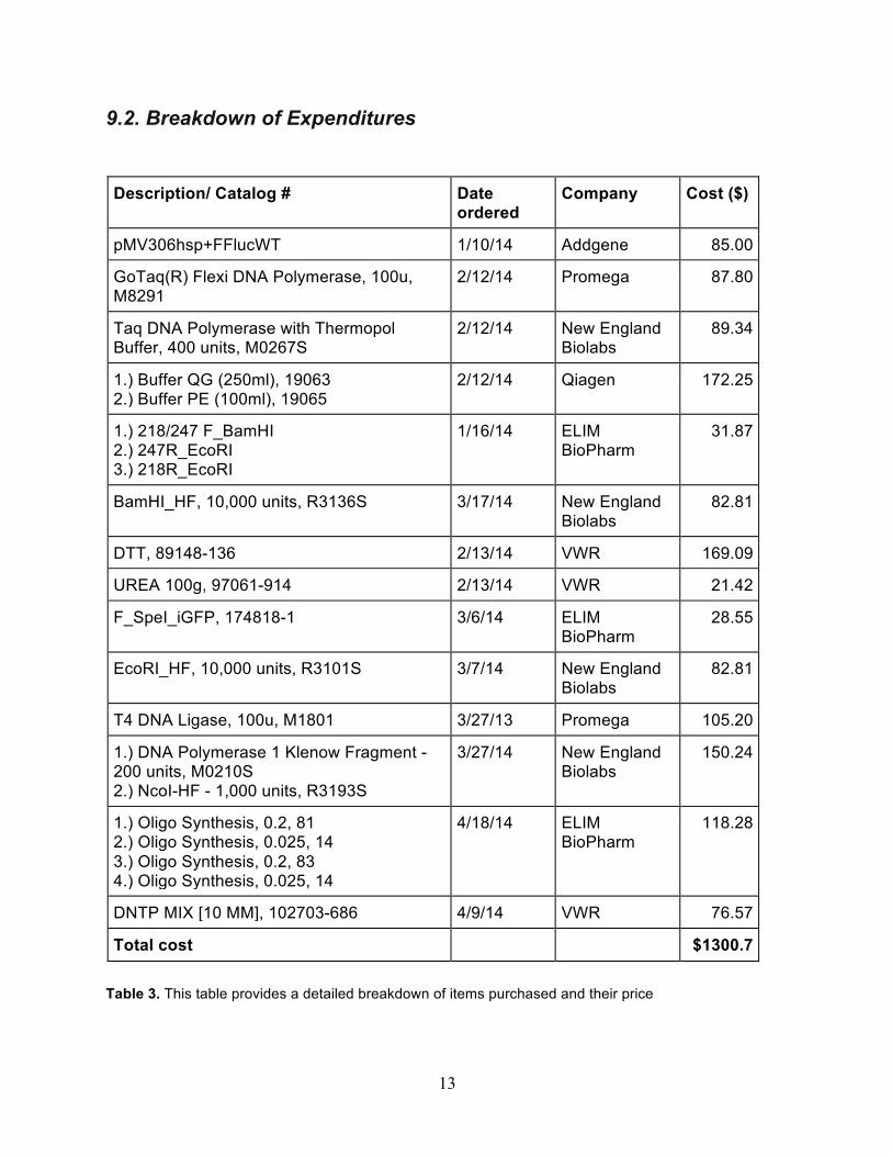
9.2. Breakdown of Expenditures Description/ Catalog # Date
ordered Company Cost ($)
pMV306hsp+FFlucWT 1/10/14 Addgene 85.00
GoTaq(R) Flexi DNA Polymerase, 100u, M8291
2/12/14 Promega 87.80
Taq DNA Polymerase with Thermopol Buffer, 400 units, M0267S
2/12/14 New England Biolabs
89.34
1.) Buffer QG (250ml), 19063 2.) Buffer PE (100ml), 19065
2/12/14 Qiagen 172.25
1.) 218/247 F_BamHI 2.) 247R_EcoRI 3.) 218R_EcoRI
1/16/14 ELIM BioPharm
31.87
BamHI_HF, 10,000 units, R3136S 3/17/14 New England Biolabs
82.81
DTT, 89148-136 2/13/14 VWR 169.09
UREA 100g, 97061-914 2/13/14 VWR 21.42
F_SpeI_iGFP, 174818-1 3/6/14 ELIM BioPharm
28.55
EcoRI_HF, 10,000 units, R3101S 3/7/14 New England Biolabs
82.81
T4 DNA Ligase, 100u, M1801 3/27/13 Promega 105.20
1.) DNA Polymerase 1 Klenow Fragment - 200 units, M0210S 2.) NcoI-HF - 1,000 units, R3193S
3/27/14 New England Biolabs
150.24
1.) Oligo Synthesis, 0.2, 81 2.) Oligo Synthesis, 0.025, 14 3.) Oligo Synthesis, 0.2, 83 4.) Oligo Synthesis, 0.025, 14
4/18/14 ELIM BioPharm
118.28
DNTP MIX [10 MM], 102703-686 4/9/14 VWR 76.57
Total cost $1300.7
Table 3. This table provides a detailed breakdown of items purchased and their price
14
10. System Integration, Tests, & Results
10.1 Experimental Methods 10.1.1 Determination of Core Sequences
The commercially obtained plasmid pMV306hsp+FFlucWT encodes the firefly luciferase
gene cloned from pGL2-basic (Promega). We refer to this form of firefly luciferase as
“wild-type” luciferase for the rest of this chapter unless otherwise noted. Upon
conducting literature search, it was apparent that there are numerous active site
residues that could assist with wild-type luciferase’s catalytic function. For the purposes
of this project, we selected a peptide region of 48 amino acid residues, S199-F247 as
the core of wild-type luciferase. For catalysis, S199, along with K206, are known to
chelate with the phosphate portion of adenosine triphosphate (ATP) and assist in the
removal of pyrophosphates. A218 and F247 are known to contribute hydrogen bonds to
luciferin, the substrate. These functions are essential to catalysis, hence the reason we
chose this region as our core. We refer to this core region as the 10K core for the rest of
this chapter unless otherwise noted.
10.1.2 Primer Design
In order to isolate the 10K core from wild-type luciferase, the polymerase chain reaction
(PCR) was employed. However, the first step was to design primers that incorporate the
10K core. Also, since we are taking the 10K core and ligating it into our vector of choice,
PET-28B, it is imperative to include restriction sites on both the forward and reverse
primers. In our case, we chose restriction sites for BamHI and EcoRI, as our forward
and reverse restriction sites, respectively. In addition, extra nucleotides flanking both
ends of the restriction site were added in order to optimize digestion efficiency
10.1.4 Library Flanking Sequences and Screening
Upon successful ligation of the 10K core to the PET28-B vector, the next step was to
ligate a library of 20 amino acid residues to the C-terminal domain of the 10K core. An
15
oligonucleotide library encoding 20 random amino acid residues by a codon NNK was
purchased commercially (Elim Biopharmaceuticals). Using K at the third position of the
codon eliminates two possible stop codons, TAA and TGA simultaneously allowing for
the gene to transcribe all 20 possible amino acids. The complementary double-stranded
oligonucleotide library was synthesized using the DNA Polymerase I, Large (Klenow)
Fragment purchased commercially (New England Biolabs). This oligonucleotide library
was then cloned at the C-terminus of the 10K core. The resulting plasmid was then
transformed into BL21 E.Coli cells purchased commercially (New England Biolabs).
Colonies from the library above were screened using a spray containing luciferin. None
of the colonies emitted light. However, since there were fewer colonies than expected
on the plates, it is a possibility that with more transformations and subsequent
screening, there will be one colony that will emit light.
10.2 Results
The overall goal of our project was to decrease the size of Firefly Luciferase, but along
the way we had several different protocols that had to have positive results in order for
our end result to be possible. The first step of our project was to successfully isolate the
core region from wild type Firefly Luciferase. We did in fact successfully isolate the core
region and to check that we got exactly what we wanted we ran our PCR sample of the
core through an electrophoresis gel. When compared to a 100 base pair ladder, the
band in the gel, produced by the core, measured to be just under 150 base pairs. Since
our core region was 144 base pairs long, we confirmed that we properly PCR isolated
our core region out of the lucierfase gene. The next significant step was to sub clone
this core region into a prepared Pet-28b vector. Again we were successful with this and
achieved ligation of the core insert into the Pet-28b vector. In order to be one hundred
percent sure we achieved successful ligation of our core insert, and not just self ligation
of the vector, we sent out our newly made plasmid for sequencing. The sequencing
16
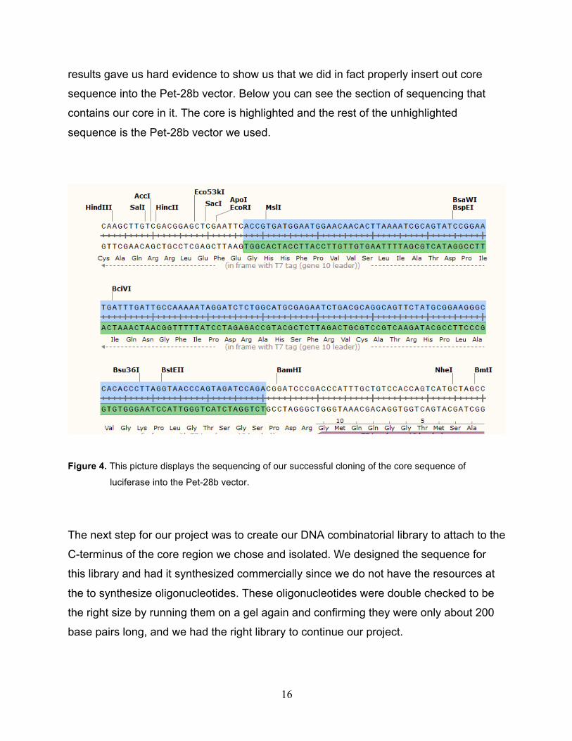
results gave us hard evidence to show us that we did in fact properly insert out core
sequence into the Pet-28b vector. Below you can see the section of sequencing that
contains our core in it. The core is highlighted and the rest of the unhighlighted
sequence is the Pet-28b vector we used.
Figure 4. This picture displays the sequencing of our successful cloning of the core sequence of
luciferase into the Pet-28b vector.
The next step for our project was to create our DNA combinatorial library to attach to the
C-terminus of the core region we chose and isolated. We designed the sequence for
this library and had it synthesized commercially since we do not have the resources at
the to synthesize oligonucleotides. These oligonucleotides were double checked to be
the right size by running them on a gel again and confirming they were only about 200
base pairs long, and we had the right library to continue our project.
17
Once confirmed, we then ligated the library to the C-terminus of our core region to
extend the sequence of the new protein, and give it more of a chance to fold properly
and function. We were successful in ligating the library to the core because we were
able to transform it into E.coli cells and plate colonies. If we did not get any colonies
after transforming the E.coli then our library ligation would not have work. But this was
not the case. We achieved successful ligation of our library and moved onto the last
step of our project. This last step included, plating 10^8 different colonies and screening
those colonies to see if any lit up, thus containing a proper functioning peptide variant of
Firefly Luciferase. We unfortunately did not get any colonies to light up and show us a
functioning peptide form of Firefly Luciferase. But there are many things that could have
gone wrong, and we will discuss these in the following section.
10.3 Discussion
10.3.1 Analysis
Unfortunately, we were not able to successfully reduce the size of firefly luciferase
within the time we had for our project. This could have happened for a number of
different reasons, and there are many things we could change to our experimental
design that would increase our chances of successful reduction of the size of firefly
luciferase. First off, we did not get to plate 10^8 colonies like we had planned too. We
ran out of time to screen our colonies and were only able to plate and screen 10^4
colonies. This means that half of our possible library combinations were not expressed.
Therefore we still have a chance to get a properly functioning protein that glows. This
process of screening takes a long time and it was difficult to try to get all this work done
in such a short time period.
Another factor that could have affected our results was the way we screened our
colonies. There are no previous protocols for screening E. coli colonies containing
luciferase, in the way we needed to screen them. We decided to mold the current
18
screening protocols to our specific needs, and ended up with a solution to spray over
the plated colonies.
Our protocol to screen the colonies included spraying the solution containing D-luciferin
over the colonies and recording them for thirty minutes to see if there was any light
emission. We had to wait thirty minutes because it can take up to thirty minutes for a
molecule to pass through the membrane of the E.coli cells. Since there are no
established protocols on how to screen E.coli colonies containing luciferase, we are
unsure that this approach even works. This means that we could have possibly had a
functioning firefly luciferase peptide variant, but it might not have responded to the spray
solution we used. A possible problem with our screening method was the D-luciferin.
We ran a control screening experiment with wildtype firefly luciferase to see if the spray
solution would cause colonies with wild type luciferase to light up. We found out that not
even wild type luciferase would light up with the D-luciferin, therefore we concluded that
the D-luciferin substrate could have been bad from the beginning.
10.3.2 Comparisons to predictions
At the beginning of our senior design project, we predicted that we would be able to
successfully reduce the size of wildtype Firefly Luciferase using reverse protein
engineering protocol. In reality we almost accomplished this prediction, but fell a little
short. This project was very advanced and difficult to perform in such a short time. Our
group knew it would take a lot of work and effort to accomplish our task. Unfortunately
even though we put in hours upon hours of work we did not achieve our prediction of
reducing Firefly Luciferase’s overall size. We did however almost make it to our
prediction. What I mean by this is that we achieved all the steps that were necessary in
our project, in order to get to the end and see if we did get a new form of Firefly
luciferase.
We achieved successful PCR isolation of our core, successful sub cloning of that core
into the Pet-28b vector, creation of our DNA combinatorial library, and ligation and
19
transformation of that library into E.coli cells. Without the success of these individual
steps, we would not have been able to proceed and make it to the end of our project. In
the end, we still had more colonies to plate so we are still unsure if we are were
completely unsuccessful with the prediction that we would reduce the size of Firefly
Luciferase. And even with that, we still were able to create and refine specific protocols
of reverse protein engineering, and these protocols can be used in future projects of
reverse protein engineering.
20
11. Summary and Conclusion
11.1 Summary and Conclusion The main goal for our senior design project was to reduce the size of firefly luciferase
using reverse protein engineering protocols. Over this past year our group has reached
many necessary milestones along the road to reducing firefly luciferase’s size. These
milestones include; successful PCR amplification of our selected luciferase core,
successful cloning of our core luciferase region into the Pet-28b vector, successful
creation of a DNA combinatorial library, and successful ligation of our DNA
combinatorial library onto the C-terminal end of our cloned luciferase core. As a group,
we had to achieve each one of the steps, one by one, in order to get to the end of our
project and have a chance to reduce the size of firefly luciferase.
Our group started by researching the key catalytic parts or the firefly luciferase protein.
Once we had chosen the core of the luciferase protein, we were able to PCR isolate
and amplify the core and clone it into our selected vector. With successful cloning, we
had our desired plasmid containing the luciferase core, and could move on to creating
the combinatorial library and attach it to the C-terminus of the core. The library was
properly and successfully attached, and we then went to the last step of our project and
screened colonies containing the new luciferase proteins. At this point we reached the
final step of our project, but as mentioned before, unfortunately we were not able to
produce a luciferase protein that folded properly and expressed bioluminescence. We
screened thousands of colonies for a bioluminescent protein but were unable to confirm
a positive glowing result.
Although our experiments did not yield any of our predicted results we accomplished all
of the steps of our reverse protein engineering protocols. The steps along the way were
difficult and time consuming, but we were able to make it through each necessary step
and successfully reach the end of our project, but did not achieve successful cloning of
wildtype firefly luciferase
21
By using reverse protein engineering protocols our group was able to attempt to reduce
the size of the bioluminescence protein firefly luciferase. Due to the complexity of the
experiments we were performing, our group was not able to successfully reduce the
size of firefly luciferase. There are many factors that complicated this project and made
it very difficult to achieve our end goal such as protein kinetics, substrate-protein
interactions, and protein folding. These topics illustrate the complexity of our project
because they all had to be considered when we altered our experiments for the size
reduction of firefly luciferase.
11.2 Future Work Our project had several variable factors, and for that reason there are protocols that can
be altered to improve the possibilities of reducing the size of firefly luciferase in future
work. Some areas that can possibly be changed or improve to alter the result of this
project are; investigate and increase the size of the chosen core, chose different
restriction enzymes for cloning of the core and sub-cloning of the library, use a different
vector, create an alternate screening method, and research more about the D-luciferin
substrate interaction with luciferase. Overall, several things could be changed and
explored more, and with more time and future work this project can yield a smaller sized
firefly luciferase.
11.3 Reflection/ Lessons learned Our group started this project with the expectation of reducing the size of wildtype firefly
luciferase, but we were unfortunately unable to achieve our final goal. Even though we
did not achieve a successful end goal with our experiment, we still gained many
valuable experiences and methods from our project. Through this project we were able
to gain valuable laboratory skills that are applicable to several industry jobs around the
country. We were faced with several challenges along the way through our experiment,
but with hard work and dedication we were able to work as a group and move pass the
challenges that arose. One example is the difficulty we had attempting to clone our
luciferase core into the Pet-28b vector. Our group had several failed attempts at this

22
step in our experiment, and it took some extra research and late nights in the lab to
finally get passed the problem and achieve successful cloning of our core. Once we
achieved successful cloning we did not have another problem with it in the entire
project. This work pushed us to critically think at a level higher than regular class, and it
was a great learning experience applicable to future jobs. This project also taught us
how to work as a team. Working as a group of four can be tough due to coordination of
everyone’s schedules and ideas. We were able to successfully work together,
cooperating and synthesizing our ideas and time in order to complete the steps of our
senior design project. Our group felt that we worked well together and were satisfied
with the work we accomplished for our project, regardless of the end result. For future
students, we suggest to work proactively on senior design projects to allow for a
prepared timeline that can accommodate for any setbacks, troubleshooting, and
material delivery times. Students should also expect to allocate a large amount of their
time working in the lab if they wish to obtain the best possible results from their projects,
as time in the lab is time well spent for success.
23
12. Engineering Standards
12.1 Ethics
For our senior design project, it is important that we conduct ourselves in an ethical
manner. As future members of the engineering profession, we are expected to learn
and exhibit the highest levels of honesty and integrity. It is crucial that we act with
honesty and integrity throughout all stages of our senior design project—from inception
to completion. Our group must act ethically with each other, as well ensure that we act
ethically with everyone involved so that the end users of our product do not encounter
any issues.
As the product from our senior design project has the potential to be used for medical
purposes, our ethical responsibility to the potential users of our product is immense. Our
project not only looks to design a single more efficient protein; the technology used also
has the potential to open up entirely new doors in the medical industry as we are
pioneering this enzymatic manipulation technique. With a potential like this we have a
great responsibility to conduct our research with the upmost ethical integrity to ensure
that we are able to gain the most we can out of this project.
12.2 Environmental
Our Project was environmentally friendly, and we did take the environment into
consideration with our project. Although our senior design project was primarily working
with biological substances that produce no toxins or pollution, we still had to keep in
mind disposing the hazardous waste properly to ensure no damage to the environment.
Since we are working with bacteria we created biohazard waste, that needs to be
properly disposed of. In specific our team would thoroughly bleach any culture plate
containing E.coli bacteria to ensure the E.coli was dead before we disposed of it in the
24
bio-hazard trash, which gets collected weekly and sent out to be burned. If we did not
bleach the plates, the E.coli bacteria could spread and mutate into a new airborne virus
that could be potentially harmful to humans. Therefore it is important to bleach all the
plates before we throw them out, so we protect the environment from any damaged that
could be caused by a mutated E.coli strain. Since the bio-hazard trash that is burned is
mainly just dead E.coli cells and culture media, little to no pollution is created in the
disposal of the bio-hazard trash. Overall, our team considered environmental issues
when disposing of our experiments waste, and were able to prevent any harm done to
the environment.
12.3 Health and Safety
Our safety for the purposes of our project, it is imperative that we take the utmost care
in ensuring our safety and the safety of others we share the lab with. Prior to beginning
our lab work, we have taken lab safety training courses sponsored by Santa Clara
University. While in the laboratory we must wear proper personal protective equipment,
such as gloves, goggles, and lab coats, at all times. Lab benches should not be
cluttered with materials, and should be wiped down after lab work with ethanol. We
should also know where the eye wash and shower locations are and how to operate
them if need be. We as a group have been acting
in accordance with Santa Clara's safety regulations thus far
12.4 Manufacturability
Manufacturability of the product is a great consideration in the research of reverse
protein engineering. Therapeutic protein molecules are manufactured by genetically
modifying cells of microorganisms—typically, E.coli—and using those cells as a vehicle
to naturally synthesis the desired protein. To genetically modify the cells to include the
25
DNA sequence coding for the protein, the protein’s DNA sequence needs to be PCR
amplified. Generally, larger biomolecules take more time, starting materials, and energy
to be PCR amplified, and thus can be more difficult to manufacture in large masses.
Because reverse protein engineering aims to create smaller biomolecules, it costs a
shorter amount of time and less resources to be amplified and makes manufacturability
of the engineered product easier to conduct. With this in mind, our team chose a protein
that is commonly used, would benefit from being reduced in size, and whose
manufacturability would be simple and straightforward. Because the ultimate goal of the
research in reverse protein engineering would be to expand into pharmaceutical
purposes, manufacturability and reproducibility of a successful product is key.
Fortunately, reverse protein engineering currently appears to improve the manufacturing
process of future peptidic biomolecules.
12.5 Social
The progression of research in reverse protein engineering is invaluable to the social
community as it has the ability to open up new doors in the field of pharmaceuticals.
The doors that will open will create new lines of drugs that have the potential to be more
efficient and effective than any existing drugs on the market. This is can lead to drugs
with less side affects that could negatively affect the patient's quality of life.
Often times people are backed into a corner where they must take a drug because the
main affect of it is that it will let them live. However, the side affects of these drugs can
deteriorate the quality of a person's life to the point where the gain or barely more than
the sacrifice. The potential to create drugs without or even with less side affects
through the technique of protein engineering renders not progressing this area of
research unethical. People owe it to each other in this world to help each other as that
is how we have progressed the world into what it is today. The field of reverse protein
engineering has the ability to help others which means it is only just to continue this
research with the hopes that someday it will benefit humanity as a whole.
26
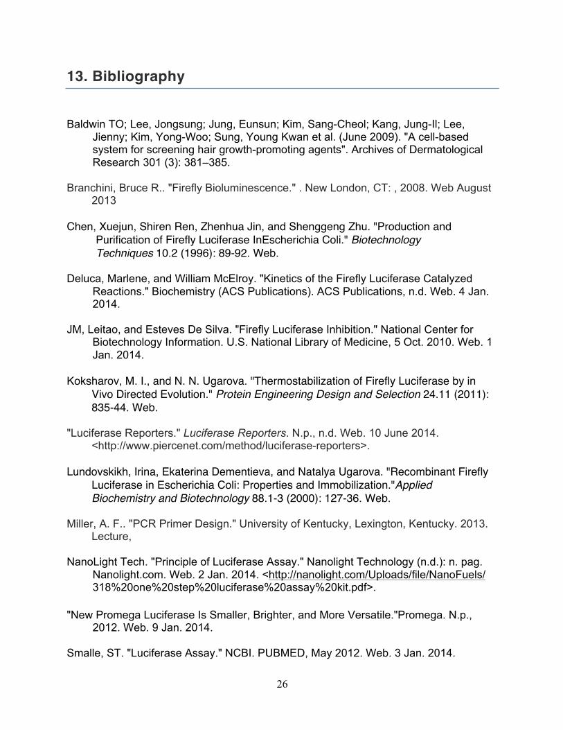
13. Bibliography
Baldwin TO; Lee, Jongsung; Jung, Eunsun; Kim, Sang-Cheol; Kang, Jung-Il; Lee, Jienny; Kim, Yong-Woo; Sung, Young Kwan et al. (June 2009). "A cell-based system for screening hair growth-promoting agents". Archives of Dermatological Research 301 (3): 381–385. Branchini, Bruce R.. "Firefly Bioluminescence." . New London, CT: , 2008. Web August
2013 Chen, Xuejun, Shiren Ren, Zhenhua Jin, and Shenggeng Zhu. "Production and
Purification of Firefly Luciferase InEscherichia Coli." Biotechnology Techniques 10.2 (1996): 89-92. Web.
Deluca, Marlene, and William McElroy. "Kinetics of the Firefly Luciferase Catalyzed Reactions." Biochemistry (ACS Publications). ACS Publications, n.d. Web. 4 Jan. 2014. JM, Leitao, and Esteves De Silva. "Firefly Luciferase Inhibition." National Center for Biotechnology Information. U.S. National Library of Medicine, 5 Oct. 2010. Web. 1 Jan. 2014. Koksharov, M. I., and N. N. Ugarova. "Thermostabilization of Firefly Luciferase by in
Vivo Directed Evolution." Protein Engineering Design and Selection 24.11 (2011): 835-44. Web.
"Luciferase Reporters." Luciferase Reporters. N.p., n.d. Web. 10 June 2014.
<http://www.piercenet.com/method/luciferase-reporters>. Lundovskikh, Irina, Ekaterina Dementieva, and Natalya Ugarova. "Recombinant Firefly
Luciferase in Escherichia Coli: Properties and Immobilization."Applied Biochemistry and Biotechnology 88.1-3 (2000): 127-36. Web.
Miller, A. F.. "PCR Primer Design." University of Kentucky, Lexington, Kentucky. 2013.
Lecture, NanoLight Tech. "Principle of Luciferase Assay." Nanolight Technology (n.d.): n. pag. Nanolight.com. Web. 2 Jan. 2014. <http://nanolight.com/Uploads/file/NanoFuels/ 318%20one%20step%20luciferase%20assay%20kit.pdf>. "New Promega Luciferase Is Smaller, Brighter, and More Versatile."Promega. N.p., 2012. Web. 9 Jan. 2014. Smalle, ST. "Luciferase Assay." NCBI. PUBMED, May 2012. Web. 3 Jan. 2014.
27
The Editors of Encyclopædia Britannica. "Biochemical Events of Light Emission." Encyclopedia Britannica Online. Encyclopedia Britannica, 19 Dec. 2013. Web. 10 Jan. 2014. Tiffen, JC. "Luciferase Expression and Bioluminescence Does Not Affect Tumor Cell Growth in Vitro or in Vivo." National Center for Biotechnology Information. U.S. National Library of Medicine, 2010. Web. 09 June 2014. Wet, J. R. D. "Cloning of Firefly Luciferase CDNA and the Expression of Active
Luciferase in Escherichia Coli." Proceedings of the National Academy of Sciences 82.23 (1985): 7870-873. Web.
28
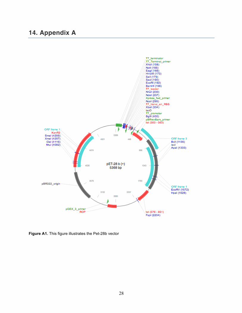
14. Appendix A
Figure A1. This figure illustrates the Pet-28b vector
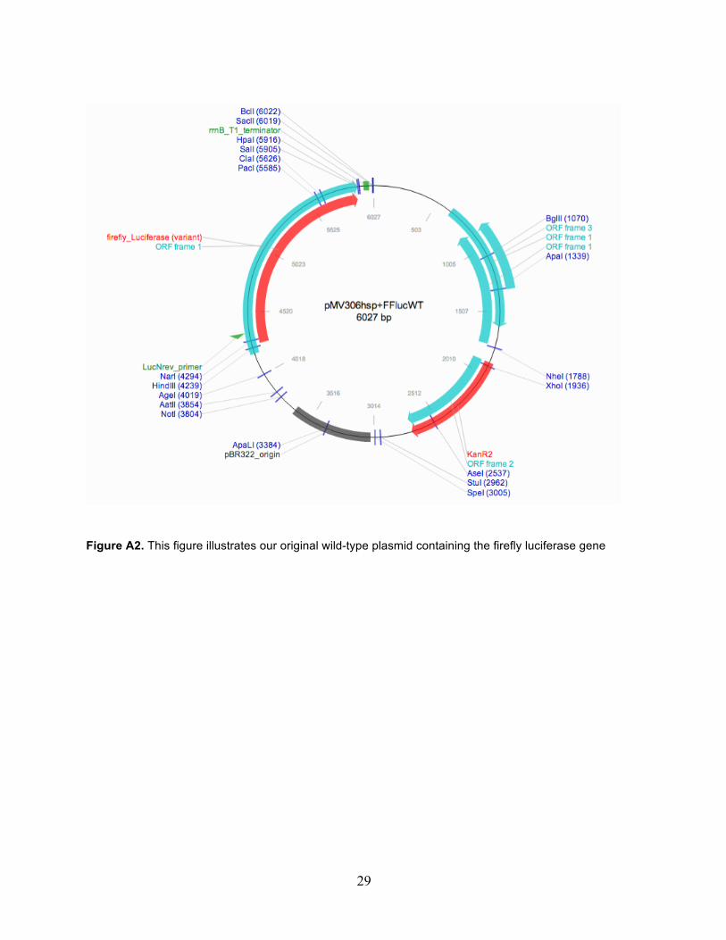
29
Figure A2. This figure illustrates our original wild-type plasmid containing the firefly luciferase gene