Embed Size (px)
Citation preview

Resuscitation in congenital heart disease
Peter C. Laussen MBBS FCICM
Department Critical Care Medicine
Hospital for Sick Children
Toronto

Evolution of Congenital Heart Disease
Extraordinary success:
Overall survival for initial hospitalization: 96-98%
Resuscitation:
Infants & children post repair
Adults with congenital heart disease (ACHD)
Corrected not cured


Delivery of Care
• Transitions
- Patient Passport
- Information & interpretation
• Regionalized ACHD centers of excellence
- Different models: within an adult hospital or part of an expanded congenital heart center / institute
- Expertise in cardiology, diagnostic and interventional catheterization, cardiac anesthesia
- No recommendations re: surgeon, perfusion, critical care or nursing
• Resuscitation:
- Consensus statement being prepared

There are hundreds of combinations and
permutations of congenital cardiac lesions

Effective CPR
• Consistent & coordinated compressions
– PALS guidelines
• Avoid overdosing with resuscitation drugs
– Epinephrine, Bicarb and Ca++
• Induce hypothermia
– Primary treatment, not post injury

CPR guidelines 2010
Early Recognition & PreventionEmphasize CPR quality:
Push hard, fast, and allow full recoil100 compressions/minuteMinimize interruptions to compressionsRotate compressorsAvoid excessive ventilation

Effective CPR
• Consistent & coordinated compressions
– PALS guidelines
• Avoid overdosing with resuscitation drugs
– Epinephrine, Bicarb and Ca++
• Induce hypothermia
– Primary treatment, not post injury
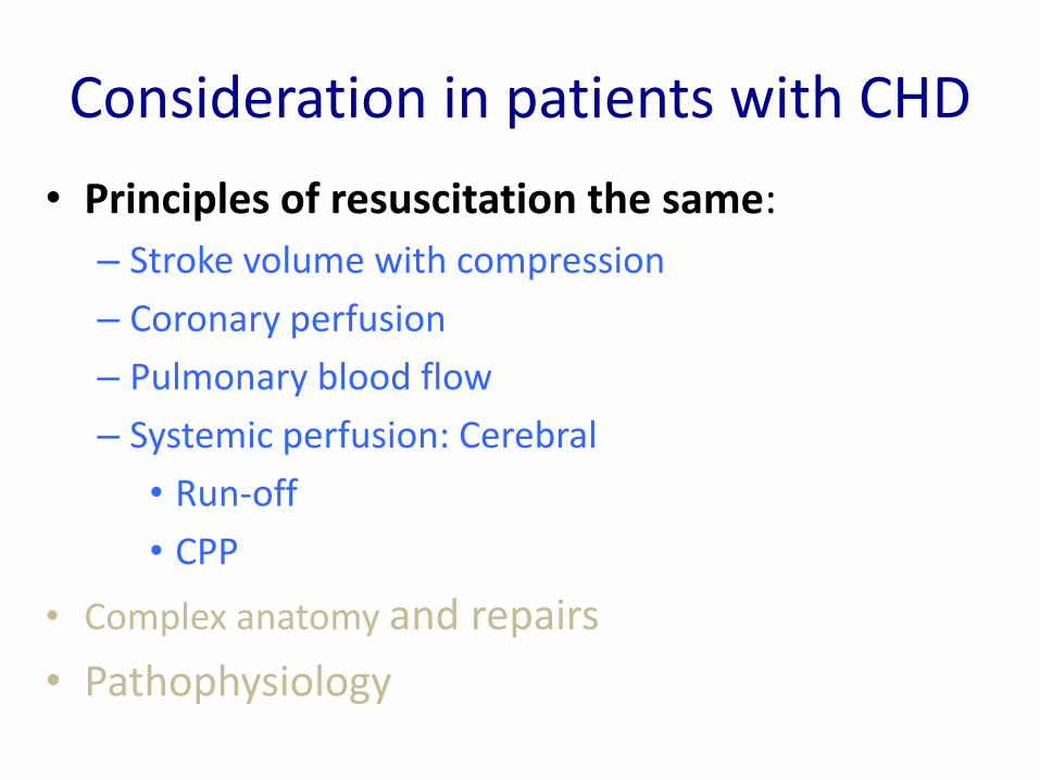
Consideration in patients with CHD
• Principles of resuscitation the same:
– Stroke volume with compression
– Coronary perfusion
– Pulmonary blood flow
– Systemic perfusion: Cerebral
• Run-off
• CPP
• Complex anatomy and repairs
• Pathophysiology

Consideration in patients with CHD
• Principles of resuscitation the same
• Complex anatomy and repairs
• Pathophysiology

Vascular Access Occluded Yes No ?
Femoral Vein
Right
Left
Femoral Artery Right
Left
Internal Jugular Vein Right
Left
ANATOMY
Aortic arch Right
Left
SVC Right
Left
Bilateral
IVC Interrupted
Dextrocardia
Heterotaxy
Asplenia
Polysplenia
Airway Difficult
intubation
• Diagram of
defect & repair
• Access
• Compressions
• Defibrillation

Consideration in patients with CHD
• Principles of resuscitation the same
• Complex anatomy and repairs
• Pathophysiology

Transposition Great Arteries:Immediate post-operative resuscitation
• Arrhythmia with LCO = ischemia
• Coronary perfusion:
– Usually mechanical
– Pacing not effective: open chest
ECMO

Arterial Switch Operation:
Longer term
Reconstructed RVOT 5-10% stenosis
Pulmonary valve in the aortic position 30% trivial AR
Coronary reimplantation up to 8% Occlusion
Aortic root dilation Uncertain
Arrhythmia Low risk
Functional class NYHA I / II

RVOT reconstruction
Restrictive RV physiology
Elevated EDP, RVH
RVOT & pulmonary stenosis
Residual atrial defect
Systolic RV dysfunction
Elevated ESV, RV dilation
Pulmonary regurgitation
Arrhythmias
Ventricular, risk for sudden death
Risk higher with EDv vs. Edp
No recommendation for prophylaxis

Restrictive LV Physiology
• LVH with increased EDp, LAp & decreased ESv
– Inadequate filling time
– Elevated PAp
– Limited ejection fraction
– Ischemia

Functional repair:
Cavo-Pulmonary connection

Positive pressure ventilation
Fontan physiology
Pulmonary
flow


Consideration in patients with CHD
• Limited stroke volume with compressions:
- Impaired preload:
Cavo-pulmonary connection
Elevated PAp or PVR
- Impaired ventricular filling:
AVVR
Ventricular hypertrophy and restrictive physiology
- Impaired ejection fraction:
Semilunar valve regurgitation
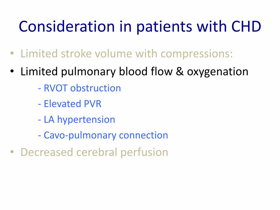
Consideration in patients with CHD
• Limited stroke volume with compressions:
• Limited pulmonary blood flow & oxygenation
- RVOT obstruction
- Elevated PVR
- LA hypertension
- Cavo-pulmonary connection
• Decreased cerebral perfusion

Consideration in patients with CHD
• Limited stroke volume with compressions:
• Limited pulmonary blood flow & oxygenation
• Decreased cerebral perfusion
- Cavo-pulmonary connection
- AoV regurgitation

Extracorporeal Membrane Oxygenation“Consider extracorporeal CPR for in-hospital cardiac arrestrefractory to initial resuscitation attempts if the conditionleading to cardiac arrest is reversible or amenable to hearttransplantation, if excellent conventional CPR has beenperformed after no more than several minutes of no-flowcardiac arrest (arrest time without CPR), and if the institutionis able to rapidly perform extracorporeal membrane oxygenation(Class IIb; LOE 561,62). Long-term survival is possibleeven after 50 minutes of CPR in selected patients.”
CPR guidelines 2005, 2010Recommend use of ECMO to support failed CPR

N = 199PCCM 2010 11: 362 - 71

• 2000-2011
• 45% cumulative survival (788/1735)
• Factors predicting non-survival:
– Duration of ECMO
– End organ injury
– NOT ECPR

Improving the quality of CPR• Leadership is key
• Control the chaos
– Role assignment
– Effective communication
• ICU structure / teams & training
– Dedicated CICU, Cardiac Code team
– Staffing levels & experience

Future
• Longer term outcomes
• Focus on ACHD risk & prevention
– Passport and algorithm
• Reliable and actionable data