Embed Size (px)
Citation preview

GROUP 9 MEMBERS:
ANIS FAZEERA BINTI AHMAD FUAD KHOR JUU YEEI MOHD ZULFIKAR BIN AHMAD FAUZI NOR FADZLIN SAKINA BINTI JAFFERI NUR MAHIRAH BINTI ABDUL MANAF TAY SUE CHYEN TENGKU NABILAH SURAYA
FAR 131 BASIC PHYSIOLOGY

By a rich network of capillaries.
RESPIRATORY SYSTEM
Lung Tube sys lead to the lungs
Conducting portionRespiratory portion
Respiratory bronchioles
Mucosa identical to mucosa of terminal bronchioles BUT numerous alveoli extend from wall.
No goblet cell.
Ciliated cuboidal cell & clora cell
L.propria(smooth muscle,elastic fibres)
Condition inspired air
Provide passage-airlung
Cartilage Elastic fibre
Smooth muscle
Cleansed
Moisted
warmed
Dust and gaseous impuritiestrap in:
-vibrissae(specialized hair)
-layer of mucous(mucous gland)
Mucous & serous secretion(moisten air)
Protect alveolar lining from dessication
Alveolar ducts
Simple squamous epithelium.
Alveolar sacs are disintended space
Elastic fibreenable aveoli-expand(inspiration),contract(expiration).
Reticular fibre prevent-overdistension & damage to capillaries.
Alveoli
Sac-like evagination.
O2,CO2 exchangedair&blood
Interalveolar septum(alveolar wall):
-2 thin squamous epithelium layer.
-Between layer(interstitium)
-Interstitium: capillaries,CT(elastic,reticular fibre & fibroblast)
Support wall
Prevent collapselumen
At periphery Lamina propria
flexibility
Encircles tube
From tracheaalveolar duct
Flow chart

RESPIRATORY Lungs SYSTEM System of tubes Leading to the lung
FUNCTIONS:Provide an intake of O2Eliminate CO2

RESPIRATORY SYSTEM


CONDUCTING PORTION

FUNCTIONS:
1) Provide a conduct through which air can travel to & from the lungs
• Cartilage ~ support wall, preventing collapse of lumen • Elastic fibres ~ flexibility ~
•Smooth muscle ~ contraction reduce of conducting tubules
(regulate air flow during inspiration & expiration)

2) Condition the inspired air (clean, moisten, warm)
• Vibrissae ~ remove dust particles & other
substance
•Mucous & serous secretions ~ moisten incoming air, protect alveolar lining from desiccation
•Rich network of blood capillaries ~ warm in-coming air

Line the mucosa of conducting portionCiliated pseudostratified columnar + goblet cells
5 TYPE OF CELLS:
1) Ciliated columnar cell- Most abundant- Each cells have about 300 cilia on its apical
surface- Beneath cilia are numerous mitochondria, supply
ATP for cilia beating.
2) Mucous goblet cell- Next most abundant- Apical cytoplasm contains mucin granules
RESPIRATORY EPITHELIUM

3) Brush cell- Numerous microvillus on its apical surfaces- Are columnar epithelial cell- Have nerve ending on basal surface (sensory
receptor)
4) Basal cells (short cells)- Small rounded cell- Lie on basal lamina but do not extend to luminal
surface of respiratory epithelium- Generative stem cell that undergo mitosis and
differentiate to other cell types
5) Small granule cell- Resemble basal cell except it contains numerous
granules- Granules control secretory activity of goblet cells
and other glands
RESPIRATORY EPITHELIUM


MUCOSAConsist of:Pseudostratified ciliated columnar epitheliumLamina propia-elastic & reticular fibers-Provide same protection against dust as the membrane lining the nasal cavity & larynx
SUBMUCOSAConsist of:Areolar connective tissue -Seromucous glands & their ducts
HYALINE CARTILAGE•From horizontal ring that resembles C-shaped.•Fxn: Provide semirigid support so that tracheal wall does not collapsed inward.The open end s of hyaline cartilage bridged by smooth muscle,trachealis muscle, and fibroelastic ligament
ADVENTITIAConsist of:Areolar connective tissue -Joins the trachea to sorrounding tissues.
Layers of Tracheal Wall
TRACHEA ~ extended from larynx
TRACHEA

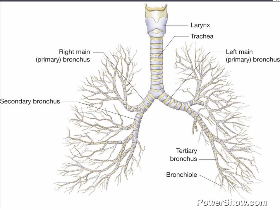
Trachea
Primary bronchi
Bronchioles
Secondary bronchi
Terminal bronchiol
es
3 in right lung
2 in left lung
BRONCHIAL TREE

BRONCHI
MUCOSA • structurally similar to trachea• Different in organization of cartilage &
smooth muscle
CARTILAGE•More irregular in shape•Large bronchi: cartilage rings completely
encircle the lumen•Bronchial diameter ↓, cartilage rings
replaced by isolated plates/ islands of hyaline cartilage
BRONCHI

LAMINA PROPRIA•Smooth muscle layer: crisscrossing bundles of spirally arranged smooth muscle•Rich in elastic fibres•Contains mucous & serous glands
BRONCHI

MUCOSA•No cartilage/glands
LAMINA PROPRIA•Consists mainly of smooth muscles & elastic fibers
BRONCHIOLES
Diameter ≤ 5mm

EPITHELIUM•Large bronchioles - have scattered goblet cells - Ciliated pseudostratified columnar epithelium
•Smaller terminal bronchioles - Ciliated simple columnar/ ciliated simple cuboidal without goblet cell - Epithelium contain Clara cells (apical cytoplasm contains secretory granules – protect the lining of bronchioles against oxidative pollutants)
BRONCHIOLES

RESPIRATORY PORTION
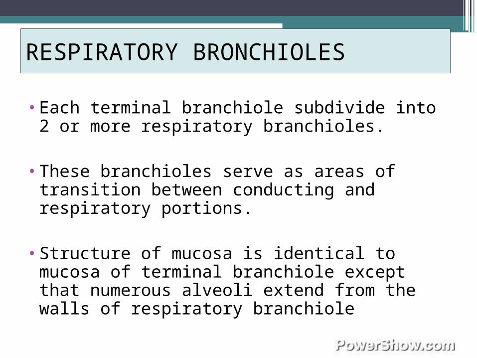
•Each terminal branchiole subdivide into 2 or more respiratory branchioles.
•These branchioles serve as areas of transition between conducting and respiratory portions.
•Structure of mucosa is identical to mucosa of terminal branchiole except that numerous alveoli extend from the walls of respiratory branchiole
RESPIRATORY BRONCHIOLES

•Some parts of respiratory bronchiole are lined with ciliated cuboidal cells and Clara cells
•Goblet cells are absent
•More distal parts of these bronchioles,the cuboidal cells do not have cilia
•Respiratory bronchioles terminate by branching into several alveolar ducts.
RESPIRATORY BRONCHIOLES
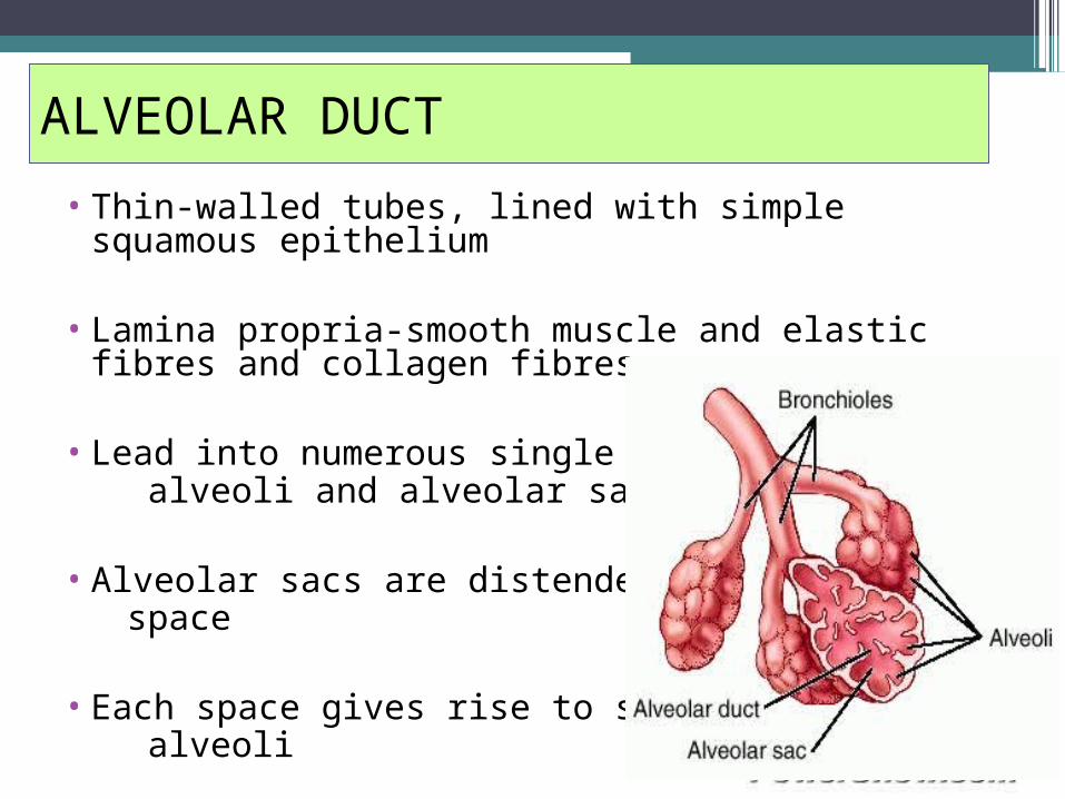
• Thin-walled tubes, lined with simple squamous epithelium
• Lamina propria-smooth muscle and elastic fibres and collagen fibres
• Lead into numerous single alveoli and alveolar sacs
• Alveolar sacs are distended space
• Each space gives rise to several alveoli
ALVEOLAR DUCT

•Elastic and reticular fibres form a complex network encircling the opening of alveolar sacs and alveoli.
•Elastic fibres enable the alveoli to expand with inspiration and to contract with expiration
•Reticular fibre serve as a walls that prevents over distention and damage to capillaries and thin alveolar duct.
ALVEOLAR DUCT

ALVEOLI

# Alveolar wall structure specialized to facilitate diffusion between external and internal environment
lies between 2 neighbouring alveoli
called interalveolar septum have 2 thin squamous epithelial layer
interalveolar wall between which lie the interstitium
# Interstitium capillaries elastic fibers connective tissue reticular fibers fibroblasts
ALVEOLI


# Blood air barrier cytoplasm of the epithelial cells fused basal lamina of the epithelial and
endothelial cells cytoplasm of endothelial cell
thickness 0.1-1.5μm
endothelial lining of the capillaries is CONTINUOUS
DON’T have fenestrae
ALVEOLI

ALVEOLAR EPITHELIUM

Consist of:
TYPE I CELLS TYPE II CELLS
Squamous alveolar cells Septal/ great alveolar cells
Extremely thin cells at alveolar surface
Scattered between type I cells
Make up 97% of alveolar surface Found in groups (2,3 cells) along alveolar surface
All have occluding junctions + desmosomes => to prevent leakage of tissue fluid into alveolar air space
Have more rounded shape, larger nucleus and foamy cytoplasm(due to lamellar bodies)
Organelle group around nuclei => reduce thickness of blood-air barrier
Discharged content from lamellar bodies spread and form thin layer on alveoli
Main role: provide barrier of minimum thickness that is readily permeable to gases.
This layer contain pulmonary surfactant which reduce surface tension within alveoli and prevent it to collapse during expiration
Divide by mitosis to replace their own population and type I cells.

ALVEOLAR MACROPHAGES

•also called ‘dust cell’•Found in interalveolar septum and surface
of alveolus•Wonder freely on surface of alveolus•clean epithelial surface of inspired
particles-by phagocytosis•alveoli (ameoboid movement) bronchioles
(via mucus layer) pharynx (swallowed)•due to a/m, the respiratory part of lungs are
normally kept sterile
ALVEOLAR MACROPHAGES

ALVEOLAR PORES

•Interalveolar septum has pores (10-15µm in diameter) that connect neighbouring alveoli
•These pores equalize air pressure in alveoli
•Provide alternative routes for air movement when there is obstruction
ALVEOLAR PORES
CONCLUSION

•Respiratory system consist of 2 parts: (1)conducting portion (2) respiratory portion
•The bronchial tree system are: Trachea 2 primary bronchi secondary
bronchi [2 in left lung;3 in
right lung] respiratory terminal bronchioles bronchiolesBronchioles
Alveoli duct alveoli sac /aveoli

THE END

![Respiratory System [โหมดความเข้ากันได้] · PATHOLOGY OF RESPIRATORY SYSTEM นพ. อรรณพ นาคะป ท Respiratory system U it](https://img.pdfslide.us/doc/110x75/5fa578efd4e80f055f6b3401/respiratory-system-aaaaaaaaaaaaaaaaaa-pathology.jpg)


![Respiratory system roadmap.pptx [Repaired] - Loginanatomical-sciences.health.wits.ac.za/roadmaps/Respiratory system... · DIVISION OF THE RESPIRATORY SYSTEM CONDUCTING PORTION Nasal](https://img.pdfslide.us/doc/110x75/5a78c3d87f8b9ae6228c9db0/respiratory-system-repaired-loginanatomical-scienceshealthwitsaczaroadmapsrespiratory.jpg)


![Anatomy and Physiology Respiratory System [Tab 2] Respiratory System](https://img.pdfslide.us/doc/110x75/56649ebd5503460f94bc631f/anatomy-and-physiology-respiratory-system-tab-2-respiratory-system.jpg)