Embed Size (px)
Citation preview
Research ArticleKlebsiella pneumoniaeOrbital Cellulitis: Clinical Manifestationsand Outcomes in a Tertiary Medical Center in Taiwan
Chieh-Hung Yen ,1,2 Shu-Ya Wu ,3 and Yi-Lin Liao 1,2
1Department of Ophthalmology, Chang Gung Memorial Hospital Linkou Branch, Taoyuan, Taiwan2College of Medicine, Chang Gung University, Taoyuan, Taiwan3Department of Ophthalmology, Taipei Tzu Chi Hospital, Buddhist Tzu Chi Medical Foundation, Taipei, Taiwan
Correspondence should be addressed to Yi-Lin Liao; [email protected]
Received 20 April 2018; Revised 9 July 2018; Accepted 15 July 2018; Published 2 October 2018
Academic Editor: Michele Figus
Copyright © 2018 Chieh-Hung Yen et al. *is is an open access article distributed under the Creative Commons AttributionLicense, which permits unrestricted use, distribution, and reproduction in any medium, provided the original work isproperly cited.
Purpose. To report six cases of Klebsiella pneumoniae orbital cellulitis without preceding endophthalmitis.Method. Retrospectivechart review. Results. We reported four females and two males admitted to our hospital for Klebsiella pneumoniae orbital cellulitisproven by computed tomographies and bacterial cultures fromMay 1995 to March 2017. Proptosis, conjunctival congestion, andchemosis and limitation of ocular motility were present in all six patients. Four patients had decreased visual acuities, and three ofthem recovered completely after treatment. *e origin of the infection was sinus in four patients, skin wound in one patient, andsepsis presumably caused by a dental procedure in one patient. *ree of all six patients had underlying diabetes mellitus. Twopatients had orbital cellulitis before they were diagnosed of diabetes during hospital stay. Conclusion. Diabetes may be a risk factorof Klebsiella pneumoniae orbital cellulitis, especially for those of nonsinus origin.
1. Introduction
Orbital cellulitis is an emergency and usually refers toinfection spread into the orbital cavity behind the orbitalseptum. Children are more likely to have this disease thanadults. *e infections may originate from the adjacentstructures such as sinuses (most commonly), periocularskin, lacrimal apparatus, the globe, and oral cavity, or fromhematogenous spread. Other causes include direct in-oculation by penetrating traumas or surgical proceduresand orbital blow-out fractures. If not properly treated, thecondition may lead to severe morbidity or even mortality[1–4]. Imaging study, especially computed tomography(CT), has become an important diagnostic tool that canhelp differentiate preseptal and orbital disease, determiningthe extent and possible etiology, and choosing the thera-peutic strategy [5–7].
*e most common microbes causing orbital cellulitisare streptococcus and staphylococcus species. Pathogens inadult patients may be more variable. Klebsiella pneumoniae
is a Gram-negative bacterium. Infectious diseases caused byKlebsiella pneumoniae are more prevalent in Asian countries.It is an important cause of lower-respiratory tract infectionin Asia and South Africa, [8] and it is also an importantpathogen of endogenous endophthalmitis in East Asiancountries. If the clinical course of KP endophthalmitisprogresses, orbital involvements will occur [9–11]. How-ever, Klebsiella pneumoniae orbital cellulitis with originsother than endophthalmitis is rarely reported [12–15].Here, we report six cases with this condition.
2. Methods
Retrospective chart review was performed on cases withculture-positive Klebsiella pneumoniae orbital cellulitis. Atotal of thirteen cases were identified between May 1995and March 2017 for detailed review. Seven cases werecollected by Dr. Wu and Dr. Liao. Six cases were identifiedfrom 113 electronic medical records with the proceduralcode of orbitotomy with drainage of orbital abscess. *ree
HindawiJournal of OphthalmologyVolume 2018, Article ID 4237573, 5 pageshttps://doi.org/10.1155/2018/4237573

cases with origins of endophthalmitis were excluded.Another four cases were excluded because of incompletemedical records. Six cases were included in this study.
3. Results
*e six patients, four females and two males, were all adultswith unilateral involvements and had CT-proven orbitalcellulitis. *e durations between the onset of symptom andthe first ophthalmic evaluations by ophthalmologists rangedfrom 1 to 7 days, with an average of 2.5 days. Symptom of eyepain was documented in four patients. Proptosis, conjunctivalcongestion, and chemosis and limitation of ocular motilitywere present in all six patients. Four patients had decreasedvisual acuities (VA) of various degrees. One patient had intactVA, and one had no available documentation on VA change.Of the four patients with VA impairment, three recovered to
normal, and one ended up with hand motion due to com-pressive optic neuropathy which was more likely to be causedby a previously long-standing fronto-ethmoidal mucocele,rather than orbital cellulitis. Intraocular pressures wereavailable in four patients and were all elevated at initialpresentation.
Leukocytosis were seen in all patients, with an averagewhite blood cell (WBC) count of 12.6×1000/µL. Five of themhad initial white count exceeding 10000/µL. C-reactive proteinlevels were available in four patients andwere elevated in three.
*ree patients had underlying diabetes mellitus, and twoof them had never been diagnosed of diabetes until theepisode of orbital cellulitis. Four patients had an origin ofinfection from the sinuses. One patient had Klebsiellapneumoniae sepsis and meningitis on presentation. One casehad a minor trauma on periocular skin and soft tissuewithout a foreign body.
(a) (b) (c)
Figure 1: CT scan of case 2. (a) Axial, (b) coronal, and (c) sagittal view. *ere was retrobulbar infiltration with possible abscess formation(arrows). *e sinuses were clear.
(a) (b)
(c) (d)
Figure 2: External eye photos and CT scan of case 6. (a, b) CT images. Orbital abscesses seen over the superior orbit and retrobulbar area(arrows).*e sinuses were clear. (c) External eye photo before incision and drainage; (d) external photo 1 month after incision and drainageand antibiotic treatment.
2 Journal of Ophthalmology
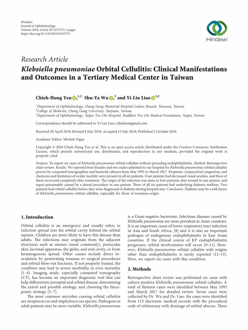
Tabl
e1:
Clin
ical
data
ofsix
patients.
Age/gender
Laterality
Oph
thalmic
chief
complaint
Initial
VA
(logM
AR)
FinalV
A(lo
gMAR)
Fever
Diabetes
HbA
1c/sugar
(ACor
rand
om)
Originof
infection
Surgery
WBC
(1000/µL
)CRP
(mg/L)
Drug
resis
tance
test
Hospitalization
days
169/F
OD
Eyepain,
redn
ess,and
swellin
gfor1
day
LPHand
motion
No
No
–/–
Sinu
sitis
(fronto-
ethm
oidal
mucocele
extend
edto
intracon
alregion
,with
second
ary
acute
infection)
Functio
nal
endo
scop
icsin
ussurgery
(FES
S)
11.5
–Ampicillin
and
ticarcillin
20
230/M
OS
Headache,eye
swellin
g,and
liddrop
for2
days
0.046
0∗Ye
sYe
s11.9/354
(rando
m)
Sepsis,
meningitis
(dental
procedure
priorto
the
episo
de)
No
15.7
285.8
Resis
tance
notfou
nd23
351/F
OD
Eyelid
swellin
g,redn
ess,and
tend
erness
for
2days
0.523∗
0∗No
No
–/–
Sinu
sitis
(pyomucocele)
FESS
orbitotomy
9.8
10.9
Resis
tance
notfou
nd9
471/F
OS
Orbita
lpain
for1day
0.222
–No
Yes
6.1/125(A
C)
Sinu
sitis
(facialb
one
fractureswith
anorbitalw
all
defect
before
theepiso
de)
FESS
sequ
estrectomy
orbitotomy
12–
Resis
tance
notfou
nd17
555/F
OS
Eyelid
swellin
gand
redn
essfor2
days
0.301
0Ye
sNo
–/114
(rando
m)
Sinu
sitis
FESS
orbitotomy
13.3
131.9
Resis
tance
notfou
nd15
648/M
OS
Lidsw
ellin
gfor7days
†0.824
0∗No
Yes
20.7/612
(rando
m)
Trauma
Incisio
nand
drainage
(atthe
office)
13.2
3.3
Resis
tance
notfou
nd10
∗VAwith
person
alglasses.
† *epatient
refusedto
beadmitted
whenhe
camethefirsttim
e;he
was
admitted
22days
afterthe
initialon
set.∗∗*
edatawereob
tained
before
admiss
ionbu
tafte
rinitia
ltreatment,
includ
ingoral
antib
iotic
andtopicala
ntiglaucom
aagents.“–”:d
atano
tavailable.
Journal of Ophthalmology 3

Two patients had nonsinusitis origin and true orbitalabscesses (not subperiosteal abscesses) (Figures 1 and 2).*ey were cured by medical treatment. *e other four pa-tients with sinusitis had undergone surgical drainage fromendonasal approaches and three of them with combinedorbitotomy. Average length of stay of the six cases was 15.67days.
*e six cases will be identified by number 1 to 6,according to the date of admission to our hospital, from pastto present. Table 1 lists greater details of the clinical data.
4. Discussion
We have found four cases of Klebsiella pneumoniae orbitalcellulitis in the literature. All four cases were unilateral. In2001, Lin and Tsai reported a 55-year-old man who had lidabscess caused by sinusitis [12]. However, the authors ofthis report stated in the discussion section that, judgingfrom the clinical signs and the CT images, they thought itmight be a preseptal cellulitis. In 2010, Yang et al. reporteda case who had orbital cellulitis with abscess formationassociated with cavernous sinus thrombophlebitis. *esource of the infection was suspected to be a hematogenousspread from her nasopharyngeal, parapharyngeal, and ret-ropharyngeal abscesses [13]. *is case had type 2 diabetesmellitus which had not been diagnosed until the episode oforbital cellulitis.*ere are also two cases in our series who hadundiagnosed diabetes before they had orbital cellulitis. In2012, Li et al. reported a case with Klebsiella pneumoniaeorbital cellulitis 9 years after repairment of orbital wallfracture with hydroxyapatite implant [14]. Another case wasreported by Murakami et al. in 2016. *e patient had un-derlying type 2 diabetes and had orbital cellulitis and septicemboli in the lungs after tooth extraction. Our case number 2also had a history of dental procedure prior to the onset of hissymptom. Table 2 summarized the previously reported 3cases. Lin’s case was not included since it should be a preseptalcellulitis case.
Klebsiella pneumoniae develops capsules composed ofpolysaccharides which inhibits phagocytosis of the host andcan cause severe invasive diseases. It has been reported thatpoor glycemic control may stimulate capsular polysaccharidebiosynthesis, further impairing the phagocytosis againstprominently virulent capsular serotypes K1 and K2 in patientswith type 2 diabetes [16–18].
In previous reports, two of the three cases had diabetes.In this series, 50% of the patients had underlying diabetesmellitus. *e two without previously recognized diabeteswere found to have high HbA1c levels during their hospitalstay for orbital cellulitis, and they also had nonsinusitisorigin of orbital cellulitis. Diabetes is a known and signif-icant risk factor of Klebsiella pneumoniae endogenousendophthalmitis, and it may also be a risk factor of Klebsiellapneumoniae orbital cellulitis, especially that without sinusitisas in Cases 2 and 6.
Case 2 had meningitis and orbital cellulitis after a dentalprocedure. Both conditions were presented upon initialevaluation, and there was no clear temporal relationshipbetween the two. It was hard to tell if one caused the otheror the Klebsiella pneumoniae bacteremia led to bothconditions independently. Moreover, the patient had noevidence of sinusitis and Klebsiella pneumoniae is nota common pathogen of odontogenic sinusitis [19–21].Diabetes may play a role in the pathogenesis of meningitisand orbital cellulitis, through salivary dysfunction, dys-biosis in the oral cavity, interactions between periodontitisand diabetes [22–24], and overall compromised immunity.In our case 2 and R. Murakami’s case, oral cavity changesmay have caused overgrowth of the bacteria, and com-promised immunity facilitated infection after bacteremiacaused by dental procedures. Case 6 had an open woundwith pus containing Klebsiella pneumoniae. Some studieson ocular flora have shown that diabetes may increasethe culture rate for Gram-negative bacteria and the pro-portion of Klebsiella pneumoniae isolates [25, 26]. *eflora change might also have occurred on the skin of thispatient’s eyelid.
5. Conclusions
Most patients with Klebsiella pneumoniae orbital cellulitishave excellent visual outcome after proper antibiotictreatment, with or without surgical treatment. No antibioticresistance against initial empiric antibiotics was encounteredin this series.
Diabetes mellitus may be a risk factor of Klebsiellapneumoniae orbital cellulitis. If a patient presents withKlebsiella pneumoniae orbital cellulitis and the origin ofinfection is not from endophthalmitis or sinusitis, an un-derlying diabetes mellitus should be suspected.
Table 2: Summary of previously reported cases.
Case report Age/gender
Ophthalmic chiefcomplaint
Initial VA(logMAR)
Final VA(logMAR) Fever Diabetes HbA1c Origin of
infectionSurgicaldrainage
Yang et al.[13] 39/F Eye pain and
swelling for 3 days – (decreased)– (reported tohave beenimproved)
Yes Yes 9.8% Deep neckinfection Yes
Li et al. [14] 52/MEye pain, blepharedema,conjunctival congestion,and proptosis for 1 week
0 – (should beintact) Yes – –
Implant related(orbital wall
fracture repair)Yes
Murakamiet al. [15] 49/M Lid swelling for 5 days 1 −0.079 Yes Yes 15.3% Tooth
extraction No
“–”: data not available.
4 Journal of Ophthalmology
Data Availability
*e clinical data used to support the findings of this studyare included within the article.
Additional Points
Summary. We reported six cases of Klebsiella pneumoniaeorbital cellulitis without preceding endophthalmitis. Fiveof them had excellent visual outcome. Four cases hadorbital infection originated from the sinuses. *e other twohad previously unrecognized diabetes mellitus which mighthave contributed to the susceptibility of the host.
Conflicts of Interest
*e authors declare that there are no conflicts of interestregarding the publication of this paper.
Acknowledgments
Radiologist Chien-Cheng Chen in Chang Gung MemorialHospital Linkou Branch helped the authors review the CTimages of the cases to confirm the findings.
References
[1] I. A. Chaudhry, F. A. Shamsi, E. Elzaridi et al., “Outcome oftreated orbital cellulitis in a tertiary eye care center in the middleEast,” Ophthalmology, vol. 114, no. 2, pp. 345–354, 2007.
[2] A. Bagheri, M. Tavakoli, M. Aletaha, H. Salour, andM. Ghaderpanah, “Orbital and preseptal cellulitis: a 10-yearsurvey of hospitalized patients in a tertiary eye hospital in Iran,”International Ophthalmology, vol. 32, no. 4, pp. 361–367, 2012.
[3] J. Y. Byeon and H. J. Choi, “Orbital cellulitis following orbitalblow-out fracture,” Journal of Craniofacial Surgery, vol. 28,no. 7, pp. 1777–1779, 2017.
[4] C. Mukherjee, A. Mitra, and B. Mushtaq, “Orbital cellulitsfollowing cataract surgery under peribulbar anaesthesia,”GMS Ophthalmology Cases, vol. 5, article Doc02, 2015.
[5] M. S. Todman and Y. R. Enzer, “Medical management versussurgical intervention of pediatric orbital cellulitis: the importanceof subperiosteal abscess volume as a new criterion,” OphthalmicPlastic&Reconstructive Surgery, vol. 27, no. 4, pp. 255–259, 2011.
[6] J. H. Son, H. B. Lim, S. H. Lee, J. W. Yang, and S. B. Lee, “Earlydifferential diagnosis of rhino-orbito-cerebral mucormycosisand bacterial orbital cellulitis: based on computed tomographyfindings,” PLoS One, vol. 11, no. 8, Article ID e0160897, 2016.
[7] A. E. Oester, P. Sahu, B. Fowler, and J. C. Fleming, “Ra-diographic predictors of visual outcome in orbital compart-ment syndrome,” Ophthalmic Plastic and ReconstructiveSurgery, vol. 28, no. 1, pp. 7–10, 2012.
[8] Y.-T. Lin, Y.-P. Wang, F.-D. Wang, and C.-P. Fung,“Community-onset Klebsiella pneumoniae pneumonia inTaiwan: clinical features of the disease and associated mi-crobiological characteristics of isolates from pneumonia andnasopharynx,” Frontiers in Microbiology, vol. 9, 2015.
[9] H. Cho, Y. U. Shin, N. H. Siegel et al., “Endogenousendophthalmitis in the American and Korean population: an8-year retrospective study,” Ocular Immunology and In-flammation, vol. 26, no. 4, pp. 496–503, 2018.
[10] B. W. Davies and R. G. Fante, “Concurrent endophthalmitisand orbital cellulitis from metastatic klebsiella pneumonialiver abscess,” Ophthalmic Plastic and Reconstructive Sur-gery, vol. 32, no. 5, pp. e118–e119, 2016.
[11] K. J. Chen, Y.-P. Chen, A.-N. Chao et al., “Prevention ofevisceration or enucleation in endogenous bacterial pan-ophthalmitis with no light perception and scleral abscess,”PLoS One, vol. 12, no. 1, Article ID e0169603, 2017.
[12] C. T. Lin and Y. Y. Tsai, “Klebsiella pneumoniae orbital cellu-litis,” Zhonghua Yi Xue Za Zhi, vol. 64, no. 9, pp. 551–554, 2001.
[13] S. J. Yang, S. Y. Park, Y. J. Lee et al., “Klebsiella pneumoniaeorbital cellulitis with extensive vascular occlusions in a patientwith type 2 diabetes,” Korean Journal of Internal Medicine,vol. 25, no. 1, pp. 114–117, 2010.
[14] J. Li, J. Ma, and X. Ge, “Late-onset orbital cellulitis withabscess formation caused by klebsiella pneumoniae,” OpenJournal of Ophthalmology, vol. 2, no. 3, pp. 89–92, 2012.
[15] R.Murakami, T. Uchida, K. Tsuzuki, and T. Nakajima, “A caseof Klebsiella pneumoniae orbital cellulitis following the toothextraction in a patient with type 2 diabetes mellitus,” Journalof the Japan Diabetes Society, vol. 59, no. 6, pp. 414–420, 2016.
[16] J. C. Lin, L. K. Siu, C.-P. Fung et al., “Impaired phagocytosis ofcapsular serotypes K1 or K2 Klebsiella pneumoniae in type 2diabetes mellitus patients with poor glycemic control,” Journalof Clinical Endocrinology & Metabolism, vol. 91, no. 8,pp. 3084–3087, 2006.
[17] K. M. Yeh, A. Kurup, L. K. Siu et al., “Capsular serotype K1or K2, rather than magA and rmpA, is a major virulencedeterminant for Klebsiella pneumoniae liver abscess inSingapore and Taiwan,” Journal of Clinical Microbiology,vol. 45, no. 2, pp. 466–471, 2007.
[18] C. H. Lee, I.-L. Chen, S.-K. Chuah et al., “Impact of glycemiccontrol on capsular polysaccharide biosynthesis and opso-nophagocytosis of Klebsiella pneumoniae: implications forinvasive syndrome in patients with diabetes mellitus,” Vir-ulence, vol. 7, no. 7, pp. 770–778, 2016.
[19] J. A. Aas, B. J. Paster, L. N. Stokes, I. Olsen, and F. E. Dewhirst,“Defining the normal bacterial flora of the oral cavity,” Journalof Clinical Microbiology, vol. 43, no. 11, pp. 5721–5732, 2005.
[20] I. Brook, “Microbiology of acute and chronic maxillary si-nusitis associated with an odontogenic origin,” Laryngoscope,vol. 115, no. 5, pp. 823–825, 2005.
[21] R. Derafshi, A. Bazargani, J. Ghapanchi, Y. Izadi, andH. Khorshidi, “Isolation and identification of nonoral path-ogenic bacteria in the oral cavity of patients with removabledentures,” Journal of International Society of Preventive &Community Dentistry, vol. 7, no. 4, pp. 197–201, 2017.
[22] D. A. Grant-*eule, “Periodontal disease, diabetes, and im-mune response: a review of current concepts,” Journal of theWestern Society of Periodontology/Periodontal abstracts,vol. 44, no. 3, pp. 69–77, 1996.
[23] A. Y. Al-Maskari, M. Y. Al-Maskari, and S. Al-Sudairy, “Oralmanifestations and complications of diabetes mellitus: a re-view,” Sultan Qaboos University Medical Journal, vol. 11,no. 2, pp. 179–186, 2011.
[24] L. Samaranayake and V. H.Matsubara, “Normal oral flora andthe oral ecosystem,” Dental Clinics of North America, vol. 61,no. 2, pp. 199–215, 2017.
[25] N. P. Moreno, R. D. Moreno, and L. B. Sousa, “Aerobicbacterial microbiota of the conjunctiva in diabetic patientswith normal and altered glycated hemoglobin levels in tworegions in Brazil,” Arquivos Brasileiros de Oftalmologia,vol. 77, no. 6, pp. 351–354, 2014.
[26] M. Adam, M. Balcı, H. A. Bayhan, A. Ç. Inkaya, M. Uyar, andC. Gurdal, “Conjunctival flora in diabetic and nondiabeticindividuals,” Turk Oftalmoloji Dergisi, vol. 45, no. 5,pp. 193–196, 2015.
Journal of Ophthalmology 5

Stem Cells International
Hindawiwww.hindawi.com Volume 2018
Hindawiwww.hindawi.com Volume 2018
MEDIATORSINFLAMMATION
of
EndocrinologyInternational Journal of
Hindawiwww.hindawi.com Volume 2018
Hindawiwww.hindawi.com Volume 2018
Disease Markers
Hindawiwww.hindawi.com Volume 2018
BioMed Research International
OncologyJournal of
Hindawiwww.hindawi.com Volume 2013
Hindawiwww.hindawi.com Volume 2018
Oxidative Medicine and Cellular Longevity
Hindawiwww.hindawi.com Volume 2018
PPAR Research
Hindawi Publishing Corporation http://www.hindawi.com Volume 2013Hindawiwww.hindawi.com
The Scientific World Journal
Volume 2018
Immunology ResearchHindawiwww.hindawi.com Volume 2018
Journal of
ObesityJournal of
Hindawiwww.hindawi.com Volume 2018
Hindawiwww.hindawi.com Volume 2018
Computational and Mathematical Methods in Medicine
Hindawiwww.hindawi.com Volume 2018
Behavioural Neurology
OphthalmologyJournal of
Hindawiwww.hindawi.com Volume 2018
Diabetes ResearchJournal of
Hindawiwww.hindawi.com Volume 2018
Hindawiwww.hindawi.com Volume 2018
Research and TreatmentAIDS
Hindawiwww.hindawi.com Volume 2018
Gastroenterology Research and Practice
Hindawiwww.hindawi.com Volume 2018
Parkinson’s Disease
Evidence-Based Complementary andAlternative Medicine
Volume 2018Hindawiwww.hindawi.com
Submit your manuscripts atwww.hindawi.com


![Mucocele Expo[1]](https://img.pdfslide.us/doc/110x75/577cdb5c1a28ab9e78a805d7/mucocele-expo1.jpg)