Embed Size (px)
Citation preview
Case ReportRenal Colic: A Red Herring for Mucocele of theAppendiceal Stump
Sameera Ganti 1 and Pothiawala Sohil 2
1SingHealth Emergency Medicine Residency Program, Singapore2Department of Emergency Medicine, Singapore General Hospital, Singapore
Correspondence should be addressed to Sameera Ganti; [email protected]
Received 24 October 2018; Accepted 13 November 2018; Published 6 December 2018
Academic Editor: Yahia A. Raja’a
Copyright © 2018 Sameera Ganti and Pothiawala Sohil. �is is an open access article distributed under the Creative CommonsAttribution License, which permits unrestricted use, distribution, and reproduction in any medium, provided the original work isproperly cited.
Flank pain with hematuria is a common presentation in the emergency department. �e commonest differential diagnosis ofthese patients is renal/ureteric calculus or pyelonephritis. �ese patients are usually treated with analgesia, antibiotics in case ofpyelonephritis, and are discharged with an outpatient referral to a urologist. �is case report describes a 51 year old male whopresented to the ED for recurrent flank pain and hematuria. Bedside ultrasonography in the ED demonstrated a cystic lesion inthe renal area. CT urography revealed an appendiceal stump mucocele and patient was transferred under surgical care. �is casehighlights the importance of the utility of bedside ultrasound in patients presenting to the ED with flank pain or abdominal painwhich can lead to expedited assessment and appropriate management.
1. Introduction
Flank pain is a common presentation in the emergencydepartment (ED). �is symptom coupled with a history ofhematuria raises the suspicion of a renal/ureteric calculusamongmost clinicians. Some patients whohave an associatedcomplaint of dysuria with or without fever are diagnosed tohave pyelonephritis. However, there are many conditions thatmimic this presentation.Wepresent the case of amiddle-agedpatient who presented with flank pain with suspicion of renalcolic.
2. Case Report
A 51 year old man presented to the ED for the third time in 2weeks with complaints of flank pain and hematuria. He hadcomplained of le� sided flank pain during the initial 2 visits.During the first visit, the patient was diagnosed to have renalcolic. X-rayKUBdid not show any renal stone.Hewas treatedsymptomatically with analgesia and discharged. During thesecond visit with complaints of persistent le� flank paindespite taking analgesia given at discharge, he was admitted
to the emergency observation ward for pain management.Bedside ultrasound done then was noted to have mild le�sided hydronephrosis. He was pain free at the end of theobservation and was then discharged with analgesia and anoutpatient follow-up with the urology department. He wasalso scheduled to have an outpatient computed tomographyscan of the kidneys, ureters, and bladder (CTKUB).However,2 days before the scheduled CT, he represented to the EDwith right sided flank pain since morning on the day ofhis visit. �e pain radiated to the right groin and wasassociatedwith hematuria.Hewas not passing blood clots.Hedenied any other complaints of fever, weight loss, vomiting,diarrhea, or constipation. He had a past medical history ofhypertension, diabetes mellitus, and hyperlipidemia. He hadprevious surgeries for appendicectomy and cholecystectomy.
His vital signs were stable. �ere was tenderness overthe right flank on physical examination. �ere was alsoa palpable tender mass measuring about 5x5cm over theright lumbar region. �ere was no renal angel tenderness.Bedside ultrasound in the ED showed an appearance of a6 cm cystic lesion around the inferior pole of the right kidneywith internal echogenicity within the cyst. �e provisional
HindawiCase Reports in Emergency MedicineVolume 2018, Article ID 2502183, 3 pageshttps://doi.org/10.1155/2018/2502183
2 Case Reports in Emergency Medicine
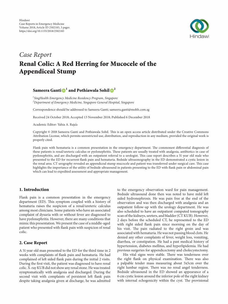
Figure 1: CT KUB showing the cystic swelling arising from the appendiceal stump (red arrow).
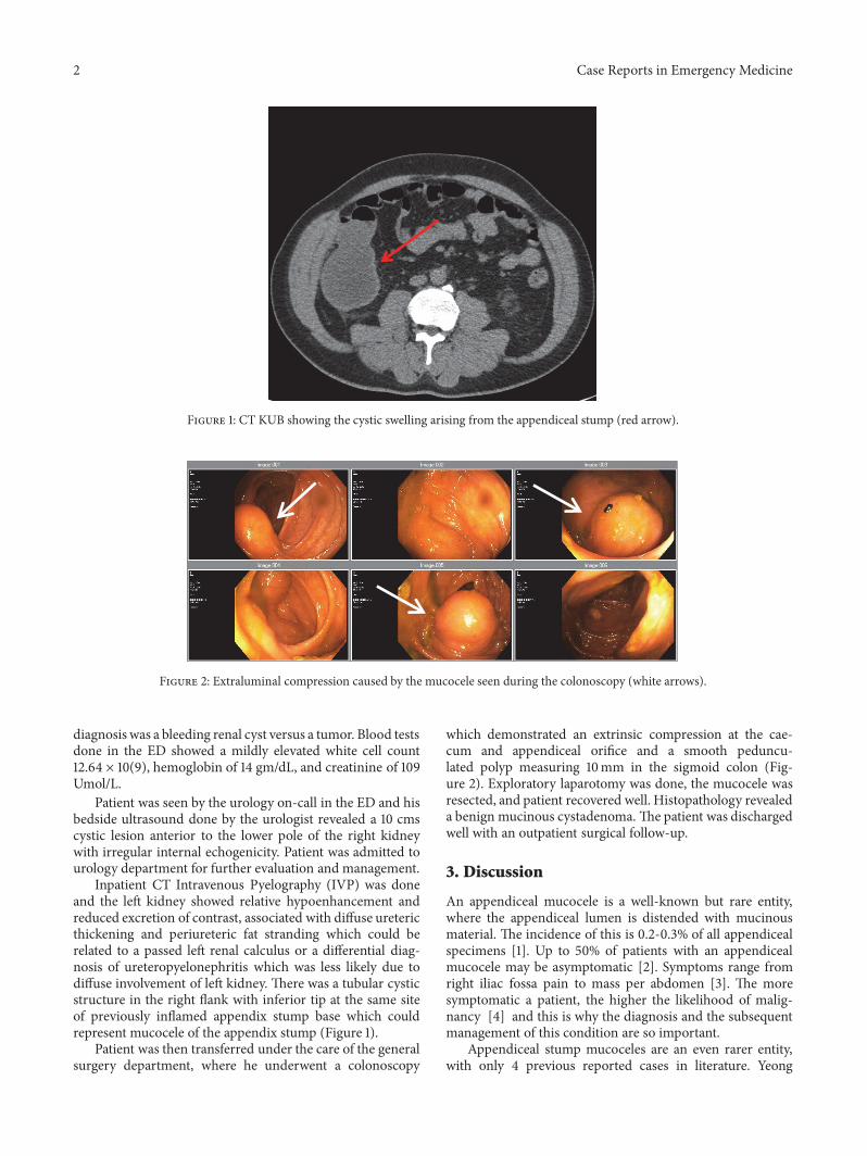
Figure 2: Extraluminal compression caused by the mucocele seen during the colonoscopy (white arrows).
diagnosis was a bleeding renal cyst versus a tumor. Blood testsdone in the ED showed a mildly elevated white cell count12.64 × 10(9), hemoglobin of 14 gm/dL, and creatinine of 109Umol/L.
Patient was seen by the urology on-call in the ED and hisbedside ultrasound done by the urologist revealed a 10 cmscystic lesion anterior to the lower pole of the right kidneywith irregular internal echogenicity. Patient was admitted tourology department for further evaluation and management.
Inpatient CT Intravenous Pyelography (IVP) was doneand the le� kidney showed relative hypoenhancement andreduced excretion of contrast, associated with diffuse uretericthickening and periureteric fat stranding which could berelated to a passed le� renal calculus or a differential diag-nosis of ureteropyelonephritis which was less likely due todiffuse involvement of le� kidney. �ere was a tubular cysticstructure in the right flank with inferior tip at the same siteof previously inflamed appendix stump base which couldrepresent mucocele of the appendix stump (Figure 1).
Patient was then transferred under the care of the generalsurgery department, where he underwent a colonoscopy
which demonstrated an extrinsic compression at the cae-cum and appendiceal orifice and a smooth peduncu-lated polyp measuring 10mm in the sigmoid colon (Fig-ure 2). Exploratory laparotomy was done, the mucocele wasresected, and patient recovered well. Histopathology revealeda benign mucinous cystadenoma. �e patient was dischargedwell with an outpatient surgical follow-up.
3. Discussion
An appendiceal mucocele is a well-known but rare entity,where the appendiceal lumen is distended with mucinousmaterial. �e incidence of this is 0.2-0.3% of all appendicealspecimens [1]. Up to 50% of patients with an appendicealmucocele may be asymptomatic [2]. Symptoms range fromright iliac fossa pain to mass per abdomen [3]. �e moresymptomatic a patient, the higher the likelihood of malig-nancy [4] and this is why the diagnosis and the subsequentmanagement of this condition are so important.
Appendiceal stump mucoceles are an even rarer entity,with only 4 previous reported cases in literature. Yeong
Case Reports in Emergency Medicine 3
et al. were the first to report a case of an appendicealstump mucocele causing pseudomyxoma peritonei [5]. �ereports by Lien et al., Korkolis et al., and El Ajmi et al.are of a benign cystadenoma of the appendiceal stumpwhere the patient presented with symptom of right lowerquadrant pain [6–8]. Our case is the first of its kind wherethe patient presents with flank pain and hematuria whichwould be most commonly diagnosed as renal or uretericcolic.
Management of both conditions, namely, appendicealmucocele and appendiceal stump mucocele, is aimed atsurgical resection and histopathological guided therapy. Inretrospective study, there was an association found betweenappendiceal mucoceles and colonic neoplasms and that studyrecommends surveillance colonoscopy in patient diagnosedwith appendiceal mucoceles [4].�is diagnostic strategy wasapplied to our patient who underwent a colonoscopy whichdid not reveal any pathology.
�e four types of pathological findings described inappendiceal mucoceles are (1) mucous hyperplasia, (2)
simple mucocele, (3) cystadenoma, and (4) cystadenocar-cinoma [9]. �e phenomenon of pseudomyxoma peri-tonei is well associated with the presence of an appen-diceal/appendiceal stump mucocele [10]. �is is a dreadedcomplication and to avoid it, prompt surgical management isindicated.
Emergency ultrasonography (EUS) has become a stan-dard of care in the management of patients in the emer-gency department. Guidelines published by the AmericanCollege of Emergency Physicians encourage the use of EUSfor therapeutic, resuscitative, and diagnostic purposes [11].Bedside ultrasonography is a quick and easy method to assesspatients with suspected nephrolithiasis and is associated withdiagnostic accuracy and less radiation exposure [12]. Marzecet al. and Garcia et al. describe a case of renal tumor detectedin the EDbyusing EUS for assessment of suspected renal colic[13, 14]. �ey suggested that EUS is cheaper and faster and agood screening tool, prior to definitive imaging such as a CTscan.
4. Conclusion
�is case highlights the importance of the utility of bedsideultrasound in patients presenting to the ED with flank painor abdominal pain. It is a quick, focused and an invaluabletool and has become the standard of care in the EDs andalso is part of the training for emergency medicine residentsin the assessment of patients presenting with abdominalpain to the ED. Further evaluation prompted by bedsideEUS done in the ED can lead to an expedited assessmentand appropriate patient management, thus improving patientoutcomes.
Conflicts of Interest
�e authors declare that they have no conflicts of interest.
References
[1] S. Dhage-Ivatury and P. H. Sugarbaker, “Update on the surgicalapproach tomucocele of the appendix,” Journal of the AmericanCollege of Surgeons, vol. 202, no. 4, pp. 680–684, 2006.
[2] J. G. D. A. Filho and E. F. D. Lira, “Mucocele of the appendix:appendectomyor colectomy?” Journal of Coloproctology, vol. 31,no. 3, pp. 276–284, 2011.
[3] P. L. Lakatos, G. Gyori, J. Halasz et al., “Mucocele of theappendix: an unusual cause of lower abdominal pain in a patientwith ulcerative colitis-. A case report and review of literature,”World Journal of Gastroenterology, vol. 11, no. 3, pp. 457–459,2005.
[4] L. Stocchi, B. G. Wolff, D. R. Larson, and J. R. Harrington,“Surgical treatment of appendiceal mucocele,” JAMA Surgery,vol. 138, no. 6, pp. 585–590, 2003.
[5] M. L. Yeong, S. P. Clark, and R. S. Stubbs, “Papillary cys-tadenocarcinoma of the appendiceal stump with mucocele andperitonealmetastases,”Pathology, vol. 21, no. 2, pp. 131–133, 1989.
[6] W.-C. Lien, K.-L. Liu,M.-Y. Kuo et al., “Mucocele of appendicealstump,” Surgery, vol. 136, no. 1, pp. 93-94, 2004.
[7] D. P. Korkolis, K. Apostolaki, G. D. Plataniotis, J. Tzorbatzoglou,I. G. Karaitianos, and P. P. Vassilopoulos, “Mucocele of theappendiceal stump due to benign mucinous cystadenoma,”Anticancer Reseach, vol. 26, no. 1 B, pp. 635–638, 2006.
[8] M. El Ajmi, W. Rebai, Z. Ben Sa�a, and E. A. Mahmoud,“Mucocele of appendiceal stump-An atypical presentation anda diagnostic dilemma,” Acta Chirurgica Belgica, vol. 109, no. 3,pp. 414-415, 2009.
[9] Z. Demetrashvili, M. Chkhaidze, K. Khutsishvili et al., “Muco-cele of the appendix: case report and review of literature,”International Surgery, vol. 97, no. 3, pp. 266–269, 2012.
[10] A. Dixit, J. H. P. Robertson, S. S. Mudan, and C. Akle, “Appen-diceal mucocoeles and pseudomyxoma peritonei,”World Jour-nal of Gastroenterology, vol. 13, no. 16, pp. 2381–2384, 2007.
[11] “ACEP Policy Statement Ultrasound Guidelines: Emergency,Point-of-Care andClinical UltrasoundGuidelines inMedicine,”Annals of Emergency Medicine, vol. 69, no. 5, pp. e27–e54, 2017.
[12] R. Smith-Bindman, C. Aubin, J. Bailitz et al., “Ultrasonographyversus computed tomography for suspected nephrolithiasis,”The New England Journal of Medicine, vol. 371, pp. 1100–1110,2014.
[13] K.Marzec, T. Mailhot, and P. Perera, “Ultrasound detection of arenal mass in a patient with flank pain and hematuria,”WesternJournal of Emergency Medicine, vol. 14, no. 2, pp. 123–126, 2013.
[14] A. A. Oviedo-Garcı́a and M. Algaba-Montes, “Patient withflank pain and intermittent hematuria. �e typical renal colic?Usefulness of ultrasonography in the emergency room,”CriticalUltrasound Journal, vol. 7, Supplement 1, no. A16, 2015.

Stem Cells International
Hindawiwww.hindawi.com Volume 2018
Hindawiwww.hindawi.com Volume 2018
MEDIATORSINFLAMMATION
of
EndocrinologyInternational Journal of
Hindawiwww.hindawi.com Volume 2018
Hindawiwww.hindawi.com Volume 2018
Disease Markers
Hindawiwww.hindawi.com Volume 2018
BioMed Research International
OncologyJournal of
Hindawiwww.hindawi.com Volume 2013
Hindawiwww.hindawi.com Volume 2018
Oxidative Medicine and Cellular Longevity
Hindawiwww.hindawi.com Volume 2018
PPAR Research
Hindawi Publishing Corporation http://www.hindawi.com Volume 2013Hindawiwww.hindawi.com
The Scientific World Journal
Volume 2018
Immunology ResearchHindawiwww.hindawi.com Volume 2018
Journal of
ObesityJournal of
Hindawiwww.hindawi.com Volume 2018
Hindawiwww.hindawi.com Volume 2018
Computational and Mathematical Methods in Medicine
Hindawiwww.hindawi.com Volume 2018
Behavioural Neurology
OphthalmologyJournal of
Hindawiwww.hindawi.com Volume 2018
Diabetes ResearchJournal of
Hindawiwww.hindawi.com Volume 2018
Hindawiwww.hindawi.com Volume 2018
Research and TreatmentAIDS
Hindawiwww.hindawi.com Volume 2018
Gastroenterology Research and Practice
Hindawiwww.hindawi.com Volume 2018
Parkinson’s Disease
Evidence-Based Complementary andAlternative Medicine
Volume 2018Hindawiwww.hindawi.com
Submit your manuscripts atwww.hindawi.com


![Mucocele Expo[1]](https://img.pdfslide.us/doc/110x75/577cdb5c1a28ab9e78a805d7/mucocele-expo1.jpg)