Embed Size (px)
Citation preview

Research ArticleMolecular Typing of Klebsiella pneumoniae Clinical Isolates byEnterobacterial Repetitive Intergenic Consensus PolymeraseChain Reaction
Parinaz Sedighi ,1,2 Omid Zarei ,3 Kiana Karimi ,1,2 Mohammad Taheri ,3
Pezhman Karami ,3 and Leili Shokoohizadeh 3
1Student Research Committee, Hamadan University of Medical Sciences, Hamadan, Iran2Universal Scientific Education and Research Network (USERN), Tehran, Iran3Department of Microbiology, Faculty of Medicine, Hamadan University of Medical Sciences, Hamadan, Iran
Correspondence should be addressed to Leili Shokoohizadeh; [email protected]
Received 6 September 2020; Revised 24 October 2020; Accepted 9 November 2020; Published 21 November 2020
Academic Editor: Simona Nardoni
Copyright © 2020 Parinaz Sedighi et al. )is is an open access article distributed under the Creative Commons AttributionLicense, which permits unrestricted use, distribution, and reproduction in any medium, provided the original work isproperly cited.
Aim. Klebsiella pneumoniae is one of the most important causes of nosocomial infections, including pneumonia, sepsis, andurinary tract infection. Enterobacterial repetitive intergenic consensus polymerase chain reaction (ERIC-PCR) technique is aquick, reliable, and cost-effective method for molecular typing of Enterobacteriaceae family members. )is study aimed to detectgenetic relatedness among K. pneumoniae isolates from hospitals in Hamadan city, using ERIC-PCR technique. Materials andMethods. A total of 72K. pneumoniae isolates were collected from patients admitted to Besat and Sina hospitals. After detectionand confirmation of K. pneumonia isolates by chemical and conventional microbiological methods, DNAs were extracted after 24hours of incubation at 37°C, using the boiling method. ERIC-PCR technique was carried out, and the ERIC patterns were analyzedby online data analysis service (inslico.ehu.es). ERIC profiles were compared using Dice method and clustered by UPGMA(unweighted pair group method with arithmetic mean) program. Also, the samples were evaluated by PCR method for thedetection of aerobactin gene within their genome. Finding.)e genetic relatedness amongK. pneumoniae isolates was studied, andresults established the genetic diversity of the clinical isolates by detecting 25 different ERIC types, including 14 common types and11 unique types. Also, none of the isolates had aerobactin gene. Discussion. )e results of this study showed high genetic diversityamong K. pneumoniae strains, indicating the polyclonal distribution of K. pneumoniae isolates in Hamadan hospitals. )isdiversity causes problems for the treatment of infections due to the circulation of diverse K. pneumoniae clones, which possiblyhave different antimicrobial susceptibility patterns.
1. Introduction
Enterobacteriaceae family is a group of rod-shaped Gram-negative bacteria. Klebsiella pneumoniae, as the most im-portant member of the Klebsiella genus, belongs to thisfamily. K. pneumoniae is a part of human gastrointestinalnormal flora and one of the most important organismsassociated with opportunistic infections, especially among
immunocompromised individuals. )e pathogenesis of in-fection stands on various virulence factors such as poly-saccharide capsule which protects bacteria against hostdefense, endotoxins, and iron-scavenging systems [1].
Nosocomial infections are developed after hospital ad-mission besides the main health problem and are known as aglobal health issue. Multidrug-resistant pathogens usuallyare involved in such infections, and K. pneumoniae is one of
HindawiInternational Journal of MicrobiologyVolume 2020, Article ID 8894727, 5 pageshttps://doi.org/10.1155/2020/8894727

the main culprits associated with pneumonia, urinary tractinfection, wound infection, bacteremia, and septicemia[2–4].
One of the important points to prevent the spread ofhealthcare-associated infections and improve infectioncontrol is identifying the genetic relatedness among thepathogenic spices. Repetitive element sequence-based po-lymerase chain reaction (PCR) is a molecular method thatinvestigates the repetitive nucleotide sequences within thebacterial genome and clusters the bacterial strains. En-terobacterial repetitive intergenic consensus (ERIC) is one ofthe repetitive elements in which the pattern and number aredifferent in the bacterial genome. Enterobacterial repetitiveintergenic consensus polymerase chain reaction (ERIC-PCR) technique is a quick, reliable, and cost-effectivemethod for molecular typing of the Enterobacteriaceaefamily and distinguishes the genetic diversity among thestrains [4, 5].
)e aerobactin gene encodes the aerobactin siderophorewhich is one of the main iron-scavenging systems producedby hypervirulent K. pneumoniae. Hypervirulent strains canlead to life-threatening infections even in healthy individuals[6, 7].
)is study aimed to detect genetic relatedness amongK. pneumoniae isolates from hospitals in Hamadan city,using ERIC-PCR technique. Also, the presence of the aer-obactin gene within their genomes was evaluated.
2. Method
2.1. Bacterial Isolation and Identification. A total number of72K. pneumoniae isolates were gathered from all throat,urine, trachea, wound, blood, sputum, and abscess drainagesamples entered to Besat and Sina Hospital laboratories fromMarch to October 2019. Isolates were confirmed asK. pneumoniae by chemical and conventional microbio-logical methods at hospital laboratories. Also, antibioticsusceptibility testing was done by Kirby–Bauer disk diffusionmethod (Mast, UK) for amikacin, ceftazidime, ciprofloxacin,cotrimoxazole, imipenem, meropenem, levofloxacin, andcolistin. )e samples were saved in microtubes and frozen at−70°C till all were gathered.)en, samples were cultured andincubated at 37°C for 24 hours. Samples’ information in-cluding the sample type (urine, tracheal discharge, sputum,blood, wound, and abscess drainage), age and sex of patients,and type of admission (inpatient or outpatient) wereextracted from laboratories’ information systems.
2.2. ERIC-PCR. Genomic DNAs of K. pneumoniae wereextracted using the boiling method. ERIC-PCR techniquewas carried out in a thermocycler (Bio-Rad, Inc. USA) usingERIC primer—forward: 5ʹ-ATG TAA GCT CCT GGG GATTCAC-3ʹ and reverse: 5ʹ-AAG TAA GTG ACT GGG GTGAGC G3ʹ (Metabion, Germany). )e PCR protocol ismentioned in Table 1 [8].
)e PCR products were loaded on a one percent agarosegel (Sigma-Aldrich) at 70 volts for one hour, and the bandingpatterns were illuminated by ultraviolet radiation (Figure 1).
)e ERIC patterns were analyzed by online data analysisservice (insilico.ehu.es). ERIC profiles were compared usingDice method and clustered by UPGMA (unweighted pairgroup method with arithmetic mean) program. Isolates witha similar ERIC pattern or with one, two, or three differentbands were considered as one ERIC type, and isolates withmore than three inconsistent bands were fallen into differentgroups. )e dendrogram was drawn according to theclusters. )e relation between different ERIC types andhospitals in which samples were taken was evaluated by theChi-square test using SPSS 22 software.
2.3. Detection of the Aerobactin Gene. )e 72 samples wereevaluated by PCR method for the detection of aerobactingene within their genome. PCR was carried out in a ther-mocycler (Bio-Rad, Inc. USA) using the primer—forward:5′-GCA TAG GCG GAT ACG AAC AT-3′ and reverse: 5′-CAC AGG GCA ATT GCT TAC CT-3′ (Metabion, Ger-many). )e PCR protocol for detection of aerobactin gene ismentioned in Table 2 [9].
3. Results
A total number of 72 samples were entered into the studyafter isolates were confirmed as K. pneumoniae by Besat andSina Hospital laboratories. 49 samples belonged to males. 6samples belonged to outpatients. 47 samples belonged toinpatients, and type of admission was not determined for 19samples. Among samples from inpatients, 40 samples weretaken from patients in the intensive care unit (ICU). Also,there was one sample from patients admitted to each ofhematology, infectious disease, orthopedics, general surgery,pediatrics and, burn wards.)eward was not determined forone of the inpatient samples. Of the total samples, 28, 15, 13,5, 5, 1, and 1 were extracted from the throat, urine, trachea,wound, blood, sputum, and abscess cultures, respectively
Table 1: ERIC-PCR protocol.
PCR step Temperature (°C) Time (min) CyclesPrimary denaturation 94 3 1Denaturation 94 1 35Annealing 48 1 35Extension 72 2 35Final extention 72 5 1
Figure 1: Agarose gel after electrophoresis and ultraviolet illu-mination. N. C: Negative control.
2 International Journal of Microbiology

(Figure 2). According to antibiotic susceptibility testing,86.5%, 84%, 80.6%, 72.5%, 72.3%, 67.3%, and 66.1% ofK. pneumoniae isolates were resistant to levofloxacin, cef-tazidime, ciprofloxacin, cotrimoxazole, meropenem, ami-kacin, and imipenem, respectively. All of isolates weresensitive to colistin.
)e genetic relatedness among K. pneumoniae isolateswas studied, and results established 25 different ERIC types,including 14 common types and 11 unique types (Figure 3).
ERIC types A, B, C, G, L, and M belonged to BesatHospital, while type N and most of type K samples belongedto Sina Hospital, and there was a significant differencebetween different ERIC groups in terms of the hospitals inwhich the samples were taken (p value< 0.001). )e largestERIC type was type L including 10 isolates that all of themwere from the intensive care unit (ICU) of Besat Hospital.
72K. pneumoniae isolates were also accessed for thedetection of aerobactin gene. Finally, none of the isolates hadaerobactin gene.
4. Discussion
Gram-negative bacilli are one of the main causes of hospital-acquired infections. K. pneumoniae is one of the Gram-negative bacilli that belong to the Enterobacteriaceae familyand the bacteria are associated with urinary tract infections,pneumonia, wound infections, and bloodstream infections[2, 4]. K. pneumoniae is known as the third leading cause ofhospital-acquired pneumonia in the United States, and alsoit is one the important causes of ventilator-associatedpneumonia (VAP) in intensive care units (ICUs) [10, 11].
In our study, approximately 60% of isolates were relatedto the respiratory system, and about 20% of isolates wereextracted from urine samples. In the same study by ParsaieMehr et al. 70% of K. pneumoniae isolates were related tourine samples and 14% were extracted from tracheal samples[5]. In a prospective cohort study by Xercavins et al. toanalyze the transmission dynamics of extended-spectrumβ-lactamase-producing K. pneumoniae, among 60 isolates,about 47% were extracted from urine samples and only onewas from respiratory tract sample [12]. )e study of biofilmformation and antibiotic resistance of K. pneumoniae iso-lates from clinical samples by Nirwati et al. revealed thatamong 167 isolates, gathered from a tertiary care hospital,51.5% were extracted from respiratory specimens [13]. Wecan conclude that respiratory and urinary tract infections aretwo important infections associated with K. pneumoniae.
Regarding the important role of K. pneumoniae innosocomial infections, genotyping the clinical isolates isbeneficial for the identification of infection sources and to
prevent hospital-acquired infections [4]. Various molecularmethods such as ribotyping, pulsed-field gel electrophoresis(PFGE), and multilocus sequence typing (MLST) can beimplemented for genotyping bacterial spices. However, theyare expensive and time-consuming. Repetitive elementpalindromic (REP) PCR method such as ERIC-PCR is an-other quick, reliable, and cost-effective technique for mo-lecular typing of the Enterobacteriaceae family so that weapplied this technique is our study. According to the results,25 different ERIC types, including 14 common types and 11unique types, were detected by ERIC-PCR which demon-strates the genetic diversity of the K. pneumoniae isolatesfrom Hamadan hospitals. Samples of the largest ERIC groupbelonged to the ICU of a specific hospital, so this resultexpresses a dominant bacterial colon and possible noso-comial infection in this unit. According to our results, therewas no significant relationship between ERIC groups andantibiotic susceptibility patterns. Studies in Iran and othercountries have discussed genetic relatedness amongK. pneumoniae isolates from different clinical settings. In thestudy by Moosavian et al. which was conducted on colistin-resistant K. pneumoniae clinical isolates in the southwest ofIran, 23 ERIC types were identified among 26 spices [2]. )eother study in Iran detected 32 different ERIC types among35K. pneumoniae spices [5]. Also, Wasfi et al. evaluatedgenetic relatedness of multidrug-resistant K. pneumoniaeisolates from Egyptian hospitals, and their results showed adiversity of the spices [14].
A study of virulence profiles of 54K. pneumoniae strainsfrom various clinical specimens by Fertas-Aissani et al.reported the prevalence of aerobactin gene which is 3.7%[15]. Also, another study by Remya et al. reported theprevalence of aerobactin gene to be 5.4% among 370 dif-ferent clinical isolates [16]. However, aerobactin gene wasdetected in none of K. pneumoniae isolates in our study.
Our results in accordance with similar studies indicategenetic diversity of K. pneumoniae clinical isolates. Diversecolons may have different antibiotic resistance patterns andso can cause difficulties for the treatment of infections. Werecommended future studies to evaluate genetic diversityamong bacteria of the Enterobacteriaceae family and find
�ro
at cu
lture
Urin
e cul
ture
Trac
heal
cultu
re
Wou
nd cu
lture
Bloo
d cu
lture
Sput
um cu
lture
Abs
cess
cultu
re
Und
eter
min
ed
Number of samples
0
5
10
15
20
25
30
Figure 2: Number of different samples types.
Table 2: PCR protocol for detection of aerobactin gene.
PCR step Temperature (°C) Time (min) CyclesPrimary denaturation 95 5 1Denaturation 95 1 40Annealing 52 1 40Extension 72 2 40Final extention 72 7 1
International Journal of Microbiology 3
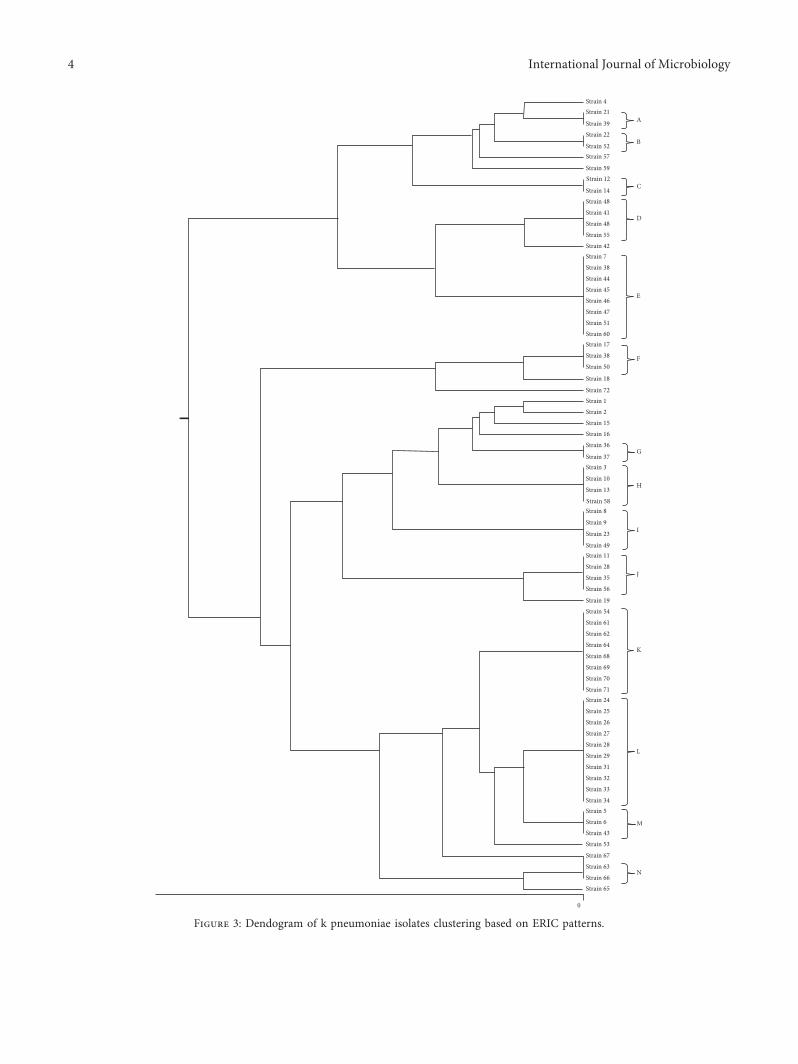
Strain 4Strain 21
A
B
C
D
E
F
G
H
I
J
K
L
M
N
Strain 39Strain 22
Strain 52Strain 57
Strain 59Strain 12
Strain 14Strain 48Strain 41Strain 48Strain 55
Strain 7Strain 42
Strain 38Strain 44Strain 45Strain 46Strain 47Strain 51Strain 60Strain 17Strain 38Strain 50
Strain 18
Strain 72Strain 1Strain 2 Strain 15Strain 16Strain 36
Strain 37Strain 3
Strain 10
Strain 13
Strain 58Strain 8
Strain 9
Strain 23
Strain 49Strain 11Strain 28Strain 35Strain 56
Strain 19Strain 54Strain 61Strain 62Strain 64Strain 68Strain 69Strain 70Strain 71Strain 24Strain 25Strain 26Strain 27Strain 28Strain 29Strain 31Strain 32Strain 33Strain 34Strain 5Strain 6Strain 43Strain 53
Strain 67Strain 63Strain 66Strain 65
0
Figure 3: Dendogram of k pneumoniae isolates clustering based on ERIC patterns.
4 International Journal of Microbiology

possible relations between ERIC type and antibiotic resis-tance pattern. Also, we suggest further studies to evaluatemore samples and the possible relations between expressionof aerobactin gene and potential virulence of K. pneumoniaeisolates.
Data Availability
Data of this manuscript are gathered from the researchentitled “Molecular Typing of Klebsiella pneumoniae Isolatesfrom Hamadan hospitals by Enterobacterial RepetitiveIntergenic Consensus (ERIC)-PCR in 2018-2019.”
Ethical Approval
)e study was approved by the ethics committee of Ham-adan University of Medical Sciences(IR.UMSHA.REC.1397.682).
Conflicts of Interest
)e authors declare that there are no conflicts of interestregarding the publication of this article.
Acknowledgments
)e authors would like to thank staff of microbiology lab-oratory at Hamadan University of Medical Sciences. )isresearch was supported by student research committee,Hamadan University of Medical Sciences (grant number:9711096717).
References
[1] S. Zhang, G. Yang, Q. Ye, Q. Wu, J. Zhang, and Y. Huang,“Phenotypic and genotypic characterization of Klebsiellapneumoniae isolated from retail foods in China,” Frontiers inMicrobiology, vol. 9, no. 289, 2018.
[2] M. Moosavian and N. Emam, “)e first report of emergingmobilized colistinresistance (mcr) genes and ERIC-PCRtyping in Escherichia coli and Klebsiella pneumoniae clinicalisolates in southwest Iran,” Infection and Drug Resistance,vol. 12, pp. 1001–1010, 2019.
[3] H. Y. P. Phoon, H. Hussin, B. M. Hussain et al., “Distribution,genetic diversity and antimicrobial resistance of clinicallyimportant bacteria from the environment of a tertiary hospitalin Malaysia,” Journal of Global Antimicrobial Resistance,vol. 14, pp. 132–140, 2018.
[4] K. SeifiH. Kazemian et al., “Evaluation of biofilm formationamong Klebsiella pneumoniae isolates and molecular char-acterization by ERIC-PCR,” Jundishapur Journal of Micro-biology, vol. 9, no. 1, 2016.
[5] V. Parsaie Mehr, L. Shokoohizadeh, M. Mirzaee, andM. Savari, “Molecular typing of Klebsiella pneumoniae iso-lates by enterobacterial repetitive intergenic Consensus(ERIC)–PCR,” Infection Epidemiology and Microbiology,vol. 3, no. 4, pp. 112–116, 2017.
[6] T. A. Russo, R. Olson, U. MacDonald, J. Beanan, andB. A. Davidson, “Aerobactin, but not yersiniabactin, sal-mochelin, or enterobactin, enables the growth/survival ofhypervirulent (hypermucoviscous) Klebsiella pneumoniae,”Infection and Immunity, vol. 83, no. 8, pp. 3325–3333, 2015.
[7] T. A. Russo, R. Olson, U. MacDonald et al., “Aerobactinmediates virulence and accounts for increased siderophoreproduction under iron-limiting conditions by hypervirulent(hypermucoviscous)Klebsiella pneumoniae ex vivo and invivo,” Infection and Immunity, vol. 82, no. 6, pp. 2356–2367,2014.
[8] O. Zarei, L. Shokoohizadeh, H. Hossainpour, andM. Y. Alikhani, “Molecular analysis of Pseudomonas aeru-ginosa isolated from clinical, environmental and cockroachsources by ERIC-PCR,” BMC Research Notes, vol. 11, no. 1,p. 668, 2018.
[9] W.-L. Yu, W.-C. Ko, K.-C. Cheng, C.-C. Lee, C.-C. Lai, andY.-C. Chuang, “Comparison of prevalence of virulence factorsfor Klebsiella pneumoniae liver abscesses between isolateswith capsular K1/K2 and non-K1/K2 serotypes,” DiagnosticMicrobiology and Infectious Disease, vol. 62, no. 1, pp. 1–6,2008.
[10] R. M. Martin and M. A. Bachman, “Colonization, infection,and the accessory genome of Klebsiella pneumoniae,” Fron-tiers in Cellular and Infection Microbiology, vol. 8, no. 4, 2018.
[11] S. S. Magill, J. R. Edwards, W. Bamberg et al., “Multistatepoint-prevalence survey of health care–associated infections,”New England Journal of Medicine, vol. 370, no. 13,pp. 1198–1208, 2014.
[12] M. Xercavins, E. Jimenez, E. Padilla et al., “High clonal di-versity of ESBL-producing Klebsiella pneumoniae isolatesfrom clinical samples in a non-outbreak situation. A cohortstudy,” Antimicrobial Resistance & Infection Control, vol. 9,no. 1, p. 5, 2020.
[13] H. Nirwati, K. Sinanjung, F. Fahrunissa et al., “Biofilm for-mation and antibiotic resistance of Klebsiella pneumoniaeisolated from clinical samples in a tertiary care hospital,Klaten, Indonesia,” BMC Proceedings, vol. 13, no. 11, p. 20,2019.
[14] R.Wasfi,W. F. Elkhatib, andH.M. Ashour, “Molecular typingand virulence analysis of multidrug resistant Klebsiellapneumoniae clinical isolates recovered from Egyptian hos-pitals,” Scientific Reports, vol. 6, no. 1, p. 38929, 2016.
[15] R. El Fertas-Aissani, Y. Messai, S. Alouache, and R. Bakour,“Virulence profiles and antibiotic susceptibility patterns ofKlebsiella pneumoniae strains isolated from different clinicalspecimens,” Pathologie Biologie, vol. 61, no. 5, pp. 209–216,2013.
[16] P. A. Remya, M. Shanthi, and U. Sekar, “Characterisation ofvirulence genes associated with pathogenicity in Klebsiellapneumoniae,” Indian Journal of Medical Microbiology, vol. 37,no. 2, pp. 210–218, 2019.
International Journal of Microbiology 5