Embed Size (px)
Citation preview
Fattore et al. Journal of Translational Medicine 2013, 11:180http://www.translational-medicine.com/content/11/1/180
RESEARCH Open Access
Activation of an early feedback survival loopinvolving phospho-ErbB3 is a general response ofmelanoma cells to RAF/MEK inhibition and isabrogated by anti-ErbB3 antibodiesLuigi Fattore1,2, Emanuele Marra3, Maria Elena Pisanu2,4, Alessia Noto1,2, Claudia de Vitis2,4, Francesca Belleudi1,5,Luigi Aurisicchio3, Rita Mancini1,2, Maria Rosaria Torrisi1,5,6, Paolo Antonio Ascierto7 and Gennaro Ciliberto7*
Abstract
Background: Treatment of advanced melanoma has been improved with the advent of the BRAF inhibitors.However, a limitation to such treatment is the occurrence of resistance. Several mechanisms have been identifiedto be responsible for the development of resistance, either MEK-dependent or MEK-independent. In order toovercome resistance due to reactivation of MEK signaling, MEK inhibitors are being clinically developed withpromising results. However, also in this case resistance inevitably occurs. It has been recently reported that ErbB3, amember of the EGFR receptor family, may be involved in the establishment of drug resistance.
Methods: Three melanoma cell lines were tested: LOX IMVI (BRAF V600E), MST-L (BRAF V600R) and WM266 (BRAFV600D). Phosphorylation of Receptor Tyrosine Kinases (RTKs) was assessed by an RTK array. Western blot analysis wasperformed on total protein extracts using anti-ErbB3, anti-AKT and anti-ERK 1/2 antibodies. The expression ofneuregulin after vemurafenib treatment was assessed by Real Time PCR and Western blotting. The growth inhibitoryeffects of vemurafenib, GSK1120212b and/or anti-ErbB3 mAbs were evaluated by in vitro colony formation assays.
Results: In the present study we demonstrate that ErbB3 is the main RTK undergoing rapidly hyperphosphorylationupon either treatment with a BRAF inhibitor or with a MEK inhibitor in a panel of melanoma cell lines harboring avariety of V600BRAF mutations and that this results in a strong activation of phospho-AKT. Importantly, ErbB3 activationis fully abrogated by the simultaneous use of anti-ErbB3 monoclonal antibodies, which are also shown to potentlysynergize with BRAF inhibitors in the inactivation of both AKT and ERK pathways and in the inhibition of melanoma cellgrowth. We show that upregulation of phospho-ErbB3 is due to an autocrine loop involving increased transcriptionand production of neuregulin by melanoma cells.
Conclusions: On the basis of these results, we propose that initial co-treatment with BRAF and/or MEK inhibitors andanti-ErbB3 antibodies should be pursued as a strategy to reduce the ErbB3-dependent feedback survival mechanismand enhance duration of clinical response.
Keywords: Melanoma, BRAF/MEK inhibitors, ErbB3 hyperphosphorylation, Anti-ErbB3 antibodies
* Correspondence: [email protected] Nazionale per lo Studio e la Cura dei Tumori “Fondazione G.Pascale”, Via Mariano Semmola, 80131 Naples, ItalyFull list of author information is available at the end of the article
© 2013 Fattore et al.; licensee BioMed Central Ltd. This is an Open Access article distributed under the terms of the CreativeCommons Attribution License (http://creativecommons.org/licenses/by/2.0), which permits unrestricted use, distribution, andreproduction in any medium, provided the original work is properly cited.
Fattore et al. Journal of Translational Medicine 2013, 11:180 Page 2 of 11http://www.translational-medicine.com/content/11/1/180
BackgroundMalignant melanoma is the deadliest form of skin cancer.Over the last 60 years there has been an approximate 700%increase in the incidence of melanoma world-wide andmortality has also increased by 165% [1]. Disease-specificsurvival curves in all stages of melanoma have a negativeslope and overall prognosis is poor with less that 5% ofstage 4 patients surviving 5 years from the manifestation ofmetastatic disease [1]. Before 2011 only three drugs wereFDA-approved for metastatic melanoma, fotemustine,dacarbazine and high-dose IL2, all of them giving rise tomodest response rates (6 to 15%) with median progressionfree survival (PFS) of 1.7 months, only 25.5% of patientsstill alive at 1 year and rare long-term regressions [2]. In re-cent years however, the scenario has completely changedthanks to the development of innovative systemic therap-ies. In first instance immunotherapy with ipilumumab hasdemonstrated improved survival in patients with advancedmelanoma in Phase III randomized trials [3]. At the sametime novel agents directed to target cell autonomousdisregulated pathways have shown remarkable clinical ef-fects. The first one is vemurafenib, a selective inhibitor ofBRAF-activating mutations which are found in more than50% of melanomas and which cause constitutive activationof the MAPK/ERK pathway driving uncontrolled melan-oma growth. In a first Phase I trial vemurafenib (previ-ously called PLX4032) achieved an objective response ratein excess of 50-60% in advanced disease [4]. Subsequentlya phase III study comparing vemurafenib to dacarbazineshowed a significant increase in survival for patients re-ceiving vemurafenib [5]. Other more potent BRAF inhibi-tors are in advanced clinical development, having achievedpromising results in early trials [6]. It is important to pointout that BRAF inhibitors are active only in tumors whereV600 BRAF mutations result in constitutively active mono-mers, whereas the same inhibitors give rise to paradoxicaltumor promoting effects in RAS mutated melanomas be-cause of their ability to induce allosteric activation throughhomo- or hetero-dimerization of wild type RAF isoforms[7,8]. Hence, the current strategy to tackle NRAS mutatedmelanomas involves the use of inhibitors of more down-stream kinases in the RAS-RAF-MAPK pathway, in particu-lar MEK [9]. In line with this, very recently MEK inhibitorshave shown clinical activity as single agents in patients bear-ing mutated NRAS [10].Although BRAF inhibitors (BRAFi) induce unprecedented
objective responses virtually all responders suffer from dis-ease progression due to the development of de novo drugresistance [11,12]. Thus, in order to significantly improvemelanoma survivability it is necessary to develop new ap-proaches to overcome or, better, avoid the development ofresistance to BRAFi. Recent investigations suggest that thereare multiple mechanisms responsible for the establishmentof resistance to BRAFi, which can be grouped into two
major modes MEK-dependent and MEK-independent[13,14]. In the first and more frequent case, reactivationof the MAPK pathway occurs, for example through theacquisition of novel N-RAS mutations or V600E BRAFtruncations resulting in RAS-independent RAF dimerizationwith other members of the same family. In the secondcase cancer growth depends upon activation of signal-ing pathways redundant to MAPK, for example viaoverexpression of RTKs, such as PDGFR or IGF1R,which promote activation of the PI3K-AKT pathway.These mechanisms have been observed both in melan-oma cell cultures exposed in vitro to continuous selec-tion with BRAF inhibitors, and in post-relapse humanmelanoma tumor samples [14,15]. Importantly, second-ary mutations in V600E BRAF have not been identified indrug-resistant tumors, thus arguing that the strategy toovercome BRAFi resistance in melanoma has to rely onthe development of combinatorial approaches.The evidence that resistance to BRAFi frequently de-
pends upon reactivation of the MAPK pathway has led tothe development of novel strategies directed to simultan-eously co-target BRAF and MEK in the attempt to mitigatethe emergence of resistance [15]. Indeed, MEK inhibitorshave been shown to increase progression free survivalwhen delivered in combination with a BRAF inhibitor ascompared to BRAF inhibitor monotherapy [16]. However,even if combinatorial treatment with BRAFi and MEKigives rise to increase in time to progression as comparedto BRAFi monotherapy, this approach is unable to com-pletely eradicate disease, most likely because of MAPK-independent adaptive changes taking place in melanomacells upon exposure to inhibitors of this pathway. There-fore additional approaches are under study which includefor example combination treatments with MEK and IGF1R/PI3K inhibitors [15].The EGFR family of receptor tyrosine kinases consists of
four closely related family members: EGFR (Her1), ErbB2(HER2), ErbB3 (Her3), and ErbB4 (Her4) [17]. These recep-tors are important regulators of normal growth and cell dif-ferentiation. Their gene amplification, overexpression ormutation is associated with tumor development and poorclinical prognosis in most of the human cancers [18]. EGFRand HER2, have been among the most extensively studiedfor the therapy of cancer over the past twenty years and awealth of drugs directed against them have been eitheralready approved or are in advanced clinical development[18]. Although ErbB3 has been disregarded for several yearsas a target for cancer therapies recent meta-analysis ofErbB3 expression in several solid tumor demonstrated thatincreased levels of receptor are constantly associated withworse survival [19]. Indeed, during the last years several evi-dences have been accumulated pointing to a key role of thisreceptor in tumorigenesis and cancer progression both as anode in ligand-induced signalling by members of the EGFR
Fattore et al. Journal of Translational Medicine 2013, 11:180 Page 3 of 11http://www.translational-medicine.com/content/11/1/180
family [20] and as a major contributor of resistance to ther-apies in lung and breast cancer [21-23], which fueled effortstowards the development of ErbB3 inhibitors [24]. We haverecently shown that melanoma cells often express ErbB3 inconcert with other ErbBs and that neuregulin, actingthrough ErbB3, activates the PI3K/AKT pathway, thus lead-ing to increased cell survival, proliferation and migration[25]. Furthermore, we have generated a set of three anti-human ErbB3 monoclonal antibodies (mAbs) in our labora-tory A2, A3 and A4 [26]. These antibodies have beencharacterized biochemically and functionally [25,26]. Thesestudies led to conclude that, at least in melanoma cell cul-tures, A3 and A4, but not A2 are able to strongly inhibitligand-induced signalling, proliferation and migration. Inthe attempt to understand their mechanism of action, wedemonstrated through a series of combined approachesthat antibody efficacy correlated with the ability to inducereceptor internalization, degradation and inhibition of re-ceptor recycling to the cell surface [25].In the present work we show that ErbB3 is central to a
feedback survival loop activated in melanoma cells uponexposure to BRAF and/or MEK inhibitors, that this activa-tion is dependent upon increased production and releaseof neuregulin by melanoma cells and, most importantly,that antibodies against ErbB3 capable to induce receptordegradation, abolish this loop and strongly potentiate theantitumor efficacy of BRAF and or MEK inhibitors whengiven in combination.
MethodsCell lines and treatmentsHuman melanoma cell lines LOX IMVI, MST-L andWM266 were cultured in RPMI supplemented with 10%FBS. To evaluate ErbB3, AKT and ERK 1/2 signaling andneuregulin expression melanoma cells were serum starvedfor 24 h and treated with vemurafenib and/or GSK1120212at different doses and times and incubated or not with20 μg/ml of anti-ErbB3 mAbs A4, A3 or A2 . To determineeffects on proliferation melanoma cell lines, seeded at1*105/ well, were treated with increasing concentrations(from 0,002 to 1 μM) of vemurafenib and/or GSK1120212alone or in combination with anti-ErbB3 mAbs for 10 days.To evaluate neuregulin (HRG) release LOX IMVI cellswere treated for 24 h with vemurafenib and then the condi-tioned medium of BRAFi treated cells (pre-incubated ornot with the anti-HRG antibody) was used to stimulatestarved LOX IMVI cells for 1 h.
Antibodies and reagentsAntibodies against AKT, ERK 1/2, phospho-ErbB3,phospho-AKT and phospho-ERK 1/2 were purchasedfrom Cell Signaling Technology. Anti-ErbB3, anti-HRGand anti-GAPDH were obtained from Santa Cruz Bio-technology. Anti-rabbit and anti-mouse were purchased
from AbCam. The anti-HRG (blocking peptide) waspurchased from Thermo Scientific. Anti ErbB3 anti-bodies A2, A3 and A4 have been described previouslyby our laboratory [25,26]. The three anti-ErbB3 anti-bodies are all of the IgG1 isotype (EM unpublishedobservation). Vemurafenib and GSK1120212 wereobtained from Selleck Chemicals. TaqMan probes forHRG and housekeeping gene 18S were purchased fromApplied Biosystems.
Phospho-RTK arrayA human phospho-RTK array (R&D Systems) was used todetect simultaneously the phosphorylation status of RTKs(n = 49) in melanoma cells. Membranes were incubatedwith cell lysates (100 μg) overnight according to themanufacturer' s protocol. After washing, the membraneswere incubated with a phosphotyrosine antibody conju-gated to horseradish peroxidase to allow the detection ofcaptured RTKs that are phosphorylated. Array data on im-ages were analyzed using Photoshop Quantity One Pro-gram (Bio-Rad LaboratoriesGmbH). Duplicate dots ineach corner are positive controls.
Western blot analysisMelanoma cells were lysed with RIPA buffer; 50 μg of totalprotein were resolved under reducing conditions by 8%SDS-PAGE and transferred to reinforced nitrocellulose(BA-S 83, Schleider and Schuell, Keene, NH, USA). Themembranes were blocked with 5% non fat dry milk in PBS0.1% Tween 20, and incubated with the different primaryantibodies. The membranes were rehydrated and probedagain with anti-GAPDH, to estimate the protein equal load-ing. Densitometric analysis was performed using QuantityOne Program (Bio-Rad Laboratories GmbH) and resultswere expressed as mean values from three independentexperiments.
RNA extraction and real-time PCR analysisRNA was extracted using TRIzol method (Invitrogen)according to manufacturer’s instruction and eluted with0,1% diethylpyrocarbonate (DEPC)- treated water. TotalRNA was quantitated by spectrophotometry. Real Time-PCR was assayed by TaqManW Gene Expression Assays(Applied Biosystems, Foster City, CA). To normalize theamount of source RNA, 18S transcript from the samesample was measured and used as internal reference.Each targeted transcript was validated using the com-parative Ct method for relative quantification (ΔΔCt)reference to the amount of a common reference gene(18S). The fold difference was calculated using the com-parative ΔΔCt and results were reported as mean valuesfrom three independent experiments.
1 2 3 4
vem
(0.
3μM
)un
trea
ted
pErbBs
LOX IMVI MST-L
0
20
40
60
80
100
120
140
160 untreated
Vemurafenib
ErbB1 ErbB2 ErbB3 ErbB4
Mea
n P
ixel
Den
sity
0
20
40
60
80
100
120
140
160 untreated
Vemurafenib
ErbB1 ErbB2 ErbB3 ErbB4
Mea
n P
ixel
Den
sity
1 2 3 4
pErbBs
a b
0 0.3 0.1 0.03 0.3 0.1 0.03
MST-L
6h 24h
1 1 1 1.1 4.3 3.4 1.9
1 0.1 0.1 0.5 0.4 0.3 0.6
1 0.8 0.7 0.8 2.2 1.9 1.4
ErbB3
ERK 1/2
AKT
GAPDH
0 0.3 0.1 0.03 0.3 0.1 0.03
6h 24h
1 1 0.9 1.1 15 7.8 6.5
1 1 1 0.8 12.1 12.7 7.2
1 0.1 0.1 0.3 0.3 0.2 0.6
pErbB3Y1289pErbB3/Erbb3
pERK 1/2T202/Y204pERK/ERK
pAKTS473pAKT/AKT
LOX IMVI
vem (μM)
c d
PDGFR β PDGFR βIGF-1 R IGF-1 R
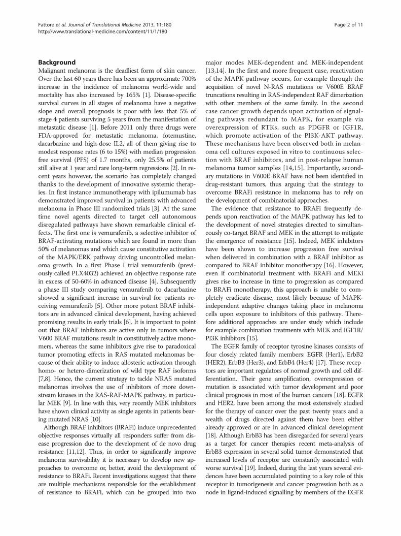
Figure 1 (See legend on next page.)
Fattore et al. Journal of Translational Medicine 2013, 11:180 Page 4 of 11http://www.translational-medicine.com/content/11/1/180
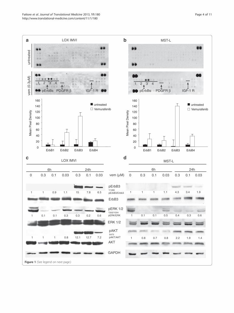
(See figure on previous page.)Figure 1 Vemurafenib treatment induces selective ErbB3 phosporylation in melanoma cells. Simultaneous detection of thephosphorylation status of RTKs (n = 49) using a human phospho-RTK array in LOX IMVI (a) and MST-L (b) melanoma cells treated or not for 24 hwith 0.3 μM vemurafenib. Membranes were incubated with cell lysates (100 μg) overnight according to the manufacturer' s protocol. The arraydetects the tyrosine-phosphorylated RTKs simultaneously in duplicate (1, pErbB1; 2, pErbB2; 3, pErbB3; 4, pErbB4). Duplicate dots in each cornerare positive controls. Array data on images were analyzed using Photoshop Quantity One Program (Bio-Rad LaboratoriesGmbH). Thephosphorylation of ErbB3 is strongly increased by vemurafenib treatment. LOX IMVI (c) and MST-L (d) cells were serum starved for 24 h, treatedor not with different doses of vemurafenib for 6 h or 24 h. Western blot analysis performed using the indicated antibodies shows thatvemurafenib induces a strong dose-dependent and time-dependent phosphorylation of ErbB3 and AKT. For densitometric analysis pErbB3/ErbB3,pERK/ERK and pAKT/ATK values are expressed as fold change with respect to the control unstimulated cells to which value = 1 was assigned.Results are reported as mean values ± standard deviation (SD) from three independent experiments.
Fattore et al. Journal of Translational Medicine 2013, 11:180 Page 5 of 11http://www.translational-medicine.com/content/11/1/180
In vitro colony formation assayCells viability was determined by crystal violet staining.Briefly, the cells were stained for 20 min at roomtemperature with staining solution (0,5% crystal violetin 30% methanol), washed four times with water andthen dried. Cells were then dissolved in a Methanol/SDS solution and the adsorbance (595 nm) was readusing a microplate ELISA reader.
Statistical analysisQuantitative analyses for curve fitting and for IC50 evalu-ation, were performed by KaleidaGraph software. p-valueswere calculated using Student’s t test and significance levelhas been defined as p < 0,05.
Results and discussionErbB3 is the only RTK rapidly phosphorylated uponexposure of BRAF mutated melanoma cells tovemurafenibIn order to identify the mechanism responsible for earlyadaptive changes of melanoma cells to BRAF inhibition,we postulated that receptor tyrosine kinases may be im-portant sensors. Hence, we utilized an RTK array to detectearly changes in the phosphorylation level of approximatelyfifty RTKs. LOX IMVI melanoma cells bearing the mostfrequent oncogenic BRAF mutation V600E [27] weretreated for 24 h with 0.3 μM vemurafenib. Surprisingly wefound that, while the phosphorylation level of most re-ceptors remained unchanged or was subjected to subtlevariations, the only receptor whose phosphorylationwas consistently upregulated 50–100 fold was ErbB3(Figure 1a). These results were confirmed in two othermelanoma cell lines, MST-L [25] bearing a V600R mu-tation (Figure 1b) and WM266 bearing a V600D [27]mutation (Additional file 1: Figure S1a). Hence, ErbB3is the major RTK undergoing hyperphosphorylationupon BRAF inhibition in melanoma cells bearing dis-tinct BRAF mutations as well as different ErbB receptorcompositions (Additional file 2: Table S1). This stronglysuggests that this is a general phenomenon taking placein melanoma when BRAF is inhibited.
Cell extracts of melanoma cell lines LOX IMVI andMST-L exposed to vemurafenib at different doses andtimes were prepared and subjected to western blotting.The results (Figure 1c and d) show that ErbB3 under-goes a strong dose- and time-dependent upregulation ofits phosphorylation in the absence of external additionof neuregulin (HRG). Feedback activation of pErbB3was accompanied by increased phosphorylation of AKT(Figure 1c and d), which suggests the activation of apro-survival loop contributing to dampen the efficacy ofBRAF inhibitors. Importantly the same findings wereconfirmed in WM266 (Additional file 1: Figure S1b). Itis important to point out that pErbB3 upregulation takesplace in the absence of increased levels of ErbB3 protein(see WBs in Figure 1c and d and Additional file 1: FigureS1b) and in the absence of increased levels of ErbB3 andFOXD3 mRNA as indicated by gene expression profilingof untreated vs vemurafenib treated melanoma cells (notshown).
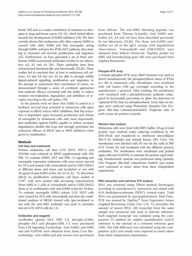
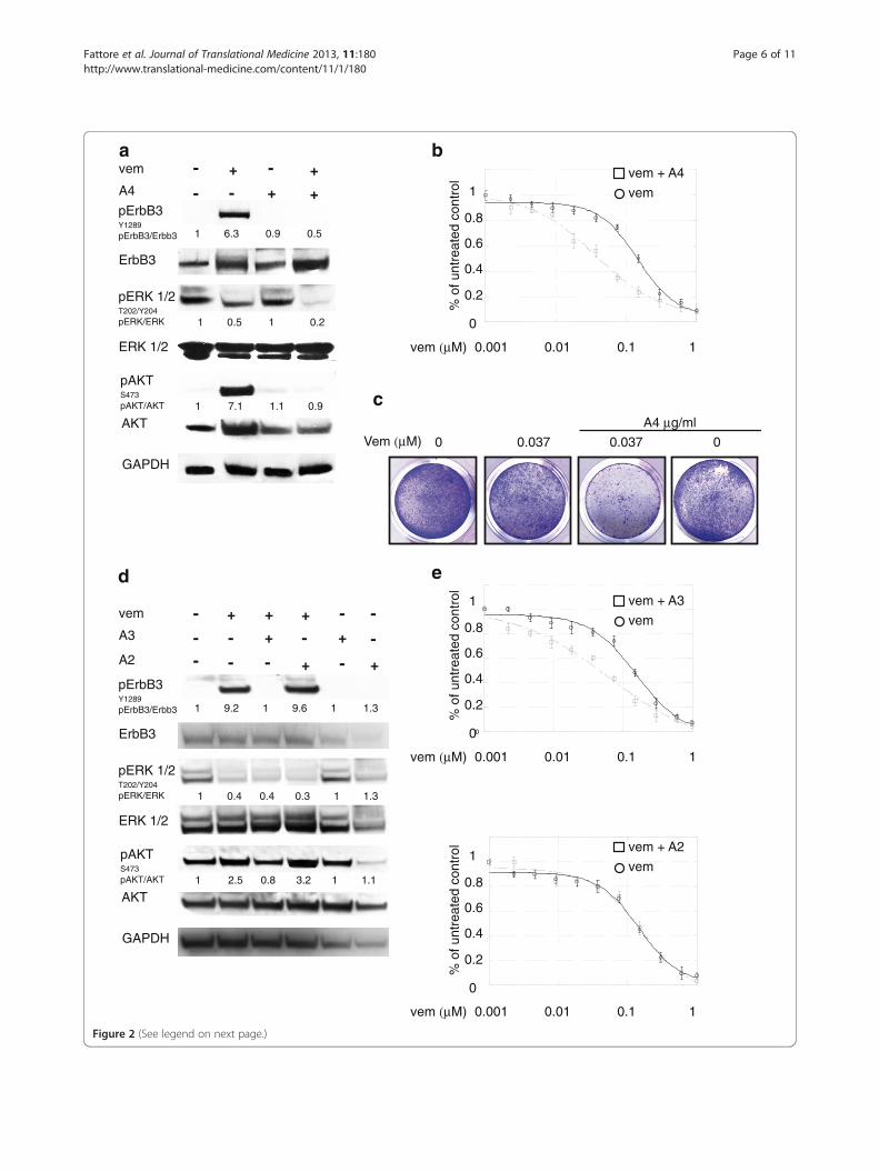
BRAF inhibitor-induced feedback survival loop isabrogated by anti-Erbb3 antibodiesWe therefore assessed the effect of the anti-ErbB3 anti-body A4 generated in our lab and able to inhibit theligand-induced signaling and to potently induce receptorinternalization and degradation [25,26], on vemurafenib-induced pErbB3 and pAKT levels and found that thiswas able to completely abrogate receptor phosphoryl-ation and AKT signaling in all cell lines tested (Figure 2a,for LOX IMVI and Additional file 3: Figure S2a and cfor MST-L and WM266 respectively). Also, it is import-ant to notice that the combined treatment led to astronger degree of pERK down regulation.In order to assess whether inhibition of pErbB3 and
pAKT could result in potentiation of the growth inhibi-tory effects of vemurafenib, in vitro colony formationassays were carried out in the presence of growing con-centrations of vemurafenib alone or in combinationwith a fixed dose of A4. Remarkably, treatment withanti-ErbB3 mAb strongly potentiated growth inhibitionby vemurafenib (Figure 2b and c for LOX IMVI andAdditional file 3: Figure S2b and d for MST-L and
A4
- + -
- +
+vem
- +
ErbB3
ERK 1/2
AKT
GAPDH
a
c
b
Vem (μM)A4 μg/ml
0 0.037 0.037 0
1 6.3 0.9 0.5
1 0.5 1 0.2
1 7.1 1.1 0.9
pErbB3Y1289pErbB3/Erbb3
pERK 1/2T202/Y204pERK/ERK
pAKTS473pAKT/AKT
1
0.8
0.6
0.4
0.2
0
0.001 0.01 0.1 1
% o
f unt
reat
ed c
ontr
ol
vem (μM)
vem + A4
vem
e
0
1
0.8
0.6
0.4
0.2
0
0.001 0.01 0.1 1
% o
f unt
reat
ed c
ontr
ol
vem (μM)
vem + A3
vem
1
0.8
0.6
0.4
0.2
0
0.001 0.01 0.1 1
% o
f unt
reat
ed c
ontr
ol
vem (μM)
vem + A2
vem
d
1 9.2 1 9.6 1 1.3
1 0.4 0.4 0.3 1 1.3
1 2.5 0.8 3.2 1 1.1
A3
- +
+
+vem
-
+
ErbB3
ERK 1/2
AKT
GAPDH
pErbB3Y1289pErbB3/Erbb3
pERK 1/2T202/Y204pERK/ERK
pAKTS473pAKT/AKT
A2 ---
+
--
-+
-
-
-
+
Figure 2 (See legend on next page.)
Fattore et al. Journal of Translational Medicine 2013, 11:180 Page 6 of 11http://www.translational-medicine.com/content/11/1/180
(See figure on previous page.)Figure 2 Anti-ErbB3 mAbs differently counteract the increase of ErbB3-dependent AKT phosphorylation and potentiate growthinhibition induced by vemurafenib. LOX IMVI melanoma cells serum starved and treated with vemurafenib (0.3 μM) for 24 h were incubated ornot with 20 μg/ml of anti-ErbB3 mAbs A4 (a), A3 or A2 (d). Western blot analysis shows that A4 and A3, but not A2 mAbs abrogate receptorphosphorylation and ATK signaling. For densitometric analysis pErbB3/ErbB3, pERK/ERK and pAKT/ATK values are expressed as fold change withrespect to the control unstimulated cells to which value = 1 was assigned. Results are expressed as mean values from three independentexperiments. LOX IMVI cells were grown in the presence of different doses of vemurafenib alone or in combination with 20 μg/ml of A4 (b), A3or A2 (e) mAbs for 10 days. Cells were then fixed and stained with crystal violet (c). Cells were then dissolved in a Methanol/SDS solution andthe adsorbance (595 nm) was read using a microplate ELISA reader (b, e). Quantitative analysis for curve fitting and for IC50 evaluation,performed by KaleidaGraph software, shows that the treatment with A4 and A3 but not with A2 enhances the inhibitory effect of vemurafenibon cell growth (IC50 vem = 155 nM; IC50 vem + A4 = 36 nM; IC50 vem + A3 = 62, IC50 vem + A2 = 146 nM). Results are reported as mean values ±standard deviation (SD) from three independent experiments. p-values were calculated using Student’s t test and significance level has beendefined as p < 0,05. For IC50 vem + A4 and IC50 vem + A3 p < 0,001 vs IC50 vem; IC50 vem + A2 NS vs IC50 vem.
Fattore et al. Journal of Translational Medicine 2013, 11:180 Page 7 of 11http://www.translational-medicine.com/content/11/1/180
WM266 respectively). In order to further confirm thespecificity of this effect we tested in LOX IMVI cellstwo other anti-ErbB3 mAbs from our collection, namelyA3 and A2 which were previously shown to be able toinhibit or not ErbB3-dependent signaling respectively[25]. As expected only A3 but not A2 was able to com-pletely abrogate vemurafenib-induced ErbB3 phosphor-ylation and AKT signaling (Figure 2d). Moreover inin vitro colony formation assays only A3 but not A2strongly potentiated growth inhibition by vemurafenib(Figure 2e).
The ErbB3 feedback survival loop is activated also uponMEK inhibitionThe evidence that one of the most frequent mechanisms re-sponsible for the development of stable resistance to BRAFis reactivation of the MAPK/ERK pathway has driven theclinical development of MEK inhibitors [14,16].We have, therefore, investigated whether the ErbB3-
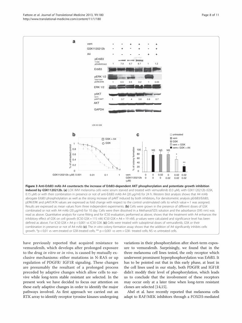
dependent feedback survival loop is activated also byMEK inhibitors. To this purpose we treated LOX-IMVIcells with GSK1120212b. As it is shown in Figure 3a andAdditional file 4: Figure S3, a strong induction of pErbB3,with concomitant increase of pAKT was observed 24 hafter cell exposure to the MEK inhibitor. Also in this casethe feedback survival loop was fully abrogated by theaddition of the anti-ErbB3 mAb A4 (Figure 3a). In vitrocolony formation assays were run in the presence of grow-ing concentrations of GSK1120212b alone or in combin-ation with a fixed dose of A4. Also in this case, co-treatmentwith anti-ErbB3 strongly potentiated growth inhibition bythe MEK inhibitor (Figure 3b). Our results clearly indicatethat targeting of the RAS-RAF-MAPK pathway at multiplelevels is unable to avoid bypass activation of the AKT-dependent adaptive mechanism centered around ErbB3,and that cell treatment with anti-ErbB3 has a dominant ef-fect on both pAKTand pERK when combined with a BRAFand MEK inhibition. Finally when cells were treated withsuboptimal doses of vemurafenib and GSK1120212b, theaddition of A4 was capable to provide a powerful synergis-tic inhibition of cell growth (Figure 3c).
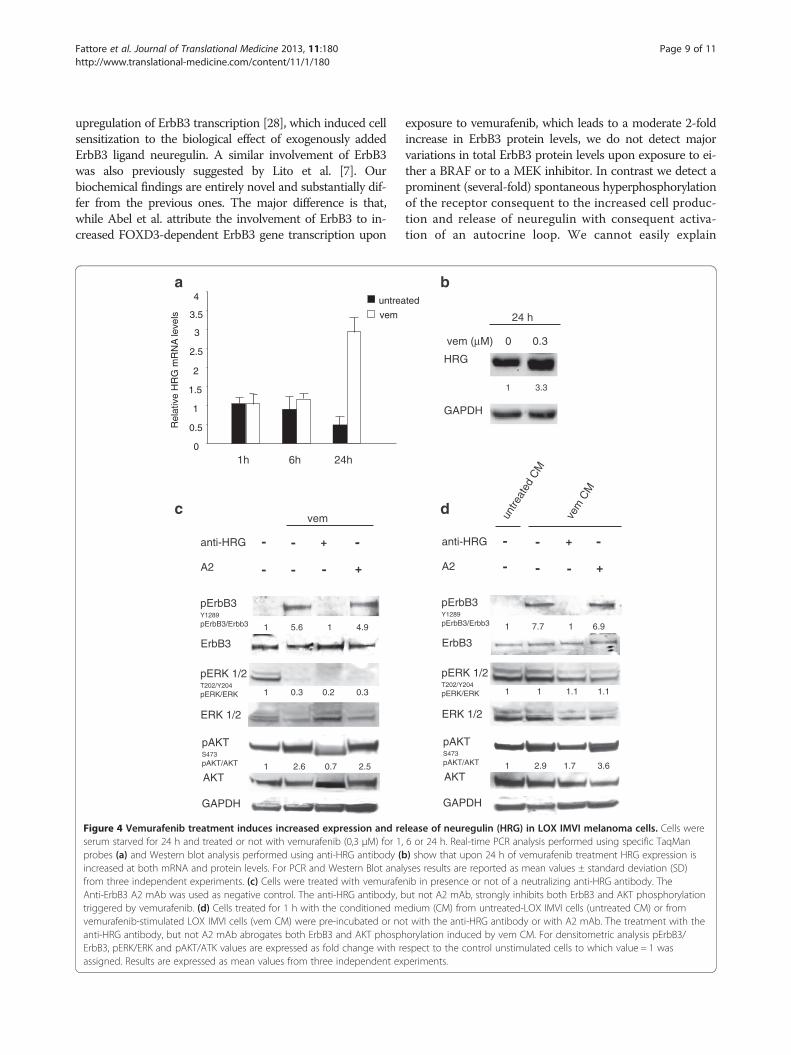
The feedback survival loop is promoted by increasedautocrine production of neuregulin by melanoma cellsWe were interested to better dissect the molecularmechanism responsible for drug-dependent pErbB3upregulation. Normally, ErbB3 is phosphorylated fol-lowing ligand-dependent hetero-dimerization withother HER-family receptor partners. Since BRAF orMEK inhibitors induce pErbB3 in melanoma cells bear-ing different HER-family receptor composition (seeAdditional file 2: Table S1) we reasoned that a commonmechanism could be the increased production and re-lease of neuregulin in the medium and activation of anautocrine loop. Indeed, real time PCR analysis of LOXIMVI cells treated at different times with vemurafenibshowed increased neuregulin mRNA levels 24 h aftertreatment (Figure 4a). This was accompanied by increasedproduction of neuregulin as detected by western blotting(Figure 4b). To confirm that the autocrine production ofneuregulin is responsible for the ErbB3-mediated survivalloop in response to BRAF inhibitor LOX IMVI cells weretreated with vemurafenib in presence or not of a neutraliz-ing antibody against neuregulin (Figure 4c). Western blotanalysis clearly showed that the anti-HRG antibodystrongly inhibit both pErbB3 and consequently pAKT. Tofurther confirm this, the conditioned medium of BRAFitreated melanoma cells was able to induce pErbB3 instarved melanoma cells with a very rapid kinetic, after1 hour exposure (Figure 4d). Finally in order to fullyprove that this mechanism is entirely dependent uponincreased production of the ligand, the conditionedmedium was pre-incubated with neutralizing antibodiesagainst neuregulin. As shown in Figure 4d, this treat-ment fully abrogated pErbB3 induction by the condi-tioned medium of drug-treated melanoma cells.
ConclusionsA current limitation of targeted therapies against meta-static melanoma with BRAF or MEK inhibitors is thedevelopment of resistance. Hence it is of utmost im-portance to identify and tackle the underlying mecha-nisms. Nazarian et al. [14] and Villanueva et al. [15]
ErbB3
ERK 1/2
AKT
GAPDH
1 7.9 6.7 8.7 1 1.2
1 3.7 4 3.8 0.8 0.7
1 0.3 0.5 0.2 1 0.03
pErbB3Y1289pErbB3/Erbb3
pERK 1/2T202/Y204pERK/ERK
pAKTS473pAKT/AKT
GSK1120212b
a
A4
-+
+vem
-
+-
-
-
-
-
+
+
-
--
+
+
+
b
1
0.8
0.6
0.4
0.2
0
0.001 0.01 0.1 1
% o
f unt
reat
ed c
ontr
ol
GSK1120212b (μM)
GSK + A4
GSK
c%
of c
olon
y fo
rmat
ion
100
80
60
40
20
0
untreated
vemGSKvem + GSKvem+ GSK + A4
0vemGSK1120212b (μM)
000
0.020 0.02
00.020.02
0.020.02
NS NS NS
*
**
Figure 3 Anti-ErbB3 mAb A4 counteracts the increase of ErbB3-dependent AKT phosphorylation and potentiate growth inhibitioninduced by GSK1120212b. (a) LOX IMVI melanoma cells were serum starved and treated with vemurafenib (0.3 μM), with GSK1120212b (GSK,0.15 μM) or with their combination in presence or not of anti-ErbB3 mAb A4 (20 μg/ml) for 24 h. Western blot analysis shows that A4 mAbabrogate ErbB3 phosphotylation as well as the strong increase of pAKT induced by both inhibitors. For densitometric analysis pErbB3/ErbB3,pERK/ERK and pAKT/ATK values are expressed as fold change with respect to the control unstimulated cells to which value = 1 was assigned.Results are expressed as mean values from three independent experiments. (b) Cells were grown in the presence of different doses of GSKcombinated or not with A4 mAb (20 μg/ml) for 10 day. Cells were then dissolved in a Methanol/SDS solution and the adsorbance (595 nm) wasread as above. Quantitative analysis for curve fitting and for IC50 evaluation, performed as above, shows that the treatment with A4 enhances theinhibitory effect of GSK on cell growth (IC50 GSK = 115 nM; IC50 GSK + A4 = 19 nM). p-values were calculated and significance level has beendefined as above. For IC50 GSK + A4 p < 0,001 vs IC50 GSK. (c) Cells were treated with suboptimal doses of vemurafenib, GSK or theircombination in presence or not of A4 mAb (c). The in vitro colony formation assay shows that the addition of A4 significantly inhibits cellsgrowth. *p < 0,01 vs vem-treated or GSK-treated cells; ** p < 0,001 vs vem + GSK- treated cells; NS vs untreated cells.
Fattore et al. Journal of Translational Medicine 2013, 11:180 Page 8 of 11http://www.translational-medicine.com/content/11/1/180
have previously reported that acquired resistance tovemurafenib, which develops after prolonged exposureto the drug in vitro or in vivo, is caused by mutually ex-clusive mechanisms: either mutations in N-RAS or upregulation of PDGFR/ IGF1R signaling. These changesare presumably the resultant of a prolonged processpreceded by adaptive changes which allow cells to sur-vive while long-term stable resistant are selected. In thepresent work we have decided to focus our attention onthese early adaptive changes in order to identify the majorpathways involved. As first approach we carried out anRTK array to identify receptor tyrosine kinases undergoing
variations in their phosphorylation after short-term expos-ure to vemurafenib. Surprisingly, we found that in thethree melanoma cell lines tested, the only receptor whichunderwent prominent hyperphosphorylation was ErbB3. Ithas to be pointed out that in this early phase, at least inthe cell lines used in our study, both PDGFR and IGF1Rdidn’t modify their level of phosphorylation, which leadsus to conclude that the involvement of these receptorsmay occur only at a later time when long-term resistantclones are selected [14,15].Abel et al. have recently reported that melanoma cells
adapt to RAF/MEK inhibitors through a FOXD3-mediated
Fattore et al. Journal of Translational Medicine 2013, 11:180 Page 9 of 11http://www.translational-medicine.com/content/11/1/180
upregulation of ErbB3 transcription [28], which induced cellsensitization to the biological effect of exogenously addedErbB3 ligand neuregulin. A similar involvement of ErbB3was also previously suggested by Lito et al. [7]. Ourbiochemical findings are entirely novel and substantially dif-fer from the previous ones. The major difference is that,while Abel et al. attribute the involvement of ErbB3 to in-creased FOXD3-dependent ErbB3 gene transcription upon
- - +
+
anti-HRG
ErbB3
ERK 1/2
AKT
GAPDH
pErbB3Y1289pErbB3/Erbb3
pERK 1/2T202/Y204pERK/ERK
pAKTS473pAKT/AKT
-
-
A2
vem
1 5.6 1 4.9
1 0.3 0.2 0.3
1 2.6 0.7 2.5
-
a
c
0
0.5
1
1.5
2
2.5
3
3.5
4 untrea
vem
Rel
ativ
e H
RG
mR
NA
leve
ls
1h 6h 24h
-
Figure 4 Vemurafenib treatment induces increased expression and reserum starved for 24 h and treated or not with vemurafenib (0,3 μM) for 1,probes (a) and Western blot analysis performed using anti-HRG antibody (bincreased at both mRNA and protein levels. For PCR and Western Blot analfrom three independent experiments. (c) Cells were treated with vemurafeAnti-ErbB3 A2 mAb was used as negative control. The anti-HRG antibody, btriggered by vemurafenib. (d) Cells treated for 1 h with the conditioned mvemurafenib-stimulated LOX IMVI cells (vem CM) were pre-incubated or noanti-HRG antibody, but not A2 mAb abrogates both ErbB3 and AKT phosphErbB3, pERK/ERK and pAKT/ATK values are expressed as fold change with rassigned. Results are expressed as mean values from three independent ex
exposure to vemurafenib, which leads to a moderate 2-foldincrease in ErbB3 protein levels, we do not detect majorvariations in total ErbB3 protein levels upon exposure to ei-ther a BRAF or to a MEK inhibitor. In contrast we detect aprominent (several-fold) spontaneous hyperphosphorylationof the receptor consequent to the increased cell produc-tion and release of neuregulin with consequent activa-tion of an autocrine loop. We cannot easily explain
- - +
+
anti-HRG
ErbB3
ERK 1/2
AKT
GAPDH
pErbB3Y1289pErbB3/Erbb3
pERK 1/2T202/Y204pERK/ERK
pAKTS473pAKT/AKT
- -
-
A2
1 7.7 1 6.9
1 1 1.1 1.1
1 2.9 1.7 3.6
-
untre
ated
CM
vem
CM
vem (μM) 0 0.3
GAPDH
HRG
24 h
1 3.3
b
d
ted
lease of neuregulin (HRG) in LOX IMVI melanoma cells. Cells were6 or 24 h. Real-time PCR analysis performed using specific TaqMan) show that upon 24 h of vemurafenib treatment HRG expression isyses results are reported as mean values ± standard deviation (SD)nib in presence or not of a neutralizing anti-HRG antibody. Theut not A2 mAb, strongly inhibits both ErbB3 and AKT phosphorylationedium (CM) from untreated-LOX IMVI cells (untreated CM) or fromt with the anti-HRG antibody or with A2 mAb. The treatment with theorylation induced by vem CM. For densitometric analysis pErbB3/espect to the control unstimulated cells to which value = 1 wasperiments.
Fattore et al. Journal of Translational Medicine 2013, 11:180 Page 10 of 11http://www.translational-medicine.com/content/11/1/180
these significant discrepancies. However it is worthpointing out that Abel et al. use melanoma cells stablytransfected with a plasmid encoding Foxd3, and whichtherefore express supraphysiological amounts of thistranscription factor [28]. In contrast we never transfectthis factor and, therefore, we believe we work in morephysiological conditions.In addition we show for the first time that neutralizing
antibodies against ErbB3 are capable to fully abrogatethis compensatory survival mechanism and to potentlysynergize with BRAF and MEK inhibitors. Therefore,we propose that initial co-treatment of melanoma pa-tients bearing BRAF mutations with an anti-ErbB3 anti-body could be a powerful strategy to enhance clinicalefficacy of BRAF and MEK inhibitors.
Additional files
Additional file 1: Figure S1. Vemurafenib treatment induces selectiveErbB3 phosphorylation in WM266 melanoma cells. (a) Simultaneousdetection of the phosphorylation status of RTKs (n = 49) using a humanphospho-RTK array in WM266 melanoma cells treated or not for 24 hwith 0.3 μM vemurafenib. Membranes were incubated with cell lysatesand array data were analyzed as reported in Figure 1. Thephosphorylation of ErbB3 is strongly increased by vemurafenib treatment.(b) WM266 cells were serum starved for 24 h, treated or not withdifferent doses of vemurafenib for 6 h or 24 h. Western blot analysisshows a strong dose-dependent and time-dependent phosphorylation ofErbB3 and AKT induced by vemurafenib. For densitometric analysispErbB3/ErbB3, pERK/ERK and pAKT/ATK values are expressed as foldchange with respect to the control unstimulated cells to which value = 1was assigned. Results are reported as mean values ± standard deviation(SD) from three independent experiments.
Additional file 2: Table S1. Flow cytometry analysis of ErbBsmembrane expression in LOX IMVI, MST-L and WM266 melanoma celllines. The percentage of positive cells was determined by staining withthe indicated primary antibodies and with the isotype-matchedandibodies as negative control. LOX IMVI, MST-L and WM266 showdifferent ErbB receptor compositions.
Additional file 3: Figure S2. Anti-ErbB3 A4mAb counteracts theincrease of ErbB3-dependent AKT phosphorylation and potentiate growthinhibition induced by vemurafenib in melanoma cells. MST-L (a) andWM266 (c) cells serum starved and treated with vemurafenib (0.3 μM) for24 h were incubated or not with A4 mAb (20 μg/ml). Western blotanalysis shows that A4 abrogate receptor phosphorylation and ATKsignaling. For densitometric analysis pErbB3/ErbB3, pERK/ERK and pAKT/ATK values are expressed as fold change with respect to the controlunstimulated cells to which value = 1 was assigned. Results are expressedas mean values from three independent experiments. MST-L (b) andWM266 (d) cells were grown in the presence of different doses ofvemurafenib alone or in combination with a fixed dose (20 μg/ml) of A4.Cells were then dissolved in a Methanol/SDS solution and theadsorbance (595 nm) was read as reported in Figure 2. Quantitativeanalysis for curve fitting and for IC50 evaluation, performed as reportedin Figure 2, shows that A4 enhances the inhibitory effect of vemurafenibon both cell lines’ growth (for MST-L cells: IC50 vem = 264 nM, IC50 vem+ A4 = 69 nM; for WM266 cells IC50 vem = 140 nM, IC50 vem + A4 = 51nM). Results are reported as mean values± standard deviation (SD) fromthree independent experiments. p-values were calculated andsignificance level has been defined as reported in Figure 2. For MST-Land WM266 cells IC50 vem + A4 p < 0,001 vs IC50 vem.
Additional file 4: Figure S3. GSK1120212b treatment induces selectiveErbB3-dependent AKT phosphorylation in LOX IMVI melanoma cells. Cellswere serum starved for 24 h, treated or not with different doses of GSK
for 6 h or 24 h. Western blot analysis performed using the indicatedantibodies shows that GSK induces a strong dose-dependent and time-dependent phosphorylation of ErbB3 and AKT. For densitometric analysispErbB3/ErbB3, pERK/ERK and pAKT/ATK values are expressed as foldchange with respect to the control unstimulated cells to which value = 1was assigned. Results are expressed as mean values from threeindependent experiments.
AbbreviationsMAPK: Mitogen-activated protein kinase; Vem: Vemurafenib;GSK: GSK1120212b; HRG: Neuregulin; MAbs: Monoclonal antibodies.
Competing interestsDr Paolo A. Ascierto participated to Advisory Board from Bristol MyersSquibb, MSD, Roche Genentech, GSK, and received honoraria from BrystolMyers Squibb, MSD and Roche-Genentech. All remaining authors declare theabsence of any competing interest.
Authors’ contributionsLF performed data acquisition, data analysis, preparation of the illustrations,and drafted the manuscript; MEP, AN and CDV contributed to acquisitionand analysis of western blotting and cell proliferation data; EM, FB, LA andRM contributed to data analysis and draft of sections of the manuscript; MRTand PAA provided melanoma cells lines, continuous advice to the study andrevised the manuscript; GC conceived and supervised the study, and revisedthe manuscript. All authors read and approved the final version of themanuscript.
AcknowledgmentsThis work was supported in part by grants from MIUR and from AIRC -Associazione Italiana per la Ricerca sul Cancro - (IG 10334) to G. Ciliberto, andby grants from MIUR and from AIRC - (IG 10272), to M.R. Torrisi.We are grateful to “Fondazione Melanoma ONLUS” for the financial supportfor this project.
Author details1Dipartimento di Medicina Clinica e Molecolare, Sapienza Università di Roma,Rome, Italy. 2Dipartimento di Chirurgia “P. Valdoni”, Sapienza Università diRoma, Rome, Italy. 3Takis srl, Rome, Italy. 4Dipartimento di MedicinaSperimentale e Clinica, Università degli Studi di Catanzaro “Magna Graecia”,Catanzaro, Italy. 5Istituto Pasteur Fondazione Cenci Bolognetti, Dipartimentodi Medicina Clinica e Molecolare, Sapienza Università di Roma, Rome, Italy.6Azienda Ospedaliera S. Andrea, Rome, Italy. 7Istituto Nazionale per lo Studioe la Cura dei Tumori “Fondazione G. Pascale”, Via Mariano Semmola, 80131Naples, Italy.
Received: 14 June 2013 Accepted: 24 July 2013Published: 27 July 2013
References1. Balch CM, Gershenwald JE, Soong SJ, Thompson JF: Update on the
melanoma staging system: the importance of sentinel node staging andprimary tumor mitotic rate. J Surg Oncol 2011, 4:379–85.
2. Korn EL, Liu PY, Lee SJ, Chapman JA, Niedzwiecki D, Suman VJ, Moon J,Sondak VK, Atkins MB, Eisenhauer EA, Parulekar W, Markovic SN, Saxman S,Kirkwood JM: Meta-analysis of phase II cooperative group trials inmetastatic stage IV melanoma to determine progression-free and overallsurvival benchmarks for future phase II trials. J Clin Oncol 2008, 4:527–34.
3. Hodi FS, O'Day SJ, McDermott DF, Weber RW, Sosman JA, Haanen JB,Gonzalez R, Robert C, Schadendorf D, Hassel JC, Akerley W, Van denEertwegh AJ, Lutzky J, Lorigan P, Vaubel JM, Linette GP, Hogg D,Ottensmeier CH, Lebbé C, Peschel C, Quirt I, Clark JI, Wolchok JD, Weber JS,Tian J, Yellin MJ, Nichol GM, Hoos A, Urba WJ: Improved survival withipilimumab in patients with metastatic melanoma. N Engl J Med 2010,8:711–23.
4. Flaherty KT, Puzanov I, Kim KB, Ribas A, McArthur GA, Sosman JA, O'DwyerPJ, Lee RJ, Grippo JF, Nolop K, Chapman PB: Inhibition of mutated,activated BRAF in metastatic melanoma. N Eng J Med 2010, 9:809–819.
5. Chapman PB, Hauschild A, Robert C, Haanen JB, Ascierto P, Larkin J,Dummer R, Garbe C, Testori A, Maio M, Hogg D, Lorigan P, Lebbe C, Jouary
Fattore et al. Journal of Translational Medicine 2013, 11:180 Page 11 of 11http://www.translational-medicine.com/content/11/1/180
T, Schadendorf D, Ribas A, O'Day SJ, Sosman JA, Kirkwood JM, EggermontAM, Dreno B, Nolop K, Li J, Nelson B, Hou J, Lee RJ, Flaherty KT, McArthurGA, BRIM-3 Study Group: Improved survival with vemurafenib inmelanoma with BRAF V600E mutation. N Engl J Med 2011, 26:2507–16.
6. Flaherty KT: BRAF inhibitors and melanoma. Cancer J 2011, 6:505–11.7. Lito P, Pratilas CA, Joseph EW, Tadi M, Halilovic E, Zubrowski M, Huang A,
Wong WL, Callahan MK, Merghoub T, Wolchok JD, De Stanchina E,Chandarlapaty S, Poulikakos PI, Fagin JA, Rosen N: Relief of profoundfeedback inhibition of mitogenic signaling by RAF inhibitors attenuatestheir activity in BRAF V600E melanomas. Cancer Cell 2012, 5:668–82.
8. Poulikakos PI, Zhang C, Bollag G, Shokat KM, Rosen N: RAF inhibitorstransactivate RAF dimers and ERK signalling in cells with wild-type BRAF.Nature 2010, 7287:427–30.
9. Sullivan RJ, Flaherty K: MAP kinase signaling and inhibition in melanoma.Oncogene 2013, 19:2373–9.
10. Ascierto PA, Schadendorf D, Berking C, Agarwala SS, Van Herpen CM,Queirolo P, Blank CU, Hauschild A, Beck JT, St-Pierre A, Niazi F, Wandel S,Peters M, Zubel A, Dummer R: MEK162 for patients with advancedmelanoma harbouring or Val600 BRAF mutations: a non-randomised,open-label phase 2 study. Lancet Oncol 2013, 3:249–56.
11. Lo RS: Combinatorial therapies to overcome B-RAF inhibitor resistance inmelanomas. Pharmacogenomics 2012, 2:125–8.
12. Corcoran RB, Settleman J, Engelman JA: Potential therapeutic strategies toovercome acquired resistance to BRAF or MEK inhibitors in BRAF mutantcancers. Oncotarget 2011, 4:336–46.
13. Sosman JA, Pavlick AC, Schuchter LM, Lewis KD, McArthur GA, Cowey CL,Moschos SJ, Flaherty KT, Kim KB, Weber J, Hersey P, Long GV, Lawrence DP,Kockx M, Spleiss O, Koehler A, Bollag G, Joe AK, Trunzer K, Ribas A: Analysisof molecular mechanisms of response and resistance to vemurafenib(vem) in BRAF V600E melanoma. J Clin Oncol 2012, 30. suppl; abstr 8503.
14. Nazarian R, Shi H, Wang Q, Kong X, Koya RC, Lee H, Chen Z, Lee MK, AttarN, Sazegar H, Chodon T, Nelson SF, McArthur G, Sosman JA, Ribas A, Lo RS:Melanomas acquire resistance to B-RAF(V600E) inhibition by RTK orN-RAS upregulation. Nature 2010, 7326:973–7.
15. Villanueva J, Vultur A, Lee JT, Somasundaram R, Fukunaga-Kalabis M, Cipolla AK,Wubbenhorst B, Xu X, Gimotty PA, Kee D, Santiago-Walker AE, Letrero R,D’Andrea K, Pushparajan A, Hayden JE, Brown KD, Laquerre S, McArthur GA,Sosman JA, Nathanson KL, Herlyn M: Acquired resistance to BRAF inhibitorsmediated by a RAF kinase switch in melanoma can be overcome bycotargeting MEK and IGF-1R/PI3K. Cancer Cell 2010, 18:683–95.
16. Flaherty KT, Infante JR, Daud A, Gonzalez R, Kefford RF, Sosman J, Hamid O,Schuchter L, Cebon J, Ibrahim N, Kudchadkar R, Burris HA 3rd, Falchook G, AlgaziA, Lewis K, Long GV, Puzanov I, Lebowitz P, Singh A, Little S, Sun P, Allred A,Ouellet D, Kim KB, Patel K, Weber J: Combined BRAF and MEK inhibition inmelanoma with BRAF V600 mutations. N Engl J Med 2012, 18:1694–703.
17. Zhang H, Berezov A, Wang Q, Zhang G, Drebin J, Murali R, Greene MI: ErbBreceptors: from oncogenes to targeted cancer therapies. J Clin Invest2007, 8:2051–8.
18. Hsieh AC, Moasser MM: Targeting HER proteins in cancer therapy and therole of the non-target HER3. Br J Cancer 2007, 4:453–457.
19. Ocana A, Vera-Badillo F, Seruga B, Templeton A, Pandiella A, Amir E: HER3overexpression and survival in solid tumors: a meta-analysis. J NatlCancer Inst 2013, 4:266–73.
20. Schoeberl B, Pace EA, Fitzgerald JB, Harms BD, Xu L, Nie L, Linggi B, Kalra A,Paragas V, Bukhalid R, Grantcharova V, Kohli N, West KA, Leszczyniecka M,Feldhaus MJ, Kudla AJ, Nielsen UB: Therapeutically targeting ErbB3: a keynode in ligand-induced activation of the ErbB receptor-PI3K axis. SciSignal 2009, 77:ra31. doi:10.1126/scisignal.2000352.
21. Sergina NV, Rausch M, Wang D, Blair J, Hann B, Shokat KM, Moasser MM:Escape from HER-family tyrosine kinase inhibitor therapy by thekinase-inactive HER3. Nature 2007, 7126:437–41.
22. Garrett JT, Olivares MG, Rinehart C, Granja-Ingram ND, Sánchez V,Chakrabarty A, Dave B, Cook RS, Pao W, McKinely E, Manning HC, Chang J,Arteaga CL: Transcriptional and posttranslational up-regulation of HER3(ErbB3) compensates for inhibition of the HER2 tyrosine kinase. Proc NatlAcad Sci USA 2011, 12:5021–6.
23. Chakrabarty A, Sanchez V, Kuba MG, Rinehart C, Arteaga CL: Feedbackupregulation of HER3 (ErbB3) expression and activity attenuatesantitumor effect of PI3K inhibitors. Proc Natl Acad Sci USA 2012, 8:2718–23.
24. Aurisicchio L, Marra E, Roscilli G, Mancini R, Ciliberto G: The promise ofanti-ErbB3 monoclonals as new cancer therapeutics. Oncotarget 2012,8:744–58.
25. Belleudi F, Marra E, Mazzetta F, Fattore L, Giovagnoli MR, Mancini R,Aurisicchio L, Torrisi MR, Ciliberto G: Monoclonal antibody-induced ErbB3receptor internalization and degradation inhibits growth and migrationof human melanoma cells. Cell Cycle 2012, 7:1455–67.
26. Aurisicchio L, Marra E, Luberto L, Carlomosti F, De Vitis C, Noto A, Gunes Z,Roscilli G, Mesiti G, Mancini R, Alimandi M, Ciliberto G: Novel anti-ErbB3monoclonal antibodies show therapeutic efficacy in xenografted andspontaneous mouse tumors. J Cell Physiol 2012, 10:3318–8.
27. Yang H, Higgins B, Kolinsky K, Packman K, Go Z, Iyer R, Kolis S, Zhao S, LeeR, Grippo JF, Schostack K, Simcox ME, Heimbrook D, Bollag G, Su F: RG7204(PLX4032), a selective BRAFV600E inhibitor, displays potent antitumoractivity in preclinical melanoma models. Cancer Res 2010, 13:5518–27.
28. Abel EV, Basile KJ, Kugel CH 3rd, Witkiewicz AK, Le K, Amaravadi RK,Karakousis GC, Xu X, Xu W, Schuchter LM, Lee JB, Ertel A, Fortina P, AplinAE: Melanoma adapts to RAF/MEK inhibitors through FOXD3-mediatedupregulation of ERBB3. J Clin Invest 2013, 5:2155–68.
doi:10.1186/1479-5876-11-180Cite this article as: Fattore et al.: Activation of an early feedback survivalloop involving phospho-ErbB3 is a general response of melanoma cellsto RAF/MEK inhibition and is abrogated by anti-ErbB3 antibodies.Journal of Translational Medicine 2013 11:180.
Submit your next manuscript to BioMed Centraland take full advantage of:
• Convenient online submission
• Thorough peer review
• No space constraints or color figure charges
• Immediate publication on acceptance
• Inclusion in PubMed, CAS, Scopus and Google Scholar
• Research which is freely available for redistribution
Submit your manuscript at www.biomedcentral.com/submit