Embed Size (px)
Citation preview
Research ArticleThe Effect of Latent Toxoplasma gondii Infection onthe Immune Response in HIV-Infected Patients
Ondrej Beran,1,2 Petr Kodym,3 Marek Maly,4 Alzbeta Davidova,1,2
Gabriela Reinvartova,1 David Jilich,1 Michal Holub,1,2 and Hanus Rozsypal1,2
1Department of Infectious and Tropical Diseases, First Faculty of Medicine, Charles University in Prague and Na Bulovce Hospital,Budınova 2, 180 81 Prague, Czech Republic2Department of Infectious Diseases, First Faculty of Medicine, Charles University in Prague and Military University Hospital Prague,Prague, Czech Republic3National Reference Laboratory for Toxoplasmosis, National Institute of Public Health in Prague, Czech Republic4Department of Biostatistics, National Institute of Public Health in Prague, Czech Republic
Correspondence should be addressed to Ondrej Beran; [email protected]
Received 2 May 2015; Revised 18 June 2015; Accepted 21 June 2015
Academic Editor: Esteban Martinez
Copyright © 2015 Ondrej Beran et al. This is an open access article distributed under the Creative Commons Attribution License,which permits unrestricted use, distribution, and reproduction in any medium, provided the original work is properly cited.
A relationship between latent toxoplasmosis and the immune system during HIV disease is poorly understood. Therefore, theaim of this follow-up study was to characterize immunological parameters in HIV-infected patients with latent toxoplasmosisand noninfected individuals. A total of 101 HIV-infected patients were enrolled in the study. The patients were classified into twogroups based on anti-Toxoplasma gondii antibodies: a group of 55 toxoplasma-positive persons (TP) and a group of 46 toxoplasma-negative persons (TN). Absolute counts of several lymphocyte subsets decreased in the TP group, namely, T cells (𝑝 = 0.007), Bcells (𝑝 = 0.002), NK cells (𝑝 = 0.009), CD4 T cells (𝑝 = 0.028), and CD8 T cells (𝑝 = 0.004). On the other hand, the percentageof CD8 T cells expressing CD38 and HLA-DR significantly increased during the follow-up in the TP group (𝑝 = 0.003, 𝑝 = 0.042,resp.) as well as the intensity of CD38 andHLA-DR expression (MFI) on CD8 T cells (𝑝 = 0.001, 𝑝 = 0.057, resp.). In the TN group,analysis of the kinetics of immunological parameters revealed no significant changes over time. In conclusion, the results suggestthat latent T. gondii infection modulates the immune response during HIV infection.
1. Introduction
Toxoplasma gondii is an intracellular protozoon causing oneof the most prevalent infections. Apart from humans, T.gondii infects various mammals and birds, with small felidsplaying a major role as definitive hosts of the parasite.Humans are infected either by consumption of undercookedmeat containing a tissue cyst or by ingestion of oocystsshed by the definitive host in feces. In women infectedduring pregnancy T. gondii infection can lead to verticaltransmission and fetal infection [1, 2].
The primary infection in immunocompetent individu-als is mostly asymptomatic or can be manifested as lym-phadenopathy and is usually followed by a lifelong latentinfection. However, from this state of latency T. gondiiinfection may be reactivated as a result of immune disorders
[3]. Despite the availability of effective antiretroviral therapy,toxoplasmosis is the most important opportunistic infectionin patients infected with human immunodeficiency virus(HIV) and can manifest as a potentially life threateningtoxoplasmic encephalitis [4, 5].
Following infection with T. gondii different immunecells were shown to have a role in host resistance to thisorganism [6]. T cells and natural killer (NK) cells help tocontrol the initial infection by production of interferon-(IFN-) 𝛾 and interleukin- (IL-)12. Long-term host resistanceis provided by T and B cells. However, the mechanisms ofimmune surveillance are not fully understood. Regarding theinfluence of latent toxoplasmosis on humans, several effectshave been documented. Latent toxoplasmosis increases therisks of Parkinson’s disease [7], behavioral changes [8–10],or autoimmune diseases [11, 12]. Moreover, latent T. gondii
Hindawi Publishing CorporationBioMed Research InternationalVolume 2015, Article ID 271842, 7 pageshttp://dx.doi.org/10.1155/2015/271842
2 BioMed Research International
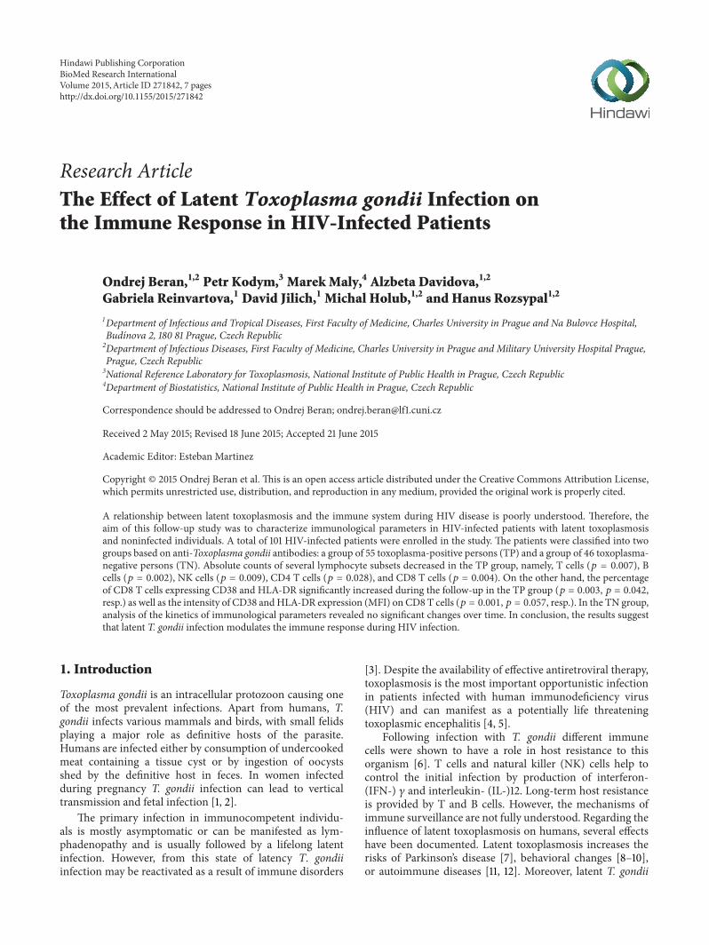
Table 1: Demographic and clinical characteristics from 46 toxoplasma-negative and 55 toxoplasma-positive HIV-infected patients upon thetime of enrollment.
Parameter Toxoplasma-negative Toxoplasma-positive 𝑝 valueAge (arithmetic mean ± SD) 42 ± 9 44 ± 11 0.382Sex (male/female) 43/3 51/4 1.000Previous AIDS diagnosis (%) 13 15 1.000Time since first positive HIV test (years)∗ 9 (8–10) 9 (8–11) 0.856Time since cART initiation (years)∗ 5.7 (4.5–7.3) 5.4 (4.3–6.9) 0.766Coinfections
HBV (%) 41 35 0.539HCV (%) 13 13 1.000CMV (%) 87 89 0.767
Antiretroviral treatment (cART) 0.7592 NRTI + 1 PI (%) 43 452 NRTI + 1 NNRTI (%) 35 272 NRTI + 1 II (%) 9 133 NRTI (%) 9 6None (%) 4 9
SD, standard deviation; AIDS, acquired immunodeficiency syndrome; HIV, human immunodeficiency virus; cART, combined antiretroviral therapy; HBV,hepatitis B virus; HCV, hepatitis C virus; CMV, cytomegalovirus; NRTI, nucleoside reverse transcriptase inhibitors; PI, protease inhibitors; NNRTI,nonnucleoside reverse transcriptase inhibitors; II, integrase inhibitors.∗Values are presented as geometric means and 95% confidence intervals.
infection may lead to immune suppression in both miceand humans [13, 14]. Despite many studies focusing on therelationship of toxoplasmosis reactivation andHIV infection,there is a lack of knowledge about the association betweenlatent toxoplasmosis and activation of the immune systemduring HIV disease.
Therefore, the aim of the study was to characterizeand compare different immunological parameters, includingexpression of activation markers on CD8 T cells of HIV-infected patients with latent toxoplasmosis and HIV-infectedindividuals without latent toxoplasmosis during a one-yearfollow-up.
2. Patients and Methods
2.1. Study Population. A total of 101 HIV-positive patients(sex ratio M/F, 94/7; mean age, 43; range, 26–74 years)registered at the AIDS Centre at Na Bulovce Hospital inPrague were enrolled in the study between December 2012and April 2013. This prospective study was conducted inaccordance with the Helsinki Declaration as revised in 2000after obtaining the approval from the local ethics committee.A written informed consent was obtained from all studyparticipants. The patients were randomly selected in orderto represent the entire cohort of HIV patients treated at thecentre. Based on anti-T. gondii antibodies, the patients wereclassified into two groups: a group of toxoplasma-positive(TP), which comprised 55 seropositive patients, and a groupof 46 toxoplasma-negatives (TN). In Table 1, demographicand clinical parameters at the time of enrollment are pre-sented for both groups. According to the CDC classification,14 (14%) patients were classified with the AIDS stage of the
HIV disease [15]. The percentages of patients with AIDSstage did not differ between the groups and these patientswere not excluded from the analysis. The patients wereevaluated regarding presence of important coinfections, thatis, hepatitis C, hepatitis B, and cytomegalovirus infection(positive serology anti-HCV, anti-HBcAg IgG, and anti-CMV). The statistical analysis demonstrated that the TPand TN groups were not different as far as the presence ofthese coinfections is concerned. Patients with clinical andlaboratory signs of active coinfectionswere excluded from thestudy. Regarding combined antiretroviral therapy (cART), 49patients (89%) were treated in the TP group and 43 patients(93%) in the TN group. The percentages of patients withdifferent treatment protocols are also presented in Table 1.Plasma viral load values were undetectable or very low ina majority of patients. Blood samples for immunologicalanalyseswere collected fromall patients upon enrollment andafter a one-year interval.
2.2. Flow Cytometric Analysis. Routine immunophenotyping(T cells, B cells, CD4 T cells, CD8 T cells, and NK cells) andan analysis of the surface expression of activation markers(CD38 and HLA-DR) on CD8 T cells were done from EDTA-treated peripheral blood using monoclonal antibodies (Ab)(Becton Dickinson, BD, Germany) [16]. Analysis was per-formed by six-color flow cytometry (FACSCanto, BD, USA)using FACSDiva software (BD). Expression is presented asmean fluorescence intensity (MFI).
2.3. Toxoplasma Serology Testing. During the follow-up, thecomplement-fixation test (CFT) and IgM/IgG ELISA testwere used for detection of anti-T. gondii antibodies [17, 18].
BioMed Research International 3
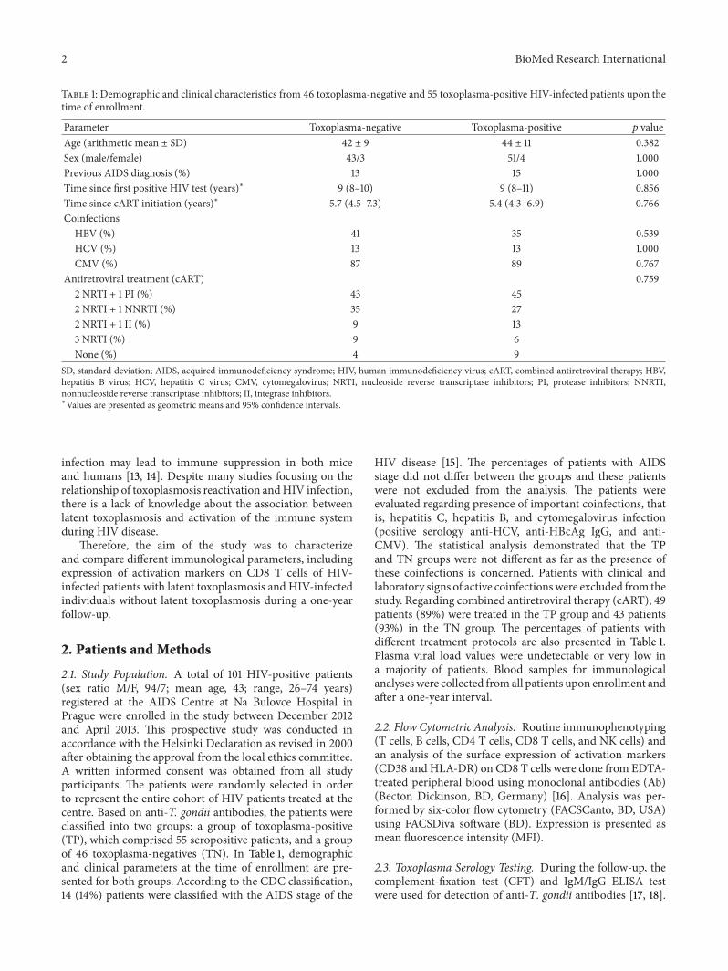
Table 2: Comparison of laboratory data from toxoplasma-negative and toxoplasma-positive HIV-infected patients upon the time ofenrollment and after a one-year follow-up.
ParameterToxoplasma-negative(baseline)
Toxoplasma-negative
(follow-up)𝑝
Toxoplasma-positive(baseline)
Toxoplasma-positive
(follow-up)𝑝
T cells (cells/mm3) 1669 (1510–1845) 1575 (1435–1729) 0.109 1814 (1656–1988) 1629 (1477–1796) 0.007CD4 T cells (cells/mm3) 666 (602–737) 630 (559–710) 0.212 688 (614–771) 632 (561–713) 0.028CD8 T cells (cells/mm3) 904 (781–1046) 839 (730–964) 0.052 1002 (880–1141) 882 (769–1012) 0.004B cells (cells/mm3) 184 (156–216) 175 (154–200) 0.361 205 (177–239) 174 (151–201) 0.002NK cells (cells/mm3) 290 (244–344) 261 (220–309) 0.063 317 (273–367) 280 (243–323) 0.009CD38+ CD8 T cells (%) 11 (9–13) 12 (10–15) 0.144 12 (9–15) 16 (13–20) 0.003CD38 expression on CD8 Tcells (MFI) 777 (667–905) 941 (777–1138) 0.036 844 (712–999) 1184 (964–1453) 0.001
HLA-DR+ CD8 T cells (%) 15 (13–19) 17 (14–20) 0.125 16 (14–20) 19 (16–23) 0.042HLA-DR expression onCD8 T cells (MFI) 258 (209–317) 281 (224–353) 0.174 266 (213–333) 333 (252–438) 0.003
MFI, mean fluorescence intensity.Values are presented as geometric means and 95% confidence intervals.
Positive results for CFT were defined as titers of ≥1 : 4 andIgG ELISA absorbance ≥0.25; IgM was positive at a positivityindex (absorbance of tested sample/absorbance of cut-offcontrol) of >1.1.
2.4. Statistical Analysis. Geometric means together withcorresponding 95% confidence intervals (95% CI) werecalculated to characterize the location and variability ofthe analyzed variables in the groups. The analysis of meanchange during follow-up in individual groups was basedon Student’s paired 𝑡-test. The comparison of the changemagnitude between TP and TN group was performed usingan analysis of variance with the interaction term betweenthe group and time. The tests were applied to logarithmicallytransformed data. All statistical tests were evaluated as two-sided at a significance level of 0.05. Stata release 9.2 (StataCorp. LP, College Station, TX) statistical software was usedfor statistical analysis.
3. Results
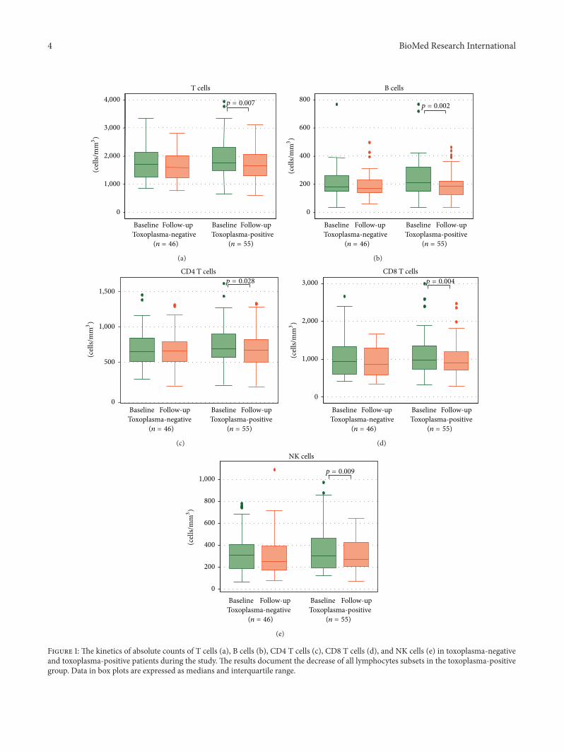
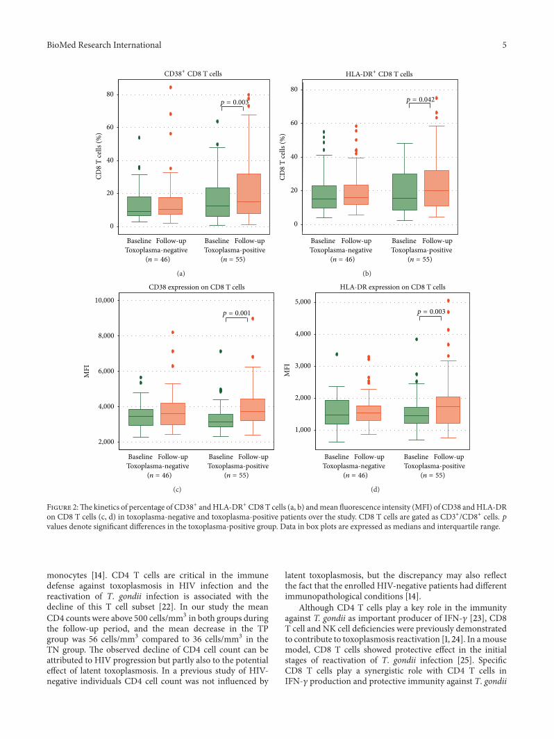
During the study follow-up, none of the 46 initially TNpatients were infected with T. gondii and seroconverted. Thecomparison of laboratory parameters between the TN andTPgroups at the time of enrollment and after a one-year follow-up is presented in Table 2. Interestingly, significant changesof evaluated parameters were observed only in the groupTP (Figures 1(a)–1(e)). The decrease was found in absolutecounts of T cells (𝑝 = 0.007), B cells (𝑝 = 0.002), NK cells(𝑝 = 0.009), CD4 T cells (𝑝 = 0.028), and CD8 T cells(𝑝 = 0.004). On the other hand, as shown also in Figures2(a)–2(d), the percentage of CD8 T cells expressing CD38andHLA-DR antigens significantly increased in the TP group(𝑝 = 0.003, 𝑝 = 0.042, resp.) similar to the intensity of CD38and HLA-DR expression (MFI) on CD8 T cells (𝑝 = 0.001,𝑝 = 0.057, resp.). In the TN group, the analysis of the kinetics
of immunological parameters revealed no significant changesover time.
4. Discussion
In this prospective follow-up study, we characterized theimmune response in HIV-infected patients latently infectedwith T. gondii in comparison with TN individuals during aone-year follow-up. Latent toxoplasmosis status was definedas the presence of anti-T. gondii antibodies without anyclinical symptoms. During the study, in none of the TPpatients latent toxoplasmosis had been reactivated. Thisfinding reflects the fact that the majority of patients hadgood immunological parameters and only a few individualsequally distributed in both groups were enrolled with HIVinfection in stage 3 (AIDS). Moreover, the incidence ofreactivation is quite low and was documented to be 3.4cases per 1000 anti-T. gondii positive HIV-infected patientsper year in the Czech Republic [1]. The prevalence of latenttoxoplasmosis inHIV-infected persons in theCzechRepublicis 40.2%. The decline of the cases with reactivation observedafter 1996 was due to improved efficacy of antiretroviraltherapy restoring cell mediated immunity including recoveryof anti-T. gondii specific CD4 and CD8 T cell responses[19]. However, previous studies documented that restorationof the immune system including antigen-specific responsesin patients treated with cART is not complete [20, 21].Despite the studies describing the effect of HIV-inducedimmunodeficiency on toxoplasmosis reactivation, the impactof latent T. gondii infection on HIV disease progression andimmune parameters is not clear.
We observed the decrease in the majority of lymphocytesubset counts in the group of HIV-infected patients withlatent T. gondii infection. This finding is in line with aprevious study in HIV-negative persons, where male patientswith latent toxoplasmosis had lower B cells, NK cells, and
4 BioMed Research International
0
1,000
2,000
3,000
4,000T cells
0.007
Baseline Follow-up Baseline Follow-upToxoplasma-negative Toxoplasma-positive
(n = 46) (n = 55)
(cel
ls/m
m3)
p =
(a)
B cells
0
200
400
600
8000.002
Baseline Follow-up Baseline Follow-upToxoplasma-negative Toxoplasma-positive
(n = 46) (n = 55)
(cel
ls/m
m3)
p =
(b)
CD4 T cells
0
500
1,000
1,500
Baseline Follow-up Baseline Follow-upToxoplasma-negative Toxoplasma-positive
(n = 46) (n = 55)
(cel
ls/m
m3)
0.028p =
(c)
CD8 T cells
0
1,000
2,000
3,000
Baseline Follow-up Baseline Follow-upToxoplasma-negative Toxoplasma-positive
0.004
(n = 46) (n = 55)
(cel
ls/m
m3)p =
(d)
NK cells
0
200
400
600
800
1,0000.009
Baseline Follow-up Baseline Follow-upToxoplasma-negative Toxoplasma-positive
(n = 46) (n = 55)
(cel
ls/m
m3)
p =
(e)
Figure 1: The kinetics of absolute counts of T cells (a), B cells (b), CD4 T cells (c), CD8 T cells (d), and NK cells (e) in toxoplasma-negativeand toxoplasma-positive patients during the study. The results document the decrease of all lymphocytes subsets in the toxoplasma-positivegroup. Data in box plots are expressed as medians and interquartile range.
BioMed Research International 5
CD8
T ce
lls (%
)
0
20
40
60
800.003
Baseline Follow-up Baseline Follow-upToxoplasma-negative Toxoplasma-positive
(n = 46) (n = 55)
p =
CD38+ CD8 T cells
(a)CD
8 T
cells
(%)
0
20
40
60
800.042
Baseline Follow-up Baseline Follow-upToxoplasma-negative Toxoplasma-positive
(n = 46) (n = 55)
p =
HLA-DR+ CD8 T cells
(b)
MFI
CD38 expression on CD8 T cells
2,000
4,000
6,000
8,000
10,000
0.001
Baseline Follow-up Baseline Follow-upToxoplasma-negative Toxoplasma-positive
(n = 46) (n = 55)
p =
(c)
MFI
HLA-DR expression on CD8 T cells
1,000
2,000
3,000
4,000
5,0000.003
Baseline Follow-up Baseline Follow-upToxoplasma-negative Toxoplasma-positive
(n = 46) (n = 55)
p =
(d)
Figure 2:The kinetics of percentage of CD38+ andHLA-DR+ CD8T cells (a, b) andmean fluorescence intensity (MFI) of CD38 andHLA-DRon CD8 T cells (c, d) in toxoplasma-negative and toxoplasma-positive patients over the study. CD8 T cells are gated as CD3+/CD8+ cells. 𝑝values denote significant differences in the toxoplasma-positive group. Data in box plots are expressed as medians and interquartile range.
monocytes [14]. CD4 T cells are critical in the immunedefense against toxoplasmosis in HIV infection and thereactivation of T. gondii infection is associated with thedecline of this T cell subset [22]. In our study the meanCD4 counts were above 500 cells/mm3 in both groups duringthe follow-up period, and the mean decrease in the TPgroup was 56 cells/mm3 compared to 36 cells/mm3 in theTN group. The observed decline of CD4 cell count can beattributed to HIV progression but partly also to the potentialeffect of latent toxoplasmosis. In a previous study of HIV-negative individuals CD4 cell count was not influenced by
latent toxoplasmosis, but the discrepancy may also reflectthe fact that the enrolled HIV-negative patients had differentimmunopathological conditions [14].
Although CD4 T cells play a key role in the immunityagainst T. gondii as important producer of IFN-𝛾 [23], CD8T cell and NK cell deficiencies were previously demonstratedto contribute to toxoplasmosis reactivation [1, 24]. In amousemodel, CD8 T cells showed protective effect in the initialstages of reactivation of T. gondii infection [25]. SpecificCD8 T cells play a synergistic role with CD4 T cells inIFN-𝛾 production and protective immunity against T. gondii
6 BioMed Research International
infection [26]. Similar to other lymphocyte subsets, reducednumbers of NK cells were previously demonstrated in HIV-infected persons with toxoplasmosis reactivation [1]. Thetrend of NK cell and CD8 cell counts decline observed in theTP group was not associated with toxoplasmosis reactivationin the patients. However, the association with the presence ofT. gondii latent infectionmay have had an influence on amorerapid long-term progression of HIV disease.
In our study, the percentages of activated CD8 T cellscharacterized by the expression of CD38 and HLA-DRincreased in the TP group during the follow-up. Similar to thelymphocyte subset counts, these changes were not observedin TN patients. Previous studies demonstrated that increasedexpression of CD38 and HLA-DR markers on T cells corre-lates with disease progression and depletion of CD4 T cells,better than the level of the viral load [27]. Also, many studiesdemonstrated that MFI of CD38 on CD8 T cells representsa reliable laboratory marker for routine monitoring of HIV-infected patients [28]. Our results may indicate a possiblemodulation of nonspecific CD8 T cell activation by latenttoxoplasmosis. This effect may be indirect and the trend ofthe CD8 T cell activation increase may be a result of the milddecrease of CD4 cell count observed during the follow-up.In previous studies, the immunomodulatory effect of latenttoxoplasmosis inHIV-negative humanswas documented andit decreased with the duration of the infection [14].
5. Conclusions
This study presents new findings about the role of latentT. gondii infection in modulation of the immune responsesduring HIV infection. The results suggest that latent tox-oplasmosis has an effect on the kinetics of lymphocytesubset counts and surface expression of immune activationmarkers in HIV-infected persons. The observed changesare mild and further studies are warranted to elucidate theimpact of highly prevalent T. gondii infection on the compleximmunopathogenesis of HIV infection.
Abbreviations
AIDS: Acquired immunodeficiency syndromecART: Combined antiretroviral therapyCFT: Complement fixation testCMV: CytomegalovirusHBV: Hepatitis B virusHCV: Hepatitis C virusHIV: Human immunodeficiency virusIFN: InterferonIL: InterleukinNK: Natural killerMFI: Mean fluorescence intensityTN: Toxoplasma-negativeTP: Toxoplasma-positive.
Conflict of Interests
The authors confirm that there is no conflict of interests orcommercial relationships regarding this study.
Acknowledgments
This study was supported by the Grant Agency of theMinistry of Health of the Czech Republic (IGA MZ CR,no. NT/11429–5) and the grants of the Charles University inPrague (PRVOUK/P24/LF1/3 and SVV-2015-260152).
References
[1] P. Kodym, M. Maly, O. Beran et al., “Incidence, immunologicaland clinical characteristics of reactivation of latent Toxoplasmagondii infection in HIV-infected patients,” Epidemiology andInfection, vol. 143, no. 3, pp. 600–607, 2015.
[2] C. Bachmeyer, G. Mouchnino, P. Thulliez, and L. Blum, “Con-genital toxoplasmosis from an HIV-infected woman as a resultof reactivation,”The Journal of Infection, vol. 52, no. 2, pp. e55–e57, 2006.
[3] S. Meers, K. Lagrou, K.Theunissen et al., “Myeloablative condi-tioning predisposes patients for toxoplasma gondii reactivationafter allogeneic stem cell transplantation,” Clinical InfectiousDiseases, vol. 50, no. 8, pp. 1127–1134, 2010.
[4] S. Abgrall, C. Rabaud, D. Costagliola, and Clinical Epidemiol-ogy Group of the French Hospital Database on HIV, “Incidenceand risk factors for toxoplasmic encephalitis in human immun-odeficiency virus-infected patients before and during the highlyactive antiretroviral therapy era,” Clinical Infectious Diseases,vol. 33, no. 10, pp. 1747–1755, 2001.
[5] D. M. Israelski, J. S. Chmiel, L. Poggensee, J. P. Phair, and J. S.Remington, “Prevalence of Toxoplasma infection in a cohort ofhomosexual men at risk of AIDS and toxoplasmic encephalitis,”Journal of Acquired Immune Deficiency Syndromes, vol. 6, no. 4,pp. 414–418, 1993.
[6] C. D. Dupont, D. A. Christian, and C. A. Hunter, “Immuneresponse and immunopathology during toxoplasmosis,” Semi-nars in Immunopathology, vol. 34, no. 6, pp. 793–813, 2012.
[7] O. Miman, O. Y. Kusbeci, O. C. Aktepe, and Z. Cetinkaya, “Theprobable relation between Toxoplasma gondii and Parkinson’sdisease,” Neuroscience Letters, vol. 475, no. 3, pp. 129–131, 2010.
[8] J. Flegr, “Influence of latent Toxoplasma infection on humanpersonality, physiology and morphology: pros and cons ofthe Toxoplasma-human model in studying the manipulationhypothesis,” The Journal of Experimental Biology, vol. 216, part1, pp. 127–133, 2013.
[9] J. Flegr, S. Zitkova, P. Kodym, and D. Frynta, “Inductionof changes in human behaviour by the parasitic protozoanToxoplasma gondii,” Parasitology, vol. 113, no. 1, pp. 49–54, 1996.
[10] J. Lindova, M. Novotna, J. Havlıcek et al., “Gender differencesin behavioural changes induced by latent toxoplasmosis,” Inter-national Journal for Parasitology, vol. 36, no. 14, pp. 1485–1492,2006.
[11] Y. Shapira, N. Agmon-Levin, C. Selmi et al., “Prevalence of anti-toxoplasma antibodies in patients with autoimmune diseases,”Journal of Autoimmunity, vol. 39, no. 1-2, pp. 112–116, 2012.
[12] S. Kankova, L. Prochazkova, J. Flegr, P. Calda, D. Springer, andE. Potlukova, “Effects of latent toxoplasmosis on autoimmune
BioMed Research International 7
thyroid diseases in pregnancy,” PLoS ONE, vol. 9, no. 10, ArticleID e110878, 2014.
[13] S. Kankova, V.Holan, A. Zajıcova, P. Kodym, and J. Flegr, “Mod-ulation of immunity in mice with latent toxoplasmosis—theexperimental support for the immunosuppression hypothesisof Toxoplasma-induced changes in reproduction of mice andhumans,” Parasitology Research, vol. 107, no. 6, pp. 1421–1427,2010.
[14] J. Flegr and I. Strız, “Potential immunomodulatory effects oflatent toxoplasmosis in humans,” BMC Infectious Diseases, vol.11, article 274, 2011.
[15] K. G. Castro, J. W.Ward, L. Slutsker et al., “1993 revised classifi-cation system forHIV infection and expanded surveillance casedefinition for AIDS among adolescents and adults,” Morbidityand Mortality Weekly Report. Recommendations and Reports,vol. 41, no. 17, pp. 1–19, 1992.
[16] Z. Bartovska, O. Beran, H. Rozsypal, and M. Holub, “Antiretro-viral treatment of HIV infection does not influence HIV-specific immunity but has an impact on non-specific immuneactivation,” Current HIV Research, vol. 9, no. 2, pp. 88–94, 2011.
[17] P. Kodym, L. Machala, H. Rohacova, B. Sirocka, and M. Maly,“Evaluation of a commercial IgE ELISA in comparison with IgAand IgM ELISAs, IgG avidity assay and complement fixation forthe diagnosis of acute toxoplasmosis,”Clinical Microbiology andInfection, vol. 13, no. 1, pp. 40–47, 2007.
[18] L. Machala, M. Maly, S. Hrda, H. Rozsypal, M. Stankova,and P. Kodym, “Antibody response of HIV-infected patients tolatent, cerebral and recently acquired toxoplasmosis,” EuropeanJournal of Clinical Microbiology & Infectious Diseases, vol. 28,no. 2, pp. 179–182, 2009.
[19] M. Lejeune, J. M. Miro, E. D. Lazzari et al., “Restoration of Tcell responses to Toxoplasma gondii after successful combinedantiretroviral therapy in patients with AIDS with previoustoxoplasmic encephalitis,” Clinical Infectious Diseases, vol. 52,no. 5, pp. 662–670, 2011.
[20] M. Connors, J. A. Kovacs, S. Krevat et al., “HIV infectioninduces changes in CD4+ T-cell phenotype and depletionswithin the CD4+ T-cell repertoire that are not immediatelyrestored by antiviral or immune-based therapies,” NatureMedicine, vol. 3, no. 5, pp. 533–540, 1997.
[21] C. G. Lange, H. Valdez, K. Medvik, R. Asaad, and M. M.Lederman, “CD4+ T-lymphocyte nadir and the effect of highlyactive antiretroviral therapy on phenotypic and functionalimmune restoration in HIV-1 infection,” Clinical Immunology,vol. 102, no. 2, pp. 154–161, 2002.
[22] B. J. Luft, R. G. Brooks, F. K. Conley, R. E. McCabe, andJ. S. Remington, “Toxoplasmic encephalitis in patients withacquired immune deficiency syndrome,” The Journal of theAmerican Medical Association, vol. 252, no. 7, pp. 913–917, 1984.
[23] E. Y. Denkers and R. T. Gazzinelli, “Regulation and functionof T-cell-mediated immunity during Toxoplasma gondii infec-tion,” Clinical Microbiology Reviews, vol. 11, no. 4, pp. 569–588,1998.
[24] R. Bhadra and I. A. Khan, “Redefining chronic toxoplasmosis—a T cell exhaustion perspective,” PLoS Pathogens, vol. 8, no. 10,Article ID e1002903, 2012.
[25] I. A. Khan, W. R. Green, L. H. Kasper, K. A. Green, and J. D.Schwartzman, “Immune CD8+ T cells prevent reactivation ofToxoplasma gondii infection in the immunocompromised host,”Infection and Immunity, vol. 67, no. 11, pp. 5869–5876, 1999.
[26] R. T. Gazzinelli, F. T. Hakim, S. Hieny, G. M. Shearer, andA. Sher, “Synergistic role of CD4+ and CD8+ T lymphocytes
in IFN-gamma production and protective immunity inducedby an attenuated Toxoplasma gondii vaccine,” The Journal ofImmunology, vol. 146, no. 1, pp. 286–292, 1991.
[27] Z. Liu, W. G. Cumberland, L. E. Hultin, H. E. Prince, R.Detels, and J. V. Giorgi, “Elevated CD38 antigen expression onCD8+ T cells is a stronger marker for the risk of chronic HIVdisease progression to AIDS and death in the multicenter AIDScohort study than CD4+ cell count, soluble immune activationmarkers, or combinations of HLA-DR and CD38 expression,”Journal of Acquired Immune Deficiency Syndromes and HumanRetrovirology, vol. 16, no. 2, pp. 83–92, 1997.
[28] O. Beran, M. Holub, J. Spala, J. Kalanin, and M. Stankova,“CD38 expression on CD8+ T cells in human immunode-ficiency virus 1-positive adults treated with HAART,” ActaVirologica, vol. 47, no. 2, pp. 121–124, 2003.

Submit your manuscripts athttp://www.hindawi.com
Stem CellsInternational
Hindawi Publishing Corporationhttp://www.hindawi.com Volume 2014
Hindawi Publishing Corporationhttp://www.hindawi.com Volume 2014
MEDIATORSINFLAMMATION
of
Hindawi Publishing Corporationhttp://www.hindawi.com Volume 2014
Behavioural Neurology
EndocrinologyInternational Journal of
Hindawi Publishing Corporationhttp://www.hindawi.com Volume 2014
Hindawi Publishing Corporationhttp://www.hindawi.com Volume 2014
Disease Markers
Hindawi Publishing Corporationhttp://www.hindawi.com Volume 2014
BioMed Research International
OncologyJournal of
Hindawi Publishing Corporationhttp://www.hindawi.com Volume 2014
Hindawi Publishing Corporationhttp://www.hindawi.com Volume 2014
Oxidative Medicine and Cellular Longevity
Hindawi Publishing Corporationhttp://www.hindawi.com Volume 2014
PPAR Research
The Scientific World JournalHindawi Publishing Corporation http://www.hindawi.com Volume 2014
Immunology ResearchHindawi Publishing Corporationhttp://www.hindawi.com Volume 2014
Journal of
ObesityJournal of
Hindawi Publishing Corporationhttp://www.hindawi.com Volume 2014
Hindawi Publishing Corporationhttp://www.hindawi.com Volume 2014
Computational and Mathematical Methods in Medicine
OphthalmologyJournal of
Hindawi Publishing Corporationhttp://www.hindawi.com Volume 2014
Diabetes ResearchJournal of
Hindawi Publishing Corporationhttp://www.hindawi.com Volume 2014
Hindawi Publishing Corporationhttp://www.hindawi.com Volume 2014
Research and TreatmentAIDS
Hindawi Publishing Corporationhttp://www.hindawi.com Volume 2014
Gastroenterology Research and Practice
Hindawi Publishing Corporationhttp://www.hindawi.com Volume 2014
Parkinson’s Disease
Evidence-Based Complementary and Alternative Medicine
Volume 2014Hindawi Publishing Corporationhttp://www.hindawi.com


![Toxoplasma gondii - PCRmax · Toxoplasma gondii is a species of parasitic protozoa in the genus Toxoplasma.[1] The definitive host of T. gondii is the cat, but the parasite can be](https://img.pdfslide.us/doc/110x75/5cc21bb288c993ed078d60e8/toxoplasma-gondii-toxoplasma-gondii-is-a-species-of-parasitic-protozoa-in.jpg)

![Research Article Toxoplasma gondii Pave the Road for Dementia?downloads.hindawi.com/journals/jpr/2020/8859857.pdf · including Toxoplasma gondii (T. gondii) [6], Herpes simplex virus-1](https://img.pdfslide.us/doc/110x75/5f9376ed91220772b35c9b7d/research-article-toxoplasma-gondii-pave-the-road-for-dementia-including-toxoplasma.jpg)